Modelling the impact of decidual senescence on embryo implantation in human endometrial assembloids
←
→
Page content transcription
If your browser does not render page correctly, please read the page content below
RESEARCH ARTICLE
Modelling the impact of decidual
senescence on embryo implantation in
human endometrial assembloids
Thomas M Rawlings1,2, Komal Makwana1,2, Deborah M Taylor1,2,3, Matteo A Molè4,
Katherine J Fishwick1, Maria Tryfonos1,2, Joshua Odendaal1,5, Amelia Hawkes1,5,
Magdalena Zernicka-Goetz4,6, Geraldine M Hartshorne1,2,3, Jan J Brosens1,2,5*,
Emma S Lucas1,2
1
Division of Biomedical Sciences, Warwick Medical School, University of Warwick,
Coventry, United Kingdom; 2Centre for Early Life, Warwick Medical School, University
of Warwick, Coventry, United Kingdom; 3Centre for Reproductive Medicine,
University Hospitals Coventry and Warwickshire NHS Trust, Coventry, United
Kingdom; 4Department of Physiology, Development and Neuroscience, University of
Cambridge, Cambridge, United Kingdom; 5Tommy’s National Centre for Miscarriage
Research, University Hospitals Coventry & Warwickshire NHS Trust, Coventry, United
Kingdom; 6Synthetic Mouse and Human Embryology Group, California Institute of
Technology (Caltech), Division of Biology and Biological Engineering, Pasadena,
United Kingdom
Abstract Decidual remodelling of midluteal endometrium leads to a short implantation window
after which the uterine mucosa either breaks down or is transformed into a robust matrix that
accommodates the placenta throughout pregnancy. To gain insights into the underlying mecha-
nisms, we established and characterized endometrial assembloids, consisting of gland-like organ-
*For correspondence:
oids and primary stromal cells. Single-cell transcriptomics revealed that decidualized assembloids
J.J.Brosens@warwick.ac.uk closely resemble midluteal endometrium, harbouring differentiated and senescent subpopulations
in both glands and stroma. We show that acute senescence in glandular epithelium drives secre-
Competing interest: The authors tion of multiple canonical implantation factors, whereas in the stroma it calibrates the emergence
declare that no competing
of anti-inflammatory decidual cells and pro-inflammatory senescent decidual cells. Pharmacolog-
interests exist.
ical inhibition of stress responses in pre-decidual cells accelerated decidualization by eliminating
Funding: See page 19 the emergence of senescent decidual cells. In co-culture experiments, accelerated decidualiza-
Preprinted: 02 March 2021 tion resulted in entrapment of collapsed human blastocysts in a robust, static decidual matrix. By
Received: 20 April 2021 contrast, the presence of senescent decidual cells created a dynamic implantation environment,
Accepted: 03 September 2021 enabling embryo expansion and attachment, although their persistence led to gradual disintegration
Published: 18 October 2021 of assembloids. Our findings suggest that decidual senescence controls endometrial fate decisions
Reviewing Editor: Thomas E
at implantation and highlight how endometrial assembloids may accelerate the discovery of new
Spencer, Fred Hutchinson Cancer treatments to prevent reproductive failure.
Research Center, United States
Copyright Rawlings et al. This
article is distributed under the
terms of the Creative Commons
Introduction
Attribution License, which Upon embryo implantation, the cycling human endometrium transforms into the decidua of preg-
permits unrestricted use and nancy to accommodate the placenta (Gellersen and Brosens, 2014). Transition between these phys-
redistribution provided that the iological endometrial states requires intensive tissue remodelling, a process termed decidualization.
original author and source are Notwithstanding that decidualization in early pregnancy cannot be studied directly, a spectrum of
credited. prevalent reproductive disorders is attributed to perturbations in this process, including recurrent
Rawlings et al. eLife 2021;10:e69603. DOI: https://doi.org/10.7554/eLife.69603 1 of 24
Research article Cell Biology
eLife digest At the beginning of a human pregnancy, the embryo implants into the uterus lining,
known as the endometrium. At this point, the endometrium transforms into a new tissue that helps
the placenta to form. Problems in this transformation process are linked to pregnancy disorders, many
of which can lead to implantation failure (the embryo fails to invade the endometrium altogether) or
recurrent miscarriages (the embryo implants successfully, but the interface between the placenta and
the endometrium subsequently breaks down).
Studying the implantation of human embryos directly is difficult due to ethical and technical
barriers, and animals do not perfectly mimic the human process, making it challenging to determine
the causes of pregnancy disorders. However, it is likely that a form of cellular arrest called senescence,
in which cells stop dividing but remain metabolically active, plays a role. Indeed, excessive senescence
in the cells that make up the endometrium is associated with recurrent miscarriage, while a lack of
senescence is associated with implantation failure.
To study this process, Rawlings et al. developed a new laboratory model of the human endome-
trium by assembling two of the main cell types found in the tissue into a three-dimensional structure.
When treated with hormones, these ‘assembloids’ successfully mimic the activity of genes in the cells
of the endometrium during implantation. Rawlings et al. then exposed the assembloids to the drug
dasatinib, which targets and eliminates senescent cells. This experiment showed that assembloids
become very robust and static when devoid of senescent cells.
Rawlings et al. then studied the interaction between embryos and assembloids using time-lapse
imaging. In the absence of dasatinib treatment, cells in the assembloid migrated towards the embryo
as it expanded, a process required for implantation. However, when senescent cells were eliminated
using dasatinib, this movement of cells towards the embryo stopped, and the embryo failed to
expand, in a situation that mimicks implantation failure.
The assembloid model of the endometrium may help scientists to study endometrial defects in
the lab and test potential treatments. Further work will include other endometrial cell types in the
assembloids, and could help increase the reliability of the model. However, any drug treatments iden-
tified using this model will need further research into their safety and effectiveness before they can
be offered to patients.
implantation failure and recurrent pregnancy loss (Dimitriadis et al., 2020; Macklon, 2017; Zhou
et al., 2019). By contrast, the sequence of events that renders the endometrium receptive to embryo
implantation has been investigated extensively, starting with obligatory oestrogen-dependent tissue
growth following menstrual repair. As a consequence of rapid proliferation of stromal fibroblasts and
glandular epithelial cells (EpCs), which peaks in the upper third of the functional layer (Ferenczy et al.,
1979), endometrial volume and thickness increases multifold prior to ovulation (Raine-Fenning et al.,
2004; Dallenbach-Hellweg, 1981). After the postovulatory rise in progesterone levels, proliferation
of EpCs first decreases and then ceases altogether in concert with the onset of apocrine glandular
secretions, heralding the start of the midluteal window of implantation (Dallenbach-Hellweg, 1981).
Concurrently, uterine natural killer (uNK) cells accumulate and endometrial stromal cells (EnSCs) start
decidualizing in a process that can be described as ‘inflammatory programming’ (Brighton et al.,
2017; Chavan et al., 2021; Erkenbrack et al., 2018; Salker et al., 2012). Morphological decidual
cells, characterized by abundant cytoplasm and enlarged nuclei, emerge upon closure of the 4 -day
implantation window, meaning that the endometrium has become refractory to embryo implantation
(Gellersen and Brosens, 2014). In pregnancy, decidual cells form a robust, tolerogenic matrix in which
invading trophoblast cells cooperate with local immune cells to form a haemochorial placenta (Aplin
et al., 2020; Vento-Tormo et al., 2018). In non-conception cycles, however, falling progesterone
levels and influx of neutrophils lead to breakdown of the superficial endometrial layer and menstrual
shedding (Jabbour et al., 2006).
Recently, we highlighted the importance of cellular senescence in endometrial remodelling during
the midluteal implantation window (Brighton et al., 2017; Lucas et al., 2020; Kong et al., 2021).
Senescence denotes a cellular stress response triggered by replicative exhaustion or other stressors
that cause macromolecular damage (Muñoz- Espín and Serrano, 2014). Activation of tumour
Rawlings et al. eLife 2021;10:e69603. DOI: https://doi.org/10.7554/eLife.69603 2 of 24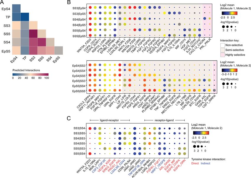
Research article Cell Biology
suppressor pathways and upregulation of cyclin-dependent kinase inhibitors p16INK4a (encoded by
CDKN2A) and p21CIP1 (CDKN1A) lead to permanent cell cycle arrest, induction of survival genes, and
production of a bioactive secretome, referred to as the senescence-associated secretory phenotype
(SASP). The composition of the SASP is tissue-specific but typically includes proinflammatory and
immunomodulatory cytokines, chemokines, growth factors, and extracellular matrix (ECM) proteins
and proteases (Birch and Gil, 2020). Acute senescence, characterized by transient SASP production
and rapid immune-mediated clearance of senescent cells, is widely implicated in processes involving
physiological tissue remodelling, including during embryo development, placenta formation, and
wound healing (Muñoz-Espín and Serrano, 2014; Van, 2014). By contrast, persisting senescent
cells cause chronic inflammation or ‘inflammaging’ (Birch and Gil, 2020), a pathological state that
underpins ageing and age-related disorders. We demonstrated that inflammatory reprogramming
of EnSC burdened by replication stress leads to the emergence of acute senescent cells during the
implantation window (Brighton et al., 2017; Lucas et al., 2020; Kong et al., 2021). Upon successful
implantation and continuous progesterone signalling, decidual cells co-opt uNK cells to eliminate
their senescent counterparts through granule exocytosis (Brighton et al., 2017; Lucas et al., 2020;
Kong et al., 2021). Clearance of senescent decidual cells likely necessitates recruitment of bone
marrow-derived decidual precursor cells, which confer tissue plasticity for rapid decidual expansion
in early pregnancy (Diniz-da-Costa et al., 2021). Importantly, lack of clonogenic decidual precursor
cells and a pro-senescent decidual response are linked to recurrent pregnancy loss (Lucas et al., 2016;
Lucas et al., 2020; Tewary et al., 2020).
Based on these insights, we hypothesized that acute senescence is integral to successful implanta-
tion by creating conditions for anchorage of the conceptus in an otherwise tightly adherent decidual
matrix. To test this hypothesis, we developed an ‘assembloid’ model, consisting of endometrial gland-
like organoids and primary EnSC, which recapitulates the complexity in cell states and gene expression
of the midluteal implantation window, improving resemblance to endometrial tissue in comparison
with existing co-culture models (Cheung et al., 2021; Rawlings, 2021). We used this model to estab-
lish co-cultures with human blastocysts and demonstrate that aspects of different pathological states
associated with implantation failure and miscarriage can be recapitulated in endometrial assembloids
by modulating decidual senescence.
Results
Establishment of endometrial assembloids
Organoids consisting of gland-like structures are established by culturing endometrial EpCs seeded in
Matrigel in a chemically defined medium containing growth factors and signal transduction pathway
modulators (Supplementary file 1: Table 1; Turco et al., 2017; Boretto et al., 2017). Gland-like
organoids grown in this medium, termed expansion medium, are genetically stable, easily passaged,
and can be maintained in long-term cultures (Boretto et al., 2017; Turco et al., 2017). Oestradiol
(E2) promotes proliferation of gland-like organoids and cooperates with NOTCH signalling to activate
ciliogenesis in a subpopulation of EpC (Haider et al., 2019). Further, treatment with a progestin (e.g.
medroxyprogesterone acetate [MPA]) and a cyclic AMP analogue (e.g. 8-bromo-cAMP) induces secre-
tory transformation of gland-like organoids in parallel with expression of luteal-phase marker genes
(Turco et al., 2017; Boretto et al., 2017).
We modified the gland-like organoid model to incorporate EnSC. To this end, midluteal endome-
trial biopsies (Supplementary file 1: Table 2) were digested and gland-like organoids established
from isolated EpC (Figure 1A). In parallel, purified EnSC were propagated in standard monolayer
cultures. At passage 2, single-cell suspensions of EnSC were combined with organoid EpC, seeded
in hydrogel, and cultured in expansion medium supplemented with E2 (Figure 1A). The hydrogel
matrix comprised 97% type I and 3% type III collagens, which are both present in midluteal endome-
trium (Oefner et al., 2015; Aplin et al., 1988; Aplin and Jones, 1989; Iwahashi et al., 1996), and
has a predicted in-use elastic modulus (Pa) of comparable magnitude to non-pregnant endometrium
(Abbas et al., 2019; Bagley, 2019). As shown in Figure 1B, gland formation was unperturbed by the
presence of EnSC and assembloids resembled the architecture of native endometrium more closely
than organoids. Further, decidualization of assembloids with 8-bromo-cAMP and MPA for 4 days
(Figure 1C) resulted in robust secretion of decidual prolactin (PRL) and C-X-C motif chemokine ligand
Rawlings et al. eLife 2021;10:e69603. DOI: https://doi.org/10.7554/eLife.69603 3 of 24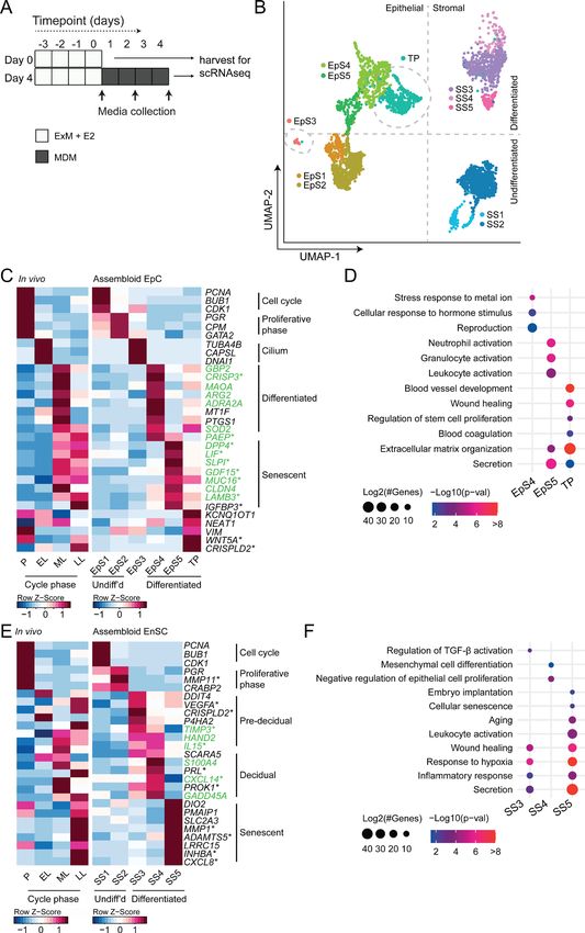
Research article Cell Biology
Figure 1. Establishment of endometrial assembloids. (A) Schematic for establishing endometrial assembloids. (B) Structural appearance of hematoxylin
and eosin stained secretory endometrium, E-cadherin labelled gland-like organoids, and E-cadherin and vimentin stained endometrial assembloids.
Scale bar = 50 µm. (C) Schematic summary of experimental design. (D) Secreted levels of PRL and CXCL14 were measured by ELISA in spent medium
at the indicated timepoints. Data points are coloured to indicate secretion in assembloids established from different endometrial biopsies (n =
3). (E) Representative immunofluorescence labelling of laminin and vimentin, progesterone receptor (PR), glycodelin, and osteopontin (OPN) in
undifferentiated (day 0, top panels) and decidualized (day 4; bottom panels) assembloids. Nuclei were counterstained with DAPI. Scale bar = 50 µm.
ELISA data in (B) are available in Figure 1—source data 1.
The online version of this article includes the following figure supplement(s) for figure 1:
Source data 1. Secretion of PRL and CXCL14 by endometrial assembloids.
14 (CXCL14) (Figure 1D). Immunofluorescence microscopy provided further evidence that decidual-
izing assembloids mimic luteal phase endometrium, exemplified by laminin deposition by decidual-
izing EnSC, induction of osteopontin (SPP1) and accumulation of glycodelin (encoded by PAEP) in the
lumen of secretory glands, and downregulation of the progesterone receptor (PGR) in both stromal
and glandular compartments (Figure 1E).
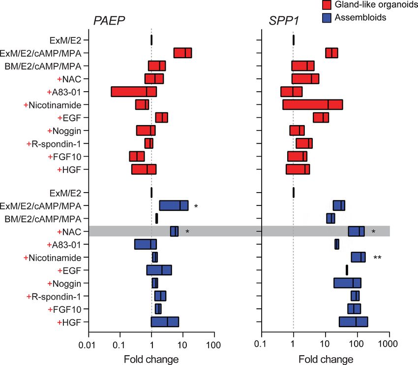
We reasoned that once established assembloids may no longer require exogenous growth factors
and pathway modulators for differentiation because of the presence of EnSC. To test this hypoth-
esis, parallel gland-like organoids and assembloids were established from three endometrial biopsies
and decidualized with E2, 8-bromo-cAMP, and MPA for 4 days in either expansion medium, base
medium (Supplementary file 1: Table 1), or base medium with each exogeneous factor added back
individually. Induction of PAEP and SPP1 was used to monitor the glandular differentiation response.
As shown in Figure 2, differentiation of gland-like organoids in base medium markedly blunted the
induction of PAEP and SPP1 when compared to expansion medium. Add-back of individual factors
did not restore the glandular response, with the exception of N-acetyl-L-cysteine (NAC). Addition
of NAC at low concentration (1.25 mM) to base medium resulted in a robust glandular response in
assembloids. Thus, in subsequent experiments assembloids were grown in expansion medium supple-
mented with E2 and then decidualized in minimal differentiation medium (MDM), consisting of base
medium containing NAC, E2, 8-bromo-cAMP, and MPA.
Rawlings et al. eLife 2021;10:e69603. DOI: https://doi.org/10.7554/eLife.69603 4 of 24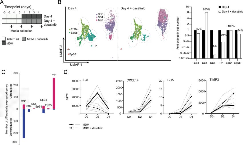
Research article Cell Biology Figure 2. Characterization of a minimal differentiation medium for endometrial assembloids. Parallel gland-like organoids (red) and assembloids (blue) were established from three endometrial biopsies and decidualized with 8-bromo-cAMP and MPA for 4 days in either expansion medium (ExM), base medium (BM), or BM with each exogeneous factor added back individually (+). Induction of PAEP and SPP1 was used to monitor the glandular differentiation. The grey bar indicates the composition of the minimal differentiation medium selected for further use (BM supplemented with NAC, E2, cAMP, and MPA). Data are presented as fold-change relative to expression levels in undifferentiated organoids or assembloids cultured in ExM+ E2. Bars present minimal, maximal, and median fold-change. * and ** indicate p
Research article Cell Biology
et al., 1999; Lucas et al., 2020). To test this hypothesis, we performed single-cell RNA sequencing
(scRNA-seq) on undifferentiated assembloids grown for 4 days in expansion medium and assem-
bloids decidualized in MDM for four additional days (Figure 3A). Eleven distinct cell clusters were
identified by Shared Nearest Neighbour (SNN) and Uniform Manifold Approximation and Projec-
tion (UMAP) analysis, segregating broadly into epithelial and stromal populations within the UMAP-1
dimension and into undifferentiated and differentiated subpopulations within the UMAP-2 dimension
(Figure 3B). Each cell cluster was annotated based on expression of curated marker genes, which
were cross-referenced with a publicly available data set (GEO: GSE4888) to determine their relative
expression across the menstrual cycle in vivo (Talbi et al., 2006).
We identified five unambiguous EpC subsets. The glandular component of undifferentiated assem-
bloids harboured actively dividing EpC (EpS1; n = 198) as well as EpC-expressing marker genes of
E2-responsive proliferative phase endometrium (EpS2; n = 692), including PGR and CPM (Figure 3C).
EpS3 (n = 29) consisted of ciliated EpC, expressing an abundance of genes involved in cilium assembly
and organization, including DNAI1 and TUBA4B (Figure 3C). Ciliated cells are the only glandular
subpopulation present in both undifferentiated and decidualized assembloids. In vivo, EpS3 marker
genes transiently peak during the early-luteal phase (Figure 3C). Decidualization of endometrial
assembloids led to the emergence of two distinct EpC subsets, EpS4 (n = 434) and EpS5 (n = 208).
Both clusters expressed canonical endometrial ‘receptivity genes’ (annotated in green in Figure 3C),
that is, genes used in a clinical test to aid the timing of embryo transfer to the window of implantation
in IVF patients (Díaz-Gimeno et al., 2011). In agreement, induction of EpS4 and EpS5 marker genes
in vivo coincides with the transition from early- to midluteal phase. However, while expression of EpS4
marker genes, including SOD2, MAOA, and PTGS1, generally peaks during the midluteal window of
implantation, EpS5 genes tend to persist or peak during the late-luteal phase (Figure 3C). Additional
mining of the data revealed that transition from EpS4 to EpS5 coincides with induction of p16INK4a and
p21CIP1 in parallel with upregulation of 56 genes encoding secretory factors (Figure 3—figure supple-
ment 1). Notably, several canonical implantation factors secreted by this subpopulation are also well-
characterized SASP components, including dipeptidyl peptidase 4 (DPP4; Kim et al., 2017), growth
differentiation factor 15 (GDF15; Basisty et al., 2020), and insulin-like growth factor binding protein 3
(IGFBP3; Elzi et al., 2012). Thus, EpS5 consists of senescent EpC producing an implantation-specific
SASP.
Decidualized endometrial assembloids also harboured a sizable population of ambiguous cells
expressing both epithelial and stromal genes (Figure 3C and Figure 3—figure supplement 2). A hall-
mark of this subset, termed ‘transitional population’ (TP; n = 472), is the induction of long non-coding
RNAs involved in mesenchymal-epithelial and epithelial-mesenchymal transition (MET/EMT), such as
NEAT1 (nuclear paraspeckle assembly transcript 1) and KCNQ1OT1 (KCNQ1 opposite strand/anti-
sense transcript 1) (Bian et al., 2019; Chen et al., 2021). GO analysis showed that both EpS5 and the
transitional population comprised secretory cells involved in ECM organization (Figure 3D). However,
while EpS5 genes are implicated in neutrophil activation (a hallmark of premenstrual endometrium),
genes expressed by the transitional population are uniquely enriched in GO terms such as ‘wound
healing’, ‘regulation of stem cell proliferation’, ‘blood coagulation’, and ‘blood vessel development’
(Figure 3D), which points towards a putative role in tissue repair and regeneration.
The stromal fraction of undifferentiated assembloids consisted of actively dividing EnSC (stromal
subpopulation 1 [SS1]; n = 434) and E2-responsive EnSC (SS2; n = 874) expressing proliferative
phase marker genes, such as PGR, MMP11, and CRABP2 (Figure 3E). As anticipated, decidualiza-
tion of assembloids for 4 days led to a preponderance of pre-decidual cells (SS3; n = 495) as well as
emerging decidual cells (SS4; n = 87) and senescent decidual cells (SS5; n = 118) (Figure 3E). Each
of these subpopulations expressed marker genes identified previously by scRNA-seq reconstruction
of the decidual pathway in standard primary EnSC cultures (Lucas et al., 2020). Pre-decidual cells in
SS3 express HAND2, a key decidual transcription factor (Marinić et al., 2021), as well as previously
identified genes encoding secreted factors, including VEGFA (vascular endothelial growth factor A),
CRISPLD2 (a progesterone-dependent anti-inflammatory response gene coding cysteine-rich secre-
tory protein LCCL domain containing 2), IL15 (interleukin 15), and TIMP3 (TIMP metallopeptidase
inhibitor 3) (Lucas et al., 2020). Novel candidate pre-decidual genes were also identified, such as
DDIT4 (DNA damage-inducible transcript 4), encoding a stress response protein intimately involved
in autophagy, stemness, and antioxidative defences (Ho et al., 2020; Miller et al., 2020). Decidual
Rawlings et al. eLife 2021;10:e69603. DOI: https://doi.org/10.7554/eLife.69603 6 of 24Research article Cell Biology
Figure 3. Decidualizing assembloids mimic midluteal endometrium. (A) Schematic overview of experimental
design. ExM: expansion medium; MDM: minimal differentiation medium. (B) Uniform Manifold Approximation
and Projection (UMAP) visualizing epithelial and stromal subsets (EpS and SS, respectively) identified by single-cell
transcriptomic analysis of undifferentiated and decidualized assembloids. A transitional population (TP) consisting
Figure 3 continued on next page
Rawlings et al. eLife 2021;10:e69603. DOI: https://doi.org/10.7554/eLife.69603 7 of 24Research article Cell Biology
Figure 3 continued
of cells expressing epithelial and stromal markers is also shown. Dotted lines indicate the separation of EpS and SS
in UMAP_1 and of undifferentiated and differentiated subpopulations in UMAP_2. Dotted circles indicate ciliated
(EpS3) and TP, which did not fit these broad segregations. (C) Composite heatmaps showing relative expression
(Z-scores) of epithelial marker genes across the menstrual cycle in vivo and in undifferentiated and decidualized
assembloids. Highlighted in green are genes that mark the midluteal window of implantation (Díaz-Gimeno et al.,
2011), whereas genes encoding secreted proteins are indicated by * (Uhlén et al., 2015). See also Figure 3—
figure supplement 1. (D) Dot plots showing GO terms related to biological processes enriched in different
epithelial populations in decidualizing assembloids. The dot size represents the number of genes in each GO term
and the colour indicates FDR-corrected p-value. (E) Composite heatmaps showing relative expression (Z-scores)
of stromal marker genes across the menstrual cycle in vivo and in undifferentiated and decidualized assembloids.
Highlighted in green are genes that mark the midluteal window of implantation (Díaz-Gimeno et al., 2011),
whereas genes encoding secreted proteins are indicated by * (Uhlén et al., 2015). (F) Dot plots showing GO
terms related to biological processes enriched in different stromal subpopulations in decidualizing assembloids.
See also Figure 3—figure supplements 1 and 2 and 3. Complete epithelial subpopulation marker lists can be
found in Figure 3—source data 1. GO analysis outputs can be found in . Complete stromal subpopulation marker
lists can be found in .
The online version of this article includes the following figure supplement(s) for figure 3:
Source data 1. Epithelial subpopulation markers.
Source data 2. GO analysis of differentiated subpopulations.
Source data 3. Stromal sub-population markers.
Figure supplement 1. Heatmap showing relative expression (Z-scores) of genes encoding the cyclin-dependent
kinase inhibitors p16INK4a and p21CIP1 as well as SASP-related genes in epithelial and transitional subpopulations in
decidualizing assembloids.
Figure supplement 2. Heatmap showing relative expression (Z-scores) of epithelial-mesenchymal
transition/mesenchymal-epithelial transition (EMT/MET), epithelial and mesenchymal marker genes in the
transitional population (TP), epithelial (EpS4-5) and stromal (SS3-5) subpopulations in decidualizing assembloids.
Figure supplement 3. Heatmap showing relative expression (Z-scores) of genes encoding the cyclin-dependent
kinase inhibitors p16INK4a and p21CIP1 as well as secretory and SASP-related genes in stromal subpopulations (SS3-5)
in decidualizing assembloids.
cells (SS4) and senescent decidual cells (SS5) express SCARA5 and DIO2, respectively (Figure 3E),
two stroma-specific marker genes identified by scRNA-seq analysis of mid- and late-luteal endome-
trial biopsies (Lucas et al., 2020). SS3 and SS4 genes mapped to the early- and midluteal phase of
the cycle, whereas SS5 genes peak in the late-luteal phase, that is, prior to menstrual breakdown.
Notably, the transcriptomic profiles of SS3 and SS5 are enriched in GO terms such as ‘Wound healing’,
‘Response to hypoxia’, and ‘Inflammatory response’, suggesting that both clusters comprise stressed
cells (Figure 3F). However, the nature of the cellular stress response differs between these popu-
lations with only senescent decidual cells (SS5) expressing genes enriched in categories such as
‘Embryo implantation’, ‘Cellular senescence’, ‘Aging’, and ‘Leukocyte activation’. By contrast, few
notable categories were selectively enriched in decidual cells (e.g. ‘Mesenchymal cell differentiation’),
rendering the lack of GO terms that pertain to stress, inflammation, or wound healing perhaps the
most striking observation. In keeping with the GO analysis, senescent decidual cells (SS5) express a
multitude of SASP-related genes (Figure 3—figure supplement 3), including matrix metallopepti-
dases (e.g. MMP3, 7, 9, 10, 11, and 14), insulin-like growth factor binding proteins (e.g. IGFBP1, 3,
6, and 7), growth factors (e.g. AREG, FGF2, FGF7, HGF, and VEGFA) and growth factor receptors
(PDGFRA and PDGFRB), cytokines (e.g. LIF, IL6, IL1A, and IL11), chemokines (e.g. CXCL8 and CXCL1),
and members of the TGF-β superfamily of proteins (e.g. GDF15, INHBA, and BMP2). By contrast,
decidual cells are characterized by expression of a unique network of secretory genes, some encoding
ECM proteins (e.g. COL1A1, COL3A1, and LAMA4) and other known decidual markers (e.g. PRL,
PROK1, and WNT4) as well as factors involved in uNK cell chemotaxis and activation (e.g. CCL2,
CXCL14, and IL15) (Figure 3—figure supplement 3).
Taken together, single-cell analysis of undifferentiated and decidualized assembloids revealed a
surprising level of cellular complexity. Each epithelial and stromal subpopulation appears function-
ally distinct and maps to a specific phase of the menstrual cycle. Transition between cellular states is
Rawlings et al. eLife 2021;10:e69603. DOI: https://doi.org/10.7554/eLife.69603 8 of 24Research article Cell Biology
Figure 4. Putative receptor-ligand interactions in decidualizing assembloids. (A) Heatmap showing the total number of cell-cell interactions predicted
by CellPhoneDB between different subpopulations in decidualizing assembloids. (B) Dot plots of representative ligand-receptor interactions between
stromal subsets (SS) and epithelial subsets (EpS) (upper panel) and EpS and SS (lower panel) in decidualizing assembloids. Circle size and colour
indicate p-value and the means of the average expression value of the interacting molecules, respectively. Shaded boxes were used to group putative
interactions by level of selectivity. (C) Dot plot of representative ligand-receptor and receptor-ligand interactions between stromal subpopulations
in decidualizing assembloids. Direct and indirect tyrosine kinase interactions are indicated by red and blue labels, respectively. Complete tables of
predicted ligand-receptor interactions can be found in Figure 4—source data 1.
The online version of this article includes the following figure supplement(s) for figure 4:
Source data 1. CellPhoneDB prediction of cell-cell interactions.
predicated on changes in cell cycle status, ranging from actively dividing cells in proliferating assem-
bloids to the emergence upon differentiation of highly secretory senescent epithelial and decidual
subpopulations, resembling premenstrual endometrium. However, the dominant subpopulations on
day 4 of decidualization are EpS4 and SS3, which map to the midluteal implantation window in vivo.
Receptor-ligand interactions in decidualizing assembloids
We used CellPhoneDB, a publicly available online repository of highly curated receptor-ligand inter-
actions, to explore putative interactions between subpopulations in decidualizing assembloids. This
computational tool also takes into account the subunit architecture of both ligands and receptors in
heteromeric complexes (Efremova et al., 2020; Vento-Tormo et al., 2018). The number of predicted
interactions is depicted in Figure 4A, showing a conspicuous lack of crosstalk between the transitional
population and any other populations. Conversely, the most abundant interactions centre around the
secretory subpopulations, EpS5 and SS5.
Rawlings et al. eLife 2021;10:e69603. DOI: https://doi.org/10.7554/eLife.69603 9 of 24Research article Cell Biology
A total of 270 significantly enriched (non-integrin) receptor-ligand interactions (FDR-corrected
pResearch article Cell Biology
Figure 5. Tyrosine kinase-dependent stress responses determine the fate of decidual cells. (A) Schematic overview of experimental design. ExM:
expansion medium; MDM: minimal differentiation medium. (B) Uniform Manifold Approximation and Projection (UMAP) visualization (left panel) and
relative proportions (right panel) of subpopulations in endometrial assembloid decidualized in the presence or absence of dasatinib. (C) Number of
differentially expressed genes (DEGs) in each subpopulation in response to dasatinib pre-treatment. (D) Secreted levels of CXCL8 and decidual cell
factors in spent medium from assembloids treated with or without dasatinib. Secreted levels in individual assembloids established from four different
endometrial assembloids decidualized with or without dasatinib are shown by dotted and solid lines, respectively. Full lists of DEGs and associated GO
analysis can be found in Figure 5—source data 1 and Figure 5—source data 2, respectively. Data used in (D) are available in Figure 5—source data
3.
The online version of this article includes the following figure supplement(s) for figure 5:
Source data 1. Differentially expressed genes for day 4 populations treated with and without dasatinib.
Source data 2. GO analysis for day 4 populations treated with and without dasatinib.
Source data 3. ELISA data.
dasatinib inhibited the expression of a network of genes enriched in GO categories such as ‘Response
to wounding’ (FDR-corrected p=3.5 × 10⁻⁵), ‘Response to stress’ (FDR-corrected p=3.8 × 10⁻⁵), and
‘Response to oxidative stress’ (FDR-corrected p=1.3 × 10⁻⁴), indicative of a blunted stress response.
To substantiate this finding, we measured the secreted levels of CXCL8 (IL-8), a potent inflammatory
mediator implicated in autocrine/paracrine propagation of cellular senescence (Acosta et al., 2008;
Kuilman et al., 2008), in assembloids decidualized with or without dasatinib. CXCL14, IL-15, and
TIMP3 levels were also measured to monitor the decidual response. As shown in Figure 5D, dasatinib
completely abrogated the release of CXCL8 by pre-decidual cells while markedly enhancing subse-
quent secretion of CXCL14, IL-15, and TIMP3, which are involved in effecting immune clearance of
senescent decidual cells (Brighton et al., 2017; Lucas et al., 2020; Kong et al., 2021). Together,
these observations not only support the CellPhoneDB predictions but also indicate that the amplitude
of the cellular stress response during the pre-decidual phase determines the subsequent decidual
trajectory, with low levels accelerating differentiation and high levels promoting cellular senescence
and MET.
Rawlings et al. eLife 2021;10:e69603. DOI: https://doi.org/10.7554/eLife.69603 11 of 24Research article Cell Biology
Modelling the impact of decidual subpopulations on human embryos
We postulated that decidual invasion by human embryos that have breached the luminal endometrial
epithelium depends on an acute cellular senescence and transient SASP production, rich in growth
factors and proteases. Conversely, we reasoned that lack of senescent decidual cells or unconstrained
SASP should simulate pathological implantation environments associated with implantation failure
and early pregnancy loss, respectively. To test this hypothesis, we constructed a simple implantation
Figure 6. Impact of decidual senescence in assembloids on co-cultured human blastocysts. (A) Diagram showing experimental design. ExM:
expansion medium; MDM: minimal differentiation medium; EM: embryo medium. (B) Schematic drawing of co-culture method. (C) Representative
time-lapse images of blastocysts embedded in assembloids following decidualization for 96 hr in the absence (upper panels) or presence (lower
panels) of dasatinib. Scale bar = 100 µm. See also Figure 6—figure supplement 1. (D) Embryo diameters (µm) measured over 72 hr when embedded
in decidualizing assembloids pre-treated with or without dasatinib. (E) OCT4 and GATA6 immunofluorescence marking the epiblast and hypoblast,
respectively, in a blastocyst attached by proliferating polar trophectoderm (arrowhead) to decidual assembloids. Scale bar = 50 µM. (F) Secreted levels
of human chorionic gonadotropin (hCG) in blastocyst-endometrial assembloid co-cultures. Individual embryo diameter measurements for biological
replicates in (D) are available in Figure 6—source data 1. Individual ELISA data used in (F) are available in Figure 6—source data 2.
The online version of this article includes the following figure supplement(s) for figure 6:
Source data 1. Embryo expansion measurement.
Source data 2. Embryo human chorionic gonadotropin (hCG) secretion.
Figure supplement 1. Stromal migration towards the polar trophectoderm of expanding embryos in differentiated endometrial assembloids.
Figure supplement 2. Dasatinib prevents disintegration of decidualizing assembloids.
Rawlings et al. eLife 2021;10:e69603. DOI: https://doi.org/10.7554/eLife.69603 12 of 24Research article Cell Biology
Video 1. Time-lapse microscopy of a human Video 2. Time-lapse microscopy of a human blastocyst
blastocyst embedded in a decidualizing assembloid. embedded in a decidualizing assembloid pre-treated
Representative video of a human blastocyst embedded with dasatinib. Representative video of a human
in an assembloid, as imaged by time-lapse microscopy blastocyst embedded in an assembloid which had been
over 72 hr with images captured every 60 min. pre-treated with dasatinib, as imaged by time-lapse
https://elifesciences.org/articles/69603/figures#video1 microscopy over 72 hr with images captured every
60 min.
https://elifesciences.org/articles/69603/figures#video2
model by embedding human embryos in endo-
metrial assembloids. To this end, assembloids
were first decidualized for 96 hr in the presence or absence of dasatinib, washed and cultured in
embryo medium, consisting of MDM with added supplements (Figure 6A and Supplementary file
1: Table 1). Day 5 human blastocysts were placed into small pockets created in the decidualized
assembloids (Figure 6B), one embryo per assembloid, and individual co-cultures imaged using time-
lapse microscopy over 72 hr. Co-cultured blastocysts (n = 5) expanded markedly when placed in
decidualized assembloids that were not pre-treated with dasatinib (Figure 6C and D). Time-lapse
microscopy revealed intense cellular movement in the stromal compartment as well as evidence that
interaction between migratory decidual cells and polar trophectoderm promotes adherence and early
invasion of the embryo (SI Video 1 and Figure 6—figure supplement 1). Retrieval and processing of
one attached embryo demonstrated proliferating polar trophectoderm and expression of OCT4 and
GATA6 in the epiblast and hypoblast, respectively (Figure 6E). A major limitation of this implantation
model is that persistence of senescent decidual cells also causes gradual disintegration of the assem-
bloids (Figure 6—figure supplement 2). By contrast, pre-treatment with dasatinib, which accelerates
decidualization and all but eliminates decidual senescence, resulted in much more robust assem-
bloids. However, all embedded blastocysts (n = 5) failed to expand in this model (Figure 6C and D).
Further, movement of the decidual matrix was greatly reduced and directed migration or attachment
of decidual cells to the blastocyst was not observed (SI Video 2). Secreted levels of human chori-
onic gonadotropin (hCG) did not differ between co-cultures (Figure 6E), suggesting that all embryos
remained viable over the 72 hr observation period. Thus, while our experimental design precluded
modelling of physiological embryo implantation, aspects of different pathological endometrial states
underlying reproductive failure, that is, implantation failure and miscarriage, were recapitulated in
assembloids.
Discussion
Here we report on the development of endometrial assembloids, consisting of gland-like organoids
surrounded by a matrix rich in primary EnSC, as novel model to parse the cellular dynamics that
govern embryo implantation in cycling human endometrium. While assembloids complement and
advance other recently described endometrial organoid models (Boretto et al., 2017; Cheung et al.,
2021; Fitzgerald et al., 2019; Luddi et al., 2020; Turco et al., 2017), they still lack the cellular
complexity of native endometrium, including uNK cells, macrophages, and vascular cells. Never-
theless, we demonstrated that aspects of pathological implantation events can be recapitulated in
assembloids, rendering them useful as novel models to study mechanisms of reproductive failure and
evaluate potential therapeutic interventions.
Rawlings et al. eLife 2021;10:e69603. DOI: https://doi.org/10.7554/eLife.69603 13 of 24Research article Cell Biology
Single-cell analysis of differentiating endometrial assembloids indicates that the sequence of events
leading up to the implantation window, and beyond, requires divergence of both glandular EpC and
EnSC into differentiated and senescent subpopulations, a process likely determined by the level of
replication stress incurred by individual cells in the preceding proliferative phase (Brighton et al.,
2017). Importantly, we demonstrate that acute senescence in glandular EpC (EpS5) underpins produc-
tion of an implantation-specific SASP, comprising canonical implantation factors and growth factors,
such as amphiregulin (AREG) and epiregulin (EREG), implicated in transforming cytotrophoblasts into
extravillous trophoblasts (Cui et al., 2020; Yu et al., 2019). On the other hand, the transcriptome
profile of differentiated EpC (EpS4) revealed a pivotal role for this subpopulation in prostaglandin and
glycodelin synthesis. Prostaglandins, and specifically PGE2, are indispensable for implantation (Ruan
et al., 2012), whereas glycodelin is an abundantly secreted, multifaceted glycoprotein involved in blas-
tocyst attachment, trophoblast differentiation, and immune modulation in early pregnancy (Lee et al.,
2016). Further, differentiated EpC highly express SLC2A1, encoding the major glucose transporter
GLUT1. Glucose is required for glycogen synthesis, an essential component of glandular secretions
that nourishes the conceptus prior to the onset of placental perfusion around 10 weeks of pregnancy
(Burton et al., 2020). The fate and function of senescent EpC in pregnancy are unknown. Arguably,
localized secretion of proteinases by senescent EpC may promote breakdown of the surrounding
basement membrane, thereby facilitating endoglandular trophoblast invasion and access to histotro-
phic nutrition in early gestation (Huppertz, 2019; Moser et al., 2010). In non-conception cycles, the
abundance of p16INK4-positive glandular EpC rises markedly during the late-luteal phase (Brighton
et al., 2017), indicating that senescent EpC are progesterone-independent and likely responsible for
glandular breakdown in the superficial endometrial layer at menstruation.
Decidual transformation of EnSC in assembloids unfolded largely as anticipated from previous
studies, that is, starting with an acute pre-decidual stress response and leading to the emergence
of both decidual and senescent decidual subpopulations (Brighton et al., 2017; Lucas et al., 2020;
Kong et al., 2021). Like their epithelial counterparts, senescent decidual cells have a conspicuous
secretory phenotype. We identified 56 and 72 genes encoding secreted factors upregulated in senes-
cent epithelial and decidual subpopulations, respectively. However, only 15 genes were shared, indi-
cating that the SASP generated in both cellular compartments is distinct. As glandular secretions drain
into the uterine cavity, the embryonic microenvironment is therefore predicted to change abruptly
upon breaching of the luminal epithelium. Recent comparative metabolomics of apical and baso-
lateral endometrial gland-like organoid secretomes also supports the prediction of an asymmetrical
profile of glandular secretions in the pre- and post-implantation microenvironments (Simintiras et al.,
2021).
Based on computational predictions of ligand-receptor interactions, we demonstrated that the
decidual response in assembloids can be targeted pharmacologically with only modest impact on
glandular function and, by extension, the preimplantation embryo milieu. Specifically, dasatinib, a
tyrosine kinase inhibitor, was highly effective in blunting the pre-decidual stress response, leading
to a dramatic expansion of anti-inflammatory decidual cells and near-total elimination of senescent
decidual cells. Dasatinib also inhibited the emergence of TP and shifted the transcriptional profile of
the remaining transitional cells towards a decidual phenotype. An analogous population of ambig-
uous cells expressing both epithelial and mesenchymal marker genes was recently identified in midlu-
teal endometrium by scRNA-seq analysis (Lucas et al., 2020). Further, based on CellPhoneDB and GO
analyses, transitional cells are predicted to be highly autonomous and involved in tissue regeneration,
in line with experimental evidence that MET drives re-epithelization of the endometrium following
menstruation and parturition (Owusu-Akyaw et al., 2019; Patterson et al., 2013). Thus, the level
of endogenous cellular stress generated by the endometrium during the window of implantation
calibrates the subsequent decidual trajectory, either promoting the formation of a robust decidual
matrix or facilitating tissue breakdown and repair. Further, an in-built feature of both trajectories is
self-enforcement as decidual cells recruit and activate uNK cells to eliminate their senescent counter-
parts (Brighton et al., 2017; Lucas et al., 2020; Kong et al., 2021), whereas senescent decidual cells
induce secondary senescence in neighbouring decidual (Brighton et al., 2017; Ozaki et al., 2017)
and, plausibly, uNK cells (Rajagopalan and Long, 2012).
Clinically, recurrent pregnancy loss, defined as multiple miscarriages, is associated with loss of
endometrial clonogenicity (Lucas et al., 2016; Diniz-da-Costa et al., 2021), uNK cell deficiency and
Rawlings et al. eLife 2021;10:e69603. DOI: https://doi.org/10.7554/eLife.69603 14 of 24Research article Cell Biology
excessive decidual senescence (Lucas et al., 2020; Tewary et al., 2020), and rapid conceptions (also
referred to as ‘superfertility’) (Dimitriadis et al., 2020; Ticconi et al., 2020). Conversely, lack of a
proliferative gene signature in midluteal endometrium and premature expression of decidual PRL have
been linked to recurrent implantation failure (Berkhout et al., 2020b; Koler et al., 2009; Koot et al.,
2016), a pathological condition defined by a failure to achieve a pregnancy following transfer of one
or more high-quality embryos in multiple IVF cycles (Polanski et al., 2014). We reasoned that these
aberrant implantation environments can be recapitulated in assembloids by manipulating the level of
decidual senescence. In line with these predictions, the presence of senescent decidual cells created
a permissive environment in which migratory decidual cells interacted with expanding blastocysts,
although continuous SASP production also promoted breakdown of the assembloids. Conversely, in
the absence of senescent decidual cells, non-expanding embryos became entrapped in a robust but
stagnant decidual matrix. These observations are in keeping with previous studies demonstrating
that implantation of human embryos depends critically on the invasive and migratory capacities of
decidual cells (Berkhout et al., 2020a; Gellersen et al., 2010; Grewal et al., 2008; Weimar et al.,
2012). Our co-culture experiments also highlighted the shortcomings of assembloids as an implan-
tation model, including the lack of a surface epithelium to create distinct pre- and post-implantation
microenvironments and the absence of key cellular constituents, such as innate immune cells.
In summary, parsing the mechanisms that control implantation has been hampered by the over-
whelming complexity of factors involved in endometrial receptivity. Our single-cell analysis of decid-
ualizing assembloids suggests that this complexity reflects the reliance of the human endometrium
on rapid E2-dependent proliferation and replicative exhaustion to generate both differentiated and
senescent epithelial and stromal subpopulations in response to the postovulatory rise in progesterone.
We demonstrate that senescent cells in both cellular compartments produce distinct bioactive secre-
tomes, which plausibly prime pre-implantation embryos for interaction with the luminal epithelium
and then stimulate encapsulation by underlying decidual stromal cells. Based on our co-culture obser-
vations, we predict that a blunted pre-decidual stress response causes implantation failure because
of a lack of senescence-induced tissue remodelling and accelerated decidualization. Conversely, a
heightened stress response leading to excessive decidual senescence may render embryo implanta-
tion effortless, albeit in a decidual matrix destined for breakdown and repair. Finally, we demonstrated
that pre-decidual stress responses can be modulated pharmacologically, highlighting the potential
of endometrial assembloids as a versatile system to evaluate new or repurposed drugs aimed at
preventing reproductive failure.
Materials and methods
Ethical approvals, endometrial samples, and human blastocysts
Endometrial biopsies were obtained from women attending the Implantation Research Clinic, Univer-
sity Hospitals Coventry and Warwickshire National Health Service Trust. Written informed consent
was obtained in accordance with the Declaration of Helsinki 2000. The study was approved by the
NHS National Research Ethics Committee of Hammersmith and Queen Charlotte’s Hospital NHS Trust
(1997/5065) and Tommy’s Reproductive Health Biobank (Project TSR19-002E, REC Reference: 18/
WA/0356). Timed endometrial biopsies were obtained 6–11 days after the post-ovulatory LH surge
using a Wallach Endocell Endometrial Cell Sampler. Patient demographics for the samples used in
each experiment are detailed in Supplementary file 1: Table 2.
The use of vitrified human blastocysts was carried out under a Human Fertilisation and Embry-
ology Authority research licence (HFEA: R0155) with local National Health Service Research Ethics
Committee approval (04/Q2802/26). Spare blastocysts were donated to research following informed
consent by couples who had completed their fertility treatment at the Centre for Reproductive Medi-
cine, University Hospitals Coventry and Warwickshire National Health Service Trust. Briefly, women
underwent ovarian stimulation and oocytes were collected by transvaginal ultrasound-guided aspi-
ration and inseminated with prepared sperm (day 0). All oocytes examined 16–18 hr after insemina-
tion and classified as normally fertilized were incubated under oil in 20–25 µl drops of culture media
(ORIGIO Sequential Cleav and Blast media, CooperSurgical, Denmark) at 5% O2, 6% CO2, 89%
N2 at 37 °C. Following culture to day 5 of development, the embryo(s) with the highest quality was
selected for transfer, whereas surplus embryos considered top-quality blastocysts were cryopreserved
Rawlings et al. eLife 2021;10:e69603. DOI: https://doi.org/10.7554/eLife.69603 15 of 24Research article Cell Biology
on day 5 or 6 by vitrification using Kitazato vitrification media (Dibimed, Spain) and stored in liquid
nitrogen. Prior to their use in the co-culture, vitrified blastocysts were warmed using the Kitazato
vitrification warming media (Dibimed, Spain) and underwent zona pellucida removal using a Saturn
5 Laser (CooperSurgical). Blastocysts were then incubated for 1 hr under oil in 20 µl drops of culture
media (ORIGIO Sequential Blast media, CooperSurgical) at 5% O2, 6% CO2, 89% N2 at 37 °C and
allowed to re-expand.
Processing of endometrial biopsies and primary EnSC cultures
Unless otherwise stated, reagents were obtained from Life Technologies (Paisley, UK). Cell cultures
were incubated at 37 °C, 5% CO2 in a humidified incubator. Centrifugation and incubation steps
were performed at room temperature unless stated otherwise. Fresh endometrial biopsies were
processed as described previously (Barros et al., 2016). Briefly, tissue was finely minced for 5 min
using a scalpel blade. Minced tissue was then digested enzymatically with 0.5 mg/ml collagenase I
(Sigma-Aldrich, Gillingham, UK) and 0.1 mg/ml deoxyribonuclease (DNase) type I (Lorne Laborato-
ries, Reading, UK) in 5 ml phenol red-free Dulbecco’s Modified Eagle Medium (DMEM)/F12 for 1 hr
at 37 °C, with regular vigorous shaking. Dissociated cells were washed with growth medium (DMEM/
F12 containing 10% dextran-coated charcoal stripped FBS [DCC-FBS], 1% penicillin-streptomycin,
2 mM L-glutamine, 1 nM E2 [Sigma-Aldrich] and 2 mg/ml insulin [Sigma-Aldrich]). Samples were
passed through a 40 µm cell sieve. EnSCs were collected from the flowthrough, while epithelial
clumps remained in the sieve and were collected by backwashing into a 50 ml Falcon tube. Samples
were resuspended in growth medium and centrifuged at 400× g for 5 min. EnSC pellets were resus-
pended in 10 ml growth medium and plated in tissue culture flasks. To isolate EnSC from other (non-
adherent) cells collected in the flowthrough, medium was refreshed after 24 hr. Thereafter, medium
was refreshed every 48 hr. Sub-confluent monolayers were passaged using 0.25% Trypsin-EDTA and
split at a ratio of 1:3.
Endometrial gland-like organoid culture
Endometrial gland-like organoids were established as described previously (Turco et al., 2017), with
adaptations. Freshly isolated endometrial gland fragments were resuspended in 500 µl phenol red-
free DMEM/F12 medium in a microcentrifuge tube and centrifuged at 600× g for 5 min. The medium
was aspirated and ice-cold, growth factor-reduced Matrigel (Corning Life Sciences B.V., Amsterdam,
Netherlands) was added at a ratio of 1:20 (cell pellet: Matrigel). Samples mixed in Matrigel were kept
on ice until plating at which point the suspension was aliquoted in 20 µl volumes to a 48-well plate,
one drop per well, and allowed to cure for 15 min. Expansion medium supplemented with E2 (Supple-
mentary file 1: Table 1; Turco et al., 2017) was then added and samples cultured for up to 7 days.
For passaging, Matrigel droplets containing gland-like organoids were collected into microcentrifuge
tubes and centrifuged at 600× g for 6 min at 4 °C. Samples were resuspended in ice-cold, phenol
red-free DMEM/F12 and subjected to manual pipetting to disrupt the organoids. Suspensions were
centrifuged again, resuspended in ice-cold additive-free DMEM/F12, and then subjected to further
manual pipetting. Suspensions were centrifuged again and either resuspended in Matrigel and plated
as described above for continued expansion or used to establish assembloid cultures.
Establishment of assembloid cultures
At passage 2, EnSC and gland-like organoid pellets were mixed at a ratio of 1:1 (v/v) and ice-cold
PureCol EZ Gel (Sigma-Aldrich) added at a ratio of 1:20 (cell pellet: hydrogel). Samples were kept
on ice until plating. The suspension was aliquoted in 20 µl volumes using ice-cold pipette tips into
a 48-well plate, one droplet per well, and allowed to cure in the cell culture incubator for 45 min.
Expansion medium supplemented with 10 nM E2 was overlaid and the medium was refreshed every
48 hr. For decidualization experiments, assembloid cultures were grown in expansion medium supple-
mented with E2 for 4 days to allow for growth and expansion. Assembloids were then either harvested
or decidualized using different media as tabulated in Supplementary file 1: Table 1 for a further
4 days. Again, the medium was refreshed every 48 hr and spent medium stored for further analysis.
For tyrosine kinase inhibition, MDM was supplemented with 250 nM dasatinib (Cell Signaling Tech-
nology, Leiden, NL).
Rawlings et al. eLife 2021;10:e69603. DOI: https://doi.org/10.7554/eLife.69603 16 of 24You can also read