The linear ubiquitin chain assembly complex (LUBAC) generates heterotypic ubiquitin chains - eLife
←
→
Page content transcription
If your browser does not render page correctly, please read the page content below
RESEARCH ARTICLE

                                     The linear ubiquitin chain assembly
                                     complex (LUBAC) generates heterotypic
                                     ubiquitin chains
                                     Alan Rodriguez Carvajal1, Irina Grishkovskaya2, Carlos Gomez Diaz1,
                                     Antonia Vogel2, Adar Sonn-Segev3, Manish S Kushwah3, Katrin Schodl1,
                                     Luiza Deszcz1,2, Zsuzsanna Orban-Nemeth2, Shinji Sakamoto4, Karl Mechtler2,
                                     Philipp Kukura3, Tim Clausen2, David Haselbach2*, Fumiyo Ikeda1,5*
                                     1
                                       Institute of Molecular Biotechnology of the Austrian Academy of Sciences (IMBA),
                                     Vienna BioCenter (VBC), Vienna, Austria; 2Research Institute of Molecular Pathology
                                     (IMP), Vienna BioCenter (VBC), Vienna, Austria; 3Department of Chemistry,
                                     University of Oxford, Chemistry Research Laboratory, Oxford, United Kingdom;
                                     4
                                       Pharmaceutical Frontier Research Labs, JT Inc., Yokohama, Japan; 5Medical
                                     Institute of Bioregulation (MIB), Kyushu University, Fukuoka, Japan

                                     Abstract The linear ubiquitin chain assembly complex (LUBAC) is the only known ubiquitin ligase
                                     for linear/Met1-linked ubiquitin chain formation. One of the LUBAC components, heme-oxidized
                                     IRP2 ubiquitin ligase 1 (HOIL-1L), was recently shown to catalyse oxyester bond formation between
                                     ubiquitin and some substrates. However, oxyester bond formation in the context of LUBAC has not
                                     been directly observed. Here, we present the first 3D reconstruction of human LUBAC obtained by
                                     electron microscopy and report its generation of heterotypic ubiquitin chains containing linear
                                     linkages with oxyester-linked branches. We found that this event depends on HOIL-1L catalytic
                                     activity. By cross-linking mass spectrometry showing proximity between the catalytic RING-in-
                                     between-RING (RBR) domains, a coordinated ubiquitin relay mechanism between the HOIL-1-
*For correspondence:                 interacting protein (HOIP) and HOIL-1L ligases is suggested. In mouse embryonic fibroblasts, these
david.haselbach@imp.ac.at (DH);      heterotypic chains were induced by TNF, which is reduced in cells expressing an HOIL-1L catalytic
fumiyo.ikeda@bioreg.kyushu-u.ac.     inactive mutant. In conclusion, we demonstrate that LUBAC assembles heterotypic ubiquitin chains
jp (FI)                              by the concerted action of HOIP and HOIL-1L.
Competing interest: See
page 23
Funding: See page 23                 Introduction
Received: 02 July 2020               Posttranslational modification of substrates with ubiquitin (ubiquitination) regulates a wide variety of
Accepted: 17 June 2021               biological functions. Ubiquitin forms chains via its seven internal Lys residues (Lys6, Lys11, Lys27,
Published: 18 June 2021              Lys29, Lys33, Lys48, and Lys63) through an isopeptide bond, or via Met1 through a peptide bond
                                     (Komander and Rape, 2012). The different ubiquitin chain types contribute to determine the fate of
Reviewing editor: Andreas
Martin, University of California,
                                     the substrate and biological outcomes regulated.
Berkeley, United States                  For substrate modification, ubiquitin can be conjugated through isopeptide bonds to Lys resi-
                                     dues, thioester bonds formed with the side chain of Cys residues, or oxyester bonds formed with
   Copyright Rodriguez Carvajal
                                     side chains of Ser and Thr residues (Carvalho et al., 2007; McClellan et al., 2019; McDowell and
et al. This article is distributed
                                     Philpott, 2013; Vosper et al., 2009; Wang et al., 2007; Williams et al., 2007). In addition, some
under the terms of the Creative
Commons Attribution License,         bacteria have evolved a ubiquitination mechanism, carried out by proteins of the SidE effector fam-
which permits unrestricted use       ily, that results in phosphoribosyl-linked ubiquitin conjugated to Ser residues of the protein substrate
and redistribution provided that     (Shin et al., 2020; Bhogaraju et al., 2016; Qiu et al., 2016). Ubiquitin also contains seven Thr (Thr7,
the original author and source are   Thr9, Thr12, Thr14, Thr22, Thr55, and Thr66) and three Ser (Ser20, Ser57, and Ser65) residues that
credited.                            could potentially act as sites for chain formation. Recently, ubiquitin polymerization through Ser or

Rodriguez Carvajal et al. eLife 2021;10:e60660. DOI: https://doi.org/10.7554/eLife.60660                                              1 of 28
Research article                                                                                 Biochemistry and Chemical Biology

                                    Thr residues by a mammalian RING-in-between-RING (RBR)-type ubiquitin ligase, heme-oxidized
                                    IRP2 ubiquitin ligase 1 (HOIL-1L), has been described (Kelsall et al., 2019). The E3 ligase MYCBP2
                                    was also shown to conjugate the ubiquitin to Thr residues through an ester bond (Pao et al., 2018).
                                    Besides these examples, all other known instances of ubiquitin-associated (UBA) oxyester bonds are
                                    found in the linkage between the ubiquitin and a non-ubiquitin substrate (McClellan et al., 2019;
                                    McDowell and Philpott, 2013).
                                        HOIL-1L is a component of the linear ubiquitin chain assembly complex (LUBAC). LUBAC is thus
                                    far the only known E3 ubiquitin ligase complex that assembles linear/Met1-linked ubiquitin chains.
                                    LUBAC consists of two RBR-containing proteins: HOIL-1-interacting protein (HOIP) and HOIL-1L
                                    (Kirisako et al., 2006). LUBAC additionally contains the accessory protein Shank-associated RH
                                    domain-interacting protein (SHARPIN) (Gerlach et al., 2011; Ikeda et al., 2011; Tokunaga et al.,
                                    2011). HOIP has a catalytic centre in its RING2 domain responsible for assembly of linear ubiquitin
                                    chains, while HOIL-1L and SHARPIN have been recognized as accessory proteins for the process. It
                                    is more recent that HOIL-1L has been shown to catalyse ubiquitination (Pao et al., 2018;
                                    Stieglitz et al., 2012; Smit et al., 2013; Tatematsu et al., 2008). Linear ubiquitin chains and the
                                    three LUBAC components are essential components in biological functions including immune signal-
                                    ling (Gerlach et al., 2011; Ikeda, 2015; Rahighi et al., 2009; Rittinger and Ikeda, 2017;
                                    Tokunaga et al., 2009; Iwai and Tokunaga, 2009), development in mice (Fujita et al., 2018;
                                    Peltzer et al., 2018; Peltzer et al., 2014), protein quality control (van Well et al., 2019), Wnt sig-
                                    nalling (Rivkin et al., 2013), and xenophagy (Noad et al., 2017; van Wijk et al., 2017). Therefore, it
                                    is important to understand how LUBAC assembles ubiquitin chains at the molecular level including
                                    how the catalytic activity of HOIL-1L contributes to the process.
                                        In a recent study, Kelsall et al. demonstrated that recombinant HOIL-1L can polymerize ubiquitin
                                    via oxyester bonds on Ser and Thr. They also showed that a HOIL-1L C458S mutation in which a pre-
                                    dicted ubiquitin-loading site is mutated results in reduction of oxyester-linked ubiquitination signals
                                    in cells suggesting their dependency on HOIL-1L (Kelsall et al., 2019). Moreover, Fuseya et al.
                                    recently demonstrated that HOIL-1L catalytic activity negatively regulates the TNF signalling cascade
                                    (Fuseya et al., 2020). However, understanding the precise mechanisms regulating this atypical form
                                    of ubiquitination and whether HOIL-1L as a part of LUBAC mediates this biochemical activity remains
                                    largely unresolved.

                                    Results
                                    Reconstitution and 3D reconstruction of LUBAC
                                    We first set out to purify high-quality recombinant LUBAC for structural characterization and bio-
                                    chemical investigation. Purifications of the three LUBAC components expressed individually in
                                    Escherichia coli consistently gave low yields and isolated proteins were co-purified with several con-
                                    taminants; this was particularly severe in purifications of full-length HOIP (Figure 1A). Given that
                                    HOIP is destabilized in cells lacking SHARPIN or HOIL-1L (Gerlach et al., 2011; Ikeda et al., 2011;
                                    Tokunaga et al., 2011; Fujita et al., 2018; Peltzer et al., 2018), we conjectured that HOIP could
                                    be unstable when recombinantly expressed in the absence of its interaction partners. To this end,
                                    we expressed HOIP (119.8 kDa), N-terminally His-tagged HOIL-1L (59.2 kDa), and N-terminally Strep
                                    (II)-tagged SHARPIN (43.0 kDa) in insect cells in order to co-purify the LUBAC holoenzyme by tan-
                                    dem affinity chromatography. Using this co-expression strategy, we were able to isolate three pro-
                                    teins of the expected molecular weights with no major contaminants as determined by SDS-PAGE
                                    followed by Coomassie staining (Figure 1B). Furthermore, we verified the identities of these proteins
                                    as the three LUBAC components by immunoblotting indicating the successful isolation of recombi-
                                    nant LUBAC (Figure 1C). Some truncation products of HOIP were detected by immunoblotting;
                                    however, these were not visible by Coomassie-based staining, indicating that they are not a promi-
                                    nent contaminant.
                                         To examine the stability of the purified complexes, we performed gel filtration chromatography
                                    (Figure 1—figure supplement 1A). The elution profile of the complex contained two peaks eluting
                                    at 0.942 ml (peak I) and 0.972 ml (peak II) as well as one minor peak eluting at 1.158 ml (peak III). All
                                    of these peaks eluted earlier than the 158 kDa molecular weight standard, which eluted at 1.246 ml
                                    (Figure 1—figure supplement 1C). Given that the monomeric mass of purified LUBAC is expected

Rodriguez Carvajal et al. eLife 2021;10:e60660. DOI: https://doi.org/10.7554/eLife.60660                                              2 of 28
Research article                                                                                                         Biochemistry and Chemical Biology

    A                                                               B                                            C

                                             1µg SHARPIN

                                                                                     0.5µg LUBAC

                                                                                                   5.0µg LUBAC
                               1µg HOIL-1L
                    1µg HOIP
                                                                                                                     kDa
             kDa                                                              kDa                                    250-
             250-                                                             250-                                   150-
             150-                                                                                                    100-
                                                                              150-
             100-
                                                                              100-                                    75-
              75-
                                                                               75-
                                                                                                                      50-
              50-                                                              50-
                                                                                                                      37-
              37-                                                              37-

                                                                               25-                                    25-
              25-
                                                                               20-                                    20-
              20-                                                                                                       IB: HOIP   IB: HOIL-1L IB: SHARPIN

Figure 1. Co-expression and purification of linear ubiquitin chain assembly complex (LUBAC) yields high-quality protein. (A) SDS-PAGE analysis of
individually purified LUBAC components. (B) SDS-PAGE analysis of co-expressed and purified LUBAC. (C) Immunoblot analysis of co-purified LUBAC.
The online version of this article includes the following figure supplement(s) for figure 1:
Figure supplement 1. Gel filtration analysis of linear ubiquitin chain assembly complex (LUBAC) showing presence of multiple populations with
different oligomeric states.

                                                           to be 222 kDa, the elution profile suggests that these peaks all correspond to assembled LUBAC in
                                                           at least three populations of different oligomeric states. However, while peaks I and II contained all
                                                           LUBAC components, peak III contained predominantly HOIL-1L and SHARPIN (Figure 1—figure
                                                           supplement 1A, lower panel) indicating the presence of partially assembled complexes. To assess if
                                                           this was a carryover from the purification or if the complex disassembles over time, we collected a
                                                           fraction from peak II and reapplied it to the same column for a second isocratic elution (Figure 1—
                                                           figure supplement 1B). The elution profile from this second tandem run contained almost exclu-
                                                           sively peaks I and II, which correspond to assembled LUBAC (Figure 1—figure supplement 1B,
                                                           lower panel). Conversely, peak III was almost entirely absent from the elution profile indicating that
                                                           the complex is not prone to dissociation after purification.
                                                               To screen the homogeneity of the sample, we imaged fractions from peak II by negative staining
                                                           electron microscopy. Micrographs show a monodisperse distribution of particles of similar size,
                                                           which could be sorted into 2D class averages showing a distinct elongated dumbbell structure thus
                                                           verifying the homogeneity of the sample (Figure 2A and Figure 2—figure supplement 1A). Further-
                                                           more, we were able to generate the first low-resolution 3D reconstruction of LUBAC from these par-
                                                           ticles (Figure 2B and Figure 2—figure supplement 1B). In accordance with the class averages, the
                                                           model displays an elongated asymmetric crescent structure with the majority of the mass concen-
                                                           trated at one end. The class averages match calculated projections of the generated model very well
                                                           showing that the model is self-consistent. (Figure 2C and Figure 2—figure supplement 2).
                                                               Collectively, we established a purification protocol to obtain high-purity and high-quality recom-
                                                           binant LUBAC that allowed us to generate the first low-resolution 3D reconstruction of the complex,
                                                           from which we find the volume of structural envelope is approximately consistent with a 230 kDa
                                                           particle.

                                                           LUBAC exists as monomers and dimers with a 1:1:1 stoichiometry
                                                           between HOIP, HOIL-1L, and SHARPIN
                                                           The precise stoichiometry and oligomerization state of LUBAC have not been established although
                                                           recent structural work has suggested a 1:1:1 stoichiometry between the three core components
                                                           (Fujita et al., 2018). To determine the stoichiometry and oligomerization state of LUBAC, we used

Rodriguez Carvajal et al. eLife 2021;10:e60660. DOI: https://doi.org/10.7554/eLife.60660                                                                      3 of 28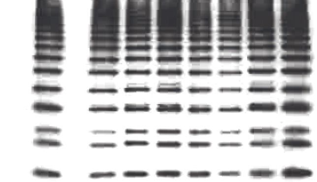
Research article                                                                                                       Biochemistry and Chemical Biology

    A                                                            B

                                                                                                                      Û

                                                                                                       Û                                     Û

                                                                                                                      Û

    C                                                            D

                                                                         Probability density                  II      Weighted average

                                                                                                                     III

                                                                                                   I

                                                                                               0       200         400      600      800      1000
      2D Class       Model         2D Class        Model                                                           Mass (kDa)
                   projection                    projection

Figure 2. First low-resolution 3D map of linear ubiquitin chain assembly complex (LUBAC) obtained by negative staining electron microscopy of the
recombinant complex. (A) Representative negative stain transmission electron micrograph of recombinant LUBAC. Scale bar: 100 nm. (B) 3D refined
model of LUBAC obtained by single particle analysis of negative stained electron micrographs. (C) LUBAC 2D class averages matched to projections
made from 3D refined map. (D) Mass photometry measurements of LUBAC indicate formation of a ternary complex with 1:1:1 stoichiometry that can
form dimers.
The online version of this article includes the following figure supplement(s) for figure 2:
Figure supplement 1. Modelling of the linear ubiquitin chain assembly complex (LUBAC) by negative staining electron microscopy.
Figure supplement 2. Projections made from 3D refined model of linear ubiquitin chain assembly complex (LUBAC).
Figure supplement 3. Independent mass photometry measurements of linear ubiquitin chain assembly complex (LUBAC).

Rodriguez Carvajal et al. eLife 2021;10:e60660. DOI: https://doi.org/10.7554/eLife.60660                                                                    4 of 28
Research article                                                                               Biochemistry and Chemical Biology

                                    mass photometry (MP), a technique that enables accurate mass measurements based on the scatter-
                                    ing of light by single macromolecules in solution (Young et al., 2018; Sonn-Segev, 2019; Figure 2D
                                    and Figure 2—figure supplement 3). MP measurements showed that the majority of species pres-
                                    ent in the samples had average masses of 231 kDa (peak II) and 454 kDa (peak III)
                                    (Supplementary file 1). An additional peak originating from a population with mass of less than 100
                                    kDa was also measured (peak I), which could arise from isolated HOIL-1L or SHARPIN present in the
                                    measured sample.
                                       With respect to the expected mass of the ternary LUBAC, the populations of peaks II and III
                                    nearly correspond with monomers (222 kDa) and dimers (444 kDa) of LUBAC with a 1:1:1 stoichiom-
                                    etry between HOIP, HOIL-1L, and SHARPIN.

                                    The RBR domains of HOIP and HOIL-1L are in close proximity
                                    Understanding the precise mechanistic action of HOIL-1L and SHARPIN within LUBAC requires
                                    knowledge of structural and functional domain interactions between the three components. Current
                                    knowledge of the interaction domains of HOIP, HOIL-1L, and SHARPIN is shown in Figure 3A. Struc-
                                    tural work of protein fragments has shown that HOIL-1L and SHARPIN interact with HOIP through
                                    their respective ubiquitin-like (UBL) domains, which bind cooperatively to the HOIP UBA domain
                                    (Fujita et al., 2018; Liu et al., 2017; Yagi et al., 2012; Figure 3A). By using truncation mutants, it
                                    has also been shown that the SHARPIN UBL domain interacts with the Npl zinc finger 2 (NZF2)
                                    domain of HOIP (Ikeda et al., 2011). More recently, structural work has revealed that SHARPIN and
                                    HOIL-1L interact with each other through their respective LUBAC-tethering motifs (LTMs)
                                    (Fujita et al., 2018). Otherwise, there is no further information available about the overall spatial
                                    arrangement of the domains of HOIP, HOIL-1L, and SHARPIN.
                                       To obtain more detailed information about the spatial arrangement of LUBAC components, we
                                    performed cross-linking mass spectrometry (XL-MS) experiments. For this purpose, we used the
                                    amine-to-carboxyl-reactive cross-linker, 4-(4,6-dimethoxy-1,3,5-triazin-2-yl)-4-methylmorpholinium
                                    tetrafluoroborate (DMTMM), a zero-length cross-linker (Leitner et al., 2014) revealing Lys and Asp/
                                    Glu contacts that are adjacent to each other. As shown in Figure 3B–E, we observed an extensive
                                    network of intra-protein and inter-protein cross-links, providing a detailed picture of LUBAC assem-
                                    bly (Figure 3B–E, Supplementary files 2 and 3).
                                       We detected some highly cross-linking residues that formed cross-links indiscriminately to all sub-
                                    units of all proteins. These were HOIL-1L K174, SHARPIN K318, and HOIP K454/458. The high
                                    degree of cross-linking formed by these residues suggests that they are located on flexible regions.
                                    This notion is supported by the cleavage of HOIL-1L in the vicinity of K174 by MALT1, which sug-
                                    gests that the residue is located on a protease-accessible structure such as a flexible loop
                                    (Elton et al., 2016). The presence of flexible regions on the three LUBAC components may be
                                    related to the difficulties in determining the structures of full-length HOIP, HOIL-1L, and SHARPIN
                                    thus far.
                                       We also observed DMTMM cross-links between LUBAC domains known to interact, such as
                                    between the HOIL-1L UBL and the SHARPIN UBL/LTM domains as well as between the HOIP UBA
                                    and HOIL-1L LTM domains, several cross-links pointed to new connections between the engaged
                                    proteins. Interestingly, we observed cross-links between the HOIL-1L RING1 and HOIP RING1/LDD
                                    domains, as well as between the HOIL-1L RING2 and the HOIP RING1/IBR/RING2/LDD domains,
                                    which suggest that the two enzymes have spatially connected catalytic activities. Additionally, HOIL-
                                    1L intra-protein cross-links were formed between its NZF domain and its RING1/IBR/RING2 domains,
                                    which could implicate the HOIL-1L NZF domain of unknown function in the catalytic action of HOIL-
                                    1L. In conclusion, the catalytic RBR domains of HOIP and HOIL1L seem to be close to each other, as
                                    well as the NZF and RBR regions of HOIL-1L. These data suggest that LUBAC may have a single cat-
                                    alytic centre containing the RBR domains of HOIP and HOIL-1L.

                                    Recombinant LUBAC assembles heterotypic ubiquitin chains containing
                                    linear and non-Lys linkages
                                    To assess the ability of recombinant LUBAC to assemble linear ubiquitin chains, we performed in
                                    vitro ubiquitination assays. As expected, LUBAC generated linear ubiquitin chains in an ATP-depen-
                                    dent manner (Figure 4A). We also observed additional signals derived from co-purified LUBAC,

Rodriguez Carvajal et al. eLife 2021;10:e60660. DOI: https://doi.org/10.7554/eLife.60660                                            5 of 28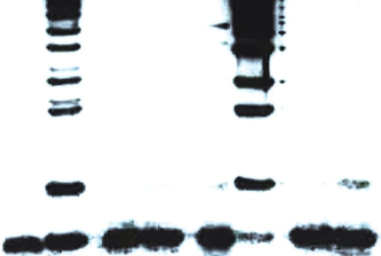
Research article Biochemistry and Chemical Biology
A HOIP
1 1072
PUB ZF NZF1 NZF2 UBA RING1 IBR RING2 LDD
1 377 1 506
PH LTM UBL NZF LTM UBL NZF RING1 IBR RING2
SHARPIN HOIL-1L
B SHARPIN 0
C
250
Domain span HOIL-1L
0
25
1000
0
Crosslink position HOIP
500 800
Domain span HOIP
600
500
400
HOIP
0 200
25
HOIL-1L
75
0
0 100 200 300 400 500
0
0
Crosslink position HOIL-1L
1000
Inter-protein crosslinks: HOIL-1L—HOIP
Inter-protein crosslinks: HOIL-1L—SHARPIN
Inter-protein crosslinks: HOIP—SHARPIN
Intra-protein crosslinks
D Domain span HOIL-1L
E Domain span HOIP
Crosslink Position SHARPIN
Crosslink Position SHARPIN
300 300
Domain span SHARPIN
Domain span SHARPIN
200 200
100 100
0 0
0 100 200 300 400 500 0 200 400 600 800 1000
Crosslink position HOIL-1L Crosslink Position HOIP
Figure 3. Cross-linking mass spectrometry (MS) analysis shows proximity between the catalytic domains of HOIL-1-interacting protein (HOIP) and heme-
oxidized IRP2 ubiquitin ligase 1 (HOIL-1L). (A) Schematic representation of linear ubiquitin chain assembly complex (LUBAC) components with their
domains and known interactions. (B) Circos plot of inter-protein cross-links formed between LUBAC components. (C) Detected inter-protein cross-links
Figure 3 continued on next page
Rodriguez Carvajal et al. eLife 2021;10:e60660. DOI: https://doi.org/10.7554/eLife.60660 6 of 28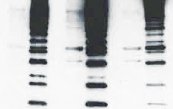
Research article                                                                                  Biochemistry and Chemical Biology

Figure 3 continued
formed between HOIL-1L and HOIP. (D) Detected inter-protein cross-links formed between HOIL-1L and Shank-associated RH domain-interacting
protein (SHARPIN). (E) Detected inter-protein cross-links formed between HOIP and SHARPIN.

                                    which were absent from reactions containing the three individually purified and mixed LUBAC com-
                                    ponents (Figure 4B: red arrows).
                                        The exclusive appearance of extra bands in the co-purified LUBAC sample indicates that the com-
                                    plex does not attain its full catalytic activity in vitro when the three LUBAC components are mixed.
                                    One possible explanation is that mixing of the three components is sufficient to activate HOIP but
                                    may not suffice for LUBAC to be adequately assembled. To determine if the mixed components
                                    could assemble the trimeric LUBAC, we carried out MP measurements of the three components
                                    mixed at an equimolar ratio (Figure 4—figure supplement 2). Our measurements showed a single
                                    prominent population of molecules with an average mass of 104 kDa. However, we were unable to
                                    detect any populations with the expected masses of 222 or 444 kDa corresponding to assembled
                                    and dimerized LUBAC. The absence of these populations indicates that mixing HOIP, HOIL-1L, and
                                    SHARPIN does not suffice to assemble LUBAC. We therefore conclude that the absence of bands in
                                    reactions carried out using mixed LUBAC components is a result of improper LUBAC assembly pre-
                                    venting the full catalytic activity of the complex.
                                        Ubiquitin chains containing different linkages resolve at different apparent molecular masses by
                                    SDS-PAGE separation (Emmerich and Cohen, 2015). Therefore, we hypothesized that the two
                                    bands in each pair contained different ubiquitin linkages. To examine the presence of different Lys-
                                    linked bonds in the heterotypic chains, we carried out in vitro ubiquitination assays using different
                                    ubiquitin Lys to Arg (KR) mutants (Figure 4C). Heterotypic ubiquitin chain assembly was observed
                                    with all the tested mutants, K6R, K11R, K27R, K29R, K33R, K48R, and K63R. To rule out the possibil-
                                    ity that mutation of a single Lys could be compensated by ubiquitination of a separate Lys residue,
                                    we also performed in vitro ubiquitination assay using ubiquitin lacking all Lys residues (K0)
                                    (Figure 4D). Despite reduced reaction efficiency, the K0 mutant could also be used by LUBAC to
                                    generate the heterotypic ubiquitin chains. At present we cannot account for the apparent difference
                                    in the ratio between branched tri- and tetra-ubiquitin chains observed across different experiments,
                                    and which is particularly pronounced with the K0 substrate. However, we speculate that these differ-
                                    ences arise from varied reaction efficiencies. We propose this is the case given that branched tri-
                                    ubiquitin is the more prominent species when K0 ubiquitin is used. With this mutant we also observe
                                    a drastic loss of overall reaction efficiency with respect to reactions containing wild-type (WT) ubiqui-
                                    tin. Nevertheless, both branched chains could be detected to some extent across all replicates of all
                                    experiments.
                                        To further analyse the linkage types of ubiquitin chains, we performed ubiquitin chain restriction
                                    (UbiCRest) (Hospenthal et al., 2015) on the ubiquitin chains generated by LUBAC (Figure 4E).
                                    UbiCRest using linkage-specific deubiquitinases (DUBs) allows detection of specific ubiquitin chains
                                    by loss of signals when the linkage is targeted. Interestingly, the additional signals from ubiquitin
                                    chains assembled by LUBAC disappeared by treatment with Cezanne, a DUB specific for the
                                    Lys11 linkage (Bremm et al., 2010); or vOTU, a DUB targeting Lys linkages (Akutsu et al., 2011;
                                    Figure 4E; upper red arrow). The linear linkage-specific DUB OTULIN (Fiil et al., 2013) hydrolysed
                                    all linear ubiquitin bonds (Figure 4E: upper panel), corresponding to most of the ubiquitin signal,
                                    but left non-linear di- and tri-ubiquitin residues (Figure 4E: lower panel).
                                        These data collectively indicate that recombinant LUBAC assembles heterotypic ubiquitin chains
                                    containing predominantly linear linkage with non-Lys-linked branches.

                                    LUBAC assembles heterotypic poly-ubiquitin chains containing linear
                                    and ester-linked bonds in vitro and in cells
                                    A recent study showed that recombinant HOIL-1L can generate di-ubiquitin linked via an oxyester
                                    bond in vitro and can also ubiquitinate substrates through oxyester bonds in cells (Kelsall et al.,
                                    2019). Therefore, we tested for the presence of oxyester bonds in the ubiquitin chains by checking
                                    their sensitivity to the nucleophile hydroxylamine (Figure 5A). Treatment with hydroxylamine
                                    resulted in the disappearance of the upper band of the linear tetra-ubiquitin chain (Figure 5A: red

Rodriguez Carvajal et al. eLife 2021;10:e60660. DOI: https://doi.org/10.7554/eLife.60660                                                7 of 28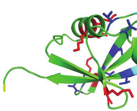
Research article Biochemistry and Chemical Biology
A ATP: - + B
0.25h
0.25h
LUBAC: Mixed Co-purified
0.5h
0.5h
0h
1h
2h
4h
0h
1h
2h
4h
ATP: - + - +
kDa
250- kDa
150- 250-
100- 150-
75- M1 100- M1
UbN 75- UbN
50-
50-
37-
M1 37-
Ub4 M1
Ub4
25- M1 M1
Ub3 25- Ub3
IB: Linear Poly-Ubiquitin IB: Linear Poly-Ubiquitin
150-
100-
75- 100-
UbN
50- IB: HOIP
37-
Ub4
50-
25- Ub3 IB: HOIL-1L
20-
50-
15- Ub2
IB: SHARPIN
10- Ub1
IB: Ubiquitin
C E
ATP: - + ATP: - +
Cezanne
K29/33 TRABID
OTULIN
OTUD1
OTUD2
OTUD3
OTUB1
Usp21
All -M1 vOTU
- -
K27R
K29R
K33R
K48R
K63R
Ubiquitin: DUB:
K11R
K6R
WT
WT
kDa
100- Target: - -
K63
K27
K48
K11
M1
K6
All
75-
M1 kDa
UbN 250-
50- 150-
100- M1
37- 75- UbN
M1 50-
Ub4
25- M1 37-
Ub3 M1
IB: Linear Poly-Ubiquitin Ub4
25- M1
Ub3
IB: Linear Poly-Ubiquitin
D
0h
2h
4h
0h
2h
4h
kDa 250-
75- 150-
100-
UbN 75- UbN
50-
50-
37-
Ub4 37-
Ub4
25- Ub3
25- Ub3
20-
20-
15- Ub2 15- Ub2
10- Ub1
10- Ub1
Short exposure Long exposure
IB: Ubiquitin
IB: Ubiquitin
Figure 4. Linear ubiquitin chain assembly complex (LUBAC) assembles heterotypic poly-ubiquitin chains containing M1 and non-Lys linkages in vitro.
(A) Time course of co-purified LUBAC in vitro ubiquitin chain assembly reaction. (B) Comparison of in vitro chain assembly between HOIL-1-interacting
protein (HOIP), heme-oxidized IRP2 ubiquitin ligase 1 (HOIL-1L), and Shank-associated RH domain-interacting protein (SHARPIN) mixed at 1:1:1 molar
ratio versus co-purified LUBAC. (C) LUBAC in vitro chain assembly using different ubiquitin K to R mutants. (D) LUBAC in vitro chain assembly using K0
Figure 4 continued on next page
Rodriguez Carvajal et al. eLife 2021;10:e60660. DOI: https://doi.org/10.7554/eLife.60660 8 of 28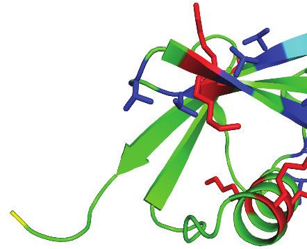
Research article                                                                                          Biochemistry and Chemical Biology

Figure 4 continued
ubiquitin. (E) Ubiquitin chain restriction (UbiCRest) analysis of poly-ubiquitin chains assembled by LUBAC in vitro. All experiments were performed in
triplicate representative results are shown.
The online version of this article includes the following figure supplement(s) for figure 4:
Figure supplement 1. Anti-linear ubiquitin antibody validation.
Figure supplement 2. Independently purified HOIL-1-interacting protein (HOIP), heme-oxidized IRP2 ubiquitin ligase 1 (HOIL-1L), and Shank-
associated RH domain-interacting protein (SHARPIN) mixed at an equimolar ratio cannot reconstitute the trimeric linear ubiquitin chain assembly
complex (LUBAC).

                                     arrow). This indicates that the chain branching is achieved by formation of an oxyester bond
                                     between ubiquitin moieties similar to the observation in the previous study (Kelsall et al., 2019).
                                         To further ascertain whether LUBAC assembles ester-linked ubiquitin polymers, we carried out in
                                     vitro ubiquitination assays using N-terminally His6-tagged ubiquitin, which cannot be used to assem-
                                     ble linear ubiquitin chains (Kirisako et al., 2006). LUBAC inefficiently assembled di- and tri-ubiquitin
                                     chains with His-ubiquitin, which are sensitive to hydroxylamine treatment (Figure 5B). These results
                                     demonstrate that when the N-terminus of ubiquitin is not available, LUBAC can still assemble oxy-
                                     ester-linked poly-ubiquitin even in the absence of linear ubiquitin chains.
                                         To determine if LUBAC was generating any other kind of ubiquitin linkage other than linear and
                                     oxyester bonds, we treated LUBAC-assembled ubiquitin chains with OTULIN and hydroxylamine
                                     (Figure 5—figure supplement 1A). Restriction with OTULIN degraded the majority of the signal
                                     from ubiquitin chains leaving short di- and tri-ubiquitin polymers. Subsequent treatment with hydrox-
                                     ylamine resulted in complete degradation of the poly-ubiquitin signals. These results indicate that
                                     LUBAC assembles exclusively OTULIN-sensitive linear and hydroxylamine-sensitive oxyester linkages
                                     between ubiquitin moieties.
                                         In our UbiCRest analysis we observed cleavage of the oxyester-linked branches from the LUBAC-
                                     assembled poly-ubiquitin chains by Cezanne and vOTU (Figure 4E). Therefore, we also tested the
                                     sensitivity of the exclusively oxyester-linked ubiquitin chains assembled by LUBAC with His6-ubiquitin
                                     to the two DUBs (Figure 5—figure supplement 1B). Both Cezanne and vOTU could fully degrade
                                     these short oxy-ester ubiquitin polymers, which is in agreement with a recent study reporting the
                                     ester-directed activity of the two DUBs (De Cesare et al., 2021).
                                         Ubiquitin contains seven Thr residues, three Ser residues, and one Tyr residue, which could theo-
                                     retically act as sites for ester bond formation. Therefore, we aimed to identify the positions of the
                                     ester-linked branches using mass spectrometry by searching for GG dipeptides covalently attached
                                     through oxyester bonds to Ser, Thr, or Tyr, as was previously done by Kelsall et al., 2019. To this
                                     end, we analysed ubiquitin chains formed by LUBAC by LC-MS. MS/MS spectra of GG-conjugated
                                     dipeptides at residues Thr12 and Thr55 of ubiquitin were detected from these samples, in which
                                     there was complete coverage of the ubiquitin amino acid sequence 1–73 (Figure 5C). Structural
                                     analysis shows that these two residues are positioned at opposite sides of ubiquitin and neither of
                                     them is located in proximity to Met1 or the C-terminal Gly76 (Figure 5D; PDB:1UBI). This suggests
                                     that branches could potentially exist on both sites of a single ubiquitin molecule located at any posi-
                                     tion of a linear ubiquitin chain without creating any steric hindrances.
                                         To further investigate if Thr12 and Thr55 are the sites of oxyester bond formation, we performed
                                     in vitro ubiquitination assays using ubiquitin mutated at Thr12 and/or Thr55 to Val (Figure 5E). Indi-
                                     vidual or concomitant mutation of Thr12 and Thr55 (Ub T12V, Ub T55V, or Ub T12/55V, respectively)
                                     did not result in loss of chain branching. These results suggest that linear chain branching is not a
                                     strictly site-specific event. We next probed the existence of the hybrid chains in cells. It has recently
                                     been reported that these chains are formed on IRAK1, IRAK2, and Myd88 in response to activation
                                     of TLR signalling (Kelsall et al., 2019). Given that LUBAC is involved in the TNF signalling cascade,
                                     we examined the induction of these heterotypic ubiquitin chains in TNF-treated cells. Linear ubiqui-
                                     tin chains were enriched by GST pull-down using the NEMO-UBAN and zinc finger domains
                                     (NEMO250-412) from total cell extracts of TNF stimulated mouse embryonic fibroblasts (MEFs). In this
                                     set-up, the GST-NEMO-UBAN was employed as a ubiquitin-binding matrix to enrich linear-linked
                                     chains and not as a substrate for LUBAC. The enriched poly-ubiquitin chains were then tested for
                                     hydroxylamine sensitivity (Figure 6A). TNF stimulation increased formation of linear ubiquitin chains
                                     detected by immunoblotting using an antibody specific for linear ubiquitin chains (Figure 6A: PD

Rodriguez Carvajal et al. eLife 2021;10:e60660. DOI: https://doi.org/10.7554/eLife.60660                                                          9 of 28
Research article Biochemistry and Chemical Biology
A B ATP: - +
NH2OH: - + - NH2OH: - + - +
Cezanne: - - + kDa
ATP: - + - + - + 37-
kDa
50-
25- Ub3
37-
M1 20-
Ub4
15- Ub2
25- M1
Ub3
IB: Linear Poly-Ubiquitin 10- Ub1
50-
IB: Ubiquitin
37-
Ub4
D Thr12
25- Ub3 Met1 Thr55
20-
15-
Ub2 Û
10- Ub1 Gly76
IB: Ubiquitin Gly76
Lys Ser Thr Met1 Gly76
C b1+ b2+ b3+ b4+ b6+ b7+ b8+ b9+ E
T L S D Y N I Q K Ub: WT T12V T55V T12/55V
+ + + + + + + + ATP: - + - + - + - +
LRGG y8 y7 y6 y5 y4 y3 y2 y1 kDa
30 b1+ 50-
y1+
20 y4+ y5+ + +
y2+b32+ b y 37-
b2+ 3 6 b4+ M1
y3+ y 7+ Ub4
10
M1
25- Ub3
Intensity [counts] (103)
b15+
0 IB: Linear Poly-Ubiquitin
V K
00
00
00
0
0
0
0
y2+ y +
20
40
60
80
10
12
14
1
37-
b1+ b2+ b3+ b4+ b6+ b7+ b8+ b9+b10+ b11+b12+b13+ b14+
Ub4
T I T L E V E P S D T E N 25-
+ + + + + + + + + + + +
GG y14 y13 y12 y11 y10 y9 y8 y7 y6 y5 y4 y3 Ub3
90 +
20-
y 9
b2+y +
15-
60 y2+ 3 Ub2
b3+ y + y10+
b1+ y4+ 5 10-
30 y1+ y11+ y12+ Ub1
y6+y7+ y13+ y + IB: Ubiquitin
14
0
00
00
0
50
10
15
m/z
Figure 5. Linear ubiquitin chain assembly complex (LUBAC) assembles heterotypic poly-ubiquitin chains containing M1 and ester bond linkages at T12
and T55. (A) Treatment of LUBAC-assembled heterotypic poly-ubiquitin chains with hydroxylamine. (B) Hydroxylamine treatment of ubiquitin polymers
assembled by LUBAC using N-terminally blocked ubiquitin. (C) MS/MS spectra of ubiquitin polymerized at T55 (top) and T12 (bottom). Poly-ubiquitin
chains assembled by LUBAC were separated by SDS-PAGE, bands were cut from the gel and subjected to mass spectrometry analysis. (D) Positions of
Thr12 and Thr55 on structure of ubiquitin (PDB:1UBI). (E) Assembly of ubiquitin chains by LUBAC using different ubiquitin Thr to Val point mutants as
substrates. All experiments were performed in triplicate representative results are shown.
The online version of this article includes the following figure supplement(s) for figure 5:
Figure supplement 1. Cezanne, vOTU, and hydroxylamine can cleave oxyester bonds in linear ubiquitin chain assembly complex (LUBAC)-assembled
heterotypic ubiquitin chains.
Rodriguez Carvajal et al. eLife 2021;10:e60660. DOI: https://doi.org/10.7554/eLife.60660 10 of 28Research article Biochemistry and Chemical Biology
A
GST empty GST-NEMO(250-412)
TNF_: - + - +
NH2OH: - + - + - + - +
kDa
250-
150-
100-
75-
PD 50-
IB: Linear Poly-Ubiquitin
250-
150-
100-
75-
50-
IB: Ubiquitin
PonceauS
GST empty GST-NEMO(250-412)
TNF_: - + - +
NH2OH: - + - + - + - +
kDa
Input 37-
IB: IgB_
150-
100-
IB: Vinculin
B
ATP: - - + +
NH2OH: - + - +
kDa
250-
150-
NEMO-UbN
100-
75-
50- NEMO
IB: NEMO
Figure 6. M1-linked/linear ubiquitin chains generated in cells and conjugated to NEMO in vitro are sensitive to
hydroxylamine treatment. (A) Hydroxylamine treatment of M1 linkage-containing poly-ubiquitin chains assembled
in response to TNF in wild-type (WT) mouse embryonic fibroblasts (MEFs). MEFs were treated with TNF for 15 min
Figure 6 continued on next page
Rodriguez Carvajal et al. eLife 2021;10:e60660. DOI: https://doi.org/10.7554/eLife.60660 11 of 28Research article Biochemistry and Chemical Biology
Figure 6 continued
and lysed, lysates were subjected to GST PD using GST or GST-NEMO(250-412), beads were treated with buffer or
hydroxylamine for 30 min, bound ubiquitin species were then analysed by immunoblotting. (B) Treatment of linear
ubiquitin chain assembly complex (LUBAC)-dependent NEMO in vitro ubiquitination reactions with hydroxylamine.
All experiments were performed in triplicate representative results are shown.
top panel lane 7). These chains proved sensitive to hydroxylamine treatment (Figure 6A: PD top
panel lane 8). In contrast, the signals from the overall population of ubiquitin chains are unchanged
by either TNF stimulation or hydroxylamine treatment (Figure 6A: PD bottom panel lanes 5–8).
Together these results suggest that ubiquitin chains containing both linear linkages and hydroxyl-
amine-sensitive ester bonds are produced in cells in response to TNF stimulation.
To determine if substrate-conjugated linear-linked ubiquitin chains are also branched, we carried
out an in vitro ubiquitination assay using NEMO as the substrate then subjected the products to
hydroxylamine treatment (Figure 6B). Treatment of ubiquitinated NEMO with the compound
resulted in enrichment of some lower molecular weight bands corresponding to ubiquitinated
NEMO when blotting with a NEMO-specific antibody. However, the majority of the signal from the
ubiquitinated NEMO was unperturbed. These results suggest that the conjugation of ubiquitin
chains to the substrate is principally through hydroxylamine-resistant isopeptide bonds with some
hydroxylamine-sensitive chain branching or substrate ubiquitination also taking place on longer lin-
ear chains.
HOIP assembles linear ubiquitin chains that are subsequently branched
with ester bonds by HOIL-1L
We next proceeded to probe how HOIP and HOIL-1L generate these heterotypic ubiquitin chains.
HOIP specifically assembles linear ubiquitin chains through the action of its RBR domains wherein
Cys885 is the catalytic residue (Smit et al., 2012; Stieglitz et al., 2013); similarly HOIL-1L catalyses
ester bond-directed ubiquitination via its RBR domain where Cys460 is the catalytic site
(Kelsall et al., 2019; Smit et al., 2013). Therefore, we examined the possibility that HOIP assembles
a linear ubiquitin chain, which is subsequently branched with ester bonds by HOIL-1L. To this end,
we purified different LUBAC containing catalytically inactive HOIP (HOIP C885A), HOIL-1L (HOIL-1L
C460A), or both HOIP C885A and HOIL-1L C460A. Mutation of these residues did not impair the
purification of LUBAC, and all variants of the complex could be isolated at similar yields and to simi-
lar degrees of purity (Figure 7—figure supplement 1). In line with our hypothesis, LUBAC contain-
ing HOIL-1L C460A generated chains with linear linkages, yet the double band indicative of
heterotypic chain assembly was absent (Figure 7A; lane 6 upper red arrow). These observations indi-
cate that HOIL-1L is responsible for the formation of the ester-linked branches. Consistent with pre-
vious reports (Smit et al., 2013; Fuseya et al., 2020), HOIP assembled longer linear ubiquitin chains
in the absence of HOIL-1L catalytic activity and did so more rapidly (Figure 7A: lanes 2 and 6). Con-
versely, LUBAC containing HOIP C885A was incapable of polymerizing ubiquitin altogether
(Figure 7A; lanes 4 and 8) as expected. These results suggest that HOIL-1L catalytic activity disturbs
linear ubiquitin chain formation but requires HOIP catalytic activity.
The gel-migration pattern differed between ubiquitin chains assembled by LUBAC with HOIL-1L
C460A and the WT complex (Figure 7A: lanes 2 and 6). Therefore, we compared the presence of
non-linear bonds in the chains assembled by the different complexes by OTULIN. The restriction
analyses showed that the ubiquitin chains assembled by LUBAC contained non-linear di- and tri-
ubiquitin chains, whereas chains assembled by HOIL-1L C460A-containing LUBAC generated exclu-
sively OTULIN-sensitive linear ubiquitin chains (Figure 7B). These results further support the claim
that chain branching is dependent on the catalytic action of HOIL-1L while HOIP exclusively assem-
bles linear ubiquitin chains.
HOIL-1L catalytic activity is required for branched ubiquitin chain
formation in cells
To further study the catalytic function of HOIL-1L in cells, we used cells derived from a HOIL-1L
C458A knock-in mouse (Rbck1C458A/C458A) generated by CRISPR/Cas9 gene-editing technology (Fig-
ure 7—figure supplement 2A–C). The expression levels of the three LUBAC components are similar
Rodriguez Carvajal et al. eLife 2021;10:e60660. DOI: https://doi.org/10.7554/eLife.60660 12 of 28Research article Biochemistry and Chemical Biology
A HOIP: WT C885A WT C885A B ATP: - +
HOIL-1L: WT WT C460A C460A HOIL-1L: WT C460A WT C460A
ATP: - + - + - + - + OTULIN: - + - + - + - +
kDa 50- kDa
50-
37- 37-
M1
Ub4 M1
Ub4
M1
25- Ub3
IB: Linear Poly-Ubiquitin 25- M1
75- Ub3
IB: Linear Poly-Ubiquitin
50- 75-
37- 50-
Ub4
37-
Ub3 Ub4
25-
20- 25- Ub3
20-
15- Ub2
15- Ub2
10- Ub1
10- Ub1
IB: Ubiquitin IB: Ubiquitin
C Rbck1+/+ Rbck1C458A/C458A D TR C
TNF: - + - + 1 506
NH2OH: - + - + - + - + WT
kDa AA C
250- T203A
150- R210A
100- TR A
75- C460A
LTM UBL NZF RING1 IBR RING2
PD 50- E T203A
IB: Linear Poly-Ubiquitin HOIL-1L: WT R210A C460A
ATP: - + - + - +
kDa
100-
250-
150- 75- M1
UbN
50-
100-
75- 37-
M1
Ub4
50-
IB: Ubiquitin 25- M1
Ub3
IB: Linear Poly-Ubiquitin
100-
75-
UbN
50-
PonceauS
Rbck1+/+ Rbck1C458A/C458A 37-
- - Ub4
TNF: + +
NH2OH: - + - + - + - + 25- Ub3
kDa
37-
20-
Input
IB: IgB_
15- Ub2
150-
100- 10- Ub1
IB: Vinculin IB: Ubiquitin
50-
IB: HOIL-1L
Figure 7. Heme-oxidized IRP2 ubiquitin ligase 1 (HOIL-1L) generates ester linkages on heterotypic chains but requires HOIL-1-interacting protein
(HOIP) catalytic activity to polymerize ubiquitin. (A) Comparison of linear ubiquitin chain assembly complex (LUBAC) in vitro chain assembly by
complexes containing different catalytically inert mutants of HOIP and HOIL-1L. (B) OTULIN restriction of poly-ubiquitin chains assembled by LUBAC
containing wild-type (WT) or catalytically inert HOIL-1L. (C) Hydroxylamine treatment of M1 linkage-containing poly-ubiquitin chains assembled in
Figure 7 continued on next page
Rodriguez Carvajal et al. eLife 2021;10:e60660. DOI: https://doi.org/10.7554/eLife.60660 13 of 28Research article Biochemistry and Chemical Biology
Figure 7 continued
response to TNF in WT and Rbck1C458A/C458A mouse embryonic fibroblast (MEF) cells. Cells were treated with TNF (50 ng/ml) for 15 min and lysed,
lysates were subjected to GST PD using GST-NEMO (250-412), beads were treated with buffer or hydroxylamine for 30 min, bound ubiquitin species
were then analysed by immunoblotting. (D) Schematic representations of different HOIL-1L mutants. (E) Comparison of LUBAC in vitro chain assembly
by complexes containing different HOIL-1L mutants. All experiments were performed in triplicate representative results are shown.
The online version of this article includes the following figure supplement(s) for figure 7:
Figure supplement 1. Purification of linear ubiquitin chain assembly complex (LUBAC) containing catalytically inert HOIL-1-interacting protein (HOIP)
and heme-oxidized IRP2 ubiquitin ligase 1 (HOIL-1L) proteins.
Figure supplement 2. Generation of Hoil-1lC458A/C458A mice.
in MEFs in Rbck1+/+ and Rbck1C458A/C458A MEFs (Figure 7—figure supplement 2D). In agreement
with numerous studies from the past, two species of HOIL-1L were detected in Rbck1+/+ MEFs by
immunoblotting; the slower migrating species originating from auto-ubiquitinated HOIL-1L was
absent in Rbck1C458A/C458A MEFs.
To determine if HOIL-1L is the responsible ligase for linear ubiquitin chain branching during TNF
signalling, TNF-treated Rbck1C458A/C458A MEF lysates were subjected to GST pull-down with GST-
NEMO250-412 followed by hydroxylamine treatment (Figure 7C). Similarly, to WT MEFs, TNF stimula-
tion led to an increase in the levels of linear ubiquitin chains in Rbck1C458A/C458A MEFs (Figure 7C:
PD top panel lanes 3 and 7). However, the enriched ubiquitin signals detected by immunoblotting
revealed a higher basal level of linear ubiquitin chains in Rbck1C458A/C458A MEFs compared to those
in Rbck1+/+ MEFs (Figure 7C: PD top panel lanes 1 and 5). Moreover, unlike in Rbck1+/+ MEFs, lin-
ear ubiquitin chains in Rbck1C458A/C458A MEFs were not sensitive to degradation by hydroxylamine
treatment (Figure 7C: PD bottom panel). However, there was no obvious difference when blotting
for general ubiquitin between Rbck1+/+ and Rbck1C458A/C458A MEFs regardless of TNF stimulation or
chain hydroxylamine treatment (Figure 7C: PD bottom panel). These results show that ester-linked
branching of linear ubiquitin chains formed during TNF signalling in MEFs is dependent on the cata-
lytic activity of HOIL-1L.
HOIL-1L Npl4 zinc finger is involved in the formation of branching
ubiquitin chains in vitro
Since the catalytic action of HOIP precedes that of HOIL-1L and the assembly of linear ubiquitin
chains precedes the appearance of the branches, we hypothesized that HOIL-1L interacts with a lin-
ear ubiquitin chain as a substrate for branching via its linear ubiquitin chain-specific binding domain
NZF (Sato et al., 2011). To test this possibility, we purified LUBAC containing a mutant in which crit-
ical residues for linear ubiquitin chain recognition are mutated (HOIL-1L T203A,R210A) (Figure 7D).
In agreement with our hypothesis, chain branching activity by LUBAC was partially impaired by the
HOIL-1L T203A,R210A mutant when compared to the WT or HOIL-1L C460A (Figure 7E). Interest-
ingly, LUBAC with HOIL-1L T203A,R210A assembled ubiquitin chains more efficiently than WT
LUBAC but less efficiently than HOIL-1L C460 (Figure 7E).
These data collectively show that HOIP assembles linear ubiquitin chains, which are subsequently
branched by HOIL-1L in a process involving its NZF domain and which requires the catalytic activity
of HOIP.
In summary, we identified that LUBAC assembles heterotypic linear/ester-linked poly-ubiquitin
chains in vitro and in cells in response to TNF stimulation. We also show that these chains are synthe-
sized through the concerted action of HOIP and HOIL-1L (Figure 8). These chains may contribute in
modulating the speed and/or efficiency of linear ubiquitin chain synthesis by LUBAC.
Discussion
We present here the first 3D reconstruction of the ternary LUBAC. We cannot determine the exact
positions of HOIP, HOIL-1L, and SHARPIN in the map at the current resolution; however, our map is
the first structure encompassing the LUBAC holoenzyme. We also made other novel structural obser-
vations that LUBAC exists as monomers and dimers of a ternary complex with 1:1:1 stoichiometry
between HOIP, HOIL-1L, and SHARPIN. This is in agreement with observations made in recent struc-
tural work (Fujita et al., 2018). We also present new data addressing the question of LUBAC
Rodriguez Carvajal et al. eLife 2021;10:e60660. DOI: https://doi.org/10.7554/eLife.60660 14 of 28Research article Biochemistry and Chemical Biology
A WT HOIL-1L HOIL-1L C460A
HOIP HOIP
RING1 RING1
IBR IBR
RING2 RING2
RING1 RING1
NZF IBR NZF IBR
RING2 RING2
Sharpin HOIL-1L Sharpin HOIL-1L
RING1 RING1
IBR IBR
RING2 RING2
RING1 RING1
NZF IBR NZF IBR
RING2 RING2
RING1 RING1
IBR IBR
RING2 RING2
RING1 RING1
NZF IBR NZF IBR
RING2 RING2
RING1 Thioester-linked ubiquitin
IBR
RING2
RING1
NZF IBR Met1-linked ubiquitin
RING2
Ester-linked ubiquitin
Figure 8. Proposed model for concerted action between HOIL-1-interacting protein (HOIP) and heme-oxidized IRP2 ubiquitin ligase 1 (HOIL-1L) in
heterotypic chain assembly. HOIP and HOIL-1L assemble heterotypic chains through a Cys relay mechanism. HOIP forms a thioester bond to ubiquitin,
which can be either transferred to a thioester bond on HOIL-1L or added to a nascent linear ubiquitin chain. HOIL-1L subsequently binds the linear
ubiquitin chain through its Npl zinc finger (NZF) domain and branches it with ester linkages. The resulting heterotypic poly-ubiquitin chains contain
predominantly linear linkages with ester-linked branches.
Rodriguez Carvajal et al. eLife 2021;10:e60660. DOI: https://doi.org/10.7554/eLife.60660 15 of 28Research article Biochemistry and Chemical Biology
oligomerization. Early gel filtration analysis of cell-derived LUBAC suggested formation of a large or
oligomeric complex with a molecular mass of over 600 kDa (Kirisako et al., 2006). However, our MP
measurements using recombinant LUBAC show that this is not the case in vitro. This could be due to
LUBAC in cells interacting with cellular components such as SPATA2 and CYLD (Wagner et al.,
2016; Schlicher et al., 2016; Kupka et al., 2016; Elliott et al., 2016). This discrepancy would also
be accounted for by a particle of non-globular structure, which would elute earlier than expected
from a gel filtration column (Siegel and Monty, 1966; Erickson, 2009). Accordingly, the 3D map we
obtained from negative stain electron micrographs shows a particle of elongated structure.
We propose a catalytic Cys relay mechanism in LUBAC: HOIP receives ubiquitin from the E2 and
uses this ubiquitin to assemble linear chains or transfers it to HOIL-1L, which then branches the
chains assisted by its NZF domain (Figure 8). One recent example of such a mechanism is the E3
ligase MYCBP2, which also conjugates the ubiquitin to Thr residues through an ester bond
(Pao et al., 2018). A relay mechanism would require spatial proximity between the catalytic sites of
the two ligases. Our XL-MS data show that this is indeed the case based on cross-linking between
the RBR domains of HOIP and HOIL-1L. We also detected proximity between the HOIL-1L NZF and
the RBR domains of both HOIP and HOIL-1L, which could contribute to the branching of the linearly
linked ubiquitin dimer. Collectively, these observations suggest that LUBAC has a single catalytic
centre containing the RBR domains of HOIP and HOIL-1L.
HOIP and HOIL-1L have distinct catalytic behaviour. HOIP exclusively targets Met1 of a ubiquitin
moiety for polymerization (Swatek et al., 2019); in contrast, HOIL-1L seems to have more flexibility
with target sites in forming ester bonds. We identified Thr12 and Thr55 in ubiquitin as target sites
by mass spectrometry; yet individual and concomitant mutation of these residues did not abolish
heterotypic chain assembly suggesting existence of secondary sites. Indeed, in ubiquitin, Thr12 is
structurally located in the vicinity of Thr7, Thr9, and Thr14, whereas Thr55 is located near Thr22,
Ser20, and Ser57. A recent study identified Thr12, Ser20, and Thr22, but not Thr55, as target sites
by isolated HOIL-1L (Kelsall et al., 2019). These differences may depend on HOIL-1L as an isolated
protein, or HOIL-1L as a part of LUBAC where HOIP and SHARPIN contribute to site selection.
We speculate that HOIP and SHARPIN contribute to the catalytic action of HOIL-1L based on the
observed loss of the HOIL-1L auto-ubiquitination signal in cells derived from SHARPIN-deficient
mice (Gerlach et al., 2011; Ikeda et al., 2011; Tokunaga et al., 2011) and HOIP knockout mice
(Peltzer et al., 2014). This effect is identical in cells derived from HOIL-1L C458S mice
(Kelsall et al., 2019), as well as HOIL-1L C458A mice from this study. Investigating the precise mech-
anism how the non-catalytic LUBAC component SHARPIN contributes to the HOIL-1L catalytic activ-
ity may shed light on its role in the complex.
Our analysis of chain branching on substrate-conjugated ubiquitin chains suggests that the oxy-
ester branch points of the chain are located distally from the substrate. Treatment of NEMO in vitro
ubiquitination reactions resulted in some change of signal, but the overall levels of ubiquitinated
substrate were unperturbed, indicating that linear ubiquitin chains are not conjugated onto NEMO
via oxyester bonds. This is consistent with a previous report showing that loss of HOIL-1L catalytic
activity impairs but does not prevent conjugation of linear ubiquitin chains to the LUBAC substrates
IRAK1, IRAK2, and MyD88 (Kelsall et al., 2019). Our results would also suggest that HOIL-1L is not
responsible for priming substrates with a mono-ubiquitin that is then extended into an linear ubiqui-
tin chain by HOIP. However, based on our data, we cannot exclude the possibility that HOIL-1L also
has Lys-directed ubiquitin conjugation capabilities as has been suggested by prior studies
(Smit et al., 2013; Fuseya et al., 2020).
Our data suggests that linear ubiquitin chains are heavily branched in cells. Hydroxylamine treat-
ment results in a near complete loss of signal from linear ubiquitin chains in cells, which suggests
that linear chains are heavily branched. In contrast, our in vitro data suggests that only small popula-
tions of linear chains are successfully branched. One explanation could be that there are other fac-
tors present in cells that assist HOIL-1L in the process of chain branching and which remain hitherto
unidentified.
DUBs targeting ester linkages remain to be identified. Our data show that a fragment of
Cezanne, a DUB specific for Lys11 linkages (Bremm et al., 2010; Mevissen et al., 2016), can cleave
the ester-linked branches in vitro. Cezanne is a known negative regulator of NF-kB (Enesa et al.,
2008; Evans et al., 2003; Evans et al., 2001; Luong et al., 2013). It is therefore tempting to specu-
late that it has esterase activity and targets the LUBAC-assembled chains during its counteraction of
Rodriguez Carvajal et al. eLife 2021;10:e60660. DOI: https://doi.org/10.7554/eLife.60660 16 of 28Research article Biochemistry and Chemical Biology
NF-kB activation. However, further work will be necessary to identify potential esterase activity in
DUBs including Cezanne.
Understanding the biological functions of the heterotypic chains is a very important aspect. A
recent study using HOIL-1L DRBR mice and HOIL-1L knock-in cell of ligase inactive mutant showed
that HOIL-1L ligase activity regulates TNF-induced signalling and apoptosis (Fuseya et al., 2020). In
line with their observations, we also observed that HOIL-1L Cys458 mutant as a part of LUBAC
increased the linear ubiquitination signal in comparison to the WT control. They also showed that
RIPK1, a known substrate of LUBAC, in the TNF complex is linearly ubiquitinated in at increased lev-
els. These data indicate that the HOIL-1L catalytic activity negatively regulates ubiquitination of the
substrates in cells, which also supports our observations in vitro. In cells, they could not detect
oxyester linkage on HOIL-1L but on HOIP in WT cells, which is abolished in HOIL-1L catalytic inactive
cells. Since they detect remaining HOIL-1L mono-ubiquitination signal even when all the Lys residues
in HOIL-1L are mutated to Arg, it is probable that there are heterogeneous population of ubiquitina-
tion sites in HOIL-1L. Furthermore, this could be due to that oxyester bond being biochemically less
stable and being a minor fraction in cells. Since HOIL-1L inactive mutant is no longer ubiquitinated in
vitro, upregulated ubiquitination of HOIL-1L mutant in cells (Fuseya et al., 2020) may depend on
interacting partners existing in cells.
To further understand the roles of these ester-linked branches in biology, it will be important to
dissect biochemical characteristics such as their interaction mode with known linear ubiquitin chain-
specific binding domains (Fennell et al., 2018), existence of DUBs, and other possible ligases that
catalyse their formation in future studies.
Materials and methods
Plasmids
pGEX-6p1-HsOTULIN, pGEX-6p1-HsHOIP, pGEX-6p1-HsHOIL-1L, and pGEX-6p1-HsSHARPIN
(Fennell et al., 2019), as well as pGEX-4t1-mNEMO250-412 Wagner et al., 2008 have been previ-
ously reported. pGEX-6p1-HsUbcH7 was kindly provided by Katrin Rittinger (Francis Crick Institute,
UK). pOPIN-K-HsOTUD1287-481 (Mevissen et al., 2013), pOPIN-K-HsOTUD2 (Mevissen et al.,
2013), pOPIN-K-HsOTUD352-209 (Mevissen et al., 2013), pOPIN-K-HsCezanne53-446 (Wagner et al.,
2008), pOPIN-K-HsTRABID245-697 (Licchesi et al., 2011), pOPIN-K-HsOTUB1 (Mevissen et al.,
2013), pOPIN-K-vOTU1-183 (Akutsu et al., 2011), and pOPIN-S-HsUsp21196-565 (Ye et al., 2011)
were kind gifts from David Komander. ORF of ubiquitin T12V, T55V, and T12/55V mutants were gener-
ated by PCR and inserted twice into a pETDuet-1 vector by isothermal assembly. pKL-der-HsLUBAC
was assembled by inserting HsHOIP, His6-HsHOIL-1L, and Strep(II)-HsSharpin coding sequences into
a pKL-derived vector using BsaI. pKL-der-HsLUBAC-HOIP(C885A), pKL-der-HsLUBAC-HOIL-1L
(C460A), pKL-der-HsLUBAC-HOIP(C885A)-HOIL-1L(C460A), and pKL-der-HsLUBAC-HOIL-1L(T203A/
R210A) were generated by a standard protocol of site-directed mutagenesis.
Antibodies
The following antibodies were used in this study: anti-HOIL-1L (clone 2E2; Merck MABC576), anti-
HOIP (Merck SAB2102031), anti-IkBa (44D4; Cell Signaling Technology 4812), anti-Ser32/36-phos-
pho-IkBa (5A5; Cell Signaling Technology 9246), anti-SHARPIN (NOVUS Biologicals NBP2-04116),
anti-ubiquitin (P4D1; Santa Cruz Biotechnology sc-8017), anti-vinculin (Merck V9131). All antibodies
were diluted in TBS 5% (w/v) BSA, 0.05% (v/v) Triton according to the manufacturer’s recommended
dilutions.
Generation of anti-linear ubiquitin monoclonal antibody
Mouse monoclonal anti-linear ubiquitin chain antibody (clone LUB4) was generated by immunizing 5-
week-old male ICR mice (Charles River Laboratory, Yokohama, Japan) with a neoepitope peptide
(LRLRGGMQIFVK) derived from the linear ubiquitin chain, which comprises a ubiquitin C-terminal
sequence (amino acids 71–76) and a ubiquitin N-terminal sequence (amino acids 1–6) conjugated to
ovalbumin. Cells isolated from the popliteal and inguinal lymph nodes were fused with a mouse mye-
loma cell line, PAI. Supernatants of the growing hybridomas were tested by direct enzyme-linked
Rodriguez Carvajal et al. eLife 2021;10:e60660. DOI: https://doi.org/10.7554/eLife.60660 17 of 28Research article Biochemistry and Chemical Biology
immunosorbent assay and western blot analysis. Specificity of the antibody clone was validated by
immunoblotting (Figure 4—figure supplement 1).
Immunoblotting
Protein samples were mixed with SDS -sample buffer containing 5% b-mercaptoethanol and dena-
tured at 96˚C for 5 min. Proteins were separated by SDS-PAGE and transferred to nitrocellulose
membranes (GE Healthcare 10600019 or 10600001). Appropriate transfer of proteins was verified by
staining of membranes with the Ponceau S solution (Roth 5938.1). Membranes were blocked with
TBS 5% BSA (w/v), 0.05% Triton (v/v), and blotted at 4˚C overnight with indicated primary antibod-
ies. Membranes were subsequently blotted with anti-mouse-IgG-HRP conjugate (Bio-Rad 1706516)
or anti-rabbit-HRP conjugate (Agilent Dako P044801-2) and visualized with Western Blotting Luminol
Reagent (Santa Cruz Biotechnology sc-2048) using Amersham Hyperfilm MP (GE Healthcare) chemi-
luminescence films.
Purification of recombinant LUBAC from insect cells
The transfer vector carrying the HsHOIP, His6-HsHOIL-1L, and Strep(II)-HsSHARPIN coding sequen-
ces was cloned using the GoldenBac cloning system (Neuhold et al., 2020) and transposed into the
bacmid backbone carried by DH10EmBacY cells (Geneva Biotech). The bacmid was then used to
generate a V0 virus stock in Sf9 cells, which was amplified once to give a V1 virus stock used to infect
1 l cultures of Sf9 cells (Expression Systems). Cells were grown in ESF 921 Insect Cell Culture
Medium Protein Free (Expression Systems 96-001-01) at 27˚C and infected at a density of 2 106
cells/ml. Cells were harvested 72 hr after growth arrest and resuspended in 100 mM HEPES, 100
mM NaCl, 100 mM KCl, 100 mM Arg, 10 mM MgSO4, 20 mM imidazole, pH 8.0 supplemented with
1 tablet of cOmplete Mini EDTA-free protease inhibitor cocktail (Merk). Cells were lysed by four
passes through a Constant Systems Cell Disruptor at 1.4 kBar and then supplemented with 100 mM
Benzonase and 10 mM PMSF. Lysates were cleared by centrifugation at 48,284 g for 45 min at 4˚C
and loaded into a HisTrap FF cartridge (GE Healthcare). Proteins were eluted with 100 mM HEPES,
100 mM NaCl, 100 mM KCl, 50 mM arginine, 500 mM imidazole, pH 8.0 and loaded into a Streptac-
tin Superflow cartridge (IBA Lifesciences 2-1238-001). Cartridge was washed with 100 mM HEPES,
100 mM NaCl, 100 mM KCl, pH 8.0, and proteins were eluted with 100 mM HEPES, 100 mM NaCl,
100 mM KCl, 5 mM D-desthiobiotin, pH 8.0. Complexes were concentrated using a Centriprep
(Merck 4311) centrifugal filter with a 50 kDa cut-off, then flash-frozen in liquid nitrogen and stored at
80˚C.
Protein purification from E. coli
Proteins were expressed in E. coli BL21(DE3) cells (Agilent) for 16 hr at 18˚C, 25˚C, or 30˚C. Cell pel-
lets were harvested and lysed by sonication with a Branson 450 Digital Sonifier (Branson) by pulsing
for 1.5 min with 1 s pulses and 2 s pauses at amplitude of 60%. Cells expressing GST-NEMO-Strep
(II), GST-HOIP, GST-HOIL-1L, GST-SHARPIN, or GST-UbcH7 were lysed in 100 mM HEPES, 500 mM
NaCl, pH 7.5 supplemented with 1 tablet of cOmplete Mini EDTA-free protease inhibitor cocktail.
Cells expressing ubiquitin mutants were lysed in 50 mM ammonium acetate, pH 4.5 supplemented
with 1 tablet of cOmplete Mini EDTA-free protease inhibitor cocktail. Cells expressing His6-SUMO-
Usp21196-565 were lysed in 50 mM Tris, 200 mM NaCl, pH 8.5 supplemented with 1 tablet of cOm-
plete Mini EDTA-free protease inhibitor cocktail. Cells expressing GST-OTUD1287-481, GST-OTUD2,
GST-OTUD352-209, GST-Cezanne53-446, GST-TRABID245-697, GST-OTUB1, GST-OTULIN, and GST-
vOTU1-183 were lysed in 50 mM Tris 200 mM NaCl, 5 mM DTT, pH 8.5 supplemented with 1 tablet
of cOmplete Mini EDTA-free protease inhibitor cocktail. Cell lysates were supplemented with 500 U
Recombinant DNAse (Merck 4716728001) and 100 mM PMSF. For purification of HOIP, HOIL-1L,
SHARPIN, NEMO-Strep(II), and UbcH7 lysates were loaded into a GSTrap HP cartridge (GE Health-
care); once loaded GST-PreScission Protease (GE Healthcare) was injected into the column and incu-
bated overnight at 4˚C. UbcH7, HOIP, HOIL-1L, and SHARPIN were eluted and further purified over
a Superdex 75 16/600 pd or a Superdex 200 16/600 pd column (GE Healthcare) equilibrated in 50
mM HEPES, 150 mM NaCl, pH 7.5. NEMO-Strep(II) was loaded into a Streptactin Superflow car-
tridge (IBA Lifesciences) and eluted in 50 mM Tris, 150 mM NaCl, 1 mM DTT, 1.5 mM D-desthiobio-
tin, pH 7.5. For ubiquitin purification, lysates were loaded into a ResourceS (GE Healthcare)
Rodriguez Carvajal et al. eLife 2021;10:e60660. DOI: https://doi.org/10.7554/eLife.60660 18 of 28You can also read