Repression by the Arabidopsis TOPLESS corepressor requires association with the core mediator complex - eLife
←
→
Page content transcription
If your browser does not render page correctly, please read the page content below
RESEARCH ARTICLE

                                    Repression by the Arabidopsis TOPLESS
                                    corepressor requires association with the
                                    core mediator complex
                                    Alexander R Leydon1, Wei Wang2†, Hardik P Gala1, Sabrina Gilmour1,
                                    Samuel Juarez-Solis1, Mollye L Zahler1, Joseph E Zemke1, Ning Zheng2,3,
                                    Jennifer L Nemhauser1*
                                    1
                                     Department of Biology, University of Washington, Seattle, United States;
                                    2
                                     Department of Pharmacology, Seattle, United States; 3Howard Hughes Medical
                                    Institute, University of Washington, Seattle, United States

                                    Abstract The plant corepressor TOPLESS (TPL) is recruited to a large number of loci that are
                                    selectively induced in response to developmental or environmental cues, yet the mechanisms by
                                    which it inhibits expression in the absence of these stimuli are poorly understood. Previously, we
                                    had used the N-terminus of Arabidopsis thaliana TPL to enable repression of a synthetic auxin
                                    response circuit in Saccharomyces cerevisiae (yeast). Here, we leveraged the yeast system to
                                    interrogate the relationship between TPL structure and function, specifically scanning for
                                    repression domains. We identified a potent repression domain in Helix 8 located within the CRA
                                    domain, which directly interacted with the Mediator middle module subunits Med21 and Med10.
*For correspondence:
                                    Interactions between TPL and Mediator were required to fully repress transcription in both yeast
jn7@uw.edu
                                    and plants. In contrast, we found that multimer formation, a conserved feature of many
Present address: †Key               corepressors, had minimal influence on the repression strength of TPL.
Laboratory of Plant Stress
Biology, State Key Laboratory of
Cotton Biology, School of Life
Science, Jinming Campus, Henan
University, Kaifeng, China
                                    Introduction
                                    Control over gene expression is essential for life. This is especially evident during development
Competing interests: The            when the switching of genes between active and repressed states drives fate determination. Muta-
authors declare that no
                                    tions that interfere with repression lead to or exacerbate numerous cancers (Wong et al., 2014) and
competing interests exist.
                                    cause developmental defects in diverse organisms (Grbavec et al., 1998; Long et al., 2006), yet
Funding: See page 22                many questions remain about how cells induce, maintain, and relieve transcriptional repression. Tran-
Received: 21 January 2021           scriptional repression is controlled in part by a class of proteins known as corepressors that interact
Accepted: 31 May 2021               with DNA-binding transcription factors and actively recruit repressive machinery. Transcriptional cor-
Published: 02 June 2021             epressors are found in all eukaryotes and include the animal SMRT (silencing mediator of retinoic
                                    acid and thyroid hormone receptor) and NCoR (nuclear receptor corepressor) complexes
Reviewing editor: Irwin
                                    (Mottis et al., 2013; Oberoi et al., 2011), the yeast Tup1 (Keleher et al., 1992; Matsumura et al.,
Davidson, Institut de Génétique
et de Biologie Moléculaire et
                                    2012; Tzamarias and Struhl, 1994), and its homologs Drosophila Groucho (Gro) and mammalian
Cellulaire, France                  transducing-like enhancer (TLE) (Agarwal et al., 2015).
                                       In plants, the role of Gro/TLE-type corepressors is played by TOPLESS (TPL), TOPLESS-RELATED
   Copyright Leydon et al. This
                                    (TPR1-4), LEUNIG (LUG) and its homolog (LUH), and High Expression of Osmotically responsive
article is distributed under the
                                    genes 15 (HOS15) (Causier et al., 2012; Lee and Golz, 2012; Liu and Karmarkar, 2008;
terms of the Creative Commons
Attribution License, which          Long et al., 2006; Zhu et al., 2008). Plant corepressors share a general structure, where at the
permits unrestricted use and        N-terminus a LIS1 homology (LisH) domain contributes to protein dimerization (Delto et al., 2015;
redistribution provided that the    Kim et al., 2004). At the C-terminus, WD40 repeats form beta-propeller structures that are involved
original author and source are      in protein-protein interactions (Collins et al., 2019; Liu et al., 2019). In TPL family corepressors, the
credited.                           LisH is followed by a C-terminal to LisH (CTLH) domain that binds transcriptional repressors through

Leydon et al. eLife 2021;10:e66739. DOI: https://doi.org/10.7554/eLife.66739                                                          1 of 28
Research article                                                                Chromosomes and Gene Expression Plant Biology

                                   an Ethylene-responsive element binding factor-associated Amphiphilic Repression (EAR) motif found
                                   in partner proteins (Causier et al., 2012; Kagale et al., 2010). The N-terminal domain also contains
                                   a CT11-RanBPM (CRA) domain, which provides a second TPL dimerization interface and stabilizes
                                   the LisH domain (Ke et al., 2015; Martin-Arevalillo et al., 2017). The beta-propellers bind to the
                                   non-EAR TPL recruitment motifs found in a subset of transcriptional regulators (RLFGV- and DLN-
                                   type motifs; Collins et al., 2019; Liu et al., 2019) and may control protein interaction with other
                                   repressive machinery. Defects in the TPL family have been linked to aberrant stem cell homeostasis
                                   (Busch et al., 2010), organ development (Gonzalez et al., 2015), and hormone signaling
                                   (Causier et al., 2012; Kagale et al., 2010), especially the plant hormone auxin (Long et al., 2006).
                                       We have previously demonstrated the recapitulation of the auxin response pathway in Saccharo-
                                   myces cerevisiae (yeast) by porting individual components of the Arabidopsis auxin nuclear response
                                   (Pierre-Jerome et al., 2014). In this Arabidopsis thaliana Auxin Response Circuit in Saccharomyces
                                   cerevisiae (AtARCSc; Figure 1A), an auxin-responsive transcription factor (ARF) binds to a promoter
                                   driving a fluorescent reporter. In the absence of auxin, the ARF protein activity is repressed by inter-
                                   action with a full-length Aux/IAA protein fused to the N-terminal domain of TPL. Upon the addition
                                   of auxin, the TPL-IAA fusion protein is targeted for degradation through interaction with a member
                                   of the Auxin Signaling F-box protein family and releases the transcriptional repression of the fluores-
                                   cent reporter. Reporter activation can be quantified after auxin addition by microscopy or flow
                                   cytometry (Pierre-Jerome et al., 2014). In the original build and characterization of AtARCSc, it was
                                   noted that the two N-terminal truncations of TPL (N100 or N300) behave differently (Pierre-
                                   Jerome et al., 2014). While both truncations are able to repress the function of a transcriptional
                                   activator fused to an Aux/IAA, only the TPLN100 fusion shows alleviation of repression after auxin
                                   addition. TPLN300 fusions to Aux/IAAs maintain strong durable repression even under high concen-
                                   trations of auxin. This disparity is not due to differential rates of protein degradation as both pro-
                                   teins appear to be turned over with equal efficiency after auxin addition (Pierre-Jerome et al.,
                                   2014).
                                       The conservation of TPL’s repressive function in yeast suggests that the protein partners that
                                   enact repression are conserved across eukaryotes. Consistent with this speculation, the series of
                                   alpha-helices that form the N-terminal portion of TPL (Figure 1B; Martin-Arevalillo et al., 2017) is
                                   highly reminiscent of naturally occurring truncated forms of mammalian TLE (Gasperowicz and
                                   Otto, 2005), such as Amino-terminal Enhancer of Split (AES) (Zhang et al., 2010), the Groucho
                                   ortholog LSY-22 (Flowers et al., 2010), and the unrelated mouse repressor protein MXI1
                                   (Schreiber-Agus et al., 1995). Gro/TLE family members are generally considered to repress by
                                   recruiting histone deacetylases (HDACs) to control chromatin compaction and availability for tran-
                                   scription (Chen and Courey, 2000; Long et al., 2006). An alternative hypothesis has been described
                                   for Tup1 in yeast, where Tup1 blocks the recruitment of RNA polymerase II (Pol II) (Wong and
                                   Struhl, 2011), possibly through contacts with Mediator complex subunits Med21 or Med3
                                   (Gromöller and Lehming, 2000; Papamichos-Chronakis et al., 2000). However, like many of these
                                   family members, multiple repression mechanisms have been described for TPL at different genetic
                                   loci. For example, TPL has been found to recruit the repressive CDK8 Mediator complex (Ito et al.,
                                   2016), chromatin remodeling enzymes such as Histone Deacetylase 19 (HD19) (Long et al., 2006),
                                   and directly bind to histone proteins (Ma et al., 2017).
                                       Here, we leveraged the power of yeast genetics to interrogate the mechanism of TPL repression.
                                   Using AtARCSc, we discovered that the N-terminal domain of TPL contains two distinct repression
                                   domains that can act independently. We mapped the first, weaker repression domain to the first 18
                                   amino acids of the LisH domain (Helix 1), and the second, more potent domain to Helix 8, which falls
                                   within the CRA domain. Full repression by Helix 8 required the Mediator complex, specifically direct
                                   interaction with Med21 and Med10. The Med21 residues that interact with TPL are the same ones
                                   required for transcriptional repression by the yeast corepressor Tup1. In addition, we found that mul-
                                   timerization of TPL was not required for repression in yeast or in plants. Our yeast results were vali-
                                   dated with plant assays and extended to include evidence that interaction with the middle domain
                                   of Mediator was required for TPL repression of the auxin-regulated development program giving
                                   rise to lateral roots. Our findings point to a conserved functional connection between Tup1/TPL cor-
                                   epressors and the Mediator complex that together create a repressed state poised for rapid
                                   activation.

Leydon et al. eLife 2021;10:e66739. DOI: https://doi.org/10.7554/eLife.66739                                                        2 of 28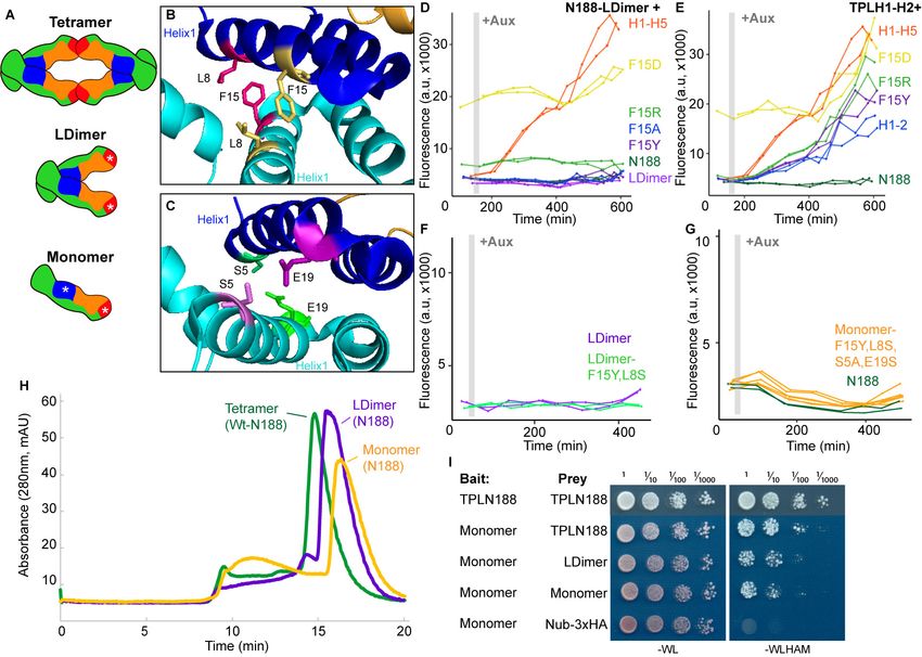
Research article                                                                        Chromosomes and Gene Expression Plant Biology

Figure 1. The N-terminal domain of TPL contains two independent repression domains. (A) Schematic of the ARCSc. The auxin-responsive promoter
driving the fluorescent protein Venus carries binding sites for the auxin-responsive transcription factor (ARF). In the absence of auxin, the IAA-TPL-N
fusion protein is bound to the ARF and maintains the circuit in a repressed state. Upon addition of auxin, the IAA-TPL protein is targeted for
ubiquitination and subsequent protein degradation, activating transcription of the fluorescent reporter. (B) TPL domains are LisH (LIS1 homology motif,
Figure 1 continued on next page

Leydon et al. eLife 2021;10:e66739. DOI: https://doi.org/10.7554/eLife.66739                                                                    3 of 28
Research article                                                                         Chromosomes and Gene Expression Plant Biology

Figure 1 continued
blue), CTLH (C-terminal LisH motif, orange), CRA (CT11-RanBPM; red, dimerization; green, foldback), and two WD40, beta-propeller motifs (purple).
N-terminal domains are indicated on the solved structure of the first 202 amino acids (Martin-Arevalillo et al., 2017, 5NQS). The termini of the
TPLN100 truncation used in the original ARCSc studies is indicated. (C) Diagram indicating the structure of constructs analyzed in experiments shown in
subsequent panels. For constructs with identical behavior (H1-H3, H1-H5, H1-H6, H1-H7), we included only a representative member (H1-H7) for
simplicity. Repression Index (Rep.) is a scaled measure of repression strength with 0 set to the level of repression observed with IAA3 and 10 set to the
level of repression by TPLN188. Auxin induction level (Aux. ind.) indicates the fold change difference between reporter expression before auxin
addition (time zero) and at the end of an experiment (~500 min). (D-F) Helix 1 and the CRA domain (Helix 3–Helix 8) can act independently to repress
transcription. Each panel represents two independent time-course flow cytometry experiments of the TPL helices indicated, all fused to IAA3. Every
point represents the average fluorescence of 5–10,000 individually measured yeast cells (a.u.: arbitrary units). Auxin (IAA-10 mM) was added at the
indicated time (gray bar, +Aux).
The online version of this article includes the following figure supplement(s) for figure 1:
Figure supplement 1. Helix 1 and the CRA domain (Helix 3-Helix 8) can act independently to repress transcription.

                                     Results
                                     To understand how TPL represses transcription, we first localized repressive activity within the pro-
                                     tein using the AtARCSc (Figure 1A). The extent of auxin-induced turnover of TPLN100 and TPLN300
                                     fusion proteins appears similar, although neither are completely degraded (Pierre-Jerome et al.,
                                     2014). In this way, auxin sensitizes the AtARCSc to even subtle differences in the strength of repres-
                                     sive activity by reducing the relative concentration of the TPL fusion proteins. To pinpoint the region
                                     conferring the strong repression of TPLN300, we generated a deletion series of the N-terminus
                                     guided by the available structural information (Figure 1B, C; Ke et al., 2015; Martin-
                                     Arevalillo et al., 2017).
                                        We started by identifying a shorter truncation, TPLN188, which behaved identically to TPLN300
                                     (Figure 1D, Pierre-Jerome et al., 2014). Subsequently, we deleted each alpha helical domain start-
                                     ing with Helix 9 (constructs are named in the format Helix x – Helix y or Hx-Hy). We found that Helix
                                     8 was required for the maximum level of repression activity and for the maintenance of repression
                                     after auxin addition (Figure 1D). All constructs lacking Helix 8 retained the ability to repress tran-
                                     scription, but this repression was lifted in the presence of auxin (Figure 1D) as had been observed
                                     for the original TPLN100 construct (Pierre-Jerome et al., 2014). In addition to the repressive activity
                                     of Helix 8, further deletions revealed that the 18 amino acids of Helix 1 were sufficient to confer
                                     repression on their own (H1, Figure 1E). To test whether Helix 8 activity depended on Helix 1, we
                                     analyzed a construct consisting of Helix 3 through Helix 9 (H3-H9, Figure 1E), which was able to
                                     repress transcription. Thus, Helix 1 (LisH) and Helix 8 (CRA) could contribute to TPL-mediated
                                     repression on their own (Figure 1D).
                                        To identify the minimal domain required for Helix 8-based repression, we generated additional
                                     deletions (Figure 1C, F, Figure 1—figure supplement 1). Helix 8 and the following linker were not
                                     sufficient for repression (Figure 1F), and removal of Helix 9 or of the linker between Helix 8 and
                                     Helix 9 slightly increased sensitivity to auxin compared to TPLN188 (H1-H8D8L, Figure 1—figure
                                     supplement 1). A deletion that removed both the LisH and Helix 8 repression domains (H3-H7) was
                                     only able to weakly repress reporter expression (Figure 1—figure supplement 1). These results
                                     demonstrate that Helix 8, in combination with the linker between Helix 8 and Helix 9 (which folds
                                     over Helix 1), was required for maintaining repression following addition of auxin. Moreover, the
                                     repressive activity of Helix 8 and the linker were only functional in the context of the larger Helix 3-
                                     Helix 8 truncation that carries the CTLH domain and a portion of the CRA domain.
                                        To determine which of the many known or predicted TPL-binding partners could mediate the
                                     repression activity of Helix8, we identified known interactors with either TPL or other Gro/TLE co-
                                     repressors, and then introduced the Arabidopsis homologs of these genes into the cytoplasmic split-
                                     ubiquitin system (cytoSUS) (Asseck and Grefen, 2018). We chose cytoSUS over yeast two-hybrid
                                     because in cytoSUS the interaction between target proteins takes place in the cytoplasm, and we
                                     had observed that the TPL N-terminus could repress activation of yeast two hybrid prototrophy
                                     reporters (Figure 2—figure supplement 1A). Putative direct interactors include HDACs (AtHDAC9,
                                     AtHDAC6; Long et al., 2006), Histone proteins (Histone H3, Histone H4; Ma et al., 2017), and the
                                     Mediator components MED13 (AtMED13; Ito et al., 2016) and MED21, which has been demon-
                                     strated to interact with Tup1, the yeast homolog of TPL (Gromöller and Lehming, 2000). We did

Leydon et al. eLife 2021;10:e66739. DOI: https://doi.org/10.7554/eLife.66739                                                                      4 of 28
Research article Chromosomes and Gene Expression Plant Biology
not observe any interactions between TPLN188 and the HDACs HDA6 and HDA9; the histone pro-
tein AtHIS4; or the Mediator subunit AtMED13 (Figure 2A, Figure 2—figure supplement 1B).
HDAC interaction with TPL has been previously hypothesized to occur through indirect interactions
with partner proteins (Krogan et al., 2012); however, direct interactions with histones and MED13
have been detected (Ito et al., 2016; Ma et al., 2017). The absence of interaction between
TPLN188 and these proteins may be due to differences between methods or interaction interfaces
in the C-terminal WD40 repeats.
Strong interaction was detected between TPLN188 and AtMED21, a component of the Mediator
middle domain (Figure 2A). MED21 is one of the most highly conserved Mediator subunits (Bour-
bon, 2008) and has a particularly highly conserved N-terminus (Figure 2—figure supplement
2A, C–E). In yeast, Tup1 interacts with the first 31 amino acids of ScMed21, with the first 7 amino
acids being absolutely required for interaction and transcriptional repression (Gromöller and Lehm-
ing, 2000). We observed that the equivalent truncation of AtMED21 (AtMED21-N31) was sufficient
for interaction with TPLN188 (Figure 2A). We next created truncations of the N-terminal domain of
AtMED21 to closely match those that had been made in yeast (Figure 2B) where deletion of the first
five amino acids of ScMed21 (ScD5Med21) severely reduce the ability of the Mediator complex to
co-purify with Pol II and CDK8 kinase complex (Sato et al., 2016). Interaction between TPLN188 and
AtMED21 similarly required the first five amino acids of AtMED21 (Figure 2B), and, as in yeast, this
was not a result of destabilization of the AtMED21 protein (Figure 2B). In fact, N-terminal Med21
deletions increased protein levels with no interaction with TPLN188; this results in high confidence
that the Med21 N-terminus is required for interaction. AtMED21 interaction was specific to the
Helix8-based repression domain as it interacted with TPLH3-H9 (Figure 2E), and not TPLH1-H7 (Fig-
ure 2—figure supplement 1C). Further screening of middle domain Mediator components identi-
fied an additional interaction with AtMED10B, the predominantly expressed MED10 isoform in
Arabidopsis (Klepikova et al., 2016; Figure 2C). Interactions between TPLN188 and both AtMED21
and AtMED10B were confirmed by immunoprecipitation (Figure 2D).
The MED21 N-terminus lies in the hinge region of the middle module and has residues that are
exterior facing and could be docking points for protein-protein interactions (Figure 2—figure sup-
plement 2C–E). A manual juxtaposition of the yeast Mediator structure with the Arabidopsis TPL
N-terminal structure shows that Helix 8 and the linker following face away from the tetramer and are
therefore optimally placed to interact with Mediator components (Figure 2F). To pinpoint which res-
idues of Helix 8 coordinate repression through interaction with MED21, we identified solution-facing
amino acids (Martin-Arevalillo et al., 2017). We reasoned that such residues were most likely not
involved in stabilizing the hydrophobic interactions between intra-TPL helical domains and could
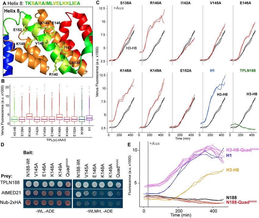
therefore be available to interact with partner proteins. Eight amino acids in Helix 8 were mutated
to alanine in the context of the H3-H8-IAA3 fusion protein (Figure 3A, light green residues) to
enable assessment of repression activity in the absence (Figure 3B) or presence (Figure 3C) of auxin.
No single amino acid was essential for repression. Two mutations (R140A and K148A) slightly
increased baseline expression of the reporter (Figure 3B, C). With the exception of E152A, which
behaved similarly to controls, all of the mutations altered the stability of repression after auxin addi-
tion, either by increasing (S138A, V145A, E146A, K149A) or decreasing (I142A) the final fluorescence
level (Figure 3C). Mutating E146 and K149 also increased the speed with which the reporter
responded to auxin (Figure 3C), suggesting that these two neighboring residues could be a critical
point of contact with co-repressive machinery. S138A had a small increase in auxin sensitivity, while
I142 reduced auxin sensitivity (Figure 3E).
We next tested whether the residues in Helix 8 that were required for repression (V145, E146,
K148, K149; Figure 3A–C) were also required for interaction with AtMED21. Single-alanine muta-
tions of these four amino acids in the context of TPLN188 significantly reduced interaction with
AtMED21, while the quadruple mutation (here called QuadAAAA) completely abrogated AtMED21
binding (Figure 3D). These mutations had little effect on interaction with AtMED10B (Figure 2—fig-
ure supplement 1D). Introduction of QuadAAAA mutations into the Helix 3 through Helix 8 context
(H3-H8-QuadAAAA) in the AtARCSc largely phenocopied a deletion of Helix 8 (yellow and pink,
Figure 3E, compare to H3-H7, Figure 1—figure supplement 1). In contrast, TPLN188-QuadAAAA
largely retained the repressive activity of wild-type N188 (red and black, Figure 3E), consistent with
the observation that Helix 1 is sufficient for full repression (Figure 1E). These results indicate that
Leydon et al. eLife 2021;10:e66739. DOI: https://doi.org/10.7554/eLife.66739 5 of 28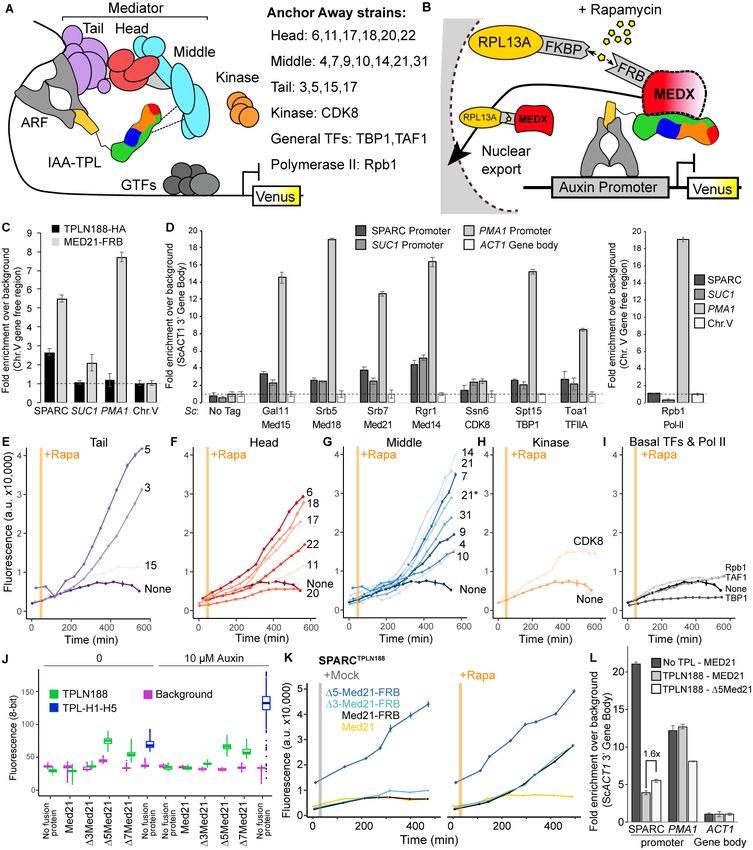
Research article                                                                      Chromosomes and Gene Expression Plant Biology

Figure 2. The Helix 8 repression domain of TPL directly interacts with AtMED21 and AtMED10B. (A–C, E) Cytoplasmic split-ubiquitin system (CytoSUS)
assays with candidate interacting proteins. Nub-3xHA is the N-terminal fragment of ubiquitin expressed with no fusion protein and is used as a
negative control. Each prey protein is from Arabidopsis. -WL, -ADE: dropout lacking Trp, Leu, and Ade (growth control); -WLMH, -ADE: dropout lacking
Trp, Leu, His, Met, and Ade (selective media). The plating for each panel was performed at the same day, white lines are provided when plates were
Figure 2 continued on next page

Leydon et al. eLife 2021;10:e66739. DOI: https://doi.org/10.7554/eLife.66739                                                                 6 of 28
Research article                                                                       Chromosomes and Gene Expression Plant Biology

Figure 2 continued
cropped for clarity. (B) Alignments of the Arabidopsis (At) and Saccharomyces (Sc) MED21 proteins are shown above cytoSUS assays with the same bait
shown in (A). Western blots below the colonies indicated that AtMED21 N-terminal D3 and D5 are well expressed in assay conditions. (C) CytoSUS
assays with selected Mediator proteins in the middle module. (D) The TPL-ProteinA-TF fusion protein can pull down TPL, AtMED21, and AtMED10B
from yeast extracts using IgG-beads. Detection of the VP16 transcriptional activator demonstrates enrichment of the fusion protein (aVP16). Each prey
protein is detected via the 3xHA tag (aHA), and efficacy of purification was judged by PGK1 depletion (aPGK1). (E) A TPL-N truncation lacking the LisH
domain (TPLH2-H9) could still interact with the AtMED21-N31 truncation. This bait construct interacted with IAA3, but only minimally with the negative
control (free Nub-3xHA). (F) Yeast Mediator (bottom, 5N9J) and AtTPL (top, 5NQV) manually juxtaposed to compare relative domain sizes and
feasibility of a TPL-MED21-MED10B interaction. TPL Helix 8–9 is colored green. MED21 is colored aqua, with the N-terminus colored red, and the
IAA27 EAR peptide in orange. MED10 is colored teal, with the C-terminus colored purple. The dotted line indicates the border between TPL and
Mediator structures.
The online version of this article includes the following figure supplement(s) for figure 2:
Figure supplement 1. The TPL-N terminal domain (TPLN188) interacts with the N-terminus of AtMED21.
Figure supplement 2. Homology and structure of the MED21 subunit of the Mediator complex.

                                    the CRA domain (H3-H8) requires contact with MED21 to repress, and that this is independent of
                                    repression via Helix 1.
                                        The large Mediator complex stabilizes the pre-initiation complex (PIC) enabling transcriptional
                                    activation (Kornberg, 2005; Nozawa et al., 2017; Roeder, 1996; Schilbach et al., 2017). The con-
                                    nection we found between TPL, two Mediator components, and transcriptional repression raised the
                                    possibility that other parts of the Mediator complex might also contribute to corepressor function.
                                    To test this, we needed a way to measure whether loss of function of individual Mediator compo-
                                    nents led to de-repression at an individual locus. In the case of the yeast corepressor Tup1, a stan-
                                    dard approach has been to test the level of transcription at target genes in the presence of deletion
                                    mutations of Tup1-interacting proteins (Gromöller and Lehming, 2000; Lee et al., 2000;
                                    Zhang and Reese, 2004). One challenge to such an approach for Mediator components is that loss-
                                    of-function mutants can be lethal or exhibit drastic physiological phenotypes (Biddick and Young,
                                    2005).
                                        To avoid these complications, we turned to the well-established Anchor Away system for induc-
                                    ible protein depletion (Haruki et al., 2008) and combined it with quantification of transcriptional
                                    activity at the synthetic locus in the AtARCS (Figure 4A, B). However, AtARCSc integrates compo-
                                    nents at four genomic locations using prototrophic markers that are not compatible with those
                                    needed for Anchor Away. To overcome this limitation, we re-created the ARC on a single plasmid
                                    (we refer to this plasmid as SPARC) using the Versatile Genetic Assembly System (VEGAS,
                                    Mitchell et al., 2015). SPARC behaved with similar dynamics to the original AtARCSc on both solid
                                    and liquid growth conditions (Figure 4—figure supplement 1A–C). As a first test of the Anchor
                                    Away system with SPARC, we fused Tup1 and its partner protein Cyc8 to two copies of the FKBP12-
                                    rapamycin-binding (FRB) domain of human mTOR (Haruki et al., 2008). Rapamycin treatment of
                                    strains targeting either of these proteins caused no release of repression on the SPARC reporter,
                                    providing confirmation of orthogonality of the synthetic system in yeast (Figure 4—figure supple-
                                    ment 1D).
                                        Before testing repressive function, we first performed chromatin immunoprecipitation using the
                                    Anchor Away FRB tag to ask whether Mediator proteins or specific Mediator modules were detect-
                                    able at the promoters of TPL-repressed genes. We began by assaying the integrated AtARCSc locus
                                    and comparing it to a Tup1-repressed locus, SUC2 (Carlson and Botstein, 1982; Fleming and Pen-
                                    nings, 2007; Trumbly, 1992), and to an active locus enriched for Mediator, PMA1 (Petrenko et al.,
                                    2017; Schmitt et al., 2006; Serrano et al., 1986). We generated a yeast strain with an integrated
                                    MED21-FRB fusion protein and an integrated AtARCSc locus with a 2xHA epitope-tagged TPLN188-
                                    IAA3 repressor protein. We observed enrichment of both MED21-FRB and the 2xHA-TPLN188-IAA3
                                    fusion protein at the AtARCSc locus (Figure 4C, Figure 4—figure supplement 2A–C). We also
                                    observed a modest enrichment of MED21-FRB at the SUC2 promoter (approximately twofold) and
                                    higher enrichment of MED21-FRB at the active PMA1 promoter (approximately eightfold). Next, we
                                    introduced the fully repressed SPARC plasmid containing TPLN188 (SPARCN188) into a library of
                                    Anchor Away yeast strains that allow specific depletion of Mediator components (see Figure 4A, B;
                                    Haruki et al., 2008; Petrenko et al., 2017). We tested representatives of the mediator complex (tail

Leydon et al. eLife 2021;10:e66739. DOI: https://doi.org/10.7554/eLife.66739                                                                   7 of 28
Research article Chromosomes and Gene Expression Plant Biology
Figure 3. Identification of critical residues within Helix 8 repression domain. (A) Sequence and structure of Helix 8 (5NQS). Helix 8 is colored green, and
amino acids chosen for mutation are highlighted in light green in both the sequence and the structure. (B) Repression activity of indicated single- and
double-alanine mutations. (C) Time-course flow cytometry of selected mutations of Helix 8 following auxin addition. TPLH3-8-IAA3 fusion proteins
(black) were compared to indicated single mutations to alanine (red). Controls: Helix 1 (H1 – blue) and TPLN188 (dark green). (D) A series of alanine
mutations (V145A, E146A, K148A, K149A, and the quadruple mutant QuadAAAA chosen from A–C) were introduced into the TPLN188 bait construct and
tested for interaction with wild-type TPLN188, AtMED21, and controls. Each single-alanine mutation reduces TPL interaction with AtMED21, while the
quad mutation abrogated interaction. (E) The Helix 8 QuadAAAA mutation was introduced into the TPLN188-IAA3 and TPLH3-8-IAA3 fusion proteins
and compared to wild-type N188 in time-course flow cytometry. For all cytometry experiments, the indicated TPL construct is fused to IAA3. Every point
represents the average fluorescence of 5–10,000 individually measured yeast cells (a.u.: arbitrary units). Auxin (IAA-10 mM) was added at the indicated
time (gray bar, +Aux). At least two independent experiments are shown for each construct.
– Med15, head – Med18, middle – Med21 and Med14, kinase – CDK8, general transcription factors
– TBP1, TFIIA, and RNA Pol II – Rpb1) by ChIP-qPCR. We observed enrichment of all tested core
mediator complex members, as well as general transcription factors, at both the SPARC and the
SUC2 loci, with very little enrichment of RNA Pol II (Figure 4D). In general, MED21 was detected in
lower levels at repressed loci than at the active PMA1 promoter (Figure 4D). Consistent with this
observation, Mediator is highly enriched at the SPARC promoter when TPL is absent (Figure 4—
Leydon et al. eLife 2021;10:e66739. DOI: https://doi.org/10.7554/eLife.66739 8 of 28Research article Chromosomes and Gene Expression Plant Biology Figure 4. Repression by TPL requires interaction with the N-terminus of MED21 at promoters. (A) Model of the proposed interaction between the TPL N-terminus with Mediator, where TPL interaction with Mediator 21 and 10 inhibits the recruitment of Pol II. Proteins in this complex that were tested by Anchor Away are listed on the right. (B) Schematic of AtARCSc combined with methods for inducible expression and nuclear depletion of MED21. In Anchor Away, the yeast ribosomal protein 13A (RPL13A) is fused to the rapamycin-binding protein FKBP. Addition of rapamycin induces dimerization Figure 4 continued on next page Leydon et al. eLife 2021;10:e66739. DOI: https://doi.org/10.7554/eLife.66739 9 of 28
Research article                                                                        Chromosomes and Gene Expression Plant Biology

Figure 4 continued
between FKBP and any target protein fused to 2xFRB, resulting in removal of the target protein from the nucleus. For these experiments, AtARCSc was
assembled into a single plasmid (SPARC) rather than being integrated into separate genomic loci (Figure 4—figure supplement 1). (C) 2xHA-
TPLN188-IAA3 and MED21-FRB association with the ARC and the ScSUC2 and ScPMA1 promoters. ChIP was performed with aHA and aFRB before
qPCR was used to quantify enrichment at specified loci. (D) Association of FRB-tagged components of Mediator and the transcriptional machinery with
the SPARC plasmid and the ScSUC2 promoters. ChIP was performed with aFRB, and qPCR was used to quantify enrichment at specified loci. (E–I)
Time-course flow cytometry analysis of SPARCN188 in Mediator Anchor Away yeast strains with rapamycin (orange bar, +Rapa). Two Med21 strains were
compared in the Middle domain (E), 21 (generated in this study) and 21* (generated in Petrenko et al., 2017). Both 21 and 21* demonstrated similar
increases in reporter expression. (J) Quantification of Venus fluorescence from SPARCN188 in wild-type and N-terminal ScMed21 deletions with and
without auxin. The x-axis indicates strain and which FRB fusion protein is being tested. Yeast was grown for 48 hr on synthetic drop out (SDO) media
with or without auxin, and colony fluorescence was quantified and plotted with the auxin-responsive SPARCH1-H5 in wild type as a reference.
Background: red autofluorescence was used as a reference for total cell density. (K) Time-course flow cytometry analysis of SPARCN188 in wild-type and
N-terminal ScMed21 deletions with and without rapamycin. Genotypes are indicated in the colored key inset into the graph. For (E–I, K) a.u.: arbitrary
units. Rapamycin was added at the indicated time (orange bar, +Rapa). Every point represents the average fluorescence of 5–10,000 individually
measured yeast cells. (L) Association of MED21-FRB or D5-MED21-FRB with SPARC plasmids. ChIP was performed with aFRB, and qPCR was used to
quantify enrichment at specified loci. (C, D, L) A region of the ACT1 gene body or a gene-free region of chromosome V (Chr.V) was arbitrarily defined
as background, and data is presented as fold enrichment over the control gene. Averages and standard errors of four replicates are shown.
The online version of this article includes the following figure supplement(s) for figure 4:
Figure supplement 1. Construction and characterization of the single locus auxin response circuit (SPARC).
Figure supplement 2. Mediator is detectable at the ARC promoter.
Figure supplement 3. N-terminal ScMed21 deletions impair auxin-responsive transcriptional activation.
Figure supplement 4. Inducible MED21 rescues rapamycin-induced yeast growth defects.

                                    figure supplement 2D). Higher enrichment of members of the middle module at repressed pro-
                                    moters (i.e., Med21, Med14, Figure 4D) may point to these subunits nucleating assembly of the
                                    entire complex.
                                       We next tested whether the association of Mediator complex components was required for TPL-
                                    mediated repression (Figure 4A, B). Nuclear depletion of Mediator components from the tail, head,
                                    and middle domain triggered clear activation of the SPARCN188 reporter (Figure 4E–I). Depletion of
                                    the Mediator kinase module component CDK8 had a more modest effect (Figure 4H). One caveat
                                    to this approach is that nuclear depletion of components that are absolutely required for transcrip-
                                    tional activation, such as RNA Pol II Anchor Away (Rpb1; Figure 4I), cannot be assayed for impacts
                                    on repression using transcription of the reporter as the output.
                                       To further interrogate the impact of Mediator on repression, we next focused on the other side
                                    of the interaction, namely which region of MED21 was required for interaction with TPL. Deletion of
                                    the first seven amino acids of ScMed21 (D7Med21) partially activates genes that are normally
                                    repressed by Tup1 (Gromöller and Lehming, 2000), so we first tested if the same held true for TPL-
                                    mediated repression. We introduced SPARCs with different TPL constructs into strains where wild-
                                    type ScMed21 or N-terminal deletions were targets of Anchor Away. Importantly, the addition of
                                    the FRB tag did not alter ScMED21 function (Figure 4J, Figure 4—figure supplement 1E, F). We
                                    observed that deletion of either five or seven of the N-terminal residues of ScMed21 increased the
                                    expression of the reporter in SPARCN188 to a level similar as what is observed with TPL H1-H5
                                    (Figure 4J, Figure 4—figure supplement 1E, F). No mutation increased the SPARC’s sensitivity to
                                    auxin. As D7ScMed21 had a noticeable impact on growth, as has been reported previously
                                    (Gromöller and Lehming, 2000; Hallberg et al., 2006), we removed it from further studies.
                                    D5ScMed21 had no observable growth defects, although this deletion is known to be sufficient to
                                    alter Mediator assembly and disrupt binding of Pol II and the CDK8 kinase module (Hallberg et al.,
                                    2006; Sato et al., 2016).
                                       The fully repressed SPARCN188 in D3ScMed21 or D5ScMed21 mutants showed elevated reporter
                                    transcription when compared to strains carrying wild-type ScMED21 (Figure 4K). The addition of
                                    rapamycin further increased reporter expression, particularly in the D3ScMed21 strains, suggesting
                                    that this deletion could only partially disrupt the TPLN188-Med21 interaction. We used chromatin
                                    immunoprecipitation to directly test whether D5Med21 showed a change in association with the
                                    SPARCN188 promoter. While this deletion would be expected to reduce Med21 association with TPL,
                                    the resulting de-repression of the locus should lead to an increase in Mediator association with the
                                    activated promoter. Indeed, we observed an ~1.6-fold increase in D5Med21-FRB promoter binding

Leydon et al. eLife 2021;10:e66739. DOI: https://doi.org/10.7554/eLife.66739                                                                  10 of 28
Research article Chromosomes and Gene Expression Plant Biology
compared to wild type (Figure 4L). While this enrichment was modest compared to a repressor-free
SPARCIAA14 (Figure 4L, no TPL; dark gray bar), it was similar to the magnitude of transcriptional acti-
vation of the reporter in the D5Med21 genotype (Figure 4J, K). The well-documented PMA1 pro-
moter had a substantial enrichment of wild-type MED21, as expected, and was unaffected by the
presence of TPL (Figure 4L, dark and light gray bars). To confirm that the interaction with TPL was
not unique to ScMed21, we replaced the first five amino acids of ScMed21-FRB with the correspond-
ing sequence from AtMED21. The strain carrying this chimeric protein had an identical repression
profile as the one with native ScMed21 (Figure 4—figure supplement 3B) and showed no differ-
ence in growth or viability (Figure 4—figure supplement 3C).
To minimize any possible off-target impact of ScMed21 deletions, we introduced estradiol-induc-
ible versions of ScMed21 (iScMed21) into the Anchor Away SPARCN188 strains (Figure 4—figure
supplement 4; McIsaac et al., 2013). The combination of all three synthetic systems – ARCSc,
Anchor Away, and estradiol inducibility – made it possible to rapidly deplete the wild-type
ScMed21-FRB from the nucleus while simultaneously inducing ScMed21 variants and visualizing the
impact on a single auxin-regulated locus. Depletion of nuclear ScMed21 by rapamycin increased lev-
els of the reporter in all genotypes examined (Figure 4G, K) while increasing cell size even in short
time courses, consistent with its essential role in many core pathways (Figure 4—figure supplement
4A; Gromöller and Lehming, 2000). When wild-type iScMed21 was induced, there was a rescue of
both reporter repression and cell size (Figure 4—figure supplement 4C, D), whereas induction of
either D3 and D5 variants resulted in significantly less reporter repression (Figure 4—figure supple-
ment 4D). iD3Med21 was induced and accumulated at a comparable level to wild-type Med21, while
iD5Med21 is less stable (Figure 4—figure supplement 4E, F). In the time courses with both rapamy-
cin and estradiol, we did not observe the cell size increases observed in the rapamycin treatments
alone (populations were evenly distributed around a single mean), suggesting that we were observ-
ing the immediate effects of the Med21 deletions (Figure 4—figure supplement 4G, H).
Several lines of evidence suggest that, in addition to interactions with other partners, homomulti-
merization modulates TPL repression potential. First, structures of the N-terminal domains of TPL
(Martin-Arevalillo et al., 2017) and a rice homolog OsTPR2 (Ke et al., 2015) reveal high conserva-
tion of residues that coordinate formation of homotetramers and connect tetramer formation to
Aux/IAA binding. Second, the dominant TPL mutant tpl-1 altered a single amino acid in the ninth
helix of the TPL-N terminus (N176H) that induces aggregation of TPL and its homologs (TPR1-4),
reducing total activity (Long et al., 2006; Ma et al., 2017). Third, TPL recruitment motifs found in
the rice strigolactone signaling repressor D53 induce higher-order oligomerization of the TPL N-ter-
minus, which increases histone binding and transcriptional repression (Ma et al., 2017). Our studies
in yeast suggest that there may be a more complex relationship between tetramer formation and
repression as we have measured strong repressive activity in several constructs that are unlikely
(TPLN100; Pierre-Jerome et al., 2014) or unable (H1, H3-8; Figure 1C) to form tetramers (compare
Figure 1B with Figure 5A). To quantify the potential for interaction among our constructs, we used
the cytoSUS assay (Asseck and Grefen, 2018). Helix 8 was required for strongest interaction
between TPL constructs (Figure 5—figure supplement 1A), although this assessment was compli-
cated by the fact that some of the shorter constructs accumulated to significantly lower levels (Fig-
ure 5—figure supplement 1B). The weak interaction we could observe between full-length TPL-N
and the Helix 1 through Helix 3 construct (H1-3) indicated that the TPL LisH domain is sufficient for
dimerization. Therefore, while auxin-insensitive repression may require multimeric TPL, this higher-
order complex was not required for auxin-sensitive repression mediated by Helix 1 (Figure 1E).
To avoid any potential artifacts from analysis of truncated forms of the N-terminus, we next gen-
erated site-specific mutations that disrupted multimerization in the context of TPLN188. Martin-Are-
valillo et al. had previously identified a quadruple mutation (K102S-T116A-Q117S-E122S) that
abrogated the ability of the CRA domain (Helix 6 and Helix 7) to form inter-TPL interactions (Martin-
Arevalillo et al., 2017). As this mutant form of TPL is only capable of dimerizing through its LisH
domain, we refer to it here as LDimer (Figure 5A). The LDimer mutations in TPLN188 retained the
same auxin-insensitive repression behavior as wild-type TPLN188 (Figure 5D), supporting the finding
from the deletion series.
To make a fully monomeric form of TPL, we introduced mutations into the dimerization interface
of the LisH domain in the context of LDimer. We first mutated one of a pair of interacting residues
(F15) to a series of amino acids (tyrosine – Y, alanine – A, arginine – R, or aspartic acid – D) in the
Leydon et al. eLife 2021;10:e66739. DOI: https://doi.org/10.7554/eLife.66739 11 of 28Research article Chromosomes and Gene Expression Plant Biology
Figure 5. Multimerization is not required for repression in yeast. (A) TPL can form a homotetramer via the CRA (red) and LisH (blue) domains. Asterisks
indicate mutations that block or diminish these interactions. (B, C) Locations of critical positions in Helix 1 are highlighted for two interacting TPL
monomers (shown in light and dark blue). Interacting amino acids share the same color (adapted from 5NQV). (D–G) Time-course flow cytometry
analysis of TPLN-IAA3 fusion proteins carrying selected single point mutations in N188-LDimer-IAA3 (D) and the TPLH1-2 truncation (E). The F15Y
mutation had little effect on repression activity for either TPL construct. Double mutations (F15Y, L8S in LDimer) (F) or the quadruple Monomer
mutations (S5A, L8S, F15Y, E19S in LDimer) (G) showed repression activity that was indistinguishable from LDimer or wild-type N188 fused to IAA3. For
all cytometry experiments, the indicated TPL construct is fused to IAA3. Every point represents the average fluorescence of 5–10,000 individually
measured yeast cells (a.u.: arbitrary units). Auxin (IAA-10 mM) was added at the indicated time (gray bar, +Aux). At least two independent experiments
are shown for each construct. (H) Size exclusion chromatography on TPLN188 wild-type (green), LDimer (purple), and Monomer (orange) tetramerization
mutants. (I) Cytoplasmic split-ubiquitin system (CytoSUS) on TPL tetramerization mutants.
The online version of this article includes the following figure supplement(s) for figure 5:
Figure supplement 1. TPL multimerization requires Helix 8.
context of either LDimer (Figure 5D), or H1-2 (Figure 5B, E). Conversion of F15 to the polar and
charged aspartic acid (D) completely abolished repression activity, while the positively charged argi-
nine was better tolerated (Figure 5D, E). The conversion of F15 to tyrosine had no effect on LDimer
(Figure 5D), and only a minimal increase in auxin sensitivity in the context of H1-2 (Figure 5E). We
then combined LDimer-F15Y with a mutation of the coordinating residue L8 to serine with the inten-
tion of stabilizing the now solvent-facing residues. The repressive behavior of this mutant was indis-
tinguishable from that of LDimer (Figure 5F).
To further push the LDimer towards a monomeric form, we introduced two additional mutations
(S5A, E19S, Figure 5C, G). Size-exclusion chromatography confirmed that this combination of
Leydon et al. eLife 2021;10:e66739. DOI: https://doi.org/10.7554/eLife.66739 12 of 28Research article Chromosomes and Gene Expression Plant Biology
mutations (S5A-L8S-F15Y-E19S-K102S-T116A-Q117S-E122S, hereafter called Monomer) successfully
shifted the majority of the protein into a monomeric state (Figure 5H); however, this shift had no
observable impact on repression strength before or after auxin addition (Figure 5G). To test
whether these mutations had a similar impact on in vivo TPL complexes, we introduced the LDimer
and Monomer mutations into the cytoSUS assay. In contrast to the in vitro chromatography results
with purified proteins, Monomer expressed in yeast retained measurable interaction with wild-type
TPL, LDimer or Monomer, albeit at a reduced level than what was observed between wild-type
TPLN188 constructs (Figure 5I). A caveat to this apparent difference between assays is that the
Monomer mutations led to a striking increase in protein concentration in yeast (Figure 5—figure
supplement 1C), likely partially compensating for the decrease in affinity.
To ascertain which of our findings about TPL required the sensitivity and simplicity of the syn-
thetic context and which could be observed in the full complexity of intact plant systems, we per-
formed a set of experiments in Nicotiana benthamiana (tobacco) and Arabidopsis. Bimolecular
fluorescence complementation (BiFC) confirmed the interaction between TPL and MED21
(Figure 6A), which was further validated by co-immunoprecipitation using tobacco extracts
(Figure 6B). We were also able to pull down MED21 and TPL using MED10B (Figure 6—figure sup-
plement 1A). BiFC also confirmed the importance of the same TPL Helix 8 residues for the TPL-
AtMed21 interaction (Figure 6A, TPLH8QuadA). Similarly, the D5AtMED21 N-terminal truncation elimi-
nated interaction with full-length TPL (Figure 6A). We next developed a quantitative repression
assay based on UAS/GAL4-VP16 (Brand and Perrimon, 1993; Figure 6C). To block potentially con-
founding interactions with endogenous TPL/TPRs or TIR1/AFBs, we engineered a variant of IAA14
with mutations in the two EAR domains (EARAAA) and in the degron (P306S) (IAA14mED; Figure 6C).
After prototyping the system in yeast (Figure 6—figure supplement 1B, C), we quantified repres-
sion strength of constructs carrying TPLN-IAA14mED variants using the well-characterized synthetic
auxin-responsive promoter DR5 (Ulmasov et al., 1997). As expected, DR5 was strongly induced by
co-transformation with AtARF19, and this induction was sharply reduced by the inclusion of UAS-
TPLN188-IAA14mED and GAL4-VP16 (Figure 6—figure supplement 1D). Overall, we observed
strong correlation in repression activity between what was observed in yeast and in tobacco.
To connect the observed differences in repression strength to a developmental context, we gen-
erated transgenic Arabidopsis lines where the UAS-TPL-IAA14mED constructs were activated in the
cells where IAA14 normally acts to regulate the initiation of lateral root primordia (Figure 6D;
Gala et al., 2021; Laplaze et al., 2005). Expression of functional TPL-IAA14mED fusion proteins in
these xylem pole pericycle cells should strongly suppress production of lateral roots, phenocopying
the solitary root (slr) mutant, which carries an auxin-resistant form of IAA14 (Fukaki et al., 2002).
Indeed, TPLN188 fusion constructs sharply decreased lateral root density (Figure 6E), while trans-
formants expressing either IAA14mED (with no TPL fusion) or TPLN188 (with no IAA14 fusion) had no
effect on lateral root production (Figure 6E). TPLH3-H9 decreased lateral root density albeit not as
effectively as TPLN188, suggesting that Helix 1 is required for full repression in a native context
(Figure 6E). Both LDimer and Monomer constructs (Figure 6E) were able to repress lateral root
development to the same extent as TPLN188, meaning that multimer formation is not required for
TPL-mediated repression in this context. The fusion containing the Helix 8 quadruple mutant demon-
strated a clear loss of repression, indicating that the TPL-MED21 interaction is critical for repression
when expressed in lateral root-forming cells (Figure 6E).
Given this result, we wanted to directly test the role of AtMed21 in auxin-regulated development
in the presence of native isoforms of TPL and Aux/IAAs. This was complicated by the fact that, as in
yeast, AtMED21 is essential in plants. While homozygous loss-of-function mutations are embryo
lethal (Dhawan et al., 2009), plants heterozygous for Atmed21 mutations appear wild type (Fig-
ure 6—figure supplement 2A, B). To overcome this obstacle, we took two approaches that relied
on the same xylem pole pericycle driver as described for the TPL functional assays. First, we
expressed N-terminal deletion variants of MED21 that should weaken or sever interaction with TPL
(Figure 6F; D3MED21, D5MED21, D7MED21). If MED21-TPL interaction is critical to maintain normal
expression of the lateral root program, reduced interaction should trigger an increase in lateral root
density. This was exactly what we observed for transgenic lines expressing any of the three dele-
tions. Second, we repressed transcription of AtMED21 by introducing a dCAS9-TPLN300 synthetic
repressor under the control of a UAS promoter along with three sgRNAs complementary to the
AtMED21 promoter. Similar to the predictions above, reduced expression of AtMED21 in xylem
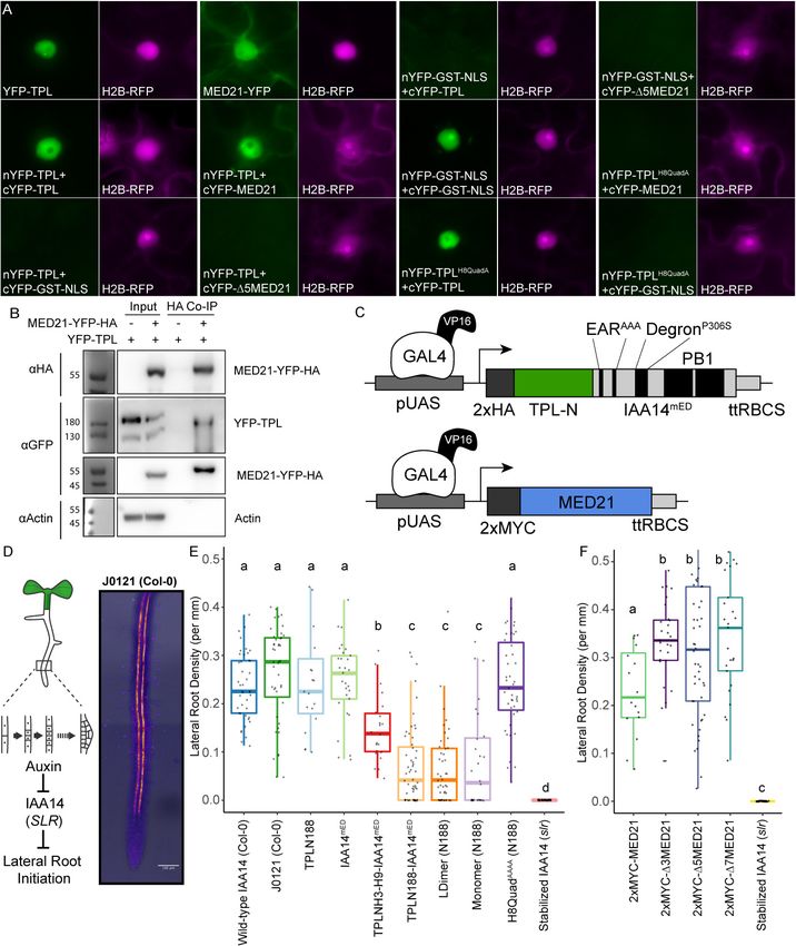
Leydon et al. eLife 2021;10:e66739. DOI: https://doi.org/10.7554/eLife.66739 13 of 28Research article Chromosomes and Gene Expression Plant Biology Figure 6. The TPL CRA repression domain behaves similarly in yeast and plants. (A) Bimolecular fluorescence complementation assay performed in tobacco. Each image is an epi-fluorescent micrograph taken at identical magnification from tobacco epidermal cells at 2 days post injection. The YFP image is colored green (left panel). p35S:H2B-RFP was used as a control and is false-colored magenta (right panel). (B) Co-immunoprecipitation of MED21 and TPL from tobacco leaves. MED21-YFP-HA was immunoprecipitated using anti-HA, and YFP-TPL was detected using the YFP fusion. Actin Figure 6 continued on next page Leydon et al. eLife 2021;10:e66739. DOI: https://doi.org/10.7554/eLife.66739 14 of 28
Research article                                                                        Chromosomes and Gene Expression Plant Biology

Figure 6 continued
was used to demonstrate that the purification had removed non-specific proteins. Numbers on the left of blots indicate sizes of protein standards in
kilodaltons. (C) Design of UAS-TPL-IAA14mED and UAS-MED21 constructs. Mutation of the conserved lysine residues in the EAR domain disrupted
potential interactions with endogenous TPL/TPR proteins. The IAA14 degron has been mutated (P306S) to render it auxin insensitive. UAS: upstream
activating sequence; ttRBCS: Rubisco terminator sequence. (D) Auxin-induced degradation of IAA14 is absolutely required for initiation of lateral root
development (cartoon, left). An enhancer trap line (J0121) expresses GAL4-VP16 and UAS-GFP in in xylem pole pericycle cells. (E) N-terminal domains
of TPL were sufficient to repress the development of lateral roots in Arabidopsis seedlings. The density of emerged lateral roots was measured in T1
seedlings at 14 days after germination. (F) N-terminal deletions in AtMED21 were sufficient to dominantly increase the development of lateral roots in
Arabidopsis seedlings. The density of emerged lateral roots was measured in T1 seedlings at 14 days after germination. (E, F) Lowercase letters
indicate significant difference (ANOVA and Tukey HSD multiple comparison test; p
Research article                                                                 Chromosomes and Gene Expression Plant Biology

                                   authors utilized the Anchor Away approach to correlate the importance of HDACs, transcriptional
                                   machinery, and chromatin remodeling enzymes to the repression state of endogenously repressed
                                   Cyc8-Tup1 target genes. They observed that Tup1 did not block the binding of transcription factors
                                   but inhibited the recruitment of one Mediator component in the tail domain, GAL11/MED15, as well
                                   as Pol II and the chromatin remodelers Snf2 and Sth1. They additionally observed that HDACs had
                                   only a supportive role in reinforcing Tup1 repression. These results led to their hypothesis that Tup1
                                   blocks the activation domains of transcription factors and suggested this was through direct binding
                                   to activation domains (Wong and Struhl, 2011).
                                       The synthetic system used here allowed us to build on this model and further refine our under-
                                   standing of TPL’s repressive activity. In our experiments, we see a similar set of conditions, with TPL
                                   recruited to the DNA-bound transcriptional activator (ARF), and several possible mechanisms of
                                   repression. Unlike Tup1, we have subdivided the TPL protein to identify interactions between TPL
                                   and individual protein interactors with no effect on yeast function. In these experiments, we can
                                   eliminate the possibility that TPL blocks ARF activation by directly blocking the transcription factor
                                   activation domains because we see a loss of repression only when TPL-MED21 binding is eliminated
                                   through specific point mutations (Figure 4J–L). Our estradiol-inducible replacement assays where
                                   different isoforms of Med21 are expressed also corroborate these findings (Figure 4—figure sup-
                                   plement 4) as the SPARC remains genetically identical in these strains, indicating that TPL-MED21
                                   interaction is regulating Mediator activity not a TPL-ARF interaction. Furthermore, our results corre-
                                   late well with findings that repressed targets are reactivated when this portion of MED21 is deleted
                                   in yeast (TPL, Figure 4; Tup1; Gromöller and Lehming, 2000). Therefore, we suggest that instead
                                   of directly binding activation domains that TPL (and likely Tup1) binds to components of Mediator
                                   (MED21, MED10B, and possibly others) recruited by the transcription factor. Indeed, it is easier to
                                   rationalize that the repressor binds the same domains of the Mediator complex recruited by the
                                   transcription factor’s activation domain (with the same affinity) as opposed to binding each diverse
                                   activation domain (with varying affinity). In this model, corepressor binding blocks formation of a fully
                                   active Mediator complex, thereby limiting Pol II recruitment and promoter escape (Petrenko et al.,
                                   2017).
                                       The Mediator complex is a multi-subunit complex that connects DNA-bound transcription factors
                                   and the RNA Pol II complex to coordinate gene expression (Flanagan et al., 1991; Kim et al., 1994;
                                   Kornberg, 2005). The yeast Mediator subunits are organized into four separate modules, head, mid-
                                   dle, tail, and kinase, with a strong conservation of module components in plants (Dolan and Chap-
                                   ple, 2017; Maji et al., 2019; Malik et al., 2017; Samanta and Thakur, 2015). Med21 forms a
                                   heterodimer with Med7 and interacts with Med10, among others, to create the central region of the
                                   middle region of the Mediator complex. The Med21 N-terminus is centered on a flexible hinge
                                   region (Baumli et al., 2005), which is required for recruitment of Pol II and the CDK8 kinase module
                                   (Sato et al., 2016). The protein interaction between TPL and MED21 occurs at the N-terminus of
                                   MED21, highlighting the importance of this region as a signaling hub (Sato et al., 2016). Other lines
                                   of evidence support this role as this region binds the yeast homolog of TPL, Tup1 (Gromöller and
                                   Lehming, 2000), through a completely different protein domain as no homology can be found
                                   between TPL Helix 8 and Tup1 in any region by primary amino acid homology (i.e., BLAST).
                                       As suggested by Ito and colleagues (Ito et al., 2016) and supported by our synthetic system,
                                   auxin-induced removal of TPL is sufficient to induce changes in the activity of the Mediator complex;
                                   however, multiple points of contact likely exist between the Mediator complex and other parts of
                                   the transcriptional machinery in both transcriptionally repressed and active states. For auxin
                                   response, specifically, there are several lines of evidence to support this model, including docu-
                                   mented association between the structural backbone of Mediator, MED14, and activated and
                                   repressed auxin loci in Arabidopsis (Ito et al., 2016). In addition, MED12 and MED13 are required
                                   for auxin-responsive gene expression in the root, and MED12 acts upstream of AUX1 in the root
                                   growth response to sugar (Raya-González et al., 2018). MED18 in the head module represses auxin
                                   signaling and positively regulates the viability of the root meristem (Raya-González et al., 2018).
                                   PFT1/MED25 regulates auxin transport and response in the root (Raya-González et al., 2014).
                                   MED7, MED21’s partner protein in the hinge domain, is required for normal root development, and
                                   loss of MED7 function impacts expression of auxin signaling components (Kumar et al., 2018). Pre-
                                   vious research identified the Mediator CDK8 module, specifically MED13 (MAB2), as an interactor
                                   with the full-length TPL protein (Ito et al., 2016). We could not observe interaction between the

Leydon et al. eLife 2021;10:e66739. DOI: https://doi.org/10.7554/eLife.66739                                                        16 of 28
Research article Chromosomes and Gene Expression Plant Biology
N-terminal domain of TPL and AtMED13, AtCYC8, or AtCYCC (Figure 2—figure supplement 1B),
suggesting that any direct interactions occur outside the N-terminal region.
The conserved interaction of both TPL and Tup1 with Mediator has implications for modeling
eukaryotic transcription (e.g., Estrada et al., 2016). By stabilizing the Mediator complex, TPL (and
by extension Tup1) may create a ‘pre-paused’ state that allows rapid recruitment of Pol II and activa-
tion once TPL is removed. This would be compatible with the multiple repression mechanisms
described for TPL at different genetic loci. TPL recruitment of the repressive CDK8 Mediator com-
plex (Ito et al., 2016), chromatin remodeling enzymes such as HD19 (Long et al., 2006), and contact
with histone proteins (Ma et al., 2017) would be removed with TPL upon relief of repression. It will
be critical in the future to understand how these various forms of repression interact, and especially
to map the dynamics of assembly and disassembly of complexes as loci transition from repressed to
active states and back to repressed once again.
Code availability statement
All codes are available through Github: https://github.com/achillobator/TPL_Structure_Function/
(Leydon, 2021 copy archived at swh:1:rev:141d7d05fe0c23be55af5050563d160f019d6d65).
Materials and methods
Key resources table
Reagent type
(species) or Source or Additional
resource Designation reference Identifiers information
Gene TOPLESS, TPL GenBank AT1G15750
(Arabidopsis
thaliana)
Gene MEDIATOR 21, MED21 GenBank AT4G04780
(Arabidopsis
thaliana)
Strain, strain Anchor Away strains EURO- SCARF HHY168 See Yeast Strain
background euroscarf.de list (Supplementary file 3)
(Saccharomyces
cerevisiae)
Strain, strain cytoSUS strains Asseck and Grefen, 2018 THY.AP4, THY.AP5 See Yeast Strain
background list (Supplementary file 3)
(Saccharomyces
cerevisiae)
Strain, strain Rosetta 2 Sigma-Aldrich 71400 Electrocompetent
background strain cells
(Escherichia coli)
Strain, strain Nicotiana GenBank NCBI: txid4100
background benthamiana (wild-type)
(Nicotiana
benthamiana)
Strain, strain GV3101 GenBank NCBI: txid358 Electrocompetent
background cells
(Agrobacterium
tumefaciens)
Genetic reagent J0121 Gala et al., 2021 J0121
(Arabidopsis (in Col-0 accession)
thaliana)
Genetic reagent slr TAIR SLR-1, AT4G14550
(Arabidopsis
thaliana)
Genetic reagent med21-1 Arabidopsis Biological WiscDsLox461-464K13
(Arabidopsis Resource Center
thaliana)
Continued on next page
Leydon et al. eLife 2021;10:e66739. DOI: https://doi.org/10.7554/eLife.66739 17 of 28You can also read