DYNAMICALLY EVOLVING NOVEL OVERLAPPING GENE AS A FACTOR IN THE SARS-COV-2 PANDEMIC - ELIFE
←
→
Page content transcription
If your browser does not render page correctly, please read the page content below

RESEARCH ARTICLE

                                    Dynamically evolving novel overlapping
                                    gene as a factor in the SARS-CoV-2
                                    pandemic
                                    Chase W Nelson1,2†*, Zachary Ardern3†*, Tony L Goldberg4,5, Chen Meng6,
                                    Chen-Hao Kuo1, Christina Ludwig6, Sergios-Orestis Kolokotronis2,7,8,9,
                                    Xinzhu Wei10,11*
                                    1
                                      Biodiversity Research Center, Academia Sinica, Taipei, Taiwan; 2Institute for
                                    Comparative Genomics, American Museum of Natural History, New York, United
                                    States; 3Chair for Microbial Ecology, Technical University of Munich, Freising,
                                    Germany; 4Department of Pathobiological Sciences, University of Wisconsin-
                                    Madison, Madison, United States; 5Global Health Institute, University of Wisconsin-
                                    Madison, Madison, United States; 6Bavarian Center for Biomolecular Mass
                                    Spectrometry (BayBioMS), Technical University of Munich, Freising, Germany;
                                    7
                                      Department of Epidemiology and Biostatistics, School of Public Health, SUNY
                                    Downstate Health Sciences University, Brooklyn, United States; 8Institute for
                                    Genomic Health, SUNY Downstate Health Sciences University, Brooklyn, United
                                    States; 9Division of Infectious Diseases, Department of Medicine, SUNY Downstate
                                    Health Sciences University, Brooklyn, United States; 10Departments of Integrative
                                    Biology and Statistics, University of California, Berkeley, Berkeley, United States;
                                    11
                                       Departments of Computer Science, Human Genetics, and Computational
                                    Medicine, University of California, Los Angeles, Los Angeles, United States
*For correspondence:
cnelson@amnh.org (CWN);
zachary.ardern@tum.de (ZA);
aprilwei@berkeley.edu (XW)          Abstract Understanding the emergence of novel viruses requires an accurate and
†
 These authors contributed          comprehensive annotation of their genomes. Overlapping genes (OLGs) are common in viruses and
equally to this work                have been associated with pandemics but are still widely overlooked. We identify and characterize
                                    ORF3d, a novel OLG in SARS-CoV-2 that is also present in Guangxi pangolin-CoVs but not other
Competing interests: The
                                    closely related pangolin-CoVs or bat-CoVs. We then document evidence of ORF3d translation,
authors declare that no
                                    characterize its protein sequence, and conduct an evolutionary analysis at three levels: between
competing interests exist.
                                    taxa (21 members of Severe acute respiratory syndrome-related coronavirus), between human
Funding: See page 23                hosts (3978 SARS-CoV-2 consensus sequences), and within human hosts (401 deeply sequenced
Received: 03 June 2020              SARS-CoV-2 samples). ORF3d has been independently identified and shown to elicit a strong
Accepted: 30 September 2020         antibody response in COVID-19 patients. However, it has been misclassified as the unrelated gene
Published: 01 October 2020          ORF3b, leading to confusion. Our results liken ORF3d to other accessory genes in emerging viruses
Reviewing editor: Antonis
                                    and highlight the importance of OLGs.
Rokas, Vanderbilt University,
United States
   Copyright Nelson et al. This
article is distributed under the
                                    Introduction
                                    The COVID-19 pandemic raises urgent questions about the properties that allow animal viruses to
terms of the Creative Commons
Attribution License, which          cross species boundaries and spread within humans. Addressing these questions requires an accu-
permits unrestricted use and        rate and comprehensive understanding of viral genomes. One frequently overlooked source of nov-
redistribution provided that the    elty is the evolution of new overlapping genes (OLGs), wherein a single stretch of nucleotides
original author and source are      encodes two distinct proteins in different reading frames. Such ‘genes within genes’ compress geno-
credited.                           mic information and allow genetic innovation via overprinting (Keese and Gibbs, 1992), particularly

Nelson, Ardern, et al. eLife 2020;9:e59633. DOI: https://doi.org/10.7554/eLife.59633                                            1 of 29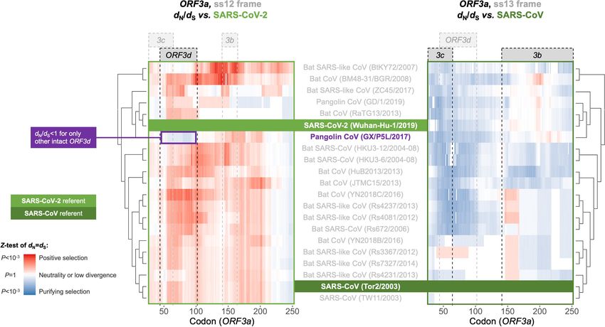
Research article                                                            Evolutionary Biology Microbiology and Infectious Disease

                                    as frameshifted sequences preserve certain physicochemical properties of proteins (Bartonek et al.,
                                    2020). However, OLGs also entail the cost that a single mutation may alter two proteins, constrain-
                                    ing evolution of the pre-existing open reading frame (ORF) and complicating sequence analysis.
                                    Unfortunately, genome annotation methods typically miss OLGs, instead favoring one ORF per
                                    genomic region (Warren et al., 2010).
                                       OLGs are known entities but remain inconsistently reported in viruses of the species Severe acute
                                    respiratory syndrome-related coronavirus (subgenus Sarbecovirus; genus Betacoronavirus;
                                    Coronaviridae Study Group of the International Committee on Taxonomy of Viruses et al.,
                                    2020). For example, annotations of ORF9b and ORF9c are absent or conflicting in SARS-CoV-2 ref-
                                    erence genome Wuhan-Hu-1 (NCBI: NC_045512.2) and genomic studies (e.g. Chan et al., 2020;
                                    Wu et al., 2020b), and OLGs are often not displayed in genome browsers (e.g. Flynn et al., 2020).
                                    Further, ORF3b, an extensively characterized OLG within ORF3a that is present in other species
                                    members including SARS-CoV (SARS-CoV-1) (McBride and Fielding, 2012), has sometimes been
                                    annotated in SARS-CoV-2 even though it contains four early STOP codons in this virus. Such inconsis-
                                    tencies complicate research, because OLGs may play key roles in the emergence of new viruses. For
                                    example, in human immunodeficiency virus-1 (HIV-1), the novel OLG asp within env is actively
                                    expressed in human cells (Affram et al., 2019) and is associated with the pandemic M group lineage
                                    (Cassan et al., 2016).

                                    Results
                                    Novel overlapping gene candidates
                                    To identify novel OLGs within the SARS-CoV-2 genome, we first generated a list of candidate over-
                                    lapping ORFs in the Wuhan-Hu-1 reference genome (NCBI: NC_045512.2). Specifically, we used the
                                    codon permutation method of Schlub et al., 2018 to detect unexpectedly long ORFs while control-
                                    ling for codon usage. One unannotated OLG candidate, here named ORF3d, scored highly
                                    (p=0.0104), exceeding the significance of two known OLGs annotated in Uniprot (ORF9b and
                                    ORF9c, both within N; https://viralzone.expasy.org/8996) (Figure 1, Figure 1—figure supplement
                                    1, and Supplementary file 1).
                                       ORF3d comprises 58 codons (including STOP) near the beginning of ORF3a (Table 1), making it
                                    longer than the known genes ORF7b (44 codons) and ORF10 (39 codons) (Supplementary file 1).
                                    ORF3d was discovered independently by Chan et al., 2020 as ‘ORF3b’ and Pavesi, 2020 as ‘hypo-
                                    thetical protein’. Due to this naming ambiguity and its location within ORF3a, ORF3d has subse-
                                    quently been conflated with the previously documented ORF3b in multiple studies (e.g. Fung et al.,
                                    2020; Ge et al., 2020; Gordon et al., 2020; Hachim et al., 2020; Helmy et al., 2020; Yi et al.,
                                    2020). Critically, ORF3d is unrelated (i.e. not homologous) to ORF3b, as the two genes occupy dif-
                                    ferent genomic positions within ORF3a: ORF3d ends 39 codons upstream of the genome region
                                    homologous to ORF3b, and the ORF3b start site encodes only 23 codons in SARS-CoV-2 due to a
                                    premature STOP (Wu et al., 2020a; Table 1, Figure 1, Figure 1—figure supplement 1, and
                                    Supplementary file 1). Furthermore, the two genes occupy different reading frames: codon position
                                    1 of ORF3a overlaps codon position 2 of ORF3d (frame ss12) but codon position 3 of ORF3b (frame
                                    ss13). ORF3d is also distinct from other OLGs hypothesized within ORF3a (Table 1). Thus, ORF3d
                                    putatively encodes a novel protein not present in previously discovered Severe acute respiratory syn-
                                    drome-related coronavirus genomes, while the absence of full-length ORF3b in SARS-CoV-2 distin-
                                    guishes it from SARS-CoV (Figure 1).

                                    ORF3d molecular biology and expression
                                    To assess the expression of ORF3d, we re-analyzed the SARS-CoV-2 ribosome profiling (Ribo-seq)
                                    data of Finkel et al., 2020. This approach allows the study of gene expression and reading frame at
                                    single nucleotide (nt) resolution by sequencing mRNA fragments bound by actively translating ribo-
                                    somes (ribosome footprints) (Ingolia et al., 2009). To do this, we focused on samples with reads reli-
                                    ably associated with ribosomes stalled at translation start sites, i.e. lactimidomycin and harringtonine
                                    treatments at 5 hr post-infection. After trimming reads to their 50 ends (first nt), we observe a consis-
                                    tent peak in read depth ~12 nt upstream of the start site of ORF3d (Figure 2A), the expected

Nelson, Ardern, et al. eLife 2020;9:e59633. DOI: https://doi.org/10.7554/eLife.59633                                                    2 of 29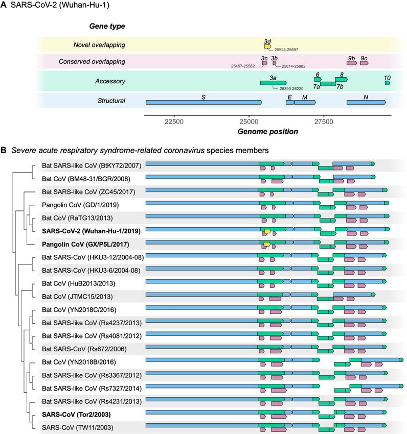
Research article                                                               Evolutionary Biology Microbiology and Infectious Disease

Figure 1. Gene repertoire and evolutionary relationships of Severe acute respiratory syndrome-related coronavirus species members. Only genes
downstream of ORF1ab are shown, beginning with the Spike gene S. (A) Four types of genes and their relative positions in the SARS-CoV-2 Wuhan-Hu-
1 genome (NCBI: NC_045512.2). Genes are colored by type: novel overlapping genes (OLGs) (gold; ORF3d only); conserved OLGs (burgundy);
accessory (green); and structural (blue). Note that ORF3b has been truncated relative to SARS-CoV genomes, whereas ORF8 remains intact (i.e. has not
been split into ORF8a and ORF8b). (B) Genes with intact ORFs in each of 21 Severe acute respiratory syndrome-related coronavirus genomes. Gene
positions are shown relative to each genome, i.e. homologous genes are not precisely aligned. Only the full-length isoforms of ORF3a and ORF3d are
shown (for shorter isoforms, see Table 1). Note that the first 20 codons of ORF3d overlap the last codons of ORF3c (Supplementary file 1), such that
Figure 1 continued on next page

Nelson, Ardern, et al. eLife 2020;9:e59633. DOI: https://doi.org/10.7554/eLife.59633                                                         3 of 29
Research article                                                               Evolutionary Biology Microbiology and Infectious Disease

Figure 1 continued
the beginning of ORF3d involves a triple overlap (ORF3a/ORF3c/ORF3d). ORF3b is full-length in only three sequences (SARS-CoV TW11, SARS-CoV
Tor2, and bat-CoV Rs7327), while the remaining sequences have premature STOP codons (Supplementary file 1). ORF8 is not novel in
SARS-CoV-2 (contra Chan et al., 2020), but is intact in all but five sequences (split into ORF8a and ORF8b in SARS-CoVs TW11 and Tor2; deleted in
bat-CoVs BtKY72, BM48-31, and JTMC15). ORF9b and ORF9c are found throughout this virus species, yet rarely annotated in genomes at NCBI.
The online version of this article includes the following source data and figure supplement(s) for figure 1:
Source data 1. SARS-related-CoV_ALN.fasta.
Source data 2. SARS-related-CoV_ALN.gtf.txt.
Figure supplement 1. Codon permutation analysis to identify candidate overlapping genes in all three forward-strand reading frames of the SARS-
CoV-2 genome.

                                    ribosomal P-site offset (Calviello and Ohler, 2017). Similar peaks are also observed for previously
                                    annotated genes (Figure 2A, Figure 2—figure supplement 1).
                                        To investigate the relationship between ribosome profiling read depth and expressed protein lev-
                                    els, we re-analyzed five publicly available SARS-CoV-2 mass spectrometry (MS) datasets
                                    (Bezstarosti et al., 2020; Bojkova et al., 2020; Davidson et al., 2020; PRIDE Project PXD018581;
                                    Zecha et al., 2020; Materials and methods). We were unable to detect ORF3c, ORF3d, ORF3b,
                                    ORF9c, or ORF10 when employing a 1% false-discovery threshold. This result may reflect the limita-
                                    tions of MS for detecting proteins that are very short, weakly expressed under the specific conditions
                                    tested, or lack detectable peptides; for example, even the envelope protein E is not detected in
                                    some SARS-CoV-2 datasets (Bojkova et al., 2020; Davidson et al., 2020). However, we do find a
                                    strong correlation between protein expression as estimated from MS and the ribosome profiling
                                    read depth observed at upstream peaks, with rS = 0.89 (p=0.0004, Spearman’s rank) (Figure 2, Fig-
                                    ure 2—figure supplement 2). This suggests that the presence and depth of an upstream peak is a
                                    reliable indicator of expression. Specifically, results from both proteomic and ribosome profiling
                                    approaches confirm that N, M, and S are the most highly expressed proteins, with N
                                    constituting ~80% of the total viral protein content. We also observe that the number of reads deter-
                                    mined to be in the correct reading frame (codon position 1) along the gene is moderately correlated
                                    with MS expression values (rS = 0.60, p=0.056), while this is not true for out-of-frame (codon position
                                    2 and 3) reads (rS = 0.39, p=0.237) (Figure 2B, Figure 2—figure supplement 2).
                                        Ribosome profiling reads have a strong tendency to map with their first nucleotide occupying a
                                    particular codon position (i.e. in-frame) (Figure 2—figure supplement 3). We therefore examined
                                    codon position mapping across ORF3a to explore whether the proportions of reads in alternative
                                    frames are higher in the ORF3c/ORF3d region. Analyses were limited to reads of length 30 nt, the
                                    expected size of ribosome-bound fragments, which exhibit positioning indicative of the correct read-
                                    ing frame (codon position 1) of annotated genes, as well as the highest total coverage (Figure 2—
                                    figure supplement 3, Figure 2—figure supplement 4). Sliding windows reveal a peak in the fraction
                                    of reads mapping to the reading frame of ORF3d at its hypothesized locus, with this frame’s maxi-
                                    mum occurring at the center of ORF3d, co-located with a dip in the fraction of reads mapping to the
                                    frame of ORF3a (Figure 2C). These observations are reproducible across treatments (Figure 2—fig-
                                    ure supplement 5), robust to sliding window size (Figure 2—figure supplement 6), and similar to
                                    the reading frame perturbations observed for the OLG ORF9b within N (Figure 2—figure supple-
                                    ment 7). At the same time, the ORF3d peak stands in contrast to the remainder of ORF3a, where
                                    the reading frame of ORF3a predominates, and smaller peaks in the frame of ORF3d are not accom-
                                    panied by a large dip in the frame of ORF3a (Figure 2—figure supplement 8). These observations
                                    suggest that ORF3d is actively translated. Similar conclusions can be drawn for ORF3c, ORF3d-2,
                                    and ORF9b, but not ORF9c (Figure 2C, Figure 2—figure supplement 7, Figure 2—figure supple-
                                    ment 9).
                                        Additional experiments have also provided evidence of ORF3d translation. Gordon et al., 2020
                                    used overexpression experiments to demonstrate that ORF3d (referred to as ‘ORF3b’; Table 1) can
                                    be stably expressed and that it interacts with the mitochondrial protein STOML2. Most compellingly,
                                    ORF3d, ORF8, and N elicit the strongest antibody responses observed in COVID-19 patient sera,
                                    with ORF3d sufficient to accurately diagnose the majority of COVID-19 cases (Hachim et al., 2020).
                                    Because ORF3d is restricted to SARS-CoV-2 and pangolin-CoV (see below), this finding is unlikely to

Nelson, Ardern, et al. eLife 2020;9:e59633. DOI: https://doi.org/10.7554/eLife.59633                                                         4 of 29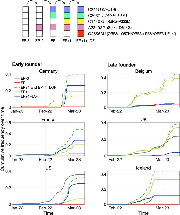
Research article                                                                           Evolutionary Biology Microbiology and Infectious Disease

Table 1. Nomenclature and reading frames for overlapping gene candidates in SARS-CoV-2 ORF3a.
              Reading    Genome positions, Wuhan-Hu-1
Gene*         frame†     (CDS positions, ORF3a)‡             Length            Description                                       References

ORF3a         ss11        25393–26220                        276 codons        Ion channel formation and virus release in        Lu et al., 2010; Cui et al., 2019
              (reference) (1-828)                            (828 nt)          SARS-CoV infection; host cell apoptosis;
                                                                               triggers inflammation; antagonizes interferon

ORF3c         ss13       25457–25582                         42 codons         Features suggestive of a viroporin                First discovered by Cagliani et al., 2020 as
                         (65-190)                            (126 nt)          (Cagliani et al., 2020); lowest pN/pS ratio       ORF3h; ORF3c in Firth, 2020; ORF3c in
                                                                               estimated for any gene in our between-host        Jungreis et al., 2020; ORF3a.iORF1 in
                                                                               selection analysis (Figure 5); overlaps codons    Finkel et al., 2020; ORF3b in Pavesi, 2020
                                                                               22–64 of ORF3a

ORF3d         ss12       25524–25697                         58 codons         Aligned to and named ORF3b by Chan et al.,        Present study; first discovered by Chan et al.,
                         (132-305)                           (174 nt)          2020 but is not homologous to ORF3b;              2020 but misclassified as ORF3b; ORF3b in
                                                                               interferon antagonism has not been                Gordon et al., 2020, Hachim et al., 2020,
                                                                               demonstrated; binds STOML2 mitochondrial          and citing studies; ‘hypothetical protein’ in
                                                                               protein (Gordon et al., 2020); contains a         Pavesi, 2020; ‘a completely different ORF’ in
                                                                               predicted signal peptide in the region            Michel et al., 2020
                                                                               encoding ORF3d-2; contains an X motif in
                                                                               pangolin-CoV but not SARS-CoV-2
                                                                               (Michel et al., 2020); may contribute to
                                                                               differences between SARS-CoV and SARS-
                                                                               CoV-2 in immune response as a unique
                                                                               antigenic target (Hachim et al., 2020; Niloufar
                                                                               Kavian, pers. comm.); overlaps codons 44–102
                                                                               of ORF3a

ORF3d-2       ss12       25596–25697                         34 codons         A shorter isoform of ORF3d that starts after the Discovered by Finkel et al., 2020 as ORF3a-
                         (204-305)                           (102 nt)          first 24 codons, where the majority of            iORF2
                                                                               premature STOP codons in SARS-CoV-2 are
                                                                               located; contains a predicted signal peptide
                                                                               (Finkel et al., 2020); likely expressed at higher
                                                                               levels than full-length ORF3d (Figure 2A)
                                                                               overlaps codons 68–102 of ORF3a

ORF3a-2       ss11        25765–26220                        152 codons        A shorter isoform of ORF3a that starts after the Discovered by Davidson et al., 2020 (pers.
              (reference) (373-828)                          (456 nt)          first 124 codons; evidence of expression         comm.) but referred to as ORF3b
                                                                               separate from that of ORF3a (Davidson et al.,
                                                                               2020); has also been conflated with ORF3b;
                                                                               equivalent to codons 124–276 of ORF3a

ORF3b         ss13       25814–26281; four ORFs at 25814–    156 codons        Full-length in some related viruses, but       Konno et al., 2020 claim functionality for the
region§                  82, 25910–84, 26072–170,            (468 nt); the     truncated by multiple in-frame STOP codons first (23-codon) ORF in SARS-CoV-2; the first
                         and 26183–281 (422-889; ORFs at     four ORFs are     in SARS-CoV-2; longer forms function as        ORF is also mentioned by Wu et al., 2020a
                         422–90, 518–92, 680–778, and 791–   23, 25, 33, and   an interferon antagonist in SARS-related
                         889)                                33 codons         viruses; may contribute to differences between
                                                             (69, 75, 99,      SARS-CoV and SARS-CoV-2 in immune
                                                             and 99 nt)        response; although aligned to ORF3d by
                                                                               Chan et al., 2020, the two are not
                                                                               homologous; overlaps codons 141–276 of
                                                                               ORF3a (first ORF overlaps codons 141–164)

*
Genes are listed by position of start site in the genome from 50 (top) to 30 (bottom).
†
    Nomenclature as described in Wei and Zhang, 2015 and Nelson et al., 2020a: ss = sense-sense (same strand); ss12 = codon position 1 of the reference
frame overlaps codon position 2 of the overlapping frame on the same strand; ss13 = codon position 1 of the reference frame overlaps codon position 3
of the overlapping frame on the same strand. Frame is indicated from the perspective of ORF3a as the reference gene, i.e. ORF3d starts at codon position
3 of ORF3a, while ORF3c and ORF3b start at codon position 2 of ORF3a.
‡
    Positions and counts include STOP codons. Positions or sequences were indicated by the original publications or verified by personal communication if
ambiguous.
§
  The SARS-CoV-2 region homologous to ORF3b of SARS-CoV contains four premature STOP codons and four distinct ORFs (AUG-to-STOP); see Figure 4,
Figure 6, and Supplementary file 1.

                                        be due to cross-reactivity with another coronavirus and provides strong independent evidence of
                                        ORF3d translation during infection.

                                        Protein sequence properties
                                        To further investigate the antigenic properties of ORF3d, we predicted linear T cell epitopes for
                                        each SARS-CoV-2 protein. We employed NetMHCpan (Jurtz et al., 2017) for MHC class I (cytotoxic

Nelson, Ardern, et al. eLife 2020;9:e59633. DOI: https://doi.org/10.7554/eLife.59633                                                                                   5 of 29
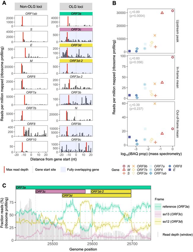
Research article                                                                Evolutionary Biology Microbiology and Infectious Disease

Figure 2. SARS-CoV-2 gene expression in ribosome profiling and mass spectrometry datasets. Ribosome profiling (Ribo-seq) data were obtained from
Finkel et al., 2020; mass spectrometry data were obtained from Davidson et al., 2020 and Bezstarosti et al., 2020. Reads were trimmed to their first
(50 ) nucleotide to minimize statistical dependence while preserving reading frame. Results are shown after pooling samples treated with harringtonine
(SRR11713360, SRR11713361) or lactimidomycin (SRR11713358, SRR11713359) at 5 hr post-infection. (A) Ribosome profiling coverage (read depth) near
translation initiation sites, measured as mean reads per million mapped reads. Only reads of length 29–31 nucleotides were used, chosen for their
enrichment at the start sites of highly expressed annotated genes (Figure 2—figure supplement 4). Light blue backgrounds denote fully (internal)
overlapping genes. Annotated genes show an accumulation of 50 ends of protected reads upstream of the gene’s start site (vertical gray dotted lines),
peaking near the ribosome P-site offset of 12 nt (red = maximum depth). Distributions are largely consistent across individual samples (Figure 2—
figure supplement 1). The ranges of the y-axes vary according to expression level, with the most highly expressed gene (N; Figure 2—figure
supplement 2) having the largest range. (B) Correlation between protein expression as estimated by mass spectrometry and ribosomal profiling. ‘iBAQ
prop’ refers to the relative (proportion of maximum) protein intensity-based absolute quantification (iBAQ) value (Schwanhäusser et al., 2011). Genes
Figure 2 continued on next page

Nelson, Ardern, et al. eLife 2020;9:e59633. DOI: https://doi.org/10.7554/eLife.59633                                                           6 of 29
Research article                                                                Evolutionary Biology Microbiology and Infectious Disease

Figure 2 continued
are denoted by shape and ordinally colored by iBAQ prop from high (red) to low (blue). ‘Upstream peak’ refers to the maximum read depth observed
at the approximate P-site offset (red bars in A), while mean read depths were measured across each gene using 30 nt reads separately for in-frame
(codon position 1) and out-of-frame (codon positions 2 and 3) sites (non-overlapping gene regions only, except for ORF9b). (C) Reading frame of
ribosome profiling reads in the ORF3c/ORF3d region of ORF3a. Solid lines show the fraction of reads in each frame, summed across samples in sliding
windows of 30 nt (step size = 1 nt; read length = 30 nt). Color denotes frame: green = reference frame (ORF3a); burgundy = ss13, the forward-strand
frame encoding ORF3c, whose codon position 3 overlaps codon position 1 of ORF3a; and gold = ss12, the forward-strand frame encoding ORF3d,
whose codon position 2 overlaps codon position 1 of ORF3a. Values are shown for the central nucleotide of each window, with shaded regions
corresponding to 95% binomial confidence intervals. Alternative frame translation is suggested where a given frame (solid line) exceeds its average
across the remainder of the gene (horizontal dashed line; non-OLG regions of ORF3a). Vertical gray dotted lines indicate gene start and end sites. Gray
bars show read depth for each window, with a maximum of 2889 reads at genome position 25442.
The online version of this article includes the following source data and figure supplement(s) for figure 2:
Source data 1. riboseq_upstream_peaks.txt.
Source data 2. expression_data_by_gene_frame.txt.
Source data 3. riboseq_ORF3d_sliding_window.txt.
Figure supplement 1. Ribosome profiling coverage near translation initiation sites for individual samples.
Figure supplement 2. Correlation between protein expression as estimated by mass spectrometry and ribosomal profiling, for individual treatments
and codon positions.
Figure supplement 3. Reading frames occupied by the 50 ends of ribosome profiling reads as a function of read length.
Figure supplement 4. Ribosome profiling read accumulation at gene start sites as a function of read length.
Figure supplement 5. Reading frame of ribosome profiling reads in the ORF3c/ORF3d region of ORF3a for individual treatments.
Figure supplement 6. Reading frame of ribosome profiling reads in the ORF3c/ORF3d region of ORF3a for individual treatments using a smaller
sliding window size of 19 nt.
Figure supplement 7. Reading frame of ribosome profiling reads in the N/ORF9b/ORF9c region.
Figure supplement 8. Reading frame of ribosome profiling reads in the full ORF3a gene.
Figure supplement 9. Reading frame of ribosome profiling reads in the ORF3c/ORF3d region of ORF3a for reads of length 29 nt.

                                     CD8+ T cells), an approach shown to accurately predict SARS-CoV-2 epitopes shared with
                                     SARS-CoV (Grifoni et al., 2020), and NetMHCIIpan (Reynisson et al., 2020) for MHC class II (helper
                                     CD4+ T cells). Specifically, we tested all 9 amino acid (MHC I) or 15 amino acid (MHC II) substrings
                                     of each viral protein for predicted weak or strong binding by MHC. Epitope density was estimated
                                     as the mean number of predicted epitopes overlapping each residue for each protein. We also
                                     tested two sets of negative controls: (1) randomized peptides generated from each protein, repre-
                                     senting the result expected given amino acid content alone and (2) short unannotated ORFs present
                                     in the SARS-CoV-2 genome, representing the result expected for ORFs that have been evolving
                                     without functional constraint.
                                        For CD8+ T cells, the lowest predicted epitope density occurs in ORF3d (1.5 per residue), which
                                     is unexpected given its own amino acid content (p=0.150) and compared to short unannotated
                                     ORFs (p=0.078; permutation tests). The next lowest densities occur in N and ORF8 (Figure 3A).
                                     Intriguingly, as previously mentioned, these three peptides (ORF3d, N, and ORF8) also elicit the
                                     strongest antibody (B cell epitope) responses measured in COVID-19 patient sera (Hachim et al.,
                                     2020), suggesting a possible balance between CD8+ T and B cell epitopes. For CD4+ T cells, again,
                                     ORF3d has one of the lowest predicted epitope densities (5.6 per residue; p0.291), with lower val-
                                     ues seen in only three other genes. Focusing instead on the shorter ORF3d-2 isoform, this protein
                                     contains zero predicted epitopes, which is a highly significant depletion given its own amino acid
                                     content (p=0.001) and compared to short unannotated ORFs (p=0.001). These observations suggest
                                     either a predisposition toward immune escape, allowing gene survival, or the action of selective
                                     pressures on ORF3d or ORF3d-2 to remove epitopes. In stark contrast to ORF3d, ORF3c has the
                                     highest predicted CD8+ T cell epitope density (8.4 per residue), apparently as a function of its amino
                                     acid content (i.e. not differing from its randomized peptides; p=0.996). The enrichment of predicted
                                     epitopes in unannotated proteins (e.g. ORF3c and randomized peptides) but not N, for which
                                     numerous epitopes are documented (Grifoni et al., 2020), demonstrates that ascertainment or
                                     methodological biases cannot account for the depletion of predicted epitopes in ORF3d.
                                        Our structural prediction for the ORF3d protein suggests a-helices connected with coils and an
                                     overall fold model that matches known protein structures (e.g. Protein Data Bank IDs 2WB7 and
                                     6A93) with borderline confidence (Figure 3—figure supplement 1), similar to the predictions of

Nelson, Ardern, et al. eLife 2020;9:e59633. DOI: https://doi.org/10.7554/eLife.59633                                                            7 of 29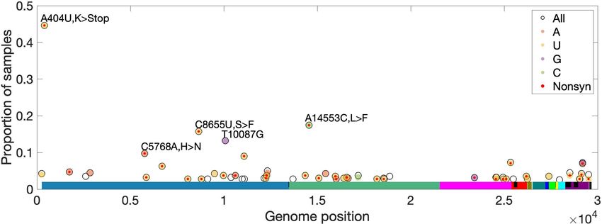
Research article                                                               Evolutionary Biology Microbiology and Infectious Disease

                                    Figure 3. SARS-CoV-2 protein sequence properties. (A) Predicted densities of MHC class I-bound CD8+ T cell 9
                                    amino acid (aa) epitopes (top) and MHC class II-bound CD4+ T cell 15 aa epitopes (bottom). Results for proteins
                                    encoded downstream of ORF1ab are shown. Mean numbers of predicted epitopes per residue (blue bars) are
                                    calculated as the number of epitopes overlapping each amino acid position divided by protein length. Error bars
                                    show 95% confidence intervals. Two sets of negative controls were also tested: (1) n = 1000 randomized peptides
                                    generated from each protein by randomly sampling its amino acids with replacement (dark gray bars),
                                    representing the result expected given amino acid content alone; and (2) short unannotated ORFs, the peptides
                                    encoded by n = 103 putatively nonfunctional ORFs present in the SARS-CoV-2 genome, representing the result
                                    expected for ORFs that have been evolving without functional constraint. For the short unannotated ORFs, the
                                    horizontal gray dotted line shows the mean number of epitopes per residue, and the gray-shaded region shows
                                    the 95% confidence interval (i.e. 2.5% to 97.5% quantiles). ORF3d, N, and ORF8 have the lowest MHC class I
                                    epitope densities; ORF3d, ORF3b, ORF7b, and ORF10 have the lowest MHC class II epitope densities. (B)
                                    Hydrophobicity profiles of amino acid sequences encoded by the three forward-strand reading frames of the
                                    ORF3a/ORF3c/ORF3d gene region, as calculated by the VOLPES server, using the unitless ‘Factor 1’ consensus
                                    hydrophobicity scale. Frame is reported using ORF3a as the reference, for example ss12 refers to the frame
                                    encoding ORF3d, for which codon position 2 overlaps codon position 1 of ORF3a (Figure 2C).
                                    The online version of this article includes the following source data and figure supplement(s) for figure 3:
                                    Source data 1. epitope_summary_MHCI_LONG.txt.
                                    Source data 2. epitope_summary_MHCII_LONG.txt.
                                    Source data 3. hydrophobicity_profiles_ORF3a_corr.txt.
                                    Figure supplement 1. Structural prediction for the ORF3d protein.
                                    Figure supplement 2. Correlations between hydrophobicity profiles of amino acid sequences encoded by the
                                    three forward-strand reading frames of the ORF3a region.
                                    Figure supplement 3. Correlation between hydrophobicity profiles in the amino acid sequences encoded by all
                                    three forward-strand frames of ORF3a, by gene subregion.

Nelson, Ardern, et al. eLife 2020;9:e59633. DOI: https://doi.org/10.7554/eLife.59633                                                         8 of 29
Research article Evolutionary Biology Microbiology and Infectious Disease
Figure 4. Amino acid variation in proteins encoded by genes overlapping ORF3a in viruses of the species Severe acute respiratory syndrome-related
coronavirus. (A) Amino acid sequences of ORF3c, ORF3d, and ORF3b, as encoded by SARS-CoV-2 (Wuhan-Hu-1; NCBI: NC_045512.2). Note that, while
ORF3b encodes a protein of 154 amino acids (aa) in SARS-CoV (NCBI: NC_004718.3), a premature STOP codon in SARS-CoV-2 has resulted in an ORF
encoding only 22 aa. (B) Amino acid alignments of ORF3c, ORF3d, and ORF3b (3b) show their sequence conservation. Black lines indicate STOP
codons in ORF3d and ORF3b, showing their restricted taxonomic ranges. Intact ORF3d is restricted to SARS-CoV-2 and pangolin-CoV GX/P5L;
however, note that ORF3d-2 (denoted 3d-2), a shorter isoform of ORF3d, could have a slightly wider taxonomic range if TTG or GTG are permitted as
translation initiation codons. Full-length ORF3b (ORF3b region) is found throughout members of this virus species, but truncated early in most
genomes outside of SARS-CoV (Supplementary file 1), with the shortest isoform (denoted 3b) found in SARS-CoV-2 and closely related viruses.
The online version of this article includes the following source data for figure 4:
Source data 1. aa_alignments_3c_3d_3b.xlsx.
Chan et al., 2020. Remarkably, biochemical properties that influence the structure of this novel pro-
tein appear to be inherited from the pre-existing ORF3a protein sequence encoded by the overlap-
ping reading frame. It has recently been shown that frame-shifted nucleotide sequences tend to
encode proteins with similar hydrophobicity profiles as a consequence of the standard genetic code
Nelson, Ardern, et al. eLife 2020;9:e59633. DOI: https://doi.org/10.7554/eLife.59633 9 of 29Research article Evolutionary Biology Microbiology and Infectious Disease
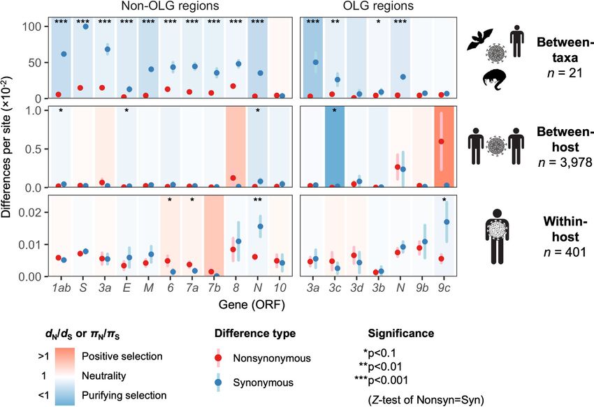
Figure 5. Natural selection analysis of viral nucleotide differences at three hierarchical evolutionary levels.
Nucleotide differences in each virus gene were analyzed at three host levels: between-taxa divergence (d) among
Severe acute respiratory syndrome-related coronavirus genomes infecting bat, human, and pangolin species;
between-host diversity (p) for SARS-CoV-2 infecting human individuals (consensus-level); and within-host diversity
(p) for SARS-CoV-2 infecting human individuals (deep sequencing). Each gene/level is shaded according to the
ratio of mean nonsynonymous to synonymous differences per site to indicate purifying selection (dN/dS < 1 or pN/
pS < 1; blue) or positive selection (dN/dS > 1 or pN/pS > 1; red). The extremely low ratio for ORF3c was artificially
adjusted to allow the display of other ratios, and a Jukes-Cantor correction was applied to dN and dS values.
Values range from a minimum of pN/pS = 0.04 (ORF3c, between-host; p=0.0410) to a maximum of 21.0 (ORF9c,
between-host; p=0.126), where significance was evaluated using Z-tests of the hypothesis that dN-dS = 0 or pN-
pS = 0 (10,000 bootstrap replicates, codon unit). The mean of all pairwise comparisons is shown for sequenced
genomes only, i.e. no ancestral sequences were reconstructed or inferred. For each gene, sequences were only
included in the between-species analysis if a complete, intact ORF (no STOPs) was present. Genes containing an
overlapping gene (OLG) in a different frame were analyzed separately for non-OLG and OLG regions using
SNPGenie and OLGenie, respectively. For ORF3b, only the region corresponding to the first ORF in SARS-CoV-2
(Table 1) was analyzed. The short overlap between ORF1a and ORF1b (nsp11 and nsp12) was excluded from the
analysis. Error bars represent the standard error of mean pairwise differences. See Materials and methods for
further details.
The online version of this article includes the following source data and figure supplement(s) for figure 5:
Source data 1. selection_three_levels.txt.
Figure supplement 1. SARS-CoV-2 between-host nucleotide diversity and allele frequencies as a function of date
during the initial period of the COVID-19 pandemic.
Figure supplement 2. Correlation between natural selection and gene expression.
(Bartonek et al., 2020). To explore whether this is the case for ORF3d, we used the VOLPES server
(Bartonek and Zagrovic, 2019) to calculate the hydrophobicity profiles (Atchley et al., 2005) of the
peptides encoded by all three frames of ORF3a. The maximum correlation observed occurs between
the frames of ORF3a (ss11) and ORF3d (ss12) in the region encoding ORF3d (ORF3a residues 64–
102), with rS = 0.87 (p=5.79 10 13, Spearman’s rank), which is much stronger than the correlation
between these frames observed for the non-OLG residues of ORF3a (rS = 0.27, p=8.70 10 4)
(Figure 3C; Figure 3—figure supplement 2). This conservation of a structure-related property with
the ORF3a protein provides further evidence for ORF3d functionality, and may predispose this
Nelson, Ardern, et al. eLife 2020;9:e59633. DOI: https://doi.org/10.7554/eLife.59633 10 of 29Research article Evolutionary Biology Microbiology and Infectious Disease
Figure 6. Between-taxa sliding window analysis of natural selection on overlapping frames of ORF3a. Pairwise sliding window analysis (window
size = 50 codons; step size = 1 codon) of selection across members of the species Severe acute respiratory syndrome-related coronavirus. OLG-
appropriate dN/dS values were computed using OLGenie (Nelson et al., 2020a), a method that is conservative (non-conservative) for detecting
purifying (positive) selection, and a Jukes-Cantor correction for multiple hits was employed. On the left-hand side, each genome is compared to SARS-
CoV-2 in the ss12 reading frame of ORF3a, which contains ORF3d (Table 1 and Figure 2C). This frame shows evidence for purifying selection specific
to the ORF3d region that is limited to the comparison with pangolin-CoV GX/P5L. On the right-hand side, this analysis is repeated for the ss13 reading
frame of ORF3a, which contains ORF3c and ORF3b (Table 1 and Figure 2C), this time with respect to SARS-CoV, where full-length ORF3b is functional.
This frame shows constraint across much of this gene and virus species, and ORF3c in particular is deeply conserved.
The online version of this article includes the following source data and figure supplement(s) for figure 6:
Source data 1. SARS-CoV-2-ref_ORF3a_ss12_windows.txt.
Source data 2. SARS-CoV-ref_ORF3a_ss13_windows.txt.
Figure supplement 1. Between-taxa sliding window of genes overlapping N.
region toward de novo gene birth. Again, ORF3c shows the opposite pattern, as the minimum corre-
lation observed between hydrophobicity profiles occurs between the frames of ORF3a (ss11) and
ORF3c (ss13) in the region encoding ORF3c (ORF3a residues 22–43), with rS = 0.40
(p=6.16 10 2). Such disparate hydrophobicity profiles may be due to the strong conservation of
ORF3c (see below).
ORF3d taxonomic distribution and origins
To assess the origin of ORF3d and its conservation within and among host taxa, we aligned 21
Severe acute respiratory syndrome-related coronavirus genomes reported by Lam et al., 2020
(Supplementary file 1), limiting our analysis to those with an annotated ORF1ab and no frameshift
variants relative to SARS-CoV-2 in the core genes ORF1ab, S, ORF3a, E, M, ORF7a, ORF7b, and N
(Supplementary file 1; SARS-related-CoV_ALN.fasta, supplementary data). All other genes are also
intact (i.e. there are no mid-sequence STOP codons) in all genomes (Supplementary file 1), with the
exception of ORF3d, ORF3b, and ORF8 (Figure 4). Specifically, ORF3d is intact in only two sequen-
ces: SARS-CoV-2 Wuhan-Hu-1 and pangolin-CoVs from Guangxi (GX/P5L). Full-length ORF3b is
intact in only three sequences: SARS-CoV TW11, SARS-CoV Tor2, and bat-CoV Rs7327, with the
Nelson, Ardern, et al. eLife 2020;9:e59633. DOI: https://doi.org/10.7554/eLife.59633 11 of 29Research article Evolutionary Biology Microbiology and Infectious Disease
remainder having premature STOP codons (Supplementary file 1). Finally, ORF8 is intact in all but
five sequences, where it contains premature STOP codons or large-scale deletions (Figure 1B).
The presence of intact ORF3d homologs among viruses infecting different host species (human
and pangolin) raises the possibility of functional conservation. However, the taxonomic distribution
of this ORF is incongruent with whole-genome phylogenies in that ORF3d is intact in the pangolin-
CoV more distantly related to SARS-CoV-2 (GX/P5L) but not the more closely related one (GD/1)
(Figure 4), a finding confirmed by the alignment of Boni et al., 2020. New sequence data reveal
similarly puzzling trends: ORF3d contains STOP codons in the closely related bat-CoV RmYN02
(GISAID: EPI_ISL_412977; data not shown), but it is intact in three more distantly related bat-CoVs
discovered in Rwanda and Uganda, where it is further extended by 20 codons (total 78 codons) but
shows no evidence of conservation with SARS-CoV-2 (Wells et al., 2020 and pers. comm; data not
shown). Further, phylogenies of the 21 Severe acute respiratory syndrome-related coronavirus
genomes built on ORF3a are incongruent with whole-genome phylogenies, likely due to the pres-
ence of recombination breakpoints in ORF3a near ORF3d (Boni et al., 2020; Rehman et al., 2020).
Recombination, convergence, or recurrent loss may therefore have played a role in the origin and
taxonomic distribution of ORF3d.
Between-taxa divergence
To estimate natural selection on ORF3d, we measured viral diversity at three hierarchical evolution-
ary levels: between-taxa, between-host, and within-host. Specifically, between-taxa refers to diver-
gence (d) among the 21 aforementioned viruses infecting bat, human, or pangolin (Figure 1B);
between-host refers to diversity (p) between consensus-level SARS-CoV-2 genomes infecting differ-
ent human individuals; and within-host refers to p in deeply sequenced ‘intrahost’ SARS-CoV-2 sam-
ples from single human individuals. At each level, we inferred selection by estimating mean pairwise
nonsynonymous (dN or pN; amino acid changing) and synonymous (dS or pS; not amino acid chang-
ing) distances among all sequences. Importantly, we combined standard (non-OLG) methods
(Nei and Gojobori, 1986; Nelson et al., 2015) with OLGenie, a new dN/dS method tailored for
OLGs (hereafter OLG dN/dS), which we previously used to verify purifying selection on a novel OLG
in HIV-1 (Nelson et al., 2020a).
The only gene to show significant evidence of purifying selection at all three evolutionary levels is
the nucleocapsid gene N (Figure 5), which undergoes disproportionately low rates of nonsynony-
mous change (dN/dS < 1 and pN/pS < 1) specifically in its non-OLG regions, evidencing strict func-
tional constraint. N is also the most highly expressed gene (Figure 2—figure supplement 2),
confirming that selection has more opportunity to act when a protein is manufactured in abundance
(Figure 2B; Supplementary file 1) (Materials and methods). Importantly, this signal can be missed if
non-OLG methods are applied to N without accounting for its internal OLGs, ORF9b and ORF9c
(e.g. at the between-host level, p=0.0268 when excluding OLG regions, but p=0.411 when including
them; Supplementary file 1).
With respect to ORF3d, comparison of Wuhan-Hu-1 to pangolin-CoV GX/P5L (NCBI:
MT040335.1) yields OLG dN/dS = 0.14 (p=0.264) (Figure 5), whereas inclusion of a third allele found
in pangolin-CoV GX/P4L (NCBI: MT040333.1) yields 0.43 (p=0.488) (Supplementary file 1). Because
this is suggestive of constraint, we performed sliding windows of OLG dN/dS across the length of
ORF3a. Pairwise comparisons of each sequence to SARS-CoV-2 reveal OLG dN/dS < 1 that is specific
to the reading frame, genome positions, and species in which ORF3d is intact (pangolin-CoV GX/
P5L) (Figure 6, left). This signal is independent of whether STOP codons are present, so its consil-
ience with the only intact ORF in this region and species is highly suggestive of purifying selection.
We note that this conclusion does not contradict studies which fail to find evidence of ORF3d con-
servation when comparing taxa where ORF3d is absent (e.g. Jungreis et al., 2020), because ORF3d
is a novel gene and would by definition lack such conservation. This contrastive signal is also similar
to what is observed for the known OLGs ORF3b (in comparisons to SARS-CoV; Figure 6, right), and
ORF9b and ORF9c (in both viruses; Figure 6—figure supplement 1).
Between-host evolution and pandemic spread
Purifying selection can be specific to just one taxon, as in the case of novel genes. Thus, to measure
selection within SARS-CoV-2 only, we obtained 3978 high-quality human SARS-CoV-2 consensus
Nelson, Ardern, et al. eLife 2020;9:e59633. DOI: https://doi.org/10.7554/eLife.59633 12 of 29Research article Evolutionary Biology Microbiology and Infectious Disease
Table 2. The mutational path to European pandemic founder haplotypes*.
Variant EP–3 EP–2 EP† EP+1† EP+1+LOF
0
C241U (5 -UTR) - - + + +
C3037U (nsp3-F106F) - - + + +
C14408U (RdRp-P323L) - - - + +
A23403G (Spike-D614G) - + + + +
G25563U - - - - +
(ORF3a-Q57H/ORF3c-R36I/ORF3d-E14*)
Earliest collection§ 24-Dec 7-Feb 28-Jan 20-Feb 21-Feb
Earliest location§ Wuhan Wuhan Munich (Shanghai)‡ Lombardy Hauts de France
Occurrence in China 233 1 1 (2)‡ 0 0
Occurrence in Europe 458 0 21 1153 310
Occurrence in Italy 1 0 0 27 0
Occurrence in Germany 15 0 1 11 21
Occurrence in Belgium 27 1 20 187 27
Occurrence in UK 210 0 0 338 38
Occurrence in Iceland 56 0 0 212 54
Occurrence in France 14 0 72 102
Occurrence in US 467 0 0 88** 326**
Total in GISAID†† 1610 2 22 1455 752
*
Haplotypes are here defined by the presence (+) or absence (-) of five high-frequency variants (rows 1–5), and other variants with lower frequencies on
these backgrounds are ignored. EP-1 is not observed in our dataset.
†
The EP haplotype is first detected in German patient #4 and is a documented founder for coronavirus spread in Germany (Rothe et al., 2020). Neither
the EP nor EP+1 haplotypes were detectable between January 28 and February 20, although they immediately became a major haplotype once EP+1 was
detectable. Failure to detect these two haplotypes during these 3 weeks could potentially be explained by ascertainment bias, for example lack of testing
for travel-independent cases.
‡
This Shanghai sample (GISAID: EPI_ISL_416327) comprises 1.32% poly-Ns and failed our quality control criteria, but is added here since it is potentially rel-
evant to the origin of the EP haplotype. Including this sample, the EP haplotype is observed in Shanghai twice.
§
The earliest collection location and time are highly subject to collection and submission bias and do not necessarily reflect where the mutation/haplotype
first occurred.
**
There is likely a testing bias in the United States, as the EP+1+LOF haplotype was often detected in Washington but EP+1 was not.
††
These numbers are based on 3853 samples from December 24 to April 1 at the time of GISAID accession that passed both our quality control criteria for
alignment and for this particular analysis (i.e. no ambiguous genotype calls among the five SNPs in this table), unless otherwise stated.
sequences from GISAID (accessed April 10, 2020; Supplementary file 1; Supplementary file 2).
Between-host diversity was sufficient to detect marginally significant purifying selection across all
genes (pN/pS = 0.50, p=0.0613, Z-test; Figure 5—figure supplement 1) but not most individual
genes (Figure 5). Therefore, we instead investigated single mutations over time, limiting to 27 high-
frequency variants (minor allele frequency 2%; Supplementary file 1).
One high-frequency mutation occurred in ORF3d: G25563U, here denoted ORF3d-LOF (ORF3d-
loss-of-function). This mutation causes a STOP codon in ORF3d (ORF3d-E14*) but nonsynonymous
changes in both ORF3a (ORF3a-Q57H) and ORF3c (ORF3c-R36I), another OLG that overlaps this site
(Figure 1A). ORF3d-LOF is not observed in any other species member included in our analysis (Fig-
ure 4; SARS-related-CoV_ALN.fasta, supplementary data). During the first months of the COVID-19
pandemic, ORF3d-LOF increased in frequency (Supplementary file 1) in multiple locations (Fig-
ure 5—figure supplement 1D; Supplementary file 1), making this mutation a candidate for natural
selection on ORF3a, ORF3c, ORF3d, or any combination thereof (Kosakovsky-Pond, 2020). How-
ever, temporal allele frequency trajectories (Figure 5—figure supplement 1D) and similar signals
from phylogenetic branch tests may also be caused by founder effects or genetic drift, and are
Nelson, Ardern, et al. eLife 2020;9:e59633. DOI: https://doi.org/10.7554/eLife.59633 13 of 29Research article Evolutionary Biology Microbiology and Infectious Disease Figure 7. Pandemic spread of the EP+1 haplotype and the hitchhiking of ORF3d-LOF. The mutational path leading to EP+1+LOF is shown in the upper panel. Cumulative frequencies of haplotypes in samples from Germany and five other countries with the most abundant sequence data are shown in the lower panel. Countries are grouped into early founder (left) and late founder (right) based on the presence or absence of SARS-CoV-2 samples from January, respectively. In the early founder group, EP–3 (gray) is observed earlier than other haplotypes in France and the US, and EP (red) is observed early in Germany, giving them the advantage of a founder effect. However, neither EP nor EP–3 dominate later spread; instead, EP+1 (yellow) and EP+1+LOF (blue) increase much faster despite their later appearance in these countries. In the late founder group, multiple haplotypes appear at approximately the same time, but EP–3 and EP spread more slowly. The green dashed line shows the combined frequencies of EP+1 and EP+1+LOF (yellow and blue, respectively). Note that EP–1 is never observed in our dataset. Nelson, Ardern, et al. eLife 2020;9:e59633. DOI: https://doi.org/10.7554/eLife.59633 14 of 29
Research article                                                            Evolutionary Biology Microbiology and Infectious Disease

                                    susceptible to ascertainment bias (e.g. preferential sequencing of imported infections and uneven
                                    geographic sampling) and stochastic error (e.g. small sample sizes).
                                       To partially account for these confounding factors, we instead constructed the mutational path
                                    leading from the SARS-CoV-2 haplotype collected in December 2019 to the haplotype carrying
                                    ORF3d-LOF. This path involves five mutations (C241U, C3037U, C14408U, A23403G, G25563U),
                                    constituting five observed haplotypes (EP–3 ! EP–2 ! EP ! EP+1 ! EP+1+LOF), shown in Table 2.
                                    Here, EP is suggested to have driven the European Pandemic (detected in German patient #4; see
                                    footnote 3 of Table 2; Korber et al., 2020; Rothe et al., 2020); EP–3 is the Wuhan founder haplo-
                                    type; EP–1 is never observed in our dataset; and LOF refers to ORF3d-LOF. We then documented
                                    the frequencies and earliest collection date of each haplotype (Table 2) to determine when ORF3d-
                                    LOF occurred on the EP background.
                                       Surprisingly, despite its expected predominance in Europe due to a founder effect, the EP haplo-
                                    type is extremely rare. By contrast, haplotypes with one additional mutation (C14408U; RdRp-P323L)
                                    on the EP background are common in Europe, and ORF3d-LOF occurred very early on this back-
                                    ground to create EP+1+LOF from EP+1. Neither of these two haplotypes was initially observed in
                                    China (Table 2), suggesting that they might have arisen in Europe. Thus, we further partitioned the
                                    samples into two groups, corresponding to countries with early founders (January samples) or only
                                    late founders (no January samples) (Figure 7). In the early founder group, EP–3 (Wuhan) is the first
                                    haplotype detected in most countries, consistent with most early COVID-19 cases being related to
                                    travel from Wuhan. Because this implies that genotypes EP–3 and EP had longer to spread in the
                                    early founder group, it is surprising that their spread is dwarfed by an increase in EP+1 and
                                    EP+1+LOF starting in late February. This turnover is most evident in the late founder group, where
                                    multiple haplotypes are detected in a narrow time window, and the number of cumulative samples is
                                    always dominated by EP+1 and EP+1+LOF. Thus, while founder effects and drift are plausible
                                    explanations, it is also worth investigating whether the early spread of ORF3d-LOF may have been
                                    caused by its linkage with another driver, either C14408U (+1 variant) or a subsequent variant(s)
                                    occurring on the EP+1+LOF background (Discussion).

                                    Within-host diversity and mutational bias
                                    Examination of within-host variation across multiple samples allows detection of recurrent mutations,
                                    which might indicate mutation bias or within-host selection. To investigate these possibilities, we
                                    obtained 401 high-depth ‘intrahost’ human SARS-CoV-2 samples from the Sequence Read Archive
                                    (Supplementary file 1) and called SNPs relative to the Wuhan-Hu-1 reference genome. Within
                                    human hosts, 42% of SNPs passed our false-discovery rate criterion (Materials and methods), with a
                                    median passing minor allele frequency of 2% (21 of 1344 reads). Using these variants to estimate
                                    within-host diversity, ORF3d does not show significant evidence of selection, with OLG pN/pS = 1.5
                                    (p=0.584) (Figure 5). We also examined six high-depth samples of pangolin-CoVs from Guangxi, but
                                    no conclusions could be drawn due to low sequence quality (Materials and methods;
                                    Supplementary file 1).
                                        To identify recurrent mutations in multiple SARS-CoV-2 samples, separately for each site, we lim-
                                    ited to samples for which the major or fixed allele is also ancestral (i.e. matches Wuhan-Hu-1). At
                                    such sites, precluding sequencing artifacts or coinfection by multiple genotypes, minor alleles occur-
                                    ring in more than one sample are expected to be derived and recurrent (i.e. identical by state but
                                    not descent). For ORF3d, we observe ORF3d-LOF as a minor allele in three (0.94%) of 320 samples
                                    (frequencies of 20.7%, 6.0%, and 2.2% in samples SRR11410536, SRR11479046, and SRR11494643,
                                    respectively). This proportion of samples with a recurrent mutation is high but not unusual, as 1.7%
                                    of observed minor variants have an equal or higher proportion of recurrence. Additionally, no muta-
                                    tions in ORF3d recur in >2.5% of samples (Figure 8). Thus, we find no evidence that within-host
                                    selection or mutation pressure are involved in the spread of ORF3d-LOF. However, a small number
                                    of other loci exhibit high rates of recurrent mutation, with five mutations independently observed
                                    in ~10% of samples or more (Materials and methods; Figure 8). Surprisingly, another STOP mutation
                                    (A404U; NSP1-L47*) is never a major allele but is observed at low frequencies in 44% of samples
                                    (Figure 8, Figure 8—figure supplement 1), unexplainable by mutational bias and warranting
                                    investigation.

Nelson, Ardern, et al. eLife 2020;9:e59633. DOI: https://doi.org/10.7554/eLife.59633                                                   15 of 29
Research article Evolutionary Biology Microbiology and Infectious Disease
Figure 8. High-frequency within-host mutations. For each site, mutations that occur in more than 2.5% of samples
are shown, limiting to samples where the major or fixed allele matches Wuhan-Hu-1 at that site. The y-axis shows
the proportion of such samples having the indicated minor (derived) allele. Each locus has up to three possible
single-nucleotide derived alleles compared to the reference background. Open circles (black outlines) show the
proportion of samples having any of the three possible derived alleles (’All’), while solid circles (color fill) show the
proportion of samples having a specific derived allele (equivalent to ’All’ if only one variant is observed). For most
sites, only one derived mutation type (e.g. C!U) is observed across all samples. Precluding co-infection by
multiple genotypes and sequencing errors, derived mutations occurring in more than one sample (y-axis) must be
identical by state but not descent (i.e. recurrent). Genome positions are plotted on the x-axis, with distinct genes
shown in different colors and overlapping genes shown as black blocks within reference genes. Nonsynonymous
and nonsense mutations (together denoted ’Nonsyn’) are indicated with a red dot. Source data are available in the
supplementary material.
The online version of this article includes the following figure supplement(s) for figure 8:
Figure supplement 1. Recurrent nonsynonymous mutations observed in multiple human hosts.
Discussion
Our analyses provide strong evidence that SARS-CoV-2 contains a novel overlapping gene (OLG),
ORF3d, that has not been consistently identified or fully analyzed before this study. The annotation
of a newly emerged virus is difficult, particularly in genome regions subject to frequent gene gain
and loss. Moreover, due to the inherent difficulty of studying OLGs, they tend to be less carefully
documented than non-OLGs; for example, ORF9b and ORF9c are still not annotated in the most-
used reference genome, Wuhan-Hu-1 (last accessed September 26, 2020). Therefore, de novo and
homology-based annotation are both essential, followed by careful expression analyses using multi-
omic data and evolutionary analyses within and between species. In particular, we emphasize the
importance of using whole-gene or genome alignments when inferring homology for both OLGs and
non-OLGs, taking into account genome positions and all reading frames. Unfortunately, in the case
of SARS-CoV-2, the lack of such inspections has led to mis-annotation and a domino effect. For
example, homology between ORF3b (SARS-CoV) and ORF3d (SARS-CoV-2) has erroneously been
implied, leading to unwarranted inferences of shared functionality (Table 1). Given the rapid growth
of the SARS-CoV-2 literature, it is likely this mistake will be further propagated. We therefore pro-
vide a detailed annotation of Wuhan-Hu-1 protein-coding genes and codons in Supplementary file
1, as a resource for future studies.
Our study highlights the highly dynamic process of frequent losses and gains of accessory genes
in the species Severe acute respiratory syndrome-related coronavirus, with the greatest functional
constraint typically observed for the most highly expressed genes (Figure 5—figure supplement 2).
With respect to gene loss, while many accessory genes may be dispensable for viruses in cell culture,
they often play an important role in natural hosts (Forni et al., 2017). Thus, their loss may represent
a key step in adaptation to new hosts after crossing a species barrier (Gorbalenya et al., 2006). For
example, the absence of full-length ORF3b in SARS-CoV-2 has received attention from only a few
authors (e.g. Lokugamage et al., 2020), even though it plays a central role in SARS-CoV infection
and early immune interactions as an interferon antagonist (Kopecky-Bromberg et al., 2007), with
Nelson, Ardern, et al. eLife 2020;9:e59633. DOI: https://doi.org/10.7554/eLife.59633 16 of 29Research article Evolutionary Biology Microbiology and Infectious Disease
effects modulated by ORF length (Zhou et al., 2012). Thus, the absence or truncation of ORF3b in
SARS-CoV-2 may be immunologically important (Yuen et al., 2020), for example in the suppression
of type I interferon induction (Konno et al., 2020; Lokugamage et al., 2020). Further, loss of func-
tion (LOF) mutations in ORF3d and any other ORFs need to be taken into account for testing or ther-
apeutics, particularly in light of predicted cellular interactions (Gordon et al., 2020).
With respect to gene gain, the apparent presence of ORF3d coincident with the inferred entry of
SARS-CoV-2 into humans from a hitherto undetermined reservoir host suggests that this gene is
functionally relevant for the emergent properties of SARS-CoV-2, analogous to asp for HIV-1-M
(Cassan et al., 2016). The mechanisms and detailed histories of gene birth, neo-functionalization,
and survival of novel genes remain unclear. However, our findings that ORF3d remarkably inherited
its hydrophobicity profile from the overlapping region of ORF3a, that ORF3d is depleted for pre-
dicted CD8+ and CD4+ T cell epitopes, and that ORF3d exhibits potential purifying selection
between SARS-CoV-2 and pangolin-CoV GX/P5L, provide strong leads for further research. Indeed,
both the structural and immunogenic properties of new viral proteins deserve further attention and
are important parts of their evolutionary story.
Failure to account for OLGs can lead to erroneous inference of (or failure to detect) natural selec-
tion in known genes, and vice versa. For example, a synonymous variant in one reading frame is very
likely to be nonsynonymous in a second overlapping frame. As a result, purifying selection in the sec-
ond frame usually lowers dS (raises dN/dS) in the first frame, increasing the likelihood of mis-inferring
positive selection (Holmes et al., 2006; Sabath et al., 2008; Nelson et al., 2020a). Such errors
could, in turn, lead to mischaracterization of the genetic contributions of OLG loci to important viral
properties such as incidence and persistence. One potential consequence is misguided countermea-
sure efforts, for example through failure to detect functionally conserved or immunologically
important genome regions.
Although our study focuses on ORF3d, other OLGs have been proposed in SARS-CoV-2 and war-
rant investigation. ORF3d-2, a shorter isoform of ORF3d (Table 1), shows evidence of higher overall
expression than ORF3d (Figure 2A). However, analysis of full-length ORF3d is confounded by its 20
codon triple overlap with both ORF3a and ORF3c, making expression and selection results difficult
to compare between the two isoforms. Although not evolutionarily novel, the recently discovered
ORF3c (Table 1) shows deep conservation among viruses of this species (Cagliani et al., 2020;
Firth, 2020; Jungreis et al., 2020), the lowest pN/pS ratio observed in our between-host analysis
(Figure 5), strong evidence for translation in SARS-CoV-2 (Figure 2), and the highest predicted
CD8+ T cell epitope density (Figure 3). Within S, S-iORF2 (genome positions 21768–21863) also
shows evidence of translation from ribosome profiling (Finkel et al., 2020), and between-host com-
parisons suggest purifying selection (pN/pS = 0.22, p=0.0278) (Table 1). We therefore suggest that
the SARS-CoV-2 genome could contain additional undocumented or novel OLGs.
Our comprehensive evolutionary analysis of the SARS-CoV-2 genome demonstrates that many
genes are under relaxed purifying selection, consistent with the exponential growth of the virus
(Gazave et al., 2013). At the between-host level, nucleotide diversity increases somewhat over the
initial period of the COVID-19 pandemic, tracking the number of locations sampled, while the pN/pS
ratio remains relatively constant at 0.46 (±0.030 SEM) (Figure 5—figure supplement 1B). Other
genes differ in the strength and direction of selection at the three evolutionary levels, for example
ORF9c (Figure 5), suggesting a shift in function or importance over time or between different host
species or individuals. ORF3d and ORF8 are among the youngest genes in SARS-CoV-2, being taxo-
nomically restricted to a subset of betacoronaviruses (Cui et al., 2019), and both exhibit high rates
of change and turnover (Figure 1; Figure 5; SARS-related-CoV_ALN.fasta, supplementary data).
High between-host pN/pS was also observed in ORF8 of SARS-CoV, perhaps due to a relaxation of
purifying selection upon entry into civet cats or humans (Forni et al., 2017). However, ORF3d and
ORF8 both exhibit strong antibody (B cell epitope) responses (Hachim et al., 2020) and predicted
T cell epitope depletion (Figure 3) in SARS-CoV-2. This highlights the important connection between
evolutionary and immunologic processes (Daugherty and Malik, 2012), as antigenic peptides may
impose a fitness cost for the virus by allowing immune detection. Thus, the loss or truncation of
these genes may share an immunological basis and deserves further attention.
The quick expansion of ORF3d-LOF (EP+1+LOF) and its background (EP+1) during this pandemic
is surprising, given that a founder effect would have favored other variants that arrived earlier. How-
ever, newer data show that ORF3d-LOF has remained at relatively low frequencies compared to EP
Nelson, Ardern, et al. eLife 2020;9:e59633. DOI: https://doi.org/10.7554/eLife.59633 17 of 29You can also read