The Different Facets of Heart Rate Variability in Obstructive Sleep Apnea
←
→
Page content transcription
If your browser does not render page correctly, please read the page content below
REVIEW
                                                                                                                                                     published: 22 July 2021
                                                                                                                                            doi: 10.3389/fpsyt.2021.642333

                                                The Different Facets of Heart Rate
                                                Variability in Obstructive Sleep Apnea
                                                Hua Qin 1*, Nicolas Steenbergen 2 , Martin Glos 1 , Niels Wessel 3 , Jan F. Kraemer 3 ,
                                                Fernando Vaquerizo-Villar 4,5 and Thomas Penzel 1,6*
                                                1
                                                  Interdisciplinary Center of Sleep Medicine, Charité-Universitätsmedizin Berlin, Berlin, Germany, 2 Imperial College London
                                                School of Medicine, London, United Kingdom, 3 Department of Physics, Humboldt Universität zu Berlin, Berlin, Germany,
                                                4
                                                  Biomedical Engineering Group, Universidad de Valladolid, Valladolid, Spain, 5 Centro de Investigación Biomédica en
                                                Red-Bioingeniería, Biomateriales y Nanomedicina, Valladolid, Spain, 6 Saratov State University, Russian Federation, Saratov,
                                                Russia

                                                Obstructive sleep apnea (OSA), a heterogeneous and multifactorial sleep related
                                                breathing disorder with high prevalence, is a recognized risk factor for cardiovascular
                                                morbidity and mortality. Autonomic dysfunction leads to adverse cardiovascular
                                                outcomes in diverse pathways. Heart rate is a complex physiological process involving
                                                neurovisceral networks and relative regulatory mechanisms such as thermoregulation,
                                                renin-angiotensin-aldosterone mechanisms, and metabolic mechanisms. Heart rate
                                                variability (HRV) is considered as a reliable and non-invasive measure of autonomic
                                                modulation response and adaptation to endogenous and exogenous stimuli. HRV
                              Edited by:        measures may add a new dimension to help understand the interplay between cardiac
                     Carolina Lombardi,
            Istituto Auxologico Italiano        and nervous system involvement in OSA. The aim of this review is to introduce the various
                           (IRCCS), Italy       applications of HRV in different aspects of OSA to examine the impaired neuro-cardiac
                       Reviewed by:             modulation. More specifically, the topics covered include: HRV time windows, sleep
                      Frederic Roche,
      Université Jean Monnet, France
                                                staging, arousal, sleepiness, hypoxia, mental illness, and mortality and morbidity. All
                Vitor Engracia Valenti,         of these aspects show pathways in the clinical implementation of HRV to screen,
     São Paulo State University, Brazil         diagnose, classify, and predict patients as a reasonable and more convenient alternative
                 *Correspondence:               to current measures.
                           Hua Qin
                  hua.qin@charite.de            Keywords: obstructive sleep apnea, heart rate variability, autonomic dysfunction, central autonomic networks,
                     Thomas Penzel              time-window analysis, time-domain analysis, frequency-domain analysis, non-linear analysis
            thomas.penzel@charite.de

                    Specialty section:          INTRODUCTION
          This article was submitted to
                       Sleep Disorders,         Obstructive sleep apnea (OSA) is closely associated with neurocognitive, behavioral,
                a section of the journal        psychophysiological states, and cardiovascular outcomes (1–3). It is estimated that globally
                 Frontiers in Psychiatry
                                                ∼1 billion adults have mild to severe sleep apnea. Some countries have a prevalence over
       Received: 15 December 2020               50% and it still is increasing. The consequent health and financial burden can be minimized
           Accepted: 14 June 2021               by effective diagnosis and treatment (4). To that effect, recently, the role of cardiovascular
           Published: 22 July 2021
                                                autonomic dysfunction has received increasing attention as an independent risk factor for
                               Citation:        clinical complications in OSA (5). Heart rate variability (HRV) has been generally accepted as a
       Qin H, Steenbergen N, Glos M,
                                                non-invasive tool to quantify cardiovascular autonomic modulation under varying healthy and
                 Wessel N, Kraemer JF,
Vaquerizo-Villar F and Penzel T (2021)
                                                pathogenic conditions (6, 7). HRV measures the variation between beat-to-beat intervals over a
    The Different Facets of Heart Rate          time series (6). It is an integrated reflection of central-peripheral neural feedback mechanisms to
Variability in Obstructive Sleep Apnea.         the heart via mediating sympathovagal inflow and outflow (8). Previous studies suggested that in
          Front. Psychiatry 12:642333.          conjunction with brain imaging, HRV analysis has been used to investigate the connection between
     doi: 10.3389/fpsyt.2021.642333             autonomic cardiac modulation and sleeping brain activity (9).

Frontiers in Psychiatry | www.frontiersin.org                                          1                                              July 2021 | Volume 12 | Article 642333
Qin et al. Heart Rate Variability in OSA
Currently, HRV analysis, including time-domain, frequency- TABLE 1 | Selected time-domain HRV measures.
domain, and non-linear analysis, is used to explore the activities
Variable Units Definition
of sympathetic and parasympathetic nervous systems (6, 10).
Time-domain analysis quantifies the magnitudes of variation. Time-domain analysis
The most relevant time-domain parameters are described in SDNN ms Standard deviation of normal to normal
Table 1. For example, the standard deviation of normal-to- (NN) interval time series
normal intervals (SDNN), a global HRV metric, is frequently SDANNX (X = 1, 5) ms Standard deviation of BBI averages in
used as a prognostic indicator of cardiovascular risk in successive X-minute intervals
different populations (11). Frequency-domain analysis is used for RMSSD ms Square root of the mean squared
partitioning the rhythms of electrocardiography (ECG) signals differences of successive NN intervals
into different frequencies (12, 13). This analysis helps gain a pNNX (X = 50, 100, 200) % NN>Xms counts divided by the total
number of all NN intervals.
better understanding of cardiac control as ECG frequencies
pNNlX (X = 10, 20, 30) % NNQin et al. Heart Rate Variability in OSA
TABLE 3 | Selected non-linear HRV parameters and methods. TIME-WINDOW ANALYSIS TECHNOLOGY
Variable Units Definition
OF HRV
Chaotic invariant analysis HRV is usually measured over a short-term (5–15 min) or long-
D2 – Correlation dimension term period (1–24 h). Long-term measurements are generally
LLE – Largest Lyapunov exponent used to assess mortality and adverse prognosis of patients, but
FD – Fractal dimension short-term measurements have been shown to be sufficiently
H – Hurst exponent stable and applicable for screening. However, 5-min recordings
Poincare plots
only had strong correlation with HF (31). Li et al. (32) assessed
SD1 ms Standard deviation around the Y-axis of the
short-term analysis to be suitable for estimation of autonomic
Poincaré plot status and tracking dynamic changes but long-term changes
SD2 ms Standard deviation around the X-axis of the to be better as an autonomic function assessor and prognostic
Poincaré plot indicator. The issue is that the cardiovascular system is in
Detrended fluctuation analysis (DFA) constant flux and thus HRV parameters constantly fluctuate at
α1 – Slope of the short-time scales of the DFA profile rest or during various conditions (33–35). The selection of the
α2 – Slope of the long-time scales of the DFA profile time window is thus a crucial aspect in HRV analysis (36, 37).
Entropy analysis Most studies use short-term time windows with their
ApEn – Approximate entropy analytic techniques; 2–5 min with Fast Fourier Transform (FFT)
SampEn – Sample entropy or autoregression, or 1–2 min with multiple trigonometric
RenyiEn – Renyi entropy regressive spectral (MTRS) analysis (6, 38, 39). New techniques
ShanEn Shannon entropy such as short time Fourier transform or Wigner-Ville transforms
REEn – Renormalized entropy (WVT) are able to return instant power spectral profiles (40,
Recurrence plots (RP) 41). Short-term windows have the advantages of being easy to
MDL – Average length of diagonal lines in RP perform, easy to control for confounding factors, require the
TT – Average length of vertical lines in RP
least data processing and describe dynamic HRV changes in short
DET – Rercentage of recurrent points forming
time periods (32). However, the constant flux of HRV values
diagonal lines in a RP means that it may not be stable and that it cannot measure
LAM – Rercentage of recurrent points forming vertical long RRI fluctuations, especially the ultra-LF (6, 37). Ultra-short
lines in a RP term HRV has shown potential for diagnostic capability within a
ENTR – Shannon entropy of the distribution of diagonal short timespan immediately after an apneic event (e.g., arousals).
lines in a RP However, it is only able to measure time-domain parameters
Symbolic dynamics and no frequency-domain parameters, severely limiting its
Fwshannon – Shannon entropy of the probabilities of informational output and, like short-term HRV, the constant flux
occurrence of the words of the symbol may mean it is unstable (42).
sequence
Longer time windows are commonly analyzed with FFT or
Forbword – Number of words of length 3 that never or only
autoregression, as they are commonly divided into 1–5-min
seldom occur
periods and averaged to provide a mean for the total time
Wsdavar – Standard deviation of the word sequence
segment (6, 36, 37). Alternatively, the entire time window is
Phvar5 – Portion of high-variability patterns in the NN
interval time series (>5ms) used as a single data segment, which yields similar results
Plvar20 – Portion of low-variability patterns in the NN
for LF and HF over 24 h (43). Its primary advantage is in
interval time series (Qin et al. Heart Rate Variability in OSA
(48–50). Studies in this particular area are particularly lacking methods: time and morphological approaches, methods based
and require further investigation. In describing change in on the morphological transformation, wavelet-based approaches,
autonomic function, both short-term and long-term analysis can empirical mode decomposition methods, and neural network
be used over a period of hours or months, whereas short-term approaches (65). Conversely, deletion, interpolation (zero-
can measure changes in minutes. In this regard, measuring degree, linear interpolation, and cubic spline methods), and
changes due to apneic episodes is a useful application of short- adaptive approaches are used to correct non-normal beats (65).
term analysis. However, this type of short-term analysis likely However, these methods can also cause measurement errors in
already falls under an overnight long-term analysis (32). Many the HRV signal, which demands more research efforts on the
OSA studies use overnight HRV with 5-min time windows. Still, development of correction methods.
more studies are needed directly comparing the two with respect
to OSA. Using HRV as a prognostic indicator is usually done via
INFLUENCE OF SLEEP STRUCTURE ON
long-term analysis. Many studies assessing mortality have used
overnight or 24-h HRV analyses to obtain a reliable prognosis HRV
and use 5-min windows within these time periods to compare
According to the American Academy of Sleep Medicine (AASM),
HRV (49–55).
sleep is categorized into non-rapid eye movement (NREM)
It is clear that the majority studies use long-term HRV analysis
stages N1, N2, N3, into stage rapid eye movement sleep (REM),
for the assessment of OSA, mostly with time-frequency domains.
and into stage Wake by visual electroencephalogram (EEG),
However, whether this is the best use of HRV is not clear as there
electrooculogram (EOG), and chin electromyogram (EMG)
is a lack of studies reporting on this particular aspect. To further
scoring (66). Collectively, studies have reported a general trend
this point, there is no agreement on a single method with which
in HRV during healthy sleep; LF and the LF/HF ratio are high
to analyze HRV in sleep apnea as a wide variety of time windows
in Wake and decrease in NREM sleep, peaking once more
within an overnight sleep study are analyzed in the literature.
during REM sleep, while HF follows the opposite trend (67–71).
Studies aimed at short-term changes potentially analyze 2-min
This corresponds to muscle sympathetic and parasympathetic
epochs around apneas and hypopneas or arousal-free windows
activity observed in sleep (72, 73). Opposingly, Ako et al. (74)
or look at the first and last 10-min segments during SDB and
reported decreasing LF and LF/HF ratio during NREM and an
stable breathing during NREM, for example (56–58). Long-term
increase during REM but no differentiation of HF during the
analysis aimed studies sometimes look at averaged consecutive
NREM and REM stages in healthy sleep. However, Abdullah et al.
5-min windows in different sleep stages (stage 2 is commonly
(75) reported a strong correlation between EEG delta, sigma,
used as a reflection of NREM sleep) or stable 5-min intervals
and beta bands with HRV parameters (LF, HF, LF/HF ratio).
from each sleep stage or the first 5-min segment of each sleep
Jurysta et al. (76) and Köhler and Schönhofer (77) reported
stage, to name a few (59). The standardization of time window
negative correlations between cardiac vagal predominance and
approach to provide a regulated and agreed upon methodology
delta sleep EEG and abnormalities in the respective power
of time window analysis that presents comparable and valuable
bands. In contrast, Yang et al. (71) reported a negative relation
ECG changes in OSA during an overnight sleep study is an area
between cardiac sympathetic regulation and depth of sleep, but
in pressing need of further study. Although time window analysis
not vagal regulation. The repeatability of the measurements in
is a potent area of research to solidify first, the current use of HRV
HRV parameter patterns in relation to the sleep stages, however,
has shown promise and accuracy in many areas, from prognosis
certifies the suggested physiological activity seen during sleep.
to sleep stage detection.
INFLUENCE OF SLEEP APNEA ON HRV
TECHNICAL FEATURES OF HRV DURING DAYTIME
MEASUREMENTS
OSA seems to have long-term effect on HRV even during
There are some important technical features that affect HRV wakefulness with the absence of sleep apnea. Limited data
analysis. In this respect, ECG sampling rate could be critical to the regarding its underlying mechanisms during daytime or
accuracy and reliability of the HRV time series. Two hundred and ambulatory wake state is reported. It is assumed that autonomic
fifty hertz or higher are recommended, however, given the minor dysfunction plays a key role in persistent OSA related outcomes,
relative errors among various ECG sampling rates, over 100 Hz leading to a blunted diurnal HRV pattern. Using 10-min
are acceptable in time-domain, frequency-domain, and non- ECG segments and muscle sympathetic nerve activity (MSNA)
linear HRV analysis (60–62). Concerning the extraction of RR recordings during daytime, Narkiewicz et al. found that the
intervals, there is a big variety of algorithms aimed at detecting magnitude of cardiovascular variability is associated with the
the R peaks (63), being the Pan and Tompkins the most well- severity of OSA. There was reduced RR variance, increased
known one (64). However, artifacts and ectopic beats are usually sympathetic tone and decreased parasympathetic tone in
present in ECG recordings, which can result in non-normal RR moderate-to-severe OSA populations compared to matched
intervals, thus affecting HRV analysis. This issue is addressed controls (25). Balachandran et al. (78) found significantly
by detecting and correcting non-normal beats. The detection of different LF, HF, and LF/HF between mild OSA without any
non-normal beats can be performed using different automatic symptoms and healthy controls in waking condition. Similarly,
Frontiers in Psychiatry | www.frontiersin.org 4 July 2021 | Volume 12 | Article 642333Qin et al. Heart Rate Variability in OSA
Hilton et al. (79) found that at daytime amount of HF power as with results from Dingli et al. (56) and Jurysta et al. (87),
marker of vagal activity is negatively correlated with the apnea- which showed an increase in sympathetic and decrease in
hypopnea index (AHI) and %HF and LF/HF were shown to parasympathetic activity during NREM apnea episodes. Bonnet
be different in OSA patients compared to controls. Respiratory and Arand (67) reported EEG arousal during Stage 2 and
sinus arrhythmia (RSA) is a natural physiological phenomenon associated HRV changes. Palma et al. reported OSA with hypoxia
reflecting cardiopulmonary coupling characterized by periodic patients had increased LF and LF/HF during N1 and N2 and
increases and decreases with heartbeat synchronized with REM compared to OSA without hypoxia patients and controls.
respiration, whereby heartbeat increases during inspiration and They also reported that OSA with and without hypoxia had
decreases during expiration. Consequently, normal respiration lower HF during NREM and REM in compared to controls
HRV is different than deep respiration HRV and apneic (88). In contrast, Jurysta et al. reported no changes in LF/HF
respiration HRV due to the inspiration-expiration pattern (80). and RRI between healthy and OSA subjects. They did however
Given the altering effect of respiration on HRV, Khoo et al. (81) suggest that sympathetic and vagal surges during apneic episodes
developed two modified spectral HRV measures (the modified may suppress the normal shifts between stages of sleep (76).
LF/HF and the average gain relating respiration to RR changes) Trimer et al. reported higher LF and LF/HF in moderate OSA
to show cardiac autonomic alternations in OSA and non-OSA subjects compared to normal subjects. Mild OSA subjects also
during in relaxed wakefulness and stage 2 sleep compared failed to show the linear HRV difference between sleep stages
to standard spectral metrics. They found that the modified present in non-OSA subjects (20). Kesek et al. studied the
spectral HRV measures are more sensitive than the traditional relationship between OSA severity and HRV in 387 women
measures, suggesting a respiration–correlated component should and found that high AHI was associated with low variation
be considered in HRV analysis. In addition, Wang et al. (24) of sympathetic activity between REM and NREM, suggesting
suggested that autonomic dysfunction was related to OSA a depressed sympathetic drive and a disability increasing it
severity. However, they mainly evaluated gender differences in during REM. These results differ from others, but the study was
frequency-domain HRV measures, rather than with different in healthy women only and gender differences in HRV have
levels of severity of OSA, showing significantly higher LF in been reported (89). Reynolds et al. found a positive correlation
male patients from wakefulness to sleep state. Park et al. (82) between apnea severity and LF in wakefulness and REM sleep,
examined the correlation between severity of OSA and overnight but LF was lower in those with a higher BMI during REM sleep
HRV during wakefulness in moderate/severe OSA. They found in 105 OSA patients. The suggestion is thus that there is possible
increased total power (TP), LF, LF/HF, and HRV triangular index autonomic dysfunction in obese apnea patients (90). On the
in the severe group compared to the moderate one. Comparably, contrary, Oh et al. (91) conducted a 27-participant study and
Qin et al. (83) found a significant relationship between 5-min concluded that OSA during REM sleep is not a major contributor
HRV measures during wakefulness prior to sleep onset and of autonomic dysfunction. However, the study was conducted on
OSA severity in a large international clinical cohort, suggesting a small cohort and requires repeated testing to confirm results.
reduced time-domain and non-linear HRV measurements in In addition, Lado et al. (92) found significant differences in
severe OSA compared to other AHI groups. Moreover, their spectral HRV in all three types of intervals (normal breathing,
findings demonstrated that OSA seems to play a significant role borderline episodes, and sleep apnea) among non-OSA control,
in obese patients, showing a shift to sympathetic predominance mild, and severe OSA subjects during sleep, suggesting that
only in obese patients with more severe OSA with increased LF patients with OSA have reduced HRV during sleep even without
and higher LF/HF compared to obese patients without OSA. the presence of sleep apnea (Figure 1). In addition, Szollosi
There are also hints that OSA therapy normalizes autonomic et al. (58) compared HRV patterns between OSA and central
balance not only during sleep but also at daytime. Glos et al. (84) sleep apnea (CSA), finding higher very low frequency (VLF)
found that both continuous positive airway pressure (CPAP) as percentage, lower LF percentage and HF percentage in CSA,
well as mandibular advancement therapy (MAD) therapy led to while no significant changes during normal breathings between
increased vagal output to the heart, indicated by increased HRV patients with OSA and CSA. Their results suggested that CSA
HF components calculated from 5-min short-time recordings and OSA have different autonomic modulation, respectively.
under conditions of controlled breathing at daytime. Overall, the research presented shows increased sympathetic
activity during apneic sleep with episodic surges in comparison
to healthy sleep, reflected via increased LF and LF/HF parameters
INFLUENCE OF SLEEP APNEA ON HRV in HRV.
DURING SLEEP In seeing the relation of parameters to apneic sleep, there
appears to be potential in using HRV as a cost-effective tool for
The normalizing effect of OSA therapy on HRV during sleep has the detection of apnea. Some studies report that cardiac changes
also been suggested. Earlier studies report higher sympathetic visibly precede EEG changes with a range of 10 beats to 5 min
activity during wake and sleep, but this has normalized, perhaps in apneic episodes (67, 76). Penzel et al. (93) reported that it
because of CPAP (73, 85). This is supported by Noda et al.’s was possible to classify apnea via HRV with 100% accuracy when
(86) study reporting that managed OSA and better sleep quality comparing to normal subjects and 90% when comparing normal
was associated with a decreased LF. Since then, Abdullah et al. and apneic minute intervals in 35 samples. Roche et al. (94)
(75) reported an increase in LF and LF/HF in Stages 2 and 3 in reached sensitivities of 83 and 89.7% and specificities of 98.1
sleep apnea compared with healthy patients. This corresponds and 96.5% when using SDNN as a marker in the detection of
Frontiers in Psychiatry | www.frontiersin.org 5 July 2021 | Volume 12 | Article 642333Qin et al. Heart Rate Variability in OSA FIGURE 1 | Depicts an example of the changes in beat-to-beat intervals (BBI) in an obstructive sleep apnea (OSA) subject with (upper) and without (middle) the presence of apneic events and a healthy subject (bottom) during stage 3 sleep in the supine position. Frontiers in Psychiatry | www.frontiersin.org 6 July 2021 | Volume 12 | Article 642333
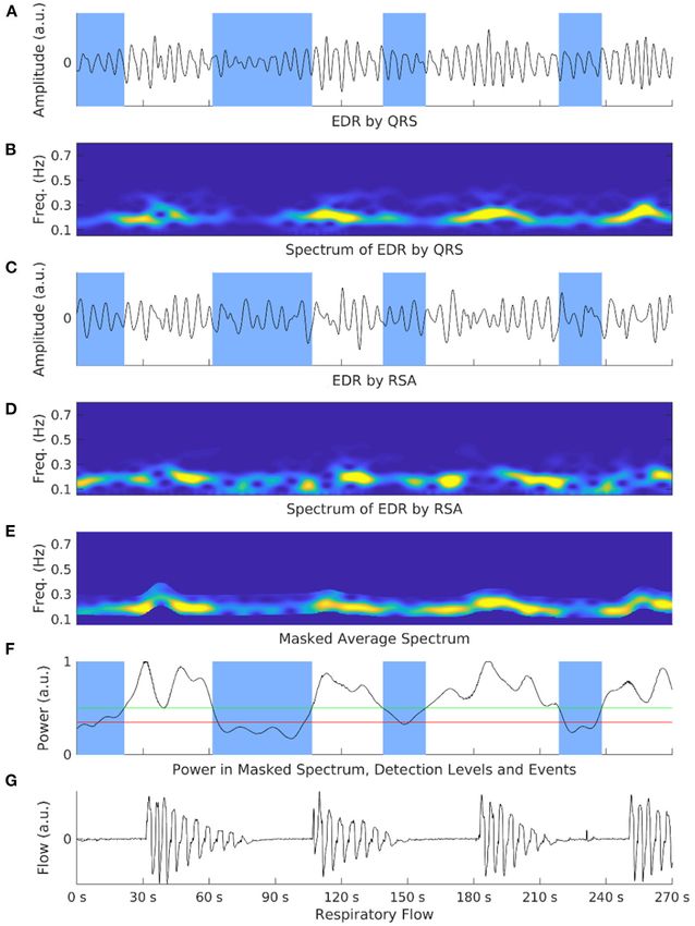
Qin et al.                                                                                                                                Heart Rate Variability in OSA

  FIGURE 2 | Shows an exemplary illustration of the respiratory power index (RPI) and electrocardiograph-derived respiration (EDR) methods in an OSA patient.
  Overnight electrocardiograph recordings are processed and cut into limited time segments. EDR signals are calculated via ECG respiration embeddings such as QRS
  complex (A) or respiratory sinus arrhythmia (RSA) (C). Spectrograms of both embeddings are also generated (B,D). These spectrograms are normalized and
  averaged to amplify the respiration-based component and mask non-respiration-related power (E). The power is calculated at each step with two selection events (F).
  A respiratory flow shows corresponding events to the power spectrum (G). The number of detected apneic events is the RPI.

OSA in groups of 91 and 52 patients, respectively. Then using                         (75). However, this study was conducted on a small population
wavelet decomposition parameters in 147 patients, Roche et al.                        and thus requires further study in order to improve upon the
(95) reached a sensitivity of 92.4% and specificity of 90.1%.                         application to classification. Gil et al. (97) used decreases in
Karasulu et al. (96) found a 90.4% sensitivity and 50% specificity                    amplitude fluctuations of photoplethysmography (PPG) with an
when using a VLF cut-off of 9.12, 80 and 76.2% when SDNN was                          accuracy of 80%, sensitivity of 87.5% and specificity of 71.4%.
higher than 83 and 73.3 and 85.7% with an SDNN cut-off of 62                          Babaeizadeh and Zhou (98) created a novel method of ECG-
in 87 patients. Offering a variant to these results, Abdullah et al.                  derived respiration (EDR) combined with HRV for an accuracy
combined EEG and HRV at 64% correct classification accuracy,                          of 88% and correct classification of 71%. Similarly, Lyons et al.
HRV alone at 56% accuracy and EEG alone at 62% accuracy                               (99) developed an ECG-derived respiratory power index (RPI) as

Frontiers in Psychiatry | www.frontiersin.org                                     7                                             July 2021 | Volume 12 | Article 642333
Qin et al.                                                                                                             Heart Rate Variability in OSA

an estimate for AHI to identify severe OSA in commercial drivers          (CNS) and autonomic nervous system (ANS) in arousal is poorly
(Figure 2).                                                               understood. Arousal may be a contributor in cardiac alternations
   There are thus a variety of tools and combinations that appear         such as heart rate changes and blood pressure fluctuation.
to have potential in the detection and classification of apnea.           HRV changes accordingly since heart rate is accelerated and
   Collectively this body of studies points to the potential of           decelerated immediately pre- and post-arousal. Animal studies
diagnosing OSA via HRV parameters reflecting sympathetic                  confirmed that transient arousal from NREM sleep is associated
hyperactivity during sleep, particularly during apneic episodes.          with acute cardiac sympathetic activation and parasympathetic
However, more research needs to be done in this area as there             withdrawal (118). The presence of arousal somehow immediately
are conflicting reports on the accuracy of using HRV alone,               leads to wakefulness that differs in autonomic changes from
as compared to coordination with other measurements such as               rested wakefulness in other conditions (118).
EEG, PPG, or EDR. Showing promise in the application of this                  Daytime cardiac vagal modulation improves due to the
idea, Le et al. (100) have made wearable device sensor technology         reduction of the frequency of arousals, suggesting arousal
predicting apneic episodes 1–5 min before onset with accuracy             may trigger cardiac vagal inhibition. OSA is strongly related
of 83.6, 80, 76.2, 66.9, and 61.1%, respectively, that could have         to hypertension, which is mainly attributed to sympathetic
many applications.                                                        hyperactivity and/or vagal withdrawal causing a surge in heart
                                                                          rate and blood pressure during apnea-arousal episodes (119,
                                                                          120). Study of autonomic arousals may help understanding why
HRV CHANGES DURING AROUSAL                                                OSA patients with daytime sleepiness are associated with a higher
                                                                          risk of developing adverse CV outcomes such as hypertension
Arousal interrupts sleep continuity to cause sleep fragmentation,         and cardiac sudden death (121). It is reported that patients with
which may contribute to cognitive impairment, excessive                   co-morbid OSA and insomnia have a significantly higher number
daytime sleepiness and adverse cardiovascular outcomes in                 of arousals during sleep than OSA alone (122). Bennett et al. (123)
OSA (101–103). Quantification of arousal would improve                    found significant correlations between the autonomic arousal
understanding of the underlying mechanism and relationship                index based on pulse transit time analysis and pretreatment
between arousal and OSA related outcomes (e.g., daytime                   objective sleepiness (r = 0.49) and nCPAP responsive objective
sleepiness and functioning) (104, 105). Currently, EEG arousal            sleepiness (r = 0.44), suggesting autonomic arousal detection
is defined as the abrupt increase in high-frequency EEG activities        should be taken into account as a sleep fragmentation index
lasting 3–15 s, following at least 10 s of sleep during NREM sleep.       to quantify sleepiness. Bartels et al. tried to define autonomic
Additionally, increased chin EMG activity is needed during REM            arousal. They found that lower blood pressure and high heart
sleep according to the AASM criteria (106, 107). However, even            rate in the 15-s window before short-term cortical arousal and
if the concept of arousal should be extended, there currently is no       cardiovascular changes shift in the opposite direction after sleep
agreement on the classification of arousal (108). Arousal could           recovery (110).
be divided into several states on the basis of specific causes. Two           HRV provides insight to the processing of arousal response
main types of arousal, physiologic (spontaneous or secondary              during sleep and improves the definition of arousal, criteria
to various stimuli), and pathologic (induced by sleep hypopnea            of detection and scoring, although it is still controversial. It
and apnea, upper airway resistance syndrome or periodic limb              should be included in the assessment of OSA for its useful
movement) are commonly accepted (108, 109). Some studies                  clinical value. EEG arousal generally does not cause behavioral
tried to classify arousal manually based on whether an arousal            awakenings. However, arousal threshold measured by esophageal
is associated with a physiological event such as cortical arousal,        pressure, a gold standard for upper airway resistance syndrome,
respiratory arousal, cardiac arousal, movement arousal, snoring           is invasive in clinical practice. On the other hand, cardiac arousal
arousal, or SpO2 arousal (110, 111). It is reported that autonomic        may reflect a neural response to stimuli. Little is known about
arousal does not have visual recognition in the way EEG arousal           the accumulation of persistent hyperarousal conditions in OSA.
does. It is plausible that some peripheral stimulations may not           HRV would be a sensitive physiological index of autonomic
be sufficient to lead to cortical visual EEG arousals but can cause       arousal requiring more investigation. Further research is needed
cardiovascular perturbation (e.g., heart rate and blood pressure          to understand the connectivity and interaction between the heart,
changes) (112–114).                                                       its intrinsic nervous system, and the brain.
    In this case, autonomic arousal may be a new entity of
arousals in OSA during sleep, possibly undetectable by EEG
(115). Thirty percent of respiratory event termination causes             DAYTIME SLEEPINESS AND HRV
are still undetermined. Some research indicates that it might
be related to apnea-related autonomic arousal, which tends to             On the other end of arousal, daytime sleepiness, a multifactorial
be ignored due to its non-visible nature compared with other              psychophysiological state, is one of the predominant symptoms
types of arousal in polysomnography (PSG) (116, 117). As a                in OSA (124, 125). Currently, the existing findings suggest
result, PSG would underestimate arousal severity if only visible          that daytime sleepiness depends on the quantity and quality of
EEG arousal counts. The occurrence of arousal induced by                  prior sleep. Patients with OSA commonly suffer from reduced
different causes varies in NREM and REM sleep (116). The                  sleep quality that is related to fragmented sleep (126). Sleep
underlying mechanisms between the central nervous system                  disturbances caused by arousal are important contributors to

Frontiers in Psychiatry | www.frontiersin.org                         8                                      July 2021 | Volume 12 | Article 642333
Qin et al. Heart Rate Variability in OSA sleepiness (123, 127, 128). Moreover, the frequency of arousal learning models to predict hypersomnolence in drivers with 90% has more impact on sleep recovery than the amount of sleep accuracy (144–146). (129). Subjective and objective sleepiness is often assessed by the It has been shown that sleepy OSA patients have a Epworth sleepiness scale (ESS) and multiple sleep latency test higher prevalence of adverse cardiovascular outcomes (e.g., (MSLT) (130, 131). ESS is a measure of a person’s general daytime hypertension) than non-sleepy OSA patients (147). Furthermore, sleepiness, where a score ≥10 could be diagnosed as excessive excessively sleepy OSA patients are at increased risk of daytime sleepiness. As a gold standard, the cut-off point of MSLT incident cardiovascular disease (CVD) compared to other OSA is still debatable based on the types of patients. According to symptom subtypes (Disturbed Sleep, Minimally Symptomatic, the AASM, a sleep latency during MSLT of
Qin et al.                                                                                                             Heart Rate Variability in OSA

in subjects normoxic at rest. Nevertheless, it is admitted that           a potential early indicator of the adverse effects of hypoxia
changes in detailed HRV parameters are not consistently similar           on OSA and identifying treatment responses. To date, the
due to the varying experimental protocols (e.g., the duration,            effect of nocturnal hypoxia on HRV patterns is unknown and
severity, and types of hypoxia).                                          correlation studies of HRV and hypoxia in HRV are limited.
   OSA generally generates a decrease in HRV during                       Those results may contribute to monitoring the progress of
normobaric hypoxia in most reported investigations. However,              chronic sustained normobaric hypoxia on the cardiovascular and
there are still underlying complex central-peripheral interactions        autonomic systems.
and modulation pathways in vulnerable populations. To address
those issues, a growing body of studies have attempted to
investigate the hypoxia burden in OSA (159–161). Time-                    HRV IN PEDIATRIC OSA
dependent static and dynamic desaturation give more insight to
the severity of hypoxia. Acute and chronic hypoxia may lead to            OSA affects 0.1–13% of children, particularly occurred in pre-
different autonomic modulation mechanisms. Hypoxia activated              school age (169). Pediatric OSA characterize by prolonged
chemoreflex leads to acutely increased short-term sympathetic             partial OSA, which usually occurred in REM sleep, preserved
tone during the occurrence of sleep apnea (54). Furthermore,              sleep architecture, uncommon OSA-related cortical arousals and
hypoxia exerted long-lasting chronic effects during the daytime           recurrent hypoxia (170). Enlarged tonsils and adenoids are the
and impaired baroreflex sensitivity (162). Meanwhile whether              leading causes of OSA in children. Unlike adult OSA manifested
or not sympathetic hyperactivity induces parasympathetic                  with excessive daytime sleepiness and cognitive dysfunction,
inhibition is still controversial. The overall reduced HRV with           pediatric OSA is more likely to have negative impact on the
increased sympathetic tone resulted from chronic hypoxia,                 development of the central nervous system and cardiovascular
while a rise in HRV with decreased vagal withdrawal occurred              system, potentially leading to neurobehavioral deficits (e.g.,
due to the subsequent adaptation and improved tolerance to                growth impairment, behavioral, and learning problems) (171).
short-term exposure to repeated hypoxic stress (163). Geovanini           Overt cardiovascular disease is not common in pediatric
et al. demonstrated a vicious circle between hypoxia-induced              OSA compared to adults (172), but early evidence shows
inflammation and cardiac autonomic abnormality with elevated              that pediatric OSA is related to left ventricular hypertrophy
sympathetic or reduced parasympathetic tone. They also found              (173, 174), abnormal blood pressure fluctuation (175, 176),
the values of SDNN, LF, and HF are closely linked to OSA                  and reduced systolic and diastolic function (177, 178). HRV
severity while only mean heart rate significantly correlated with         analysis is increasingly explored in assessment for cardiovascular
augments in neutrophils (164). In an OSA children study, Walter           autonomic control, the screening and diagnosis of sleep apnea
et al. (165) found that OSA may have negative influence on                and efficacy of treatments in pediatric OSA during daytime
cerebral blood flow due to the attenuated central autonomic               and nighttime due to its feasibility (179, 180). Current findings
control by mediating HRV. Therefore, it is reasonable to                  suggested that altered HRV patterns during daytime and sleep
believe that different cardiac autonomic modulation responses             are also found in childhood OSA (181). Not surprising, there are
occur either due to reduced vagal modulation, sympathetic                 more discrepancies in the results of frequency domain analysis
predominance, or even a combination of these responses.                   than in time domain analysis due to diverse subject samples
   The possibility of an increasing risk in the mortality and             and the different methodologies (182, 183). Chaicharn et al.
morbidity in hypoxic OSA patients with autonomic dysfunction              (179) tried to quantify daytime autonomic function in non-
requires further evidence. In addition, both hypoxia and arousal          OSA and OSA children with spectral HRV analysis, showing
have confounding effects on respiratory-cardiac coupling. Which           OSA children have significant elevated sympathetic tone but
one is the determinant of cardiac autonomic dysfunction                   normal parasympathetic control, with less reactive response to
in OSA is controversial in animal and human studies (5).                  autonomic tests compared to controls. Liao et al. (182) found
It seems that in prospective animal studies, OSA-induced                  autonomic imbalance with increased LF/HF during sleep among
hypoxia has a persistent impact on daytime hypertension                   groups with different levels of AHI. Similarly, Baharav et al. (183)
compared to acoustic arousal-induced control models, which                were able to show sympathetic augmentation with increased
exerted nocturnal elevations in blood pressure. However, in               LF both during wake before sleep onset and during sleep. By
humans, the answer to that question is uncertain. Norman                  contrast, Kwok et al. (180) demonstrated no changes in most
et al. suggested that CPAP therapy, which reduced both the                of the important time-domain and HRV measures between
intermittent hypoxia and arousals, plays a more important role in         non-OSA and OSA children using 1-h ECG data. Impaired
improving cardiovascular autonomic function than elimination              baroreflex adaptation is also found in OSA as it is associated
of nighttime intermittent hypoxia by comparing the results of             with a decrease in nighttime baroreflex gain (184). Autonomic
24-h ambulatory blood pressure in moderate-to-severe OSA                  activity may play a key role in pharyngeal compliance of
patients who received either CPAP therapy or sham-CPAP with               childhood OSA (185). Another application of HRV in childhood
supplemental oxygen (166).                                                OSA is to evaluate treatment response. Muzumdar et al. (186)
   Some studies indicated that certain damages of autonomic               reported that HRV improved with decreased sympathetic and
function are reversible after eliminating physiological influences        increased vagal tones after adenotonsillectomy in children with
(e.g., arousal, hypoxia, and respiratory events) in OSA population        OSA, while no changes showed in HRV in moderate-severe
with CPAP treatment (81, 167, 168). Thus, HRV maybe become                pediatric OSA with 1-year non-invasive ventilation (187). The

Frontiers in Psychiatry | www.frontiersin.org                        10                                      July 2021 | Volume 12 | Article 642333
Qin et al.                                                                                                            Heart Rate Variability in OSA

results of long-term effect of OSA on HRV are debated.                    predominance in cardiac regulation. Men have a higher LF, and
Vlahandonis et al. (188) failed to show significant differences in        a lower HF/LF ratio than women. Those results demonstrate
autonomic regulation determined by using HRV analysis among               white and male populations have higher sympathetic activity,
children with habitual snoring, and those with and without                which is considered as a major contributor to cardiovascular
OSA regardless of intervention during 4-year follow-up visits.            diseases (e.g., hypertension). In contrast, Sloan et al. (200)
However, Walter et al. (189) found improved HRV in preschool-             reported that there is a higher standard deviation of RR intervals
aged children with resolved OSA, showing decreased LF and HF,             in white subjects compared to black subjects, and in men
while increased HF in those with unresolved OSA during 3-year             compared to women with age between 33 and 47 years old.
period. It is noteworthy that age, obesity, sleep stage, and AHI          No ethnicity- and sex- special differences were found in HF-
severity are independently correlated with HRV measurements               HRV. Comparatively, Choi et al. found significant ethnically
in children (190, 191). Explanations on these results need to be          related differences and age-related differences (in Caucasian
cautious with those confounding factors. Whether or not HRV               Americans but not in African Americans) in short-term daytime
measures could be the reliable maker of disease severity and              spectral HRV. Young African Americans showed a similar
risk stratification in children with OSA is still unproven. The           HRV profile to older Caucasian Americans, leading Choi et al.
clinical implication of cardiac autonomic alternation in pediatric        (204) to suggest the presence premature autonomic nervous
OSA and how it disrupts the maturation of autonomic control               system aging in young African Americans. A few studies related
and affects the nervous and cardiovascular functioning need               to those correlates on HRV during sleep are available. Hall
further investigation.                                                    et al. suggested that ethnicity is associated with HRV during
                                                                          sleep. They found white women have decreased parasympathetic
                                                                          tone and elevated sympathetic tone during NREM stage 2 and
EFFECT OF AGE, ETHNICITY AND SEX ON                                       REM sleep compared to their African American and Chinese
HRV                                                                       counterparts after controlling for confounding factors such as
                                                                          recording length and respiratory rate (205). Huang et al. (206)
Previous studies have demonstrated age-, ethnicity-, and sex-             have shown heart rate profiles in a larger cohort of adults
specific differences in HRV in the healthy general population             without sleep apnea in order to develop heart rate phenotypes
and under certain conditions. It is generally accepted there is an        regarding sleep physiology. They implied that heart rate dipping
inverse association between age and HRV. However, it is unclear           and spectral HRV metrics could contribute to sleep phenotyping
whether the effects of OSA on cardiac autonomic modulation in             due to their significant correlations to sleep measures (e.g.,
elderly subjects (>60 yr) are different from those in other age           sleep stage, total sleep time and sleep quality). Interpreting the
groups (young and mid-aged adults). Trimer et al. compared the            clinical relevance between ethnicity, sex, and HRV should be
differences in HRV among the elderly and the young population             approached with caution due to the plethora of confounding
with and without OSA. They found the elderly with OSA have                factors, such as physiological, psychological, behavioral, and
significantly lower LF/HF ratio only during wakefulness at night          sociodemographic factors. To date, there is limited data reporting
than the young with OSA but not during other sleep stages (192).          on the influence of age, ethnicity, and sex on HRV in the OSA
Sforza et al. (140, 193) suggested age may have more devastating          population. It is unclear which OSA phenotypes are most likely
effect on HRV in the elderly, which possibly undermines the               to develop cardiovascular diseases and thus, which patients are
application of HRV in those population.                                   most likely to benefit from CPAP or other forms of therapy
    Findings on sex and ethnic differences in HRV are less                for OSA (207). Those findings in cardiac heterogeneity might
consistent (194). Nonetheless, reduced HRV is related to higher           lead to a better understanding of the underlying cardiovascular
cardiovascular morbidity and mortality, where decreased cardiac           pathophysiology and cardiovascular risk stratification in patients
vagal control is considered an important contributor. Currently,          with OSA. Additionally, it would facilitate the development
a majority of studies report females are characterized by higher          of effective strategies for treatment decision of OSA according
vagal control assessed by HF-HRV and lower sympathetic control            to cardiac phenotypic characterization in order to improve
assessed by LF-HRV (194). Furthermore, women exhibit more                 treatment efficacy and predict treatment outcomes.
complex heart rate dynamics (195). Several studies found no
difference between men and women in HF-HRV or that men
have a higher HRV (196–200). These results contradict previous            HRV AND OSA COMORBIDITY WITH
findings that women are less likely to develop progressively              PSYCHIATRY DISEASES
cardiovascular diseases compared to men (201, 202).
    In terms of interaction associations between HRV and age,             Psychophysiological disturbances have significant impacts on
sex, and ethnicity, Liao et al. (203) found changes in autonomic          the autonomic nervous system (ANS) (3, 208–211). Depression
function have close associations with age, ethnicity, and gender          and anxiety are considered as psychosocial risk factors for
in a community-based cohort by spectral analysis of HRV.                  cardiovascular comorbidity (212). HRV analysis has been used
They found that the sympathetic and parasympathetic tone                  to quantify autonomic dysregulation in insomnia, depression,
decrease with increasing age in a general population. White               anxiety, and schizophrenia (208, 211). Epidemiological data
populations have a higher LF, HF, and lower HF/LF than black              has shown that 39–58% of patients with insomnia and 5–63%
populations, suggesting that white populations show sympathetic           of patients with depression had accompanying OSA diagnoses

Frontiers in Psychiatry | www.frontiersin.org                        11                                     July 2021 | Volume 12 | Article 642333
Qin et al. Heart Rate Variability in OSA (213–215). Additionally, it was found that co-morbid OSA expect more perspectives and possible application of HRV in and insomnia patients are at a higher risk of developing OSA in neuropsychiatric alternations could be discussed in psychiatric disorders such as anxiety and depression than OSA future studies. patients without insomnia (122, 216). Interestingly, OSA and insomnia are more likely to show opposing clinical symptoms related to sleepiness and alertness (215). Nevertheless, increased HRV AND CARDIOVASCULAR MORTALITY sympathetic activity and depressed parasympathetic activity were AND MORBIDITY exhibited both in OSA and insomnia (25, 217). It is reported that untreated OSA aggravate insomnia in the disturbed sleep cluster Due to HRV being a marker of autonomic innervation of the due to hyperarousal (218). Augmentation in heart rate and heart, it has been suggested that increased sympathetic activity sympathetic tone, which is thought to be essential to the alertness during sleep due to OSA may be a link to cardiovascular disease and motivation, may play a key role in the pathophysiology of (54). Sympathetic dominance during sleep has been shown in insomnia (215). However, interaction mechanisms between OSA those with ischemic heart disease (53), coronary artery disease and insomnia of autonomic control evaluated by HRV measures (CAD) (55) and post-MI (224). Consequently, HRV parameters remain unclear. are markers for adverse CVD prognoses (49, 51, 52). Reduced global HRV is consistently reported in depression Several cardiovascular disease studies have reported an and anxiety disorders. Specifically, depression is characterized increased risk of mortality in relation to altered HRV parameters. by increased cardiac rhythmicity and reduced heart rate Kleiger et al. found that a 24-h SDNN of
Qin et al.                                                                                                               Heart Rate Variability in OSA

lacked power for definitive conclusion (236). Yaggi et al. (232)            provides hidden information on cardiopulmonary coupling,
found that OSA independently increases risk of stroke and all-              which transfers from heart rate to respiration and improves the
cause mortality with a hazard ratio of 1.97 post-adjustment. In a           accuracy of sleep apnea detection compared to either method
systematic review, Lavie (234) also concluded that sleep apnea is           alone. The cognitive consequences and the daytime outcomes of
a mortality risk that can be reduced via CPAP, which is especially          ANS alternation during sleep in patients with OSA are unclear.
crucial in younger patients, as they carry a higher mortality risk.         The use of HRV in the prognosis of OSA independent of CVD
   In the linking of OSA and CVD, HRV, and OSA mortality and                is also unclear. However, HRV has shown a close association
mortality, the exact physiological pathway through which these              to mortality and co-morbidities. Additionally, overlapping
are connected is not well-understood. In animal models, Iturriaga           conditions increase progressively in OSA, requiring reliable tools
(237) proposed that intermittent hypoxia induces carotid body               to manage those conditions at an early stage. Further studies
potentiation, and that current evidence indicated that this alters          are required to explore the implications of integrated cardiac
the sympathetic, vascular, and ventilatory response to hypoxia.             physiology in regulatory networks between the central brain and
Whether this is the exact mechanism and whether it increases                heart. In particular, following this investigation, several research
CVD risk is not definitively known. However, repetitive oxygen              topics have been found to be of value:
desaturation episodes are associated with HRV parameters
                                                                            • Prospective studies using HRV to accurately predict
suggestive of cardiac sympathetic predominance. In a group of
                                                                              cardiovascular outcomes in OSA should be as a priority
CAD patients, those with LVEF >50% had a higher LF:HF ratio
                                                                              for clinical application of HRV research
than those with LVEF ≤35% during cyclic oxygen desaturation
                                                                            • Studies investigating cardiac OSA phenotypes on the basis of
episodes but not during control episodes (55). This suggests
                                                                              HRV profiles to facilitate the definition of OSA subtypes and
that hypoxia worsens pre-existing cardiovascular conditions. A
                                                                              implement tailored treatment approaches in clinical practice
few results of the secondary analyses using ECG data from the
                                                                            • New sophisticated methods of HRV analysis to analyze the
Sleep Heart Health Study (SHHS) or the Wisconsin Sleep Cohort
                                                                              inevitable instationarities of OSA’s transitional nature that
Study are reported. Bradicich et al. (238) and Wang et al. (24)
                                                                              prove challenging for current algorithms and models
demonstrated associations between HRV and characteristics of
                                                                            • Context-dependent analyses of HRV (i.e., age, BMI, gender,
polysomnographic parameters, however, they did not attempt
                                                                              sleep stages) to better understand the association between
to use HRV as a CVD risk predictor in this part of the SHHS
                                                                              anthropometric and sleep characteristics and autonomic
dataset. Sankari et al. (239) suggested beat-to-beat intervals index
                                                                              function in OSA
(RRDI) during sleep is closely correlated to new-diagnosis CVD
                                                                            • Investigation and standardization of the time window
(hazard ratio of 1.21 per 10-unit increment in RRDI) in OSA
                                                                              segments analyzed to provide comparable and valuable ECG
patients from the Wisconsin Sleep Cohort, but they did not utilize
                                                                              data in OSA during an overnight sleep study
other linear and non-linear HRV measures to show the further
relationship between CV risk and OSA.                                       HRV is showing promise in clinical application and due to the
   However, despite the clear association between OSA and                   already large and increasing prevalence of OSA, these further
mortality and CVD, more studies need to be done to determine                studies are imperative to the advancement of diagnostic and
the exact physiological mechanisms by which this occurs, and                treatment approaches needed to minimize the existing and future
if OSA is an independent causal factor of increased mortality               health and financial burden.
and CVD risk as suggested. From the current data, altered HRV
features such as SDNN are good predictors of cardiovascular                 AUTHOR CONTRIBUTIONS
mortality. There appears to be a correlation of higher mortality
risk and lower SDNN, but the cut-off point varies depending                 HQ and TP were responsible for the manuscript concept and
on the populations and the length of ECG segments. Therefore,               design. HQ, NS, and TP prepared the manuscript draft. JFK
determination of a clear cut-off value of SDNN requires further             prepared the figures. HQ, NS, TP, MG, NW, JFK, and FV-V
investigation (240).                                                        contributed to critical revision of the manuscript. All authors
                                                                            contributed to the article and approved the submitted version.
CONCLUSION
                                                                            FUNDING
With more sophisticated analytical approaches and techniques
developing, HRV measures could provide additional                           We acknowledge support from the German Research Foundation
electrophysiological information on impaired cardiovascular                 (DFG) and the Open Access Publication Fund of Charité-
alternation, which might be related to subclinical cardiovascular           Universitätsmedizin Berlin.
outcomes in patients with OSA. It is already known that the
determination of time window (ECG segment length and SDB-                   ACKNOWLEDGMENTS
related events) is critical to HRV analysis, but a standardized
analytical approach is lacking. HRV is proving to be accurate               We appreciate Dr. Mike Mutschelknaus for the editing of the
in sleep staging and particularly screening and diagnosing                  manuscript and HQ is a mentee of World Sleep Society’s
OSA. However, a combinatorial method of HRV and EDR                         International Sleep Research Training Program (ISRTP) 2020.

Frontiers in Psychiatry | www.frontiersin.org                          13                                      July 2021 | Volume 12 | Article 642333
Qin et al. Heart Rate Variability in OSA
REFERENCES 20. Trimer R, Mendes RG, Costa FS, Sampaio LM, Delfino A Jr, Arena R,
et al. Is there a chronic sleep stage-dependent linear and nonlinear cardiac
1. McNicholas WT, Bonsigore MR. Sleep apnoea as an independent risk factor autonomic impairment in obstructive sleep apnea? Sleep Breath. (2014)
for cardiovascular disease: current evidence, basic mechanisms and research 18:403–9. doi: 10.1007/s11325-013-0900-x
priorities. Eur Respir J. (2007) 29:156–78. doi: 10.1183/09031936.50027406 21. Wessel N, Malberg H, Bauernschmitt R, Kurths J. Nonlinear methods of
2. Bilyukov RG, Nikolov MS, Pencheva VP, Petrova DS, Georgiev OB, cardiovascular physics and their clinical applicability. Int J Bifurcation Chaos.
Mondeshki TL, et al. Cognitive impairment and affective disorders in (2007) 17:3325–71. doi: 10.1142/S0218127407019093
patients with obstructive sleep apnea syndrome. Front Psychiatry. (2018) 22. Bonnet MH, Arand DL. Heart rate variability: sleep stage, time of night,
9:357. doi: 10.3389/fpsyt.2018.00357 arousal influences. Electroencephalogr Clin Neurophysiol. (1997) 102:390–6.
3. Alvares GA, Quintana DS, Hickie IB, Guastella AJ. Autonomic nervous doi: 10.1016/S0921-884X(96)96070-1
system dysfunction in psychiatric disorders and the impact of psychotropic 23. Dempsey JA, Veasey SC, Morgan BJ, O’Donnell CP. Pathophysiology of sleep
medications: a systematic review and meta-analysis. J Psychiatry Neurosci. apnea. Physiol Rev. (2010) 90:47–112. doi: 10.1152/physrev.00043.2008
(2016) 41:89–104. doi: 10.1503/jpn.140217 24. Wang W, Tretriluxana S, Redline S, Surovec S, Gottlieb DJ, Khoo MC.
4. Benjafield AV, Ayas NT, Eastwood PR, Heinzer R, Ip MSM, Morrell MJ, Association of cardiac autonomic function measures with severity of sleep-
et al. Estimation of the global prevalence and burden of obstructive sleep disordered breathing in a community-based sample. J Sleep Res. (2008)
apnoea: a literature-based analysis. Lancet Respir Med. (2019) 7:687–98. 17:251–62. doi: 10.1111/j.1365-2869.2008.00652.x
doi: 10.1016/S2213-2600(19)30198-5 25. Narkiewicz K, Montano N, Cogliati C, van de Borne PJ, Dyken ME, Somers
5. Khoo MC, Blasi A. Sleep-related changes in autonomic control in obstructive VK. Altered cardiovascular variability in obstructive sleep apnea. Circulation.
sleep apnea: a model-based perspective. Respir Physiol Neurobiol. (2013) (1998) 98:1071–7. doi: 10.1161/01.CIR.98.11.1071
188:267–76. doi: 10.1016/j.resp.2013.05.017 26. Aydin M, Altin R, Ozeren A, Kart L, Bilge M, Unalacak M. Cardiac
6. Heart rate variability: standards of measurement, physiological autonomic activity in obstructive sleep apnea: time-dependent and spectral
interpretation and clinical use. Task Force of the European Society analysis of heart rate variability using 24-hour Holter electrocardiograms.
of Cardiology and the North American Society of Pacing and Tex Heart Inst J. (2004) 31:132–6.
Electrophysiology. Circulation. (1996) 93:1043–65. 27. Gates GJ, Mateika SE, Basner RC, Mateika JH. Baroreflex sensitivity
7. Mazzotti DR, Lim DC, Sutherland K, Bittencourt L, Mindel JW, Magalang in nonapneic snorers and control subjects before and after nasal
U, et al. Opportunities for utilizing polysomnography signals to characterize continuous positive airway pressure. Chest. (2004) 126:801–7.
obstructive sleep apnea subtypes and severity. Physiol Meas. (2018) doi: 10.1378/chest.126.3.801
39:09tr01. doi: 10.1088/1361-6579/aad5fe 28. Idiaquez J, Santos I, Santin J, Del Rio R, Iturriaga R. Neurobehavioral and
8. Thayer JF, Lane RD. A model of neurovisceral integration in emotion autonomic alterations in adults with obstructive sleep apnea. Sleep Med.
regulation and dysregulation. J Affect Disord. (2000) 61:201–16. (2014) 15:1319–23. doi: 10.1016/j.sleep.2014.05.030
doi: 10.1016/S0165-0327(00)00338-4 29. Müller A, Riedl M, Penzel T, Bonnemeier H, Kurths J, Wessel N. Coupling
9. Chouchou F, Desseilles M. Heart rate variability: a tool to explore the sleeping analysis of transient cardiovascular dynamics. Biomed Tech. (2013) 58:131–9.
brain? Front Neurosci. (2014) 8:402. doi: 10.3389/fnins.2014.00402 doi: 10.1515/bmt-2012-0030
10. Sassi R, Cerutti S, Lombardi F, Malik M, Huikuri HV, Peng CK, et al. 30. Wessel N, Riedl M, Kramer J, Muller A, Penzel T, Kurths J.
Advances in heart rate variability signal analysis: joint position statement Synchronisation and coupling analysis: applied cardiovascular
by the e-Cardiology ESC Working Group and the European Heart Rhythm physics in sleep medicine. Annu Int Conf IEEE Eng Med
Association co-endorsed by the Asia Pacific Heart Rhythm Society. Europace. Biol Soc. (2013) 2013:6567–70. doi: 10.1109/EMBC.2013.
(2015) 17:1341–53. doi: 10.1093/europace/euv015 6611060
11. Hillebrand S, Gast KB, de Mutsert R, Swenne CA, Jukema JW, Middeldorp 31. Min KB, Min JY, Paek D, Cho SI, Son M. Is 5-minute heart rate variability
S, et al. Heart rate variability and first cardiovascular event in populations a useful measure for monitoring the autonomic nervous system of workers?
without known cardiovascular disease: meta-analysis and dose-response Int Heart J. (2008) 49:175–81. doi: 10.1536/ihj.49.175
meta-regression. Europace. (2013) 15:742–9. doi: 10.1093/europace/eus341 32. Li K, Rüdiger H, Ziemssen T. Spectral analysis of heart rate variability: time
12. Akselrod S, Gordon D, Ubel FA, Shannon DC, Berger AC, Cohen window matters. Front Neurol. (2019) 10:545. doi: 10.3389/fneur.2019.00545
RJ. Power spectrum analysis of heart rate fluctuation: a quantitative 33. Ziemssen T, Reimann M, Gasch J, Rüdiger H. Trigonometric regressive
probe of beat-to-beat cardiovascular control. Science. (1981) 213:220–2. spectral analysis: an innovative tool for evaluating the autonomic
doi: 10.1126/science.6166045 nervous system. J Neural Transm. (2013) 120(Suppl. 1):S27–33.
13. Akselrod S, Gordon D, Madwed JB, Snidman NC, Shannon DC, Cohen RJ. doi: 10.1007/s00702-013-1054-5
Hemodynamic regulation: investigation by spectral analysis. Am J Physiol. 34. Dietrich A, Rosmalen JG, Althaus M, van Roon AM, Mulder LJ,
(1985) 249:H867–75. doi: 10.1152/ajpheart.1985.249.4.H867 Minderaa RB, et al. Reproducibility of heart rate variability and baroreflex
14. Rajendra Acharya U, Paul Joseph K, Kannathal N, Lim CM, Suri JS. sensitivity measurements in children. Biol Psychol. (2010) 85:71–8.
Heart rate variability: a review. Med Biol Eng Comput. (2006) 44:1031–51. doi: 10.1016/j.biopsycho.2010.05.005
doi: 10.1007/s11517-006-0119-0 35. Parati G. Blood pressure variability: its measurement and
15. Parati G, Di Rienzo M, Bonsignore MR, Insalaco G, Marrone O, Castiglioni significance in hypertension. J Hypertens Suppl. (2005) 23:S19–25.
P, et al. Autonomic cardiac regulation in obstructive sleep apnea syndrome: doi: 10.1097/01.hjh.0000165624.79933.d3
evidence from spontaneous baroreflex analysis during sleep. J Hypertens. 36. Li K, Konofalska U, Akgün K, Reimann M, Rüdiger H, Haase R, et al.
(1997) 15:1621–6. doi: 10.1097/00004872-199715120-00063 Modulation of cardiac autonomic function by fingolimod initiation and
16. Wessel N, Berg K, Kraemer JF, Gapelyuk A, Rietsch K, Hauser T, et al. predictors for fingolimod induced bradycardia in patients with multiple
Cardiac autonomic dysfunction and incidence of de novo atrial fibrillation: sclerosis. Front Neurosci. (2017) 11:540. doi: 10.3389/fnins.2017.00540
heart rate variability vs. heart rate complexity. Front Physiol. (2020) 11:1583. 37. Kleiger RE, Stein PK, Bigger JT Jr. Heart rate variability: measurement
doi: 10.3389/fphys.2020.596844 and clinical utility. Ann Noninvasive Electrocardiol. (2005) 10:88–101.
17. Seely AJE, Macklem PT. Complex systems and the technology of variability doi: 10.1111/j.1542-474X.2005.10101.x
analysis. Critical Care. (2004) 8:R367. doi: 10.1186/cc2948 38. Ziemssen T, Gasch J, Ruediger H. Influence of ECG sampling frequency
18. Stein PK, Reddy A. Non-linear heart rate variability and risk stratification in on spectral analysis of RR intervals and baroreflex sensitivity using
cardiovascular disease. Indian Pacing Electrophys J. (2005) 5:210–20. the EUROBAVAR data set. J Clin Monit Comput. (2008) 22:159–68.
19. Al-Angari HM, Sahakian AV. Use of sample entropy approach to study heart doi: 10.1007/s10877-008-9117-0
rate variability in obstructive sleep apnea syndrome. IEEE Trans Biomed Eng. 39. Singh B, Bharti N. Software tools for heart rate variability analysis. Int J
(2007) 54:1900–4. doi: 10.1109/TBME.2006.889772 Recent Sci Res. (2015) 6:3501–6.
Frontiers in Psychiatry | www.frontiersin.org 14 July 2021 | Volume 12 | Article 642333You can also read