Sleep Disorders in Children With Autism Spectrum Disorder: Insights From Animal Models, Especially Non-human Primate Model - Frontiers
←
→
Page content transcription
If your browser does not render page correctly, please read the page content below
REVIEW
                                                                                                                                              published: 20 May 2021
                                                                                                                                     doi: 10.3389/fnbeh.2021.673372

                                            Sleep Disorders in Children With
                                            Autism Spectrum Disorder: Insights
                                            From Animal Models, Especially
                                            Non-human Primate Model
                                            Shufei Feng 1,2† , Haoyu Huang 1† , Na Wang 3 , Yuanyuan Wei 3 , Yun Liu 1* and
                                            Dongdong Qin 1,2,3*
                                            1
                                             Department of Pediatric Rehabilitation Medicine, Kunming Children’s Hospital, Kunming, China, 2 State Key Laboratory of
                                            Primate Biomedical Research, Institute of Primate Translational Medicine, Kunming University of Science and Technology,
                                            Kunming, China, 3 School of Basic Medical Sciences, Yunnan University of Chinese Medicine, Kunming, China

                                            Autism Spectrum Disorder (ASD) is a heterogeneous neurodevelopmental disorder with
                           Edited by:
                      Rafael S. Maior,      deficient social skills, communication deficits and repetitive behaviors. The prevalence
           University of Brasilia, Brazil   of ASD has increased among children in recent years. Children with ASD experience
                     Reviewed by:           more sleep problems, and sleep appears to be essential for the survival and integrity of
                     Kyungmin Lee,
       Kyungpook National University,
                                            most living organisms, especially for typical synaptic development and brain plasticity.
                       South Korea          Many methods have been used to assess sleep problems over past decades such as
                      Hiroki Toyoda,        sleep diaries and parent-reported questionnaires, electroencephalography, actigraphy
            Osaka University, Japan
                                            and videosomnography. A substantial number of rodent and non-human primate models
                    *Correspondence:
                                Yun Liu     of ASD have been generated. Many of these animal models exhibited sleep disorders
                         liuyun@etyy.cn     at an early age. The aim of this review is to examine and discuss sleep disorders in
                         Dongdong Qin
                 qindong108@163.com
                                            children with ASD. Toward this aim, we evaluated the prevalence, clinical characteristics,
     † These   authors have contributed
                                            phenotypic analyses, and pathophysiological brain mechanisms of ASD. We highlight
                    equally to this work    the current state of animal models for ASD and explore their implications and prospects
                                            for investigating sleep disorders associated with ASD.
                   Specialty section:
         This article was submitted to      Keywords: autism, sleep, non-human primate, brain development, animal model
              Pathological Conditions,
                a section of the journal
 Frontiers in Behavioral Neuroscience       INTRODUCTION
        Received: 27 February 2021
           Accepted: 16 April 2021          Sleep appears to be essential for most living organisms’ survival and integrity, especially for
           Published: 20 May 2021           typical synaptic development and brain plasticity (Fogel et al., 2012; Hartsock and Spencer,
                            Citation:       2020). Over the past decades, the role of sleep in learning and memory has been probed by
   Feng S, Huang H, Wang N, Wei Y,          many studies at behavioral, systemic, cellular, and molecular levels (Walker and Stickgold, 2006;
         Liu Y and Qin D (2021) Sleep       Rawashdeh et al., 2007; Sawangjit et al., 2018; Kim et al., 2019). The American Academy of Sleep
    Disorders in Children With Autism
                                            Medicine (AASM) has recently released the third edition of the International Classification of
    Spectrum Disorder: Insights From
            Animal Models, Especially
                                            Sleep Disorders (ICSD-3) in 2014. This guideline grouped sleep disorders into seven basic types:
          Non-human Primate Model.          insomnia disorders, central disorders of hypersomnolence, circadian rhythm sleep-wake disorders,
  Front. Behav. Neurosci. 15:673372.        sleep-disordered breathing, movement disorders, parasomnias, and other sleep disorders (Sateia,
    doi: 10.3389/fnbeh.2021.673372          2014; Ito and Inoue, 2015). There are universal physiologic changes during sleep, and some

Frontiers in Behavioral Neuroscience | www.frontiersin.org                        1                                            May 2021 | Volume 15 | Article 673372
Feng et al. Sleep Disorders in ASD Children biologic, environmental, psychological, social as well as genetic THE ROLE OF SLEEP factors can affect change in the sleep pattern (Jenni and O’Connor, 2005; Bathory and Tomopoulos, 2017). The sleep- In most mammalian species, sleep amounts are highest during wake circadian rhythm is regulated through both circadian the neonatal period (Weber and Dan, 2016). Sleep loss can and homeostatic processes. Arousal and sleep are active and significantly affect a child’s health-related quality and activities involved in neurophysiologic processes, including both activation of daily living (Maski and Kothare, 2013). The brain is one of and suppression of neural pathways. Sleep disorders can be the organs most impacted by sleep or the lack thereof while an early symptom of the disease, and the presence of rapid adolescence is a critical period for brain reorganization. It is eye movement (REM) sleep behavior disorder (RBD) can be beyond doubt that sleep disorders during this period exert used as an early diagnostic indicator for neurodegenerative irreversible effects on children’s brain development (Roffwarg diseases (Galland et al., 2012; Kotterba, 2015). Besides, sleep et al., 1966; Klein et al., 2000; Weber et al., 2018). REM sleep disorders have observable effects on physical and mental health of can prune newly formed postsynaptic dendritic spines during children with autism spectrum disorder (ASD) and their parents motor learning (Li et al., 2017), and the balance of newly (Zhang et al., 2021). formed and original dendritic spines is crucial for neuronal Autism spectrum disorder is a neurodevelopmental disorder circuit development and behavioral improvement in children. and the prevalence of ASD is increasing, with 1 in 59 children Two studies found that sleep enhance cortical plasticity in the in the United States. diagnosed with ASD (Orefice, 2019). ASD visual cortex during the developmental critical period (Frank is approximately four times more prevalent among males than et al., 2001; Aton et al., 2009). In conclusion, sleep seems to females (Christensen et al., 2016; Satterstrom et al., 2020). be important for brain development, learning, and memory According to the DSM-V, the previous categories of pervasive consolidation by selectively eliminating and maintaining newly developmental disorders, pervasive developmental disorder-not formed synapses (Li et al., 2017). otherwise specified (PDD-NOS) and Asperger disorder were Sleep deprivation may cause physical diseases and combined into ASD. The new diagnostic criteria from DSM- developmental problems. During early life, sleep deprivation has V defined ASD as a heterogeneous spectrum disorder with been shown to have long-term implications for social behaviors deficits in social interaction and communication, restricted in adulthood (Hudson et al., 2020). Neural substrates can be and repetitive interests, and stereotyped behaviors (American affected by sleep deprivation, including the prefrontal cortex, Psychiatric Association, 2013; Devnani and Hegde, 2015). basal ganglia, and amygdala. Furthermore, sleep deprivation It is not surprising when considering the numerous health may cause difficulties in executive functioning, reward learning and behavioral issues that sleep disturbance are commonly as well as emotional reactivity. Such issues may contribute to observed in the clinical progression of ASD. Children with difficulties in judgment, resolution of problems, challenging ASD experience more sleep problems compared with the behaviors, emotional control, and public health concerns, such general population, particularly insomnia. Sleep-wake cycle as depression, suicide, and risk-taking behavior (Maski and abnormalities are associated with communication deficits, Kothare, 2013). These findings indicate that insufficient sleep stereotyped behaviors, and autism severity (Tudor et al., during early life has persistent effects on brain development and 2012). Disrupted sleep may exacerbate the daily dysfunction later behavioral performance. of ASD children, such as social and communication skills, It has been assumed that sleep can clear out brain’s toxins, behavioral performance, stereotypical behaviors, and motor such as beta-amyloid which was associated with Alzheimer’s output on non-verbal performance tasks (Schreck et al., 2004; disease (Xie et al., 2013). Sleep is essential for maintaining the Limoges et al., 2013). body’s physical health and is associated with neurodegeneration, As we gain deeper knowledge of the neural mechanisms metabolic diseases, cancer, and aging. The processes of growth of ASD and sleep, more contributions from sleep-related and development are related to sleep quality. The abnormal biomarkers to the study of neurophysiology in ASD. sleep and circadian also affect hormones and metabolism (Li Prospectively, the emergence of digital technologies and et al., 2018a). Getting adequate sleep can help the immune devices is making studies of sleep physiology more flexible and system to better react against infection (Grigg-Damberger, 2009; convenient. The sleep study provided new insights for research Herculano-Houzel, 2013; Welberg, 2013). on the children with ASD when compared with the other behavioral tests currently used in human subjects and animal experimental models. This review’s main objective is to explore CLINICAL CHARACTERISTICS OF animal models’ role, especially non-human primate (NHP) SLEEP DISORDERS IN ASD CHILDREN models, as a useful tool to investigate sleep disorders in ASD children. Firstly, we present data on sleep disorders in autistic Many neurodevelopmental processes have been reported in the children, emphasizing their prevalence, clinical characteristics, children with ASD, such as synaptic plasticity, neurogenesis phenotypic analyses, and pathophysiological mechanisms. Next, and migration of neuron (Gilbert and Man, 2017). About 40– we highlight the current state of animal models for ASD and 80% of children with ASD exhibit at least one sleep-related explore their implications and future prospects in translational problems (Verhoeff et al., 2018), including irregular sleeping and research. We suggest that using NHP animal models may provide waking patterns, decreased sleep efficiency, reductions in total insights into sleep disorders in ASD. sleep time and REM sleep time, sleep onset delays, decreased Frontiers in Behavioral Neuroscience | www.frontiersin.org 2 May 2021 | Volume 15 | Article 673372
Feng et al. Sleep Disorders in ASD Children
sleep efficiency, increased wakening after sleep onset, bedtime humans, particularly in children related to the lively side of their
resistance, and daytime sleepiness (Humphreys et al., 2014). nature. Even though several new technological developments
Studies utilizing Actigraphy (ACT) and Polysomnogram (PSG) have been brought to reduce inconvenience, pain, and further
have found that increased sleep latency, and decreased sleep damage of these methods, expensive and burdensome must
duration and sleep efficiency in ASD children (Elrod and Hood, also be considered, especially for long-term studies that include
2015). A comprehensive review in children with ASD reported large samples. Some assessments were developed to monitor
that insomnia is one of the most common sleep problems sleep through observation of body motion and posture. These
(Souders et al., 2009). Another study also documented that the methods could obviate the need for direct contact and even
predominant sleep disorder included insomnia, difficulty falling, avoid surgery or electrode implantation. It is non-invasive and
and staying asleep (Malow et al., 2006). Mutluer et al. (2016) low cost. Nevertheless, the behavioral observation does not
found that the most common symptoms reported were troubles provide sufficient information compared to those provided by
falling asleep, sleep after waking up and tired after sleeping. electroencephalogram (EEG) and electromyogram (EMG). In
general, both humans’ and animals’ sleep analyses include sleep
patterns, locomotor activity, temperature, and food intake. The
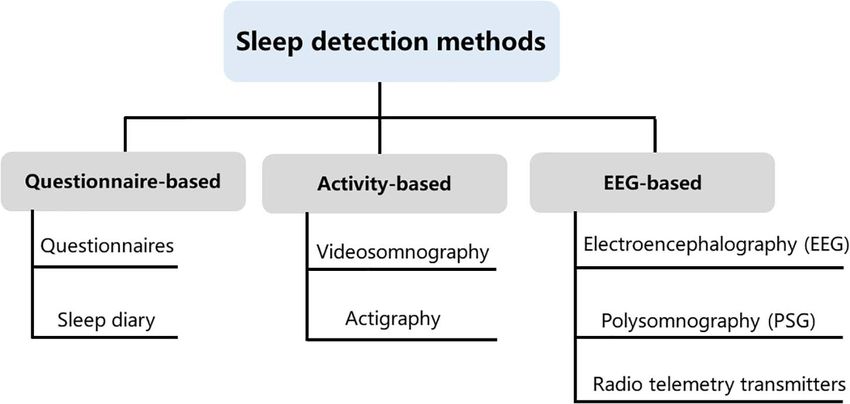
PREVALENCE OF SLEEP PROBLEMS IN current study summarized phenotype analyses for sleep disorders
CHILDREN WITH ASD obtained from sleep diaries, parent-reported questionnaires,
electroencephalography, actigraphy, and videosomnography.
Childhood sleep disorders which are mostly reported by We summarized the main types of experimental approaches
parents are associated with emotional, cognitive, and behavioral applicable to assessment methods of sleep studies and all of
disturbances. Sleep disturbances occur in approximately 20– these methods have advantages and disadvantages (Figure 1).
30% of preschool children, including bedtime resistance, sleep The selection of clinical sleep assessment should be tailored to
onset delays, night terrors or nightmares, and repetitive rhythmic children’s unique characteristics, and safety and feasibility must
behaviors (Lozoff et al., 1985; Krakowiak et al., 2008; Knappe also be taken into consideration.
et al., 2020). The abnormalities of ASD may predispose children
to various threaten of sleep and make them especially susceptible Sleep Diaries and Parent-Reported
to sleep problems (Morgenthaler et al., 2007; Maxwell-Horn
and Malow, 2017). Sleep problems have become one of the
Questionnaires
most common symptoms among ASD children (Richdale, 1999; Subjective measures including sleep diaries and parent-reported
Wiggs, 2001; Liu et al., 2006; Uren et al., 2019). Two studies questionnaires are the most common analyses in human studies.
compared sleep behaviors of ASD with typically developing They have several advantages such as the non-invasive ease of
(TD) children, they found that 66% of ASD children exhibited acquisition and low cost. Parents are usually quick to recognize
moderate sleep disturbances (Souders et al., 2009) and 71% in any changes in their child’s behavior and mood, and these
another study (Malow et al., 2016). A parent-reported study observations should be recorded (Bhargava, 2011). One of the
found that 35% of ASD children had at least one sleep dysfunction most common parent-reported questionnaires is Children’s Sleep
(Krakowiak et al., 2008). The risk of sleep disturbance is 2.8-fold Habits Questionnaire (CSHQ), a parent-report sleep screening
higher in children with ASD (Köse et al., 2017). A recent study instrument designed for school-aged children. The CSHQ score
repeated sleep measures at different age in 5,151 children, and has eight measures, evaluating the behavioral and psychological
found that ASD children have an increase in sleep problems with symptoms of sleep disorders in children (Owens et al., 2000).
age, whereas TD children decrease (Verhoeff et al., 2018). Many factors affect the reliability of sleep analysis. These
factors include the aspect of sleep assessed, the period of sleep
aggregated, and the sample population and so forth (Acebo et al.,
PHENOTYPE ANALYSES FOR SLEEP 1999; Camerota et al., 2018). Most retrospective studies on sleep
are reported by proxies, such data are limited by problems related
DISORDERS to recall bias and subject selection bias (Short et al., 2017). When
Over the past years, many different sleep analysis methods have parents have to estimate the sleep habits of their children, it has
been reported (Lomeli et al., 2008; Kelly et al., 2012; Ibáñez been shown that parents tended to estimate with more accurate
et al., 2018; de Zambotti et al., 2019). Infection, pain as well sleep schedule variables than time awake in bed (Werner et al.,
as trauma can disrupt sleep and activity (Vandekerckhove and 2008). Moreover, the consistency of their reports decreased when
Cluydts, 2010; Doufas et al., 2012), even some issues that might the monitoring lasted a long time (Short et al., 2013). Although
seem minor to us are often very significant to a child. As biases are introduced when utilizing these methods, sleep diaries,
children progress from infancy to adolescence, sleep structure, and parent reports are commonly used in monitoring children
sleep behavior and sleep duration will also change (Williams with sleep problems because of their low cost and ease of
et al., 2013), it is crucial to take into account the specificity of administration (Honomichl et al., 2002).
different ages of children when investigating sleep states. Some
sleep studies require an intimate contact of the electrode with Electroencephalography
the skin and even require surgical implantation of electrodes, The influence of technology advances becomes increasingly
which are difficult to apply in freely moving animals and evident in the study of neuroscience. EEG can provide the
Frontiers in Behavioral Neuroscience | www.frontiersin.org 3 May 2021 | Volume 15 | Article 673372Feng et al. Sleep Disorders in ASD Children FIGURE 1 | Sleep detection methods. Current methods available for measuring sleep in young children include questionnaire-based, activity-based, and electroencephalogram (EEG)-based methods. These three types of assessments are not interchangeable, as each method contains its own idiosyncrasies that can influence the quality and meaning of the data that are collected. temporal and spatial characteristics of subject. A sleep EEG is Actigraphy a recording of the electrical activity of the brain while you are Actigraphy is a non-invasive method that measures limb awake and then asleep. It involves having small disks (electrodes) movement by a watch-size accelerometer to determine which record the activity attached to the subject’s scalp (Feinberg sleep and wake episodes. It allows for multiple-day data et al., 1967; Weber and Dan, 2016). Compared with a single- collection in natural environments. One study compared channel EEG, one technique named polysomnogram (PSG) the validity of actigraphy and PSG, found that intraclass is considered as the gold standard to objectively assess sleep correlations between PSG and actigraphy variables were strong (O’Donnell et al., 2018). PSG can be used in a diverse range (>0.80) for sleep latency, sleep duration, and sleep efficiency of monitoring, such as brain electrical activity, muscle activity, (Bélanger et al., 2013). Nevertheless, lack of correspondence eye movements, respiratory rate and other channels relying of circadian sleep-wake cycles between actigraphy and PSG on experimental design (Boulos et al., 2019). It integrates was confirmed in school-age children (Meltzer et al., 2016). both normal and abnormal physiological indicators of brain Actigraphy assessments may severely underestimate the true electrical activity, sleeps architecture, sleep stages, quality of sleep statements in children with significantly elevated sleep sleep, eye movements, and physical activities during the sleep disorders (Sadeh, 2011). period. Wakefulness, NREM sleep, and REM sleep can be clearly The next generation multisensory consumer sleep trackers are distinguished making sleep a directly quantifiable behavior, which different from the first motion-based generation of consumer could be introduced more easily into clinical routine and less wearables (actigraphy). New generation sleep trackers apply stressful for patients (Blume et al., 2015). The main drawback of algorithms to achieve functions approximately similar to PSG. PSG is the need of electrodes attached to the skin surface, and Fitbit (Montgomery-Downs et al., 2012; Meltzer et al., 2015; not convenient to use in clinical sleep monitoring for children de Zambotti et al., 2016; Mantua et al., 2016; Cook et al., (Lucey et al., 2016). The children cannot be sedated by given 2017; de Zambotti et al., 2018) and Jawbone (de Zambotti medicine such as tranquilizers or sleep aids during the PSG et al., 2015; Toon et al., 2016; Cook et al., 2018) sleep trackers sleep study, and thus doctors may use a blanket or papoose are most frequently tested wearables and their performance board to keep the child from rolling around on the bed or has always been compared with PSG. Boe et al. (2019) pulling on the wires. However, this issue may restrict children’s recently presented a wireless, wearable sensor measuring hand normal sleep as we don’t expect. And also, PSG instruments are acceleration, electrocardiography (ECG), and skin temperature bulky and expensive and may be difficult to monitor changes that outperforms the ActiWatch (one common equipment of in patients for long-term studies (Stepnowsky et al., 2013; Qin actigraphy), detecting wake and sleep with a recall of 74.4 and et al., 2020). Recently, telemetry transmitters have been used for 90.0%, respectively. long-term measuring of EEG and electromyography signals in rodent and NHP animals, it could collect data from conscious, freely moving laboratory animals without skin-electrode contact Videosomnography impedance and reduce animals’ stress (Ishikawa et al., 2017; Qiu For centuries, many videosomnography monitoring systems have et al., 2019). This strategy can be potentially applied for future been used to measure predefined daily activities continuously clinical applications. (von Ziegler et al., 2021). Like actigraphy, the advantages of Frontiers in Behavioral Neuroscience | www.frontiersin.org 4 May 2021 | Volume 15 | Article 673372
Feng et al.                                                                                                         Sleep Disorders in ASD Children

videosomnography lie in its objective documentation for long-              connection between genetic risk for autism and specific brain
term interval (Goodlin-Jones et al., 2001; Burnham et al., 2002).          structures (Alarcón et al., 2008). A linkage study reported
It can also be used for capturing abnormal events such as                  an increased familial risk for autism with mutations of the
parasomnias during night. However, there are several challenges            CNTNAP2 gene (Arking et al., 2008). Cntnap2 knockout
using videosomnography in sleep research for children. First of            (KO) mice have very similar presentations as with ASD
all, the portable systems that capture time-lapse video recording          including hyperactivity and epileptic seizures. Analyses of these
are expensive and often need laborious and subjective human                mice indicated abnormal neuronal migration and synchrony
labeling. Additionally, camera is placed in fixed positions, the           (Peñagarikano et al., 2011).
angle of review and the motion of children may affect the quality             Neuroligins (NLs) are a diverse class of proteins distributed
of video recording. Finally, ethical concerns and privacy issues           molecules with functions of excitatory or inhibitory synapse
of videosomnography surveillance system must be considered                 specification (Ichtchenko et al., 1995; Ichtchenko et al., 1996;
(Sadeh, 2015; Schwichtenberg et al., 2018). Videosomnography               Graf et al., 2004; Blundell et al., 2010). Neuroligin-1 (NLG-
is now widely used in animal sleep research. Most non-invasive             1) is enriched preferentially at excitatory synapses (Song et al.,
rodent sleep assessments depend on gross body movement (Pack               1999), neuroligin-2 (NLG-2) is enriched at inhibitory synapses
et al., 2007; Fisher et al., 2012). Three-state sleep staging can be       (Varoqueaux et al., 2004; Levinson and El-Husseini, 2005),
recorded by using electric field sensors to capture both gross body        and neuroligin-3 (NLG-3) appears to be present at both
movement and respiration-related measures (McShane et al.,                 (Fekete et al., 2015). The activity of NLG1 is impaired by
2012; Mingrone et al., 2020). For NHP study, sleep states are              prolonged wakefulness. Neuroligin-1 is related to neuronal
judged by focusing on two major behavioral features: whether               activity and associated with regulation of sleep and wake (El
the eyes were open or closed, and whether gross movements                  Helou et al., 2013). Janine et al. found that NLG-1 knockout
were present or absent (Prechtl, 1974; Mizuno et al., 2006; Chen           mice can hardly sustain wakefulness and spend more time
et al., 2017). Over the past years, software packages based on deep        in NREM sleep. Neuroligin-2 knock-out mice have less total
learning/neural networks allow marker less tracking of multiple,           sleep time and exhibit abnormal spike and wave discharges
hand-picked body points with astonishing performance.                      and behavioral arrests characteristic of absence seizures (Cao
                                                                           et al., 2020). Neuroligin-3 knock-out mice exhibit reduced fear
                                                                           conditioning, olfactory impairments and hyperactivity, as well
ANIMAL MODELS USED IN THE STUDY                                            as reduced ultrasound vocalization and social novelty preference
OF ASD                                                                     (Radyushkin et al., 2009; Liu et al., 2017).
                                                                              It has been proven that SHANK3 may induce sleep difficulties
Numerous animal models of ASD have been generated in the                   in patients with ASD. SHANK proteins are important organizers
last decade (Peñagarikano et al., 2011; Li et al., 2015; Kazdoba           for signaling proteins in the post-synapse of excitatory neurons.
et al., 2016; Sacai et al., 2020). Many ASD-associated genes such as       In neurons, SHANK2 and SHANK3 have a positive effect on
Neuroligins play a crucial role in regulation of synaptic adhesion         the induction and maturation of dendritic spines, whereas
and keeping imbalance between excitatory and inhibitory control            SHANK1 induces the enlargement of spine heads. Patients
in brain circuits (Wintler et al., 2020). The gene editing tools           with an ASD-associated condition called Phelan-McDermid
have rapidly been adopted by scientists to parse the role of               syndrome (PMS) are often missing the SHANK3 gene and
genetic abnormalities in the etiology and symptomology of ASD.             they also often have sleep problems (Phelan and McDermid,
Because the more established gene editing technologies were used           2012; Bro et al., 2017; De Rubeis et al., 2018). A recent meta-
in the mice, mice have become the primary animal model of                  analysis of SHANK mutations suggested that SHANK3 mutations
genetic diseases (Crawley, 2012). Growing studies of NHP models            have a higher frequency and penetrance in individuals with
have been generated because their close phylogenetic relatedness           ASD, compared to SHANK1 and SHANK2 (Leblond et al.,
to humans (Gadad et al., 2013). Moreover, mounting evidence                2014). Shank3 mutant mice show a variety of features of both
suggests that environmental factors during early development               ASD and PMS (Jaramillo et al., 2017; Ingiosi et al., 2019).
is important. Animal models of maternal exposure to valproic               In Shank3 heterozygous mice, there was a reduction in basal
acid and maternal immune activation appear to be the most                  neurotransmission (Bozdagi et al., 2010). Shank3 knockout mice
commonly used. Frequent blood draws and PSG recordings,                    exhibit many autistic-like behaviors such as repetitive grooming,
which are difficult procedures for children with ASD, also make            social deficits, reduced activity, anxiety-related behavior, as well
the ASD models becoming ideal candidates. Here, we summarize               as learning and memory impairments (Jaramillo et al., 2016;
some rodent (Table 1) and NHP (Table 2) models of ASD,                     Dhamne et al., 2017). Shank3 KO mice have reduced sleep
which may have potential value to investigate the causes and               intensity and delayed sleep onset.
effects of ASD, as well as their effects on brain development and             Overall, there were many other rodent models of ASD
sleep disorders.                                                           displaying reduced sleep time: 16p11.2, Fmr1, Mecp2, Ube3a,
                                                                           Rims1, Scn1a, Scn8a, Disc1, Gabrb3,Camk2a, Cacna1g, and
Rodent Models for ASD                                                      Npas2 (Dudley et al., 2003; Lee et al., 2004; Anderson et al.,
The CNTNAP2 gene encodes cortactin-associated protein-like                 2005; Lonart et al., 2008; Kimura et al., 2010; Papale et al.,
2 (CASPR2), which is a cell adhesion molecule and receptor                 2010; Johnston et al., 2014; Zhou et al., 2014; Ehlen et al., 2015;
(Jackman et al., 2009). Research of CNTNAP2 demonstrated a                 Kalume et al., 2015; Kumar et al., 2015; Jaramillo et al., 2016;

Frontiers in Behavioral Neuroscience | www.frontiersin.org             5                                      May 2021 | Volume 15 | Article 673372
Feng et al. Sleep Disorders in ASD Children
TABLE 1 | Autism-relevant phenotypes in selected rodent models.
Models Methods Ages Phenotypes Sleep disorders Brain development References
Genetic rodent Cntnap2 7 days to 6 months Abnormal social Wake Impaired neuron migration Peñagarikano et al.,
models knockout contact, fragmentation and and abnormal neural 2011; Thomas
hyperactivity and reduced spectral network connectivity et al., 2017
epileptic seizures. power in the alpha
Increased repetitive (9–12 Hz) range
behaviors and during wake
reduced juvenile
ultrasonic
vocalizations
Genetic rodent Neuroligin-1 2–8 months Impaired social NLG1 knockout Abnormal long-term Blundell et al.,
models (NLG1) approach, repetitive mice do not sustain potentiation in hippocamp 2010; El Helou
knockout behavior and wakefulness and and decreased ratio of et al., 2013
deficits in spatial spend more NREM NMDA/AMPA glutamate
learning sleep. Low receptor at cortico-striatal
theta/alpha activity synapses
during wakefulness
and altered delta
synchrony during
sleep
Genetic rodent Neuroligin-2 5–8 weeks Increased More wakefulness Reduced inhibitory synaptic Blundell et al.,
models (NLG2) anxiety-like and less NREM and puncta and impaired 2009; Wöhr et al.,
knockout behavior, REM sleep. synaptic neurotransmission 2013; Seok et al.,
decreased pain Abnormal “hyper 2018
sensitivity, motor synchronized” EEG
coordination, events during
exploratory activity wakefulness and
and ultrasonic pup REM sleep
vocalizations.
Developmental
milestone delays
Genetic rodent Neuroligin-3 50–70 days Reduced fear Significantly Increased inhibitory Radyushkin et al.,
models (NLG3) conditioning. impaired EEG neuro-transmission in the 2009; Liu et al.,
knockout Olfactory power spectral barrel cortex, enhanced 2017
impairments and profiles during long-term potentiation in
hyperactivity. wake and sleep the hippocampus.
Reduced Decrease of total brain
ultrasound volume
vocalization and
social novelty
preference
Genetic rodent Shank3 4–88 days Repetitive Reduced sleep Impaired long-term Jaramillo et al.,
models knockout grooming, intensity and potentiation. Impaired 2016; Dhamne
Abnormal social delayed sleep onset transmission and plasticity et al., 2017
interactions and in hippocampus. Deficits in
vocalizations, and baseline NMDA
reduced open field receptor-mediated synaptic
activity responses
Environmentally- exposure to 7–40 days Social behavioral More wake and Decreased cortical levels of Tsujino et al., 2007;
induced valproic acid deficits, increased NREM sleep, GAD65 and Nicolini and
models (VPA) during repetitive behavior, disrupt sleep GAD67—markers of Fahnestock, 2018
pregnancy and impaired architecture. GABAergic synapses.
communication Decreased theta Increased basal levels of
and increased serotonin
gamma power
during REM sleep
Environmentally- Pregnant mice 7–12 weeks Reduced social Abnormal EEG Deficits in synaptic strength Li et al., 2018b;
induced infected with behavior and power and of prefrontal to amygdala Missig et al., 2018
models virus or increased spontaneous neural circuits. Increases in
synthetic anxiety-like epileptiform activity microglia and
dsRNA, behavior neuro-inflammatory
poly(I:C) markers
Frontiers in Behavioral Neuroscience | www.frontiersin.org 6 May 2021 | Volume 15 | Article 673372Feng et al. Sleep Disorders in ASD Children
TABLE 2 | Autism-relevant phenotypes in selected primate models.
Models Methods Ages Phenotypes Sleep disorder Brain development References
Rett Syndrome MECP2 mutations 7–8 months Increased sensory Sleep in mutants Significantly reduced Chen et al., 2017
mediated by threshold and was more cortical gray matter and
TALENs stereotypical fragmented. white matter. Reduced
behaviors, social Significantly longer total cortical volumes
communication awake durations and thicknesses
deficits and and shorter total
abnormal sleep durations
eye-tracking
MECP2 duplication MECP2 12–18 months and Increased repetitive N/A Reduced Liu et al., 2016; Cai
syndrome overexpression by then to 55 months behavior and stress β-synchronization et al., 2020
lentivirus-based responses. within
transgenic Reduced social frontal-parieto-occipital
contact networks.
Hypoconnectivity in
prefrontal and cingulate
networks
Maternal immune Poly IC injection 6–24 months Increased repetitive N/A Altered dendritic Bauman et al.,
activation behaviors, morphology. Reduces 2014; Machado
communication in both gray matter and et al., 2015
deficits, abnormal white matter.
social interactions Alterations of dendritic
and affiliative calls morphology
Maternal immune Valproic acid (VPA) 17–21 months Abnormal social N/A Severe neurogenesis Zhao H. et al., 2019
activation explored interaction, defects and abnormal
increased neurogenesis
stereotypies, and
abnormal
eye-tracking
SHANK3 mutation CRISPR/Cas9 1–12 months Motor deficits and Increased sleep Decreased gray matter. Zhou et al., 2019
increased repetitive latency and Dysregulated
behaviors. Social nocturnal waking. resting-state brain
and learning Reduced sleep connectivity
impairments efficiency
Tatsuki et al., 2016; Dittrich et al., 2017; Lu et al., 2019). Although pregnancy would make their offspring exhibiting autism relevant
the majority of these mutant rodent models exhibit reduced behavioral and physiological indicators (Schneider et al., 2008).
activity, which could be indicative of decrease sleep duration, the Several studies have reported correlation between maternal
prevalence of serious sleep problems such as sleep fragmentation antibody reactivity toward fetal brain proteins and ASD in
is far less than what has been observed in the clinical population. the children (Braunschweig et al., 2008; Croen et al., 2008;
While there is strong genetic effect, the etiology of ASD seems Brimberg et al., 2013). In the rodent maternal immune activation
to be multifactorial. Environmental factors including toxins, model of ASD (Smith et al., 2007; Malkova et al., 2012; Choi
pesticides, infection, and drugs also have a strong correlation. et al., 2016; Kim et al., 2017), offspring from pregnant mice
Environmental exposure during preconception, prenatal, and which were infected with virus or injected intra-peritoneally with
postnatal pregnancy can impact the immune system and the synthetic dsRNA [poly(I: C)], exhibited behavioral symptoms
developing nervous system, and may cause neurodevelopmental such as social deficits, communication deficits, and repetitive
disorders including ASD. behaviors. For brain neuropathology, the offspring of maternally
Valproic acid (VPA) is a drug used in humans primarily infected mice displayed significantly fewer Purkinje cells.
for epilepsy and seizure control. VPA is currently considered These data are quite similar to both ASD behavioral and
to be a risk factor for ASD and is also known teratogenicity neuropathological phenotypes.
(Balfour and Bryson, 1994). It has been demonstrated that
exposure to VPA during pregnancy would increase the risk of
autism in children based on several studies in humans (Laegreid Non-human Primate Models
et al., 1993; Christianson et al., 1994) and experimental evidence Non-human primates are among the optimal animal models, in
in animals (Lin et al., 2013). Furthermore, rodents prenatally large part because of their close phylogenetic relatedness with
exposed to this drug exhibit autism-like behavior including social humans (Zhang et al., 2014; Nunn and Samson, 2018). With
behavioral deficits, repetitive and stereotypic behaviors, and the rapid advances in gene-editing technologies, researchers have
impaired communication (Mychasiuk et al., 2012; Nicolini and established several NHP models for ASD (Liu et al., 2016; Sato
Fahnestock, 2018). Intraperitoneal injection of VPA to rats with et al., 2016; Chen et al., 2017; Tu et al., 2019). It would be valuable
Frontiers in Behavioral Neuroscience | www.frontiersin.org 7 May 2021 | Volume 15 | Article 673372Feng et al. Sleep Disorders in ASD Children for researchers to be attentive to study of many kinds of disease The mother and the fetus exploit several mechanisms in order by using NHP animal models (Anderson, 2000). to avoid fetal rejection and to maintain an immunotolerant MECP2 duplication syndrome is an X-linked recessive environment during pregnancy. The placenta is an important syndrome resulting from abnormal genomic rearrangement. organ that facilitates nutrient exchange. It has been reported The two major clinical symptoms are intellectual disability that the anatomy of the placenta is varied across species, and and anxiety. MECP2 overexpressed monkey models exhibited it is highest in humans, intermediate in rhesus macaques, and characteristic features of ASD such as social deficits, repetitive minimal in rodents (Carter, 2007). Thus, the role of the NHP behaviors, and increased anxiety (Liu et al., 2016). Cai et al. animal model in this field of research is important. reported a combination of gene-circuit-behavior analyses, including MECP2 co-expression network, locomotive and cognitive behaviors, and EEG and fMRI findings in MECP2 overexpressed monkeys. Whole-genome expression analysis MONKEYS AS AN IDEAL ANIMAL revealed MECP2 co-expressed genes were significantly MODEL FOR STUDYING SLEEP IN ASD enriched in GABA-related signaling pathways, whereby reduced β-synchronization within frontal-parietal-occipital networks was An ideal animal model of human disease should show tight associated with abnormal locomotive behaviors (Cai et al., 2020). junctions with clinical characteristics of the disease. The statistics Rett syndrome caused by mutations in MECP2 is a from United States government in 2010 indicated that almost prototypical neurodevelopmental disorder. Researchers 90% of the laboratory animals used in science research are mice, demonstrated that MECP2 mutant monkeys could well rats, and other rodents. NHP only represents 0.28% among mimic autism-associated abnormalities in physiology and social all animals (Phillips et al., 2014). However, rodents diverged behavior (Chen et al., 2017). The mutant monkeys exhibited from humans by more than 70 million years of evolution. significantly increased total awake time and more fragmental There are significantly evolutionary differences in brain anatomy, sleep during night, which have also been found in Mecp2 mutant cognitive capacity, and social behavior between humans and mice (Li et al., 2015). rodents (Kumar and Hedges, 1998; Gibbs et al., 2004). Compared Feng et al. used CRISPR/Cas9 to generate SHANK3, a top with rodents, rhesus macaque (Macaca mulatta), most common autism gene mutant monkey. SHANK3 mutant monkeys tend NHP used in study, are separated from humans approximately to be less active and have troubles sleeping that they take longer 25 million years ago and are more similar to humans in time to fall asleep and wake up more often. Monkeys in this study genetics, neurobiology, and behavior. Thus, NHP have reasonable have severe repetitive movement, deficient social skills, and show behavioral correlates to the characteristics of patients in ASD, brain-activity patterns similar to those seen in autistic people such as repetitive behaviors, communication deficits, and (Le Bras, 2019; Tu et al., 2019). SHANK3-deficient monkeys stereotyped behavior (Watson and Platt, 2012; Parker et al., showed reduced spine density and impaired development of 2018). As mentioned previously, prenatal environment and mature neurons in the prefrontal cortex (Zhao et al., 2017). It has gestational timing may impact neurodevelopment of offspring. also been found that some rhesus macaques carried spontaneous The gestational period of rhesus monkeys (165 days) and humans mutation of SHANK3 (Vegué and Roxin, 2015). Spontaneous (280 days) is much longer than mouse (18–23 days) (Clancy mutations in NHPs may have the potential to be used as a suitable et al., 2001). Besides, the prenatal immune challenge and neuron animal model to figure out the relationships between genetic development of primates occur mostly during the third trimester variants and behaviors (Haus et al., 2014; Zhao et al., 2018). of prenatal and during early postnatal period (Careaga et al., Rodent animal models of maternal exposure to VPA provided 2017). The mouse is becoming increasingly popular for genetic evidence that environmental risk factors in ASD. Recently, Zhao studies. However, the mouse’s brain weighs a few grams, and H. et al., 2019 reported the neurodevelopmental and behavioral ours weighs one and a half kilos. Can we use the mouse to learn outcomes of maternal VPA exposure in NHP for the first time. something about our brain? The region of the neocortex is almost Offspring from maternal exposure to VPA has significantly 80% in the human brain, which is just 28% in the rat (Roberts impaired neuronal development. VPA-exposed monkey offspring and Clarke, 2019). Human prefrontal cortex includes granular showed impaired social interaction, communication disabilities, and agranular cortex, while rat prefrontal cortex only contains and abnormal eye-tracking (Zhao H. et al., 2019). agranular cortex (Ongür and Price, 2000; Uylings et al., 2003). It When rhesus monkeys were given the viral mimicking has been proposed that the prefrontal cortex has a substantial role synthetic double-stranded RNA (polyinosinic:polycytidylic acid in social processing, and its potential dysfunction may cause ASD stabilized with poly-L-lysine) during pregnancy, and their (Sugranyes et al., 2011). The temporal lobe is a morphological offspring could exhibit abnormal repetitive behaviors, altered brain region which is unique to primates (Colombo et al., communication, impaired social interactions and abnormal gaze 2000; Bryant and Preuss, 2018). The major areas of the human patterns to salient social information (Bauman et al., 2014; brain classified by Brodmann have also been identified in NHP. Machado et al., 2015). These offspring with autism-like behaviors Structure and function of the amygdala are nearly the same also have reduced gray matter in most of the cortex and decreased in the human and non-human primate (Gowen et al., 2007; white matter in the parietal cortex (Short et al., 2010). Novel Rutishauser et al., 2015; Schumann et al., 2016), but remarkably evidence implicating MIA exposure with alterations of NHP different from the rodent brain (Chareyron et al., 2011). The close dendritic morphology have been found (Weir et al., 2015). relationship of development and evolution between NHP and Frontiers in Behavioral Neuroscience | www.frontiersin.org 8 May 2021 | Volume 15 | Article 673372
Feng et al.                                                                                                                          Sleep Disorders in ASD Children

human show that great prospects to mimic clinical realities by                      and sleep states in NHP may provide a better understanding of
designing NHP animal models.                                                        sleep disorders in children with ASD compared with rodents.
   As mentioned previously, sleep problems in children with
ASD are caused by multi-factorial risks such as abnormal
neurodevelopment and environmental factors (Bourgeron, 2007;                        CURRENT CHALLENGES
Owens and Mindell, 2011). Modeling clinical disorders in
animals provide an opportunity to improve translational research                    Autism spectrum disorder is a neurodevelopmental disorder and
although the human disorder’s clinical phenotype is complex                         the origins of ASD remain unresolved. The potential estimates
and heterogeneous and lacks objective homologous endpoints                          including genetic, maternal, and environmental effects (Bai et al.,
across species (Missig et al., 2020). Many previous studies of ASD                  2019). At first, there are various types of genetic variation, such as
animal models exhibited several hallmark features which have                        single-nucleotide polymorphism (SNP) or rare genetic mutations
been documented in humans.                                                          (Weiner et al., 2017). Animal model studies have shown that the
   Sleeping studies in humans must be done in accessible                            impact of genetic on behavior is complex and not completely
samples, predominantly saliva or blood, and confounded by                           correspond to specific behavior. Brain development can be
environmental factors. Species-specific differences including light                 influenced by not only the expression of genes, but also modified
and biological rhythm, as well as sleep features have been noted                    by environmental factors during the pregnancy and postnatal
in studies of sleep (Campbell and Tobler, 1984). For example,                       period. Therefore, the application of gene editing technology
rodents are commonly thought to awake during the dark                               in animal models of disease may not completely mimic clinical
phase and asleep during the light phase. However, researchers                       phenotypes of humans. Secondly, although the NHP animal
found that mice are not explicitly nocturnal, and they have                         model has been used to study the impact of environmental
diurnal feeding activity. Researchers also reported that seasonal                   modifications on the brain development. Genetic influences
influences were demonstrated to be more potent on activity than                     may also affect individual responses to different situations
specific genes which was generally considered to control sleep                      and different types of environmental challenges (Machado and
(Daan et al., 2011). Effective use of animals to study normal                       Bachevalier, 2003). In this area, rodent models may be more
sleep and sleep disorders must consider known similarities                          appropriate which have more identical genetic backgrounds
and differences between human and animals. Likewise, sleep is                       compared with NHPs. Besides, NHP exhibit significant and stable
important to keep health and can significantly influence daily                      individual differences in social commination (Capitanio, 1999;
activity schedules in NHP (Fruth et al., 2018; Qiu et al., 2019).                   Capitanio and Widaman, 2005).
Sleep structure and EEG patterns of NHP are closely related                            Non-human primates have much longer reproductive cycle
to the consolidated and monophasic organization observed in                         and lower reproduction efficiency compared with rodents, which
humans (Reite et al., 1965; Hsieh et al., 2008), which contrasts                    may bring the difficulties to prepare an adequate quantity
with the more fragmented sleep patterns in rodents (Fifel and                       of experimental animals. Furthermore, the giant body size
Cooper, 2014) (Table 3). NHP is also a diurnal animal to better                     of NHP may cause a significant challenge for experimental
recapitulate clinical conditions with behavioral and metabolic                      design. Limitations associated with the gene-editing technique,
properties closer to humans. Humans pass through 4–6 cycles                         including editing efficiency, chimeras, and off-target effects,
of NREM and REM within a night’s sleep, which are much                              should also be brought to attention (Niu et al., 2014; Chen
shorter in rats and mice (Fuller et al., 2006; Toth and Bhargava,                   et al., 2016). Lastly, NHP, rather than other animals, require
2013). Nunn and Samson compared sleep patterns in 30 different                      more significant ethical consideration because its significant
species of primates, including humans. Most species generally                       cognitive capacity and complex social behavior. Researchers
sleep between 9 and 15 h, while humans averaged just 7 h                            have moral responsibility to ensure that experimental animals
(Nunn and Samson, 2018). In summary, measuring behavioral                           receive reduced negative effects and suffering (Bentham, 1996;

TABLE 3 | Different sleep pattern between human and animals.

                                 Human                         Monkey                           Rat                                Mice

Primary circadian sleep          Dark                          Dark                             Light                              Light
phase
Sleep pattern                    Monophasic or diphasic        Monophasic or diphasic           Polyphasic                         Polyphasic
Total sleep duration (24 h)      6–8 h                         9–12 h                           12–15 h                            12–15 h
Sleep efficiency (%) (12 h       95%                           88%                              55%                                33%
dark)
REM sleep (%) (12 h dark)        20–25%                        28%                              7–9%                               3–5%
NREM sleep (%) (12 h dark)       60–83%                        76–80%                           26–30%                             22–29%
References                       Roffwarg et al., 1966;        Hsieh et al., 2008; Authier      Zepelin et al., 1972; Seelke       Vyazovskiy et al., 2006;
                                 Campbell and Tobler, 1984;    et al., 2014; Rachalski          and Blumberg, 2008                 Hasan et al., 2012
                                 Carskadon and Dement,         et al., 2014; Ishikawa et al.,
                                 2005                          2017; Qin et al., 2020

Frontiers in Behavioral Neuroscience | www.frontiersin.org                      9                                              May 2021 | Volume 15 | Article 673372
Feng et al. Sleep Disorders in ASD Children
Beauchamp and Frey, 2011). Animal experiments should follow oversimplified and have many issues. Recently, Liu et al. (2019)
the principles of the 3Rs, including replacement, reduction, and demonstrated that an approach to generate cloned monkeys
refinement (Russell and Burch, 1959; Jennings et al., 2009). by somatic cell nuclear transfer (SCNT), which can creatively
solve the problem of generating NHP models with uniform
genetic backgrounds. This study is profoundly improving the
CONCLUSION AND PERSPECTIVES overall reproducibility of the model. Continued research used
this technique to generate five BMAL1 knockout monkeys for
Autism spectrum disorder is a neurodevelopmental disorder and sleeping study, and these monkeys exhibited more activities and
with the increasing incidence of ASD, it is essential to understand reduced sleep during night (Qiu et al., 2019). The development
what has changed in our genes and environments that may of genome editing technologies (such as CRISPR/Cas9 and
contribute to these disorders. It has been showed that ASD base editing, etc.) has opened up the revolutionary ways to
is not a single disease, but rather several conditions including directly target and modify genomic sequences in animals (Kang
genetic, maternal, and environmental effects that ultimately cause et al., 2019; Zhao J. et al., 2019; Li et al., 2020). We anticipate
similar behavioral impairments. The abnormalities of ASD may greater numbers of applications will materialize shortly, such
predispose children to various threaten of sleep and make them as genome-edited NHP combined with SCNT. Although some
especially susceptible to sleep problems. Sleep disorders have issues still need to be solved, studying the sleep disorder across
been reported as one of the most common symptoms and in up to multiple biological scales can offer the hope in the field of
80% of children with ASD have sleep problems which may even translational medicine for ASD and other human diseases. The
contribute to the altered brain structure and activity (Blakemore role of NHP animal model in this process is irreplaceable and
et al., 2006). Thus, understanding how sleep affected children must be recognized.
with ASD by specific mechanisms such as brain development
and synaptic plasticity will enable a broader understanding of the
disorders’ causes and provide insights into specific treatments. AUTHOR CONTRIBUTIONS
Over the years, many different sleep analysis methods have been
reported. The selection of sleep assessment method should be All authors listed have made a substantial, direct and intellectual
tailored to specific subjects and taken into consideration of their contribution to the work, and approved it for publication.
unique characteristics.
Animal models hold great potential values to investigate the
causes and treatments for sleep problems in children with ASD. FUNDING
Numerous animal models of ASD have been generated in the
last decade. An ideal animal model should show tight junctions This work was supported by the National Natural Science
with clinical characteristics of the disease. Rodents are the most Foundation of China (31960178), the Applied Basic Research
common experimental animals and growing studies of NHP Programs of Science and Technology Commission Foundation of
models have been generated because their close phylogenetic Yunnan Province (2018FB053 and 2019FA007), the Key Realm
relatedness to humans. R&D Program of Guangdong Province (2019B030335001), the
Because of the complexity and heterogeneity of the ASD, it is China Postdoctoral Science Foundation (2018M631105), and
still inadequate to understand how genes control and influence the Yunnan Provincial Academician and Expert Workstation
complex behavior. The animal models of ASD are currently (202005AF150017 and 2019IC051).
REFERENCES CNTNAP2 increases familial risk of autism. Am. J. Hum. Genet. 82, 160–164.
doi: 10.1016/j.ajhg.2007.09.015
Acebo, C., Sadeh, A., Seifer, R., Tzischinsky, O., Wolfson, A. R., Hafer, A., et al. Aton, S. J., Seibt, J., Dumoulin, M., Jha, S. K., Steinmetz, N., Coleman,
(1999). Estimating sleep patterns with activity monitoring in children and T., et al. (2009). Mechanisms of sleep-dependent consolidation of
adolescents: how many nights are necessary for reliable measures? Sleep 22, cortical plasticity. Neuron 61, 454–466. doi: 10.1016/j.neuron.2009.
95–103. doi: 10.1093/sleep/22.1.95 01.007
Alarcón, M., Abrahams, B. S., Stone, J. L., Duvall, J. A., Perederiy, J. V., Bomar, Authier, S., Bassett, L., Pouliot, M., Rachalski, A., Troncy, E., Paquette, D., et al.
J. M., et al. (2008). Linkage, association, and gene-expression analyses identify (2014). Effects of amphetamine, diazepam and caffeine on polysomnography
CNTNAP2 as an autism-susceptibility gene. Am. J. Hum. Genet. 82, 150–159. (EEG, EMG, EOG)-derived variables measured using telemetry in Cynomolgus
doi: 10.1016/j.ajhg.2007.09.005 monkeys. J. Pharmacol. Toxicol. Methods 70, 86–93. doi: 10.1016/j.vascn.2014.
American Psychiatric Association (2013). Diagnostic and Statistical Manual of 05.003
Mental Disorders (DSM-5§). Virginia: American Psychiatric Pub. Bai, D., Yip, B. H. K., Windham, G. C., Sourander, A., Francis, R., Yoffe, R., et al.
Anderson, J. R. (2000). Sleep-related behavioural adaptations in free-ranging (2019). Association of genetic and environmental factors with Autism in a 5-
anthropoid primates. Sleep Med. Rev. 4, 355–373. doi: 10.1053/smrv.2000.0105 country cohort. JAMA Psychiatry 76, 1035–1043. doi: 10.1001/jamapsychiatry.
Anderson, M. P., Mochizuki, T., Xie, J., Fischler, W., Manger, J. P., Talley, E. M., 2019.1411
et al. (2005). Thalamic Cav3.1 T-type Ca2+ channel plays a crucial role in Balfour, J. A., and Bryson, H. M. (1994). Valproic acid. CNS Drugs 2, 144–173.
stabilizing sleep. Proc. Natl. Acad. Sci. U.S.A. 102, 1743–1748. doi: 10.1073/ Bathory, E., and Tomopoulos, S. (2017). Sleep regulation, physiology and
pnas.0409644102 development, sleep duration and patterns, and sleep hygiene in infants,
Arking, D. E., Cutler, D. J., Brune, C. W., Teslovich, T. M., West, K., Ikeda, M., toddlers, and preschool-age children. Curr. Probl. Pediat. Adolesc. Health Care
et al. (2008). A common genetic variant in the neurexin superfamily member 47, 29–42. doi: 10.1016/j.cppeds.2016.12.001
Frontiers in Behavioral Neuroscience | www.frontiersin.org 10 May 2021 | Volume 15 | Article 673372Feng et al. Sleep Disorders in ASD Children Bauman, M. D., Iosif, A. M., Smith, S. E., Bregere, C., Amaral, D. G., and Patterson, Cao, F., Liu, J. J., Zhou, S., Cortez, M. A., Snead, O. C., Han, J., et al. P. H. (2014). Activation of the maternal immune system during pregnancy (2020). Neuroligin 2 regulates absence seizures and behavioral arrests through alters behavioral development of rhesus monkey offspring. Biol. Psychiatry 75, GABAergic transmission within the thalamocortical circuitry. Nat. Commun. 332–341. doi: 10.1016/j.biopsych.2013.06.025 11:3744. Beauchamp, T. L., and Frey, R. G. (2011). The Oxford Handbook of Animal Ethics. Capitanio, J. P. (1999). Personality dimensions in adult male rhesus macaques: Oxford: Oxford University Press. prediction of behaviors across time and situation. Am. J. Primatol. 47, 299–320. Bélanger, M., Bernier, A., Paquet, J., Simard, V., and Carrier, J. (2013). Validating doi: 10.1002/(sici)1098-2345(1999)47:43.0.co;2-p actigraphy as a measure of sleep for preschool children. J. Clin. Sleep Med. 9, Capitanio, J. P., and Widaman, K. F. (2005). Confirmatory factor analysis of 701–706. doi: 10.5664/jcsm.2844 personality structure in adult male rhesus monkeys (Macaca mulatta). Am. J. Bentham, J. (1996). An Introduction to the Principles of Morals and Legislation: The Primatol. 65, 289–294. doi: 10.1002/ajp.20116 Collected Works of Jeremy Bentham. Oxford: Oxford University Press. Careaga, M., Murai, T., and Bauman, M. D. (2017). Maternal immune activation Bhargava, S. (2011). Diagnosis and management of common sleep problems in and autism spectrum disorder: from rodents to nonhuman and human children. Pediatr. Rev. 32, 91–98. doi: 10.1542/pir.32-3-91 primates. Biol. Psychiatry 81, 391–401. doi: 10.1016/j.biopsych.2016.10.020 Blakemore, S.-J., Tavassoli, T., Calò, S., Thomas, R. M., Catmur, C., Frith, U., Carskadon, M. A., and Dement, W. C. (2005). Normal human sleep: an overview. et al. (2006). Tactile sensitivity in Asperger syndrome. Brain Cogn. 61, 5–13. Principles Pract. Sleep Med. 4, 13–23. doi: 10.1016/b0-72-160797-7/50009-4 doi: 10.1016/j.bandc.2005.12.013 Carter, A. M. (2007). Animal models of human placentation–a review. Placenta Blume, C., Del Giudice, R., Wislowska, M., Lechinger, J., and Schabus, M. (2015). 28(Suppl. A), S41–S47. Across the consciousness continuum-from unresponsive wakefulness to sleep. Chareyron, L. J., Banta Lavenex, P., Amaral, D. G., and Lavenex, P. (2011). Front. Hum. Neurosci. 9:105. doi: 10.3389/fnhum.2015.00105 Stereological analysis of the rat and monkey amygdala. J. Comp. Neurol. 519, Blundell, J., Blaiss, C. A., Etherton, M. R., Espinosa, F., Tabuchi, K., Walz, C., et al. 3218–3239. doi: 10.1002/cne.22677 (2010). Neuroligin-1 deletion results in impaired spatial memory and increased Chen, Y., Niu, Y., and Ji, W. (2016). Genome editing in nonhuman primates: repetitive behavior. J. Neurosci. 30, 2115–2129. doi: 10.1523/jneurosci.4517-09. approach to generating human disease models. J. Intern. Med. 280, 246–251. 2010 doi: 10.1111/joim.12469 Blundell, J., Tabuchi, K., Bolliger, M. F., Blaiss, C. A., Brose, N., Liu, X., et al. Chen, Y., Yu, J., Niu, Y., Qin, D., Liu, H., Li, G., et al. (2017). Modeling rett (2009). Increased anxiety-like behavior in mice lacking the inhibitory synapse syndrome using TALEN-edited MECP2 mutant cynomolgus monkeys. Cell 169, cell adhesion molecule neuroligin 2. Genes Brain Behav. 8, 114–126. doi: 10. 945.e10–955.e10. 1111/j.1601-183x.2008.00455.x Choi, G. B., Yim, Y. S., Wong, H., Kim, S., Kim, H., Kim, S. V., et al. Boe, A. J., McGee Koch, L. L., O’Brien, M. K., Shawen, N., Rogers, J. A., Lieber, (2016). The maternal interleukin-17a pathway in mice promotes autism- R. L., et al. (2019). Automating sleep stage classification using wireless, wearable like phenotypes in offspring. Science 351, 933–939. doi: 10.1126/science.a sensors. NPJ Digital Med. 2:131. ad0314 Boulos, M. I., Jairam, T., Kendzerska, T., Im, J., Mekhael, A., and Murray, B. J. Christensen, D. L., Baio, J., Van Naarden Braun, K., Bilder, D., Charles, J., (2019). Normal polysomnography parameters in healthy adults: a systematic Constantino, J. N., et al. (2016). Prevalence and characteristics of autism review and meta-analysis. Lancet Respir. Med. 7, 533–543. doi: 10.1016/s2213- spectrum disorder among children aged 8 years–autism and developmental 2600(19)30057-8 disabilities monitoring network, 11 sites, United States, 2012. MMWR Surveill. Bourgeron, T. (2007). The possible interplay of synaptic and clock genes in autism Summ. 65, 1–23. doi: 10.15585/mmwr.ss6802a1 spectrum disorders. Cold Spring Harb. Symp. Quant. Biol. 72, 645–654. doi: Christianson, A. L., Chester, N., and Kromberg, J. G. (1994). Fetal valproate 10.1101/sqb.2007.72.020 syndrome: clinical and neuro-developmental features in two sibling pairs. Dev. Bozdagi, O., Sakurai, T., Papapetrou, D., Wang, X., Dickstein, D. L., Takahashi, N., Med. Child Neurol. 36, 361–369. doi: 10.1111/j.1469-8749.1994.tb11858.x et al. (2010). Haploinsufficiency of the autism-associated Shank3 gene leads to Clancy, B., Darlington, R. B., and Finlay, B. L. (2001). Translating developmental deficits in synaptic function, social interaction, and social communication. Mol. time across mammalian species. Neuroscience 105, 7–17. doi: 10.1016/s0306- Autism 1:15. doi: 10.1186/2040-2392-1-15 4522(01)00171-3 Braunschweig, D., Ashwood, P., Krakowiak, P., Hertz-Picciotto, I., Hansen, R., Colombo, J. A., Fuchs, E., Härtig, W., Marotte, L. R., and Puissant, V. (2000). Croen, L. A. I, et al. (2008). Autism: maternally derived antibodies specific for Rodent-like and primate-like types of astroglial architecture in the adult fetal brain proteins. Neurotoxicology 29, 226–231. cerebral cortex of mammals: a comparative study. Anat. Embryol. 201, 111–120. Brimberg, L., Sadiq, A., Gregersen, P. K., and Diamond, B. (2013). Brain-reactive doi: 10.1007/pl00008231 IgG correlates with autoimmunity in mothers of a child with an autism Cook, J. D., Prairie, M. L., and Plante, D. T. (2017). Utility of the Fitbit spectrum disorder. Mol. Psychiatry 18, 1171–1177. doi: 10.1038/mp.2013.101 Flex to evaluate sleep in major depressive disorder: a comparison against Bro, D., O’Hara, R., Primeau, M., Hanson-Kahn, A., Hallmayer, J., and Bernstein, polysomnography and wrist-worn actigraphy. J. Affect. Disord. 217, 299–305. J. A. (2017). Sleep disturbances in individuals with phelan-McDermid doi: 10.1016/j.jad.2017.04.030 syndrome: correlation with caregivers’ sleep quality and daytime functioning. Cook, J. D., Prairie, M. L., and Plante, D. T. (2018). Ability of the multisensory Sleep 40:zsw062. jawbone UP3 to quantify and classify sleep in patients with suspected Bryant, K., and Preuss, T. (2018). A Comparative Perspective on the Human central disorders of hypersomnolence: a comparison against polysomnography Temporal Lobe BT-Digital Endocasts: From Skulls to Brains. Cham: Springer, and actigraphy. J. Clin. Sleep Med. 14, 841–848. doi: 10.5664/jcsm. 239–258. 7120 Burnham, M. M., Goodlin-Jones, B. L., Gaylor, E. E., and Anders, T. F. (2002). Crawley, J. N. (2012). Translational animal models of autism and Nighttime sleep-wake patterns and self-soothing from birth to one year of neurodevelopmental disorders. Dialog. Clin. Neurosci. 14, 293–305. age: a longitudinal intervention study. J. Child Psychol. Psychiatry 43, 713–725. doi: 10.31887/dcns.2012.14.3/jcrawley doi: 10.1111/1469-7610.00076 Croen, L. A., Braunschweig, D., Haapanen, L., Yoshida, C. K., Fireman, B., Grether, Cai, D. C., Wang, Z., Bo, T., Yan, S., Liu, Y., Liu, Z., et al. (2020). MECP2 J. K., et al. (2008). Maternal mid-pregnancy autoantibodies to fetal brain duplication causes aberrant GABA pathways, circuits and behaviors in protein: the early markers for autism study. Biol. Psychiatry 64, 583–588. doi: transgenic monkeys: neural mappings to patients with autism. J. Neurosci. 40, 10.1016/j.biopsych.2008.05.006 3799–3814. doi: 10.1523/jneurosci.2727-19.2020 Daan, S., Spoelstra, K., Albrecht, U., Schmutz, I., Daan, M., Daan, B., et al. Camerota, M., Tully, K. P., Grimes, M., Gueron-Sela, N., and Propper, C. B. (2018). (2011). Lab mice in the field: unorthodox daily activity and effects of a Assessment of infant sleep: how well do multiple methods compare? Sleep 41, dysfunctional circadian clock allele. J. Biol. Rhythms 26, 118–129. doi: 10.1177/ zsy146. 0748730410397645 Campbell, S. S., and Tobler, I. (1984). Animal sleep: a review of sleep duration De Rubeis, S., Siper, P. M., Durkin, A., Weissman, J., Muratet, F., Halpern, D., et al. across phylogeny. Neurosci. Biobehav. Rev. 8, 269–300. doi: 10.1016/0149- (2018). Delineation of the genetic and clinical spectrum of Phelan-McDermid 7634(84)90054-x syndrome caused by SHANK3 point mutations. Mol. Autism 9:31. Frontiers in Behavioral Neuroscience | www.frontiersin.org 11 May 2021 | Volume 15 | Article 673372
You can also read