Impact of traumatic brain injury on amyotrophic lateral sclerosis: from bedside to bench
←
→
Page content transcription
If your browser does not render page correctly, please read the page content below
J Neurophysiol 122: 1174 –1185, 2019.
First published May 22, 2019; doi:10.1152/jn.00572.2018.

REVIEW            International Motoneuron Society

Impact of traumatic brain injury on amyotrophic lateral sclerosis: from
bedside to bench

              Colin K. Franz,1,2,3* Divya Joshi,1 Elizabeth L. Daley,3 Rogan A. Grant,3 Kyriakos Dalamagkas,4
              Audrey Leung,1,2 John D. Finan,5 and Evangelos Kiskinis3,6*
              1
               Biologics Laboratory, Shirley Ryan AbilityLab, Chicago, Illinois; 2Department of Physical Medicine and Rehabilitation,
              Northwestern University Feinberg School of Medicine, Chicago, Illinois; 3The Ken & Ruth Davee Department of Neurology,
              Northwestern University Feinberg School of Medicine, Chicago, Illinois; 4Department of Physical Medicine and
              Rehabilitation, McGovern Medical School, TIRR Memorial Hermann, Houston, Texas; 5Department of Neurosurgery,
              NorthShore University HealthSystem, Evanston, Illinois; and 6Department of Physiology, Northwestern University Feinberg
              School of Medicine, Chicago, Illinois
              Submitted 27 August 2018; accepted in final form 14 May 2019

                                   Franz CK, Joshi D, Daley EL, Grant RA, Dalamagkas K, Leung A, Finan
                                JD, Kiskinis E. Impact of traumatic brain injury on amyotrophic lateral sclerosis:
                                from bedside to bench. J Neurophysiol 122: 1174 –1185, 2019. First published May
                                22, 2019; doi:10.1152/jn.00572.2018.—Amyotrophic lateral sclerosis (ALS) is a
                                neurodegenerative disease characterized by the loss of upper and lower motor
                                neurons, which manifests clinically as progressive weakness. Although several
                                epidemiological studies have found an association between traumatic brain injury
                                (TBI) and ALS, there is not a consensus on whether TBI is an ALS risk factor. It
                                may be that it can cause ALS in a subset of susceptible patients, based on a history
                                of repetitive mild TBI and genetic predisposition. This cannot be determined based
                                on clinical observational studies alone. Better preclinical models are necessary to
                                evaluate the effects of TBI on ALS onset and progression. To date, only a small
                                number of preclinical studies have been performed, mainly in the superoxide
                                dismutase 1 transgenic rodents, which, taken together, have mixed results and
                                notable methodological limitations. The more recent incorporation of additional
                                animal models such as Drosophila flies, as well as patient-induced pluripotent stem
                                cell-derived neurons, should facilitate a better understanding of a potential func-
                                tional interaction between TBI and ALS.
                                amyotrophic lateral sclerosis (ALS); concussion; induced pluripotent stem cell
                                (iPSC); superoxide dismutase 1 (SOD1); TAR DNA-binding protein 43 (TDP-43);
                                traumatic brain injury (TBI)

INTRODUCTION                                                                    the fact that a unifying mechanism of disease has eluded ALS
   Patients with amyotrophic lateral sclerosis (ALS) may have                   researchers and clinicians. Approximately 10 –15% of patients
a variety of initial presenting symptoms, but the vast majority                 suffer from heritable, familial forms with functionally diverse
will suffer a progressive deterioration that includes skeletal                  genetic etiologies including RNA metabolism, protein degra-
muscle weakness and atrophy, difficulties in swallowing and                     dation, trafficking, and cytoskeletal homeostasis (Corcia et al.
movement, and eventual death, usually resulting from neuro-                     2017; Ling et al. 2013). The majority of ALS cases are
muscular respiratory failure (Kiernan et al. 2011). The progres-                characterized as sporadic in nature, with recent evidence sug-
sion of disease is attributable to the loss upper and lower motor               gesting that a proportion of these can be explained by de novo
neurons (MNs). Current treatments only have a small effect on                   mutations in known ALS-causing genes (Renton et al. 2014).
progression, and, once diagnosed, patients typically live for                   Additionally, ALS may be oligogenic in nature (resulting from
another 2–5 yr. Therapeutic development has been stymied by                     variants in ⬎1 gene), and mutations in ALS-causing genes may
                                                                                exhibit pleiotropic effects, thereby confounding diagnosis.
  * C. K. Franz and E. Kiskinis are co-senior authors.                          This, coupled with the limited penetrance of some known
  Address for reprint requests and other correspondence: E. Kiskinis, Ken and
Ruth Davee Department of Neurology, Northwestern University Feinberg
                                                                                mutations, has led to a gradual departure from the classical
School of Medicine, 303 East Chicago Ave., Chicago, IL 60654 (e-mail:           familial/sporadic divisions, in favor of a multihit model that
evangelos.kiskinis@northwestern.edu).                                           may require more than one genetic or environmental insult to
1174                                        0022-3077/19 Copyright © 2019 the American Physiological Society                          www.jn.org
                       Downloaded from www.physiology.org/journal/jn at Northwestern Univ (165.124.224.157) on October 8, 2019.
TBI IMPACT ON ALS: BEDSIDE TO BENCH                                                      1175

elicit ALS disease symptoms (Hardiman et al. 2017; Ling et al.            socioeconomic status-matched controls found no correlation
2013; Renton et al. 2014).                                                between a single injury and rates of ALS, but a threefold
   The epidemiological factors that might increase susceptibil-           increase in ALS risk was observed for subjects with a history
ity to ALS are poorly understood. Mild traumatic brain injury             of repeated injuries (Chen et al. 2007). More recently, the
(TBI) has been proposed to be a risk factor (McKee et al.                 European ALS consortium reviewed the cases of 575 ALS
2010); however, this association remains disputed (Armon and              patients and 1,150 healthy controls and again noted that a
Nelson 2012). The purpose of this review is to summarize what             history of two ⫹ TBI events was associated with an almost
is known about the relationship between TBI and ALS from a                threefold increased risk of ALS, and perhaps even greater risk
clinicopathological perspective, as well as to explore the les-           when the injury occurred between the ages 35 and 54 yr
sons we can learn from preclinical models that may eventually             (Pupillo et al. 2017). While TBI experienced in mid or late
clarify whether a functional interaction truly exists. In recent          adulthood may have a greater impact on risk, there are other
years, there has been great attention paid to mild TBI, which is          potential risk factors in need of consideration. For example, a
the most frequent type of TBI and still often referred to as a            previous study reported that ALS patients were approximately
concussion (Dixon 2017). The precise definition of mild TBI               twice as likely as controls to have always been slim or to have
remains a subject of debate among neuroscientists and clini-              been varsity athletes (Scarmeas et al. 2002). However, the link
cians, so to simplify the nomenclature we will refer to most              between high levels of physical activity and ALS remains
forms of head trauma as “TBI” and whenever possible make                  controversial (Harwood et al. 2016; Lacorte et al. 2016). It is
note of the different severities from mild to severe. In specific         possible that high levels of physical activity are only harmful
cases with neuropathology-based diagnosis of chronic trau-                in association with TBI in vulnerable individuals who carry
matic encephalopathy (CTE), a progressive neurodegenerative               other risk factors for ALS such as genetic predisposition
disorder linked to repetitive head impacts, this term may be              (Fig. 1).
used instead of TBI.                                                         At the same time, there have been a number of studies that
                                                                          did not find a clear association between TBI and ALS. A large
EPIDEMIOLOGY AND CLINICAL CORRELATIONS                                    retrospective study from the United Kingdom compared the
   Anecdotal accounts relating TBI to the development of ALS              rates of ALS in a cohort of patients who had a history of trauma
are prevalent in clinical practice, with some of the earliest case        (n ⫽ 106,593) against a large reference cohort (n ⫽ 511,831)
series reported more than a century ago (Woods 1911). Since               and concluded that a remote history of TBI was not a signifi-
then, the association between head trauma and ALS has been a              cant risk factor for developing ALS (Turner et al. 2010).
focus of many small to medium-size epidemiological studies.               However, the study noted a significant association between
Early reports of a probable link between war veterans and ALS             acute or subacute TBI and ALS, if it occurred within one year
led to the assessment of ALS disease incidence within 690,000             of the ALS diagnosis. The interpretation of this result is not
young veterans of the 1991 Gulf War (Haley 2003). This study              straightforward, because it may reflect reverse causation, in
was well controlled and ultimately concluded that a war-related           that traumatic injuries that occur within the year of formal
environmental trigger increased the incidence of ALS by as                diagnosis of ALS may simply reflect the early motor impair-
much as threefold relative to the expected frequency of cases.            ments of undiagnosed ALS, rather than trigger its onset.
While the specific environmental trigger (i.e., injury, exposure          Another notable study from a single ALS clinic looked at 100
to chemicals, etc.) was not identified, a follow-up study                 patients, of whom 24 had a documented history of head injury
showed that the spike in ALS cases within veterans was                    and 47 underwent autopsy (9 with head injury). The study
restricted to the decade following the Gulf War (Horner et al.            concluded that a history of TBI was not a significant contrib-
2008). Another study in military veterans examined ones that              utor to ALS progression (Fournier et al. 2015). This group
had suffered a TBI of unspecified severity (n ⫽ 241 cases                 went on to examine the expression patterns of pathological tau
versus n ⫽ 597 controls) and found a greater than a twofold               and TAR (transactive response) DNA-binding protein 43
higher incidence of ALS in cases, further reinforcing a poten-            (TDP-43) in the 47 autopsied brains but did not identify any
tial correlation between injury and ALS (Schmidt et al. 2010).            substantial differences in expression of these neuropathological
Intriguingly, this risk was strongest in carriers of the apolipo-         hallmarks of disease (Mackenzie et al. 2007; Maekawa et al.
protein E type 4 allele (i.e., APOE-4) (Schmidt et al. 2010),             2009) based on TBI history. Interestingly, extensive TDP-43
which by itself has been shown to not be a risk factor for                and tau pathology has been shown in the brains and spinal
sporadic ALS or to affect disease onset and progression (Mui              cords of athletes who had documented history of both repeti-
et al. 1995; Siddique et al. 1998). Several single nucleotide             tive mild TBI and MN disease (McKee et al. 2009, 2013).
polymorphisms, including APOE-4, have been associated with                   Neither the study by Turner et al. (2010) nor that by Fournier
clinical outcomes after TBI (Weaver et al. 2012). Recently,               et al. (2015) was designed to exclude repetitive mild TBI, such
whole exome sequencing has been used to identify genetic                  as occurs most frequently in collision sports like football or
mutations that appear to confer increased risk for developing             soccer (Pfister et al. 2016; Prien et al. 2018), as an ALS risk
ALS (Cirulli et al. 2015), and even genetic mutations known to            factor. This might account for some of the discrepant results to
be causal for ALS seem to have incomplete penetrance for                  date. For example, a positive correlation between the duration
unclear reasons (Al-Chalabi and Lewis 2011). This raises the              of professional football play and the more extensive expression
possibility that an epidemiological factor, such as mild TBI,             of pathological tau and TDP-43 has also been reported (McKee
might play a role in onset of disease for at least some patients          et al. 2013). In a systematic retrospective chart review of 1,835
with ALS.                                                                 ALS and primary lateral sclerosis (PLS) patients in Germany
   A 2007 study comparing 109 documented cases of head                    and Switzerland, 18 patients (14 ALS and 4 PLS) with remote
injury in New England soccer players against 55 age-, sex-, and           history of frontal contusions or other frontal intracranial lesions
                                             J Neurophysiol • doi:10.1152/jn.00572.2018 • www.jn.org
                    Downloaded from www.physiology.org/journal/jn at Northwestern Univ (165.124.224.157) on October 8, 2019.
1176 TBI IMPACT ON ALS: BEDSIDE TO BENCH
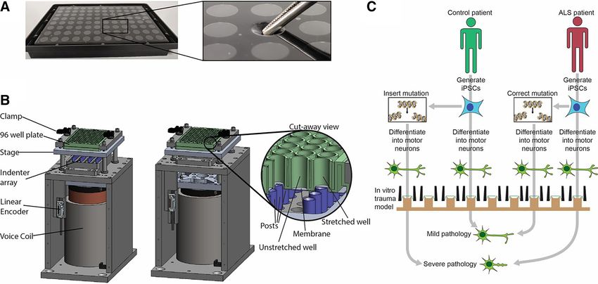
Fig. 1. The corticomotor pathway may be vulnerable
to degeneration after traumatic brain injury (TBI),
especially in people with high activity levels. A:
amyotrophic lateral sclerosis (ALS) is a disease
characterized by degeneration of both upper and
lower motor neurons (MNs). Upper MNs reside in
the motor cortex of the brain and have direct con-
nections to lower MNs in the brainstem (not shown)
and spinal cord. Lower MNs receive synaptic inputs
from upper MNs as well as project their axons to
muscle. Given the well-described focal clinical on-
set of ALS that often affects a single body region
before spreading to anatomically contiguous regions
with either type of MN within the neuroaxis, this
clinical pattern is compatible with the concept that
focal stress/damage after TBI may trigger neurode-
generative diseases like ALS. While the majority of
people who suffer a TBI will not develop ALS, both
the risk of TBI and ALS have been linked to
participation in high intensity physical activities
such as a collision sport (soccer, football) or military
combat. These clinical associations support a mul-
tihit model in which occupational exposure to TBI
and high-intensity activities, combined with patient-
specific factors such as genetic predisposition, may
determine whether MN health can be maintained (B)
or whether ALS befalls an individual (C).
confirmed by brain MRI were analyzed (Rosenbohm et al. HISTOPATHOLOGY IN TBI AND ALS
2014). Remarkably, it was noted that focal onset of their motor The clinical histopathology of ALS and TBI are largely
symptoms occurred contralateral to the cortical lesion site in distinct. CTE is associated with repeated mild TBI and char-
the majority of cases (15/18; 83.3%), suggesting spread might acterized by an extensive tauopathy, particularly in the outer
be mediated through connectivity of the corticomotor tracts. If layers of the cortex. Inclusions of amyloid beta are also
true, this would fit with one proposed model of disease prop- observed in a large subset of patients, postmortem (Johnson et
agation, via corticospinal/bulbar connectivity from upper to al. 2012; McKee et al. 2009). Conversely, tauopathy is rarely
lower MNs, perhaps related to a focal area of trauma (Fig. 1). observed in ALS, and is largely associated with the related
This mechanism is associated with cortical hyperexcitability disorders frontotemporal dementia (FTD) (Ling et al. 2013) or
(Seeger et al. 2017) and perhaps propagation via perturbed ALS with severe cognitive impairment (Strong et al. 2006;
synaptic connectivity or transmission of misfolded proteins Yang et al. 2003). The majority of ALS cases are characterized
such as TDP-43 through a prionlike mechanism. by typically cytosolic inclusions containing TDP-43, p62, and
The conflicting clinical results to date could be attributed FUS, with the exception of specific ALS subtypes such as
to a number of factors, including the possible underdiagno- familial ALS with superoxide dismutase 1 (SOD1) mutations,
sis and underreporting of milder TBI, as patients are likely which appear to have unique features (Shibata et al. 1996).
to not choose to seek medical attention for transient symp- Ubiquitin-positive inclusions appear to be a universal feature
toms. This is further compounded by the inconsistent his- of all sporadic and familial ALS (Deng et al. 2010; Nakano et
tory-taking practices between different physicians when it al. 2004). However, ubiquitin reactivity does not appear to be
comes to the incidence of TBI, during evaluation in ALS a feature in the few published cases of CTE with ALS overlap
specialty clinics. There is also the very wide range of normal (McKee et al. 2010).
for ALS disease onset and progression, which adds substan- TDP-43 inclusions have also been observed in CTE with
tial variability between individual patients. As with many ALS (McKee et al. 2010, 2013), while a single TBI is sufficient
studies relying on clinical observation and epidemiology to increase TDP-43 immunoreactivity, postmortem (Johnson et
data, it is therefore difficult to definitively prove causation. al. 2011). Aside from rare ALS cases caused by mutations in
In general, the studies performed to date were often small or the TDP-43 gene itself (Sreedharan et al. 2008), the mecha-
moderate in sample size and were at risk for both recall bias nisms responsible for the cytosolic accumulation of TDP-43 in
and high rates of type II statistical error (i.e., “false nega- sporadic ALS or CTE remain poorly understood. The repeat
tive”). Therefore, an increased emphasis on preclinical mod- expansion in C9orf72, which is the largest genetic contributor
els should facilitate a better understanding of the potential to ALS as well as FTD (DeJesus-Hernandez et al. 2011;
interaction between TBI and ALS. Renton et al. 2011), has been associated with disruptions in
J Neurophysiol • doi:10.1152/jn.00572.2018 • www.jn.org
Downloaded from www.physiology.org/journal/jn at Northwestern Univ (165.124.224.157) on October 8, 2019.TBI IMPACT ON ALS: BEDSIDE TO BENCH 1177
nucleocytoplasmic trafficking, which may contribute to cyto- unclear that this assumption is valid in the context of a
solic accumulation of TDP-43 (Freibaum et al. 2015; Jovičić et superimposed repetitive injury paradigm. TBI may induce
al. 2015; Zhang et al. 2015). Whether nucleocytoplasmic dysphagia or depression, which can directly contribute to the
disruption also plays a role in TDP-43 accumulation after TBI early weight disturbance. Importantly, the authors also reported
remain to be seen. Overall, while the histopathology features of that the same repetitive mild TBI paradigm led to motor
TBI and ALS are distinct, the common neuropathological deficits in wild-type rats (Thomsen et al. 2016).
feature of cytoplasmic TDP-43 aggregation suggests a poten- There has been one study of TBI in presymptomatic SOD1
tial mechanistic link that needs to be explored further. At the (G93A) mice, that used a closed-head TBI paradigm (Evans et
same time, although autopsy cases from subjects with clinical al. 2015). The authors noted an initial decline in body mass,
diagnoses of ALS and CTE show coexisting pathologic find- much of which seemed to occur in the first 3 days after closed
ings consistent both with ALS and CTE, these studies have no
TBI, but the weight then proceeded to rise for several weeks
ability to examine cause and effect or the role of head injuries
before starting to progressively decrease in a similar fashion to
in an unselected ALS population.
controls. After TBI, the mutant SOD1 mice, but not the
wild-type mice, showed significantly decreased grip strength
TBI IN ALS ANIMAL MODELS compared with sham control at 14 and 30 days. The authors
Few studies to date have attempted to address the influence also performed electromyography to evaluate for abnormal
of TBI on disease progression in ALS animal models. Trans- spontaneous activity such as fibrillation potentials or positive
genic mice and rats that overexpress mutant, human SOD1 sharp waves, which was present even in wild-type mice acutely
protein demonstrate phenotypes that reproduce progressive after closed TBI. Generally, this type of activity is thought to
MN disease, with features reminiscent of ALS in human be generated by muscle fibers that are not innervated, either
patients, including selective vulnerability of MNs, impaired due to motoneurogenic or myopathic processes (Daube and
motor function, and death from neuromuscular respiratory Rubin 2009). The presence of either MN or muscle degenera-
failure (Turner and Talbot 2008). Consequently, mutant SOD1 tion as a result of TBI is hard to account for in wild-type mice.
transgenic rodents remain the most well studied ALS model Therefore, the significantly greater spontaneous activity pattern
and are a standard bearer for animal modeling of neurodegen- noted in mutant SOD1 mice with TBI, at 1 and 7 days
erative disease in general (Van Damme et al. 2017). Two postinjury is hard to interpret. The mutant SOD1 mice also
studies have examined the effect of TBI in SOD1 (G93A) exhibited an upregulation of inflammatory and oxidative stress
transgenic rat models. In the first study, adult, presymptomatic biomarkers after TBI; however, there was no change in disease
rats were subjected to a focal controlled, cortical impact of onset, as measured by motor behavioral evaluation, or overall
“moderate” to “severe” magnitude centered over the motor survival.
cortex (Thomsen et al. 2015). Rats with the mutant SOD1 Recent groups have employed the Drosophila melanogaster
protein subjected to this form of TBI did not exhibit earlier fruit fly to develop in vivo models of TBI. The flies are fairly
disease onset, altered locomotor function, or shortened lifes- advantageous for animal studies as they have a short lifespan,
pan, compared with ones that underwent a sham procedure. It a wealth of genetic tools available, and relatively straightfor-
should be noted that, despite histological confirmation of ward outcome measures related to neurodegeneration. In this
extensive cortical tissue damage, there were no significant paradigm, adult flies are placed in a vial, affixed to a standard
motor deficits detected after TBI, which is in stark contrast to compression spring. The spring is then bent to a predetermined
motor deficits known to occur with motor cortex damage in angle and released against a semirigid surface to produce
human TBI patients. This may be a reflection of the well- impact injury (Katzenberger et al. 2015). This method has
known anatomical and functional differences between rodent previously been shown to reduce lifespan with repeated insult
and human pyramidal systems (Lemon 2008). and can produce significant lesions in Drosophila brains (Kat-
A follow-up study modified the injury paradigm such that zenberger et al. 2013). Strikingly, repeated injury also leads to
each controlled cortical impact was reduced to mild severity increased levels of phospho-tau and markers of immune acti-
but was performed on five separate occasions (weekly) starting vation in brain tissue, suggesting that it may recapitulate some
in a young adult group of presymptomatic SOD1 (G93A) rats, of the hallmarks of CTE (Barekat et al. 2016; Katzenberger et
in an attempt to model repetitive mild TBI (Thomsen et al. al. 2013).
2016). The authors reported that rats subjected to repetitive Unique among the studies is a recent publication examining
mild TBI in the mutant SOD1 genotype background demon- the direct interaction between TBI and multiple models of ALS
strated earlier ALS symptom onset, as defined by a decline in in Drosophila (Anderson et al. 2018). Using a sublethal injury
body weight and compared with sham control mutant rats. scheme, the authors found that flies overexpressing ALS gene
Unfortunately, there are a number of key limitations to this mutations in FUS or C9orf72 exhibited an increase in mortality
study as it pertains to understanding the relationship between after injury, with concomitant increases in ubiquitinated pro-
TBI and ALS. The group sizes were relatively small (n ⫽ 7 tein species. Injury also unmasked persistent locomotor defects
TBI versus n ⫽ 9 sham), there were no motor functional that were not observable in control animals. Interestingly, the
measures reported, and the animals were not followed to authors also observed dramatic increases in TDPH-positive
disease end stage. The early disease onset upon mild TBI was stress granules upon injury, which is the Drosophila homolog
not supported by any motor function assay or direct histopatho- of TDP-43. While this phenomenon has been observed after
logical assessment. While tracking body weight has been axotomy in mouse peripheral nerves (Moisse et al. 2009), it is
shown to be a good measure of disease onset in mutant SOD1 not yet a known hallmark of TBI. Given that stress granules
rats under standard conditions (Matsumoto et al. 2006), it is have been observed in some models of ALS (Mackenzie et al.
J Neurophysiol • doi:10.1152/jn.00572.2018 • www.jn.org
Downloaded from www.physiology.org/journal/jn at Northwestern Univ (165.124.224.157) on October 8, 2019.1178 TBI IMPACT ON ALS: BEDSIDE TO BENCH
2017; Taylor et al. 2016), this may suggest a common patho- membrane that is stretched to create a mechanical insult. Early
genic mechanism. in vitro TBI models induced stretch by applying air pressure to
Further studies will be necessary to definitively determine all (Ellis et al. 1995) or part of the silicone membrane (Smith
whether TBI can modulate ALS onset or progression. Inher- et al. 1999). More recently, indentation with a rigid piston
ently, animal model data must be interpreted with caution, as it driven by an electromagnetic voice coil has been used to
is challenging to distinguish an actual alteration of ALS pro- induce stretch (Morrison et al. 2003). Indentation systems
gression from any additive motor deficits caused by MN loss, achieve shorter pulse durations than pneumatic systems (Ellis
which are known to occur early in presymptomatic ALS et al. 1995; Morrison et al. 2006). These shorter pulses are
rodents (Franz et al. 2009; Frey et al. 2000; Pun et al. 2006). more biofidelic (Hardy et al. 2007), which is significant be-
This is particularly important for ALS/TBI paradigms, as mild cause the trauma response is rate sensitive (Ahmadzadeh et al.
TBI may (Thomsen et al. 2016) or may not (Thomsen et al. 2014; Elkin and Morrison 2007). Also, the indentation ap-
2015) cause detectable behavioral deficits in wild-type ani- proach has been scaled up to a 96-well format (Sherman et al.
mals. Establishing a model of spinal MN degeneration in 2016) but technical challenges make it difficult to scale the
wild-type animals after a TBI paradigm is unlikely to be of pneumatic approach up to a multiwell format (Magou et al.
high yield by itself, as the clinical experience has been that 2011).
even upon induction of CTE-like disease only a small subset
would go on to develop concurrent MN disease (McKee et al.
HUMAN IPSC MODELS FOR STUDYING THE RELATIONSHIP
2013). Still, there remains ample opportunity to determine a
BETWEEN TBI AND ALS
cause-and-effect relationship between TBI and ALS through
the use animal models. Larger study groups, detailed histopa- In vitro modeling has proven to be a powerful preclinical
thology and electrophysiology, and variations in the timing of tool in ALS. Experiments with patient-specific induced pluri-
injury relative to ALS phenotypic onset should be considered. potent stem cell (iPSC)-derived neurons have uncovered dis-
Additionally, over the last decade or so there have been dozens ease mechanisms (Barmada et al. 2014; Bilican et al. 2012;
of new mouse lines described with ALS gene causing muta- Chen et al. 2014; Devlin et al. 2015; Donnelly et al. 2013;
tions beyond SOD1, including some that lack major motor Egawa et al. 2012; Kiskinis et al. 2014, 2018; Mitne-Neto et al.
deficits or even MN degeneration (De Giorgio et al. 2019). 2011; Sareen et al. 2013; Serio et al. 2013; Wainger et al. 2014;
Detailed neuromuscular studies that combine a TBI paradigm Yang et al. 2013), and enable relatively rapid clinical innova-
with one of these recent mouse models that may have genetic tion through repurposing of clinically approved drugs (Mc-
vulnerability, rather than a predetermined fate, for lower MN Neish et al. 2015). Human iPSCs are easily generated after
degeneration could be highly insightful. primary cells (typically skin cells or mononuclear, blood cells),
which are harvested from a human patient, are converted by
molecular reprogramming and then differentiated into relevant
MODELING TBI IN VITRO neural subtypes (Hunsberger et al. 2015). The utility of
Although TBI pathology shares several mechanisms with CRISPR/Cas9 gene editing for the generation of isogenic
other neurological disorders, it begins in a unique way: with control iPSC lines (i.e. experiments comparing iPSC-derived
rapid deformation of the tissue. Brain tissue is soft and incom- neurons with genomes that differ only by a single genetic
pressible, i.e., it is easy to change its shape but very difficult to variant) can conclusively prove that a particular genetic variant
change its volume (Holbourn 1943). Tension, compression, causes a particular functional deficit. The combination of in
and shear are always coupled in an incompressible material, a vitro trauma experiments with patient-specific and isogenic
phenomenon known as the Poisson effect. For example, if a iPSC-derived neurons have the potential to address the ques-
piece of tissue is compressed on a vertical plane, it stretches on tion of gene-trauma interactions in ALS pathology (Fig. 2).
a horizontal plane and shears along a diagonal plane. If it is They could also be applied to a multihit, gene-trauma interac-
stretched vertically, it compresses horizontally and again tion model, i.e., the hypothesis that a given mutation is harm-
shears along a diagonal plane. In fact, these two situations are less in the absence of neurotrauma but leads to ALS in the
not on average different from the perspective of a randomly wake of neurotrauma. This opportunity is particularly exciting
oriented neuron inside the tissue. Neurons are long, slender in light of ongoing efforts to bank stem cells from up to 1,000
structures. Slender structures can generally accommodate any ALS patients (Progress & Updates: Answer ALS Research
type of loading except tension without failure because they can 2018).
curl up without damage. Therefore, while compression, ten- At the same time cell culture models have limitations. They
sion, and shear occur simultaneously on different planes provide simple approximations of the likely complex in vivo
through any point, neurons oriented along the plane of maxi- disease processes, because they lack the cellular diversity and
mum tension are the most likely to fail. These are the basic structural organization of an intact nervous system. This is
principles underlying most in vitro models of TBI. particularly relevant in the case of TBI, which is known to
There are several well-established in vitro models of TBI involve interconnected dysfunction in all three components of
(Morrison et al. 2011), including multiple 2D culture systems the neurovascular unit, which consists of neurons, glia and
that apply tension and 3D culture system that apply compres- associated vasculature (Xing et al. 2012). CNS glia, in partic-
sion (Bar-Kochba et al. 2016) or shear (LaPlaca et al. 2005). ular, appear to play an essential role in neuronal degeneration
Organotypic slice cultures (Morrison et al. 2006) and 2D or 3D and regeneration upon injury (Myer et al. 2006; Neumann et al.
cultures (Ahmed et al. 2000; Cullen et al. 2007) of dissociated 2009), while other non-cell-autonomous mechanisms such as
primary or immortalized cells have been employed in these propagation of misfolded proteins may play a key role in TBI
models. Typically, 2D cultures are maintained on a silicone pathogenesis (Hawkins et al. 2013). Phosphorylated, oligo-
J Neurophysiol • doi:10.1152/jn.00572.2018 • www.jn.org
Downloaded from www.physiology.org/journal/jn at Northwestern Univ (165.124.224.157) on October 8, 2019.TBI IMPACT ON ALS: BEDSIDE TO BENCH 1179
Fig. 2. Understanding the relationship between amyotrophic lateral sclerosis (ALS) and neurotrauma with patient-specific induced pluripotent stem cells
(iPSCs)-based neurons. Induced pluripotent stem cell-derived neurons could be used in combination with an in vitro model of neurotrauma to understand
gene-trauma interaction in ALS as follows. A: stretchable 96-well plates are fabricated by bonding a layer of flexible, transparent silicone to bottomless 96-well
plates. The inset shows tweezers gently depressing on of the well bottoms to illustrate the flexibility of the growth substrate. B: a custom-built device is used
to apply a rapid, repeatable, equibiaxial stretch to the bottom of the wells. Stretch is produced by pressing the plate rapidly down against an array of lubricated,
Teflon-coated, cylindrical indenters. The cut-away view shows an example of a stretched well alongside a well that is not stretched because the corresponding
post is not present. C: schematic of an experimental design to study gene-trauma interactions in ALS using this human in vitro model. A mutation associated
with ALS can be engineered into an iPSC line derived from a healthy control individual or alternatively can be corrected in an iPSC line derived from a patient
with a known disease-causing genetic variant. Both lines are then subjected to identical trauma in vitro, and subsequent pathology is quantified and compared with
test the hypothesis that the ALS-associated mutation amplifies the pathology of trauma.
meric tau protein has been shown to accumulate in the brains developments in direct neuronal transdifferentiation from adult
of rats exposed to a fluid percussion model of TBI (Hawkins et somatic cell types appear to preserve aspects of cellular age
al. 2013), while the capacity of certain misfolded tau species to (Abernathy et al. 2017; Huh et al. 2016; Victor et al. 2018; Yoo
then propagate through prion-like mechanisms can influence et al. 2011) and may facilitate the development of more
the long-term functional deficits observed after TBI (Ahmed et accurate in vitro models of TBI pathogenesis.
al. 2014; Gerson et al. 2016; Kfoury et al. 2012).
In some ways, these potential limitations can be of experi-
THE COMMON PATHOPHYSIOLOGY IN TBI AND ALS
mental value. The use of isolated neuronal models of TBI
allows for the detection of neuron-specific, cell-autonomous Due to the limited number of studies designed to directly
mechanisms of neurotoxicity in TBI and/or ALS. In addition, examine the link between TBI and ALS in a controlled,
rapid advances in organoid, 3D cell culture systems provide laboratory setting, the pathophysiological link between these
increasing levels of cellular diversity and structural organiza- two disease states remains largely unclear. Any proposed link
tion (Arlotta and Paşca 2019; Lancaster and Knoblich 2014). is therefore highly speculative. TDP-43 and stress-granule
Furthermore, various insult mechanisms, such as stretch or dysregulation may be a potential molecular overlap between
fluid percussion, can effectively isolate pathologies resulting the two disorders, but this link must be examined more directly
from single aspects of TBI, which otherwise entails a highly (Anderson et al. 2018). Recent evidence directly links stress-
complex array of tissue damage mechanisms. granule formation to disruptions on nucleocytoplasmic trans-
The major hurdle that these models do face, however, is the port (Zhang et al. 2018), which is a hallmark of C9orf72 ALS
relative immaturity of the neurons in culture (Ho et al. 2016). (Chou et al. 2018; Zhang et al. 2015), further suggesting that
Both TBI (Sendroy-Terrill et al. 2010) and ALS (Hardiman et this may be an important overlap between TBI and ALS.
al. 2017) are strongly influenced by aging. In the case of TBI, TDP-43 has also been demonstrated to be particularly vulner-
both age at injury and time postinjury negatively predict patient able to protease degradation in various neurotoxic states, in-
outcomes (Sendroy-Terrill et al. 2010). By nature, iPSCs cluding TBI, which may worsen loss-of-function effects in
effectively revert back to an embryonic state after reprogram- affected cells (Yang et al. 2014). We therefore hypothesize that
ming (Takahashi and Yamanaka 2006), largely irrespective of TBI may contribute to the disruption of proteostasis seen in
donor age, despite some genetic signatures (Lo Sardo et al. ALS patients, thereby leading to insurmountable proteotoxic
2017; Miller et al. 2013). Although methods of iPSC-derived stress.
cell aging exist (Miller et al. 2013), it is unlikely that they TBI appears to focally induce several pathological processes
faithfully recapitulate all mechanisms of human aging. Recent that may overlap with ALS. Most immediate among these
J Neurophysiol • doi:10.1152/jn.00572.2018 • www.jn.org
Downloaded from www.physiology.org/journal/jn at Northwestern Univ (165.124.224.157) on October 8, 2019.1180 TBI IMPACT ON ALS: BEDSIDE TO BENCH
appears to be excitotoxic firing, due to transient ion dysregu- CONCLUSIONS
lation near the site of injury (Palmer et al. 1993; Wagner et There is growing evidence that TBI, particularly of a repet-
al. 2004). Notably, dysregulation of the glial excitatory itive nature with mild severity (McKee et al. 2013; Pupillo et
amino acid transporter 2 (EAAT2) has been observed in al. 2017), might be a risk factor for developing ALS. If true,
both ALS patients (Rothstein et al. 1995) and rodent models this would be of special significance to those individuals who
of ALS (Howland et al. 2002), and this loss appears to be engage in high-risk activities such as collision sports. The facts
specific to regions typically lost in ALS. Homeostatic reg- that mutations in TDP-43 can cause ALS and that both spo-
ulation of the GluA2 subunit of AMPA receptors on MNs is radic ALS and CTE cases are frequently characterized by
also lost in mutant SOD1 models of ALS, leading to an TDP-43 proteinopathy (Mackenzie et al. 2007; McKee et al.
increased vulnerability to excitotoxicity (Taylor et al. 2016; 2013) imply a potentially shared mechanism of neurodegen-
Van Damme et al. 2007). Spinal MNs may therefore be eration.
Animal models have been extremely useful for modeling
particularly vulnerable to excitotoxic damage in ALS, and
ALS disease mechanisms and addressing the potential interac-
TBI-induced excitotoxic signaling may ultimately tip the
tion with TBI but may have critical limitations ranging from
scales in favor of cell death. their inability to capture the genetic complexity of human
Oxidative stress also appears to be a common pathological patients to fundamentally different corticomotor function and
signature between ALS and TBI (Ansari et al. 2008; Evans et connectivity. Patient-specific iPSC technologies have been
al. 2015; Readnower et al. 2010; Turner and Talbot 2008). rapidly improving over the last decade and have become
How this may preferentially lead to MN damage remains another component of our preclinical tool set for understanding
unclear, but the combined toxicity in both disease states may neurodegenerative disease (Ichida and Kiskinis 2015). At the
hasten cytotoxicity (Barber and Shaw 2010). Similarly, sus- same time, the development of instrumentation that allows for
tained, diffuse neuroinflammation has been well documented controlled delivery of biofidelic trauma to human neurons in
in both TBI (Acosta et al. 2013; Johnson et al. 2013) and ALS culture (Sherman et al. 2016) enables an additional platform
(Hall et al. 1998; Keizman et al. 2009). While this may initially that can be used to address the conflicting clinical observations
be a protective response to neurotoxic damage, sustained and animal studies on the effects of TBI on ALS incidence and
inflammation may lead to reactive gliosis and further contrib- progression. There is no doubt that these human preclinical
ute to neuronal damage through pathological alterations to the model systems will help us understand aspects of ALS that
extracellular milieu. Necroptosis, or programmed necrotic cell may be unique to patients or patient subgroups, but they will
death with autophagic induction (Degterev et al. 2005; Van- not ever fully reproduce the in vivo context of the human CNS.
denabeele et al. 2010), has recently been identified as a major Therefore, corroboration of key findings between multiple
mechanism of cell death in both ALS (Ito et al. 2016; Re et al. preclinical models (e.g., human and rodent, in vitro and in
2014) and TBI (Liu et al. 2016; Wang et al. 2012; You et al. vivo, etc.), along with careful clinical correlation to bedside
2008). Because necroptosis is activated, in part, by cell non- and histopathological data should be the path to a better
autonomous mechanisms such as inflammation (Vandenabeele understanding of disease mechanisms and the development of
et al. 2010), it is possible that TBI may increase necroptotic effective treatments for ALS.
signaling above threshold for MNs to remain viable in some
ACKNOWLEDGMENTS
individuals (Fig. 1).
Another potential pathological overlap between the two We acknowledge Caitlyn Hofer and Michael Gallagher (subQstudio.com)
for the artwork used in Fig. 1.
disease states is cytoskeletal damage and dysregulation. Mu- Present address for C. K. Franz: Physical Medicine and Rehabilitation,
tations in genes associated with cytoskeletal homeostasis, in- Shirley Ryan AbilityLab and Northwestern University Feinberg School of
cluding profilin-1 (PFN1) (Wu et al. 2012) and tubulin al- Medicine, Chicago, IL, 355 E. Erie St, Chicago, IL 60611.
pha-4A (TUBA4A) (Smith et al. 2014), have been shown to
cause ALS. Other cytoskeletal gene mutations such as neuro- GRANTS
filament heavy chain (Al-Chalabi et al. 1999) and dynactin We are grateful to the following funding sources: US National Institutes of
(Münch et al. 2004) have been associated with increased Health (NIH)/National Institute on Neurological Disorders and Stroke (NINDS)
and National Institute on Aging (NIA) R01NS104219 (E. Kiskinis), NIH/NINDS
susceptibility to ALS. Moreover, markers of cytoskeletal dam- grants R21NS098129 (J. D. Finan), R21NS107761 (E. Kiskinis), R21NS111248
age have been observed in the cerebrospinal fluid (CSF) of (E. Kiskinis), Muscular Dystrophy Association (E. Kiskinis), the Les Turner ALS
sporadic ALS patients (Brettschneider et al. 2006), while Foundation (E. Kiskinis), the Craig H. Nielsen Foundation (C. K. Franz), and the
aggregation of neurofilament and reduced expression of neu- Foundation for Physical Medicine and Rehabilitation (C. K. Franz). C. K. Franz is
a member of the Shirley Ryan AbilityLab’s Center for Translational Biomedicine,
rofilament, light-chain have also been described in iPSC-based Northwestern’s Skin Disease Research Center, as well as a physician in the Lois
models of SOD1-related ALS (Chen et al. 2014). The accumu- Insolia ALS Clinic at the Les Turner ALS Center. E. Kiskinis is a member of the
lation of neurofilament polypeptides (both light and heavy), Simpson Querrey Institute for BioNanotechnology and a Les Turner ALS Re-
has also been observed in the blood and CSF of patients with search Center Investigator.
TBI (Zetterberg et al. 2013). Diffuse axonal injury in TBI has
been shown to induce localized cytoskeletal damage (Kilinc et DISCLOSURES
al. 2008), which in turn can induce neurofilament compaction No conflicts of interest, financial or otherwise, are declared by the authors.
and mislocalization (Povlishock and Pettus 1996). Thus, dys-
regulation of cytoskeletal homeostasis in ALS patients may AUTHOR CONTRIBUTIONS
render them less able to overcome cytoskeletal damage after C.K.F. conceived and designed research; C.K.F. and J.D.F. prepared fig-
mild TBI, leading to worsened clinical outcomes. ures; C.K.F., D.J., E.L.D., R.A.G., K.D., A.L., J.D.F., and E.K. drafted
J Neurophysiol • doi:10.1152/jn.00572.2018 • www.jn.org
Downloaded from www.physiology.org/journal/jn at Northwestern Univ (165.124.224.157) on October 8, 2019.TBI IMPACT ON ALS: BEDSIDE TO BENCH 1181
manuscript; C.K.F., D.J., E.L.D., R.A.G., K.D., A.L., J.D.F., and E.K. edited vulnerability. Proc Natl Acad Sci USA 109: 5803–5808, 2012. doi:10.1073/
and revised manuscript; C.K.F., D.J., E.L.D., R.A.G., K.D., A.L., J.D.F., and pnas.1202922109.
E.K. approved final version of manuscript. Brettschneider J, Petzold A, Süssmuth SD, Ludolph AC, Tumani H.
Axonal damage markers in cerebrospinal fluid are increased in ALS.
Neurology 66: 852– 856, 2006. doi:10.1212/01.wnl.0000203120.85850.54.
REFERENCES Chen H, Qian K, Du Z, Cao J, Petersen A, Liu H, Blackbourn LW IV,
Huang CL, Errigo A, Yin Y, Lu J, Ayala M, Zhang SC. Modeling ALS
Abernathy DG, Kim WK, McCoy MJ, Lake AM, Ouwenga R, Lee SW, with iPSCs reveals that mutant SOD1 misregulates neurofilament balance in
Xing X, Li D, Lee HJ, Heuckeroth RO, Dougherty JD, Wang T, Yoo AS. motor neurons. Cell Stem Cell 14: 796 – 809, 2014. doi:10.1016/j.stem.2014.
MicroRNAs induce a permissive chromatin environment that enables neu- 02.004.
ronal subtype-specific reprogramming of adult human fibroblasts. Cell Stem Chen H, Richard M, Sandler DP, Umbach DM, Kamel F. Head injury and
Cell 21: 332–348.e9, 2017. doi:10.1016/j.stem.2017.08.002. amyotrophic lateral sclerosis. Am J Epidemiol 166: 810 – 816, 2007. doi:10.
Acosta SA, Tajiri N, Shinozuka K, Ishikawa H, Grimmig B, Diamond 1093/aje/kwm153.
DM, Sanberg PR, Bickford PC, Kaneko Y, Borlongan CV. Long-term Chou CC, Zhang Y, Umoh ME, Vaughan SW, Lorenzini I, Liu F, Sayegh
upregulation of inflammation and suppression of cell proliferation in the M, Donlin-Asp PG, Chen YH, Duong DM, Seyfried NT, Powers MA,
brain of adult rats exposed to traumatic brain injury using the controlled Kukar T, Hales CM, Gearing M, Cairns NJ, Boylan KB, Dickson DW,
cortical impact model. PLoS One 8: e53376, 2013. [Erratum in PLoS One Rademakers R, Zhang YJ, Petrucelli L, Sattler R, Zarnescu DC, Glass
8: 10.1371/annotation/a04a7468-d105-42f3-ba47-263ea2864681.] doi: JD, Rossoll W. TDP-43 pathology disrupts nuclear pore complexes and
10.1371/journal.pone.0053376.
nucleocytoplasmic transport in ALS/FTD. Nat Neurosci 21: 228 –239, 2018.
Ahmadzadeh H, Smith DH, Shenoy VB. Viscoelasticity of tau proteins leads
doi:10.1038/s41593-017-0047-3.
to strain rate-dependent breaking of microtubules during axonal stretch
injury: predictions from a mathematical model. Biophys J 106: 1123–1133, Cirulli ET, Lasseigne BN, Petrovski S, Sapp PC, Dion PA, Leblond CS,
2014. doi:10.1016/j.bpj.2014.01.024. Couthouis J, Lu YF, Wang Q, Krueger BJ, Ren Z, Keebler J, Han Y,
Ahmed SM, Rzigalinski BA, Willoughby KA, Sitterding HA, Ellis EF. Levy SE, Boone BE, Wimbish JR, Waite LL, Jones AL, Carulli JP,
Stretch-induced injury alters mitochondrial membrane potential and cellular Day-Williams AG, Staropoli JF, Xin WW, Chesi A, Raphael AR,
ATP in cultured astrocytes and neurons. J Neurochem 74: 1951–1960, 2000. McKenna-Yasek D, Cady J, Vianney de Jong JM, Kenna KP, Smith
doi:10.1046/j.1471-4159.2000.0741951.x. BN, Topp S, Miller J, Gkazi A, Al-Chalabi A, van den Berg LH, Veldink
Ahmed Z, Cooper J, Murray TK, Garn K, McNaughton E, Clarke H, J, Silani V, Ticozzi N, Shaw CE, Baloh RH, Appel S, Simpson E,
Parhizkar S, Ward MA, Cavallini A, Jackson S, Bose S, Clavaguera F, Lagier-Tourenne C, Pulst SM, Gibson S, Trojanowski JQ, Elman L,
Tolnay M, Lavenir I, Goedert M, Hutton ML, O’Neill MJ. A novel in McCluskey L, Grossman M, Shneider NA, Chung WK, Ravits JM,
vivo model of tau propagation with rapid and progressive neurofibrillary Glass JD, Sims KB, Van Deerlin VM, Maniatis T, Hayes SD, Ordureau
tangle pathology: the pattern of spread is determined by connectivity, not A, Swarup S, Landers J, Baas F, Allen AS, Bedlack RS, Harper JW,
proximity. Acta Neuropathol 127: 667– 683, 2014. doi:10.1007/s00401-014- Gitler AD, Rouleau GA, Brown R, Harms MB, Cooper GM, Harris T,
1254-6. Myers RM, Goldstein DB; FALS Sequencing Consortium. Exome se-
Al-Chalabi A, Andersen PM, Nilsson P, Chioza B, Andersson JL, Russ C, quencing in amyotrophic lateral sclerosis identifies risk genes and pathways.
Shaw CE, Powell JF, Leigh PN. Deletions of the heavy neurofilament Science 347: 1436 –1441, 2015. doi:10.1126/science.aaa3650.
subunit tail in amyotrophic lateral sclerosis. Hum Mol Genet 8: 157–164, Corcia P, Couratier P, Blasco H, Andres CR, Beltran S, Meininger V,
1999. doi:10.1093/hmg/8.2.157. Vourc’h P. Genetics of amyotrophic lateral sclerosis. Rev Neurol (Paris)
Al-Chalabi A, Lewis CM. Modelling the effects of penetrance and family size 173: 254 –262, 2017. doi:10.1016/j.neurol.2017.03.030.
on rates of sporadic and familial disease. Hum Hered 71: 281–288, 2011. Cullen DK, Simon CM, LaPlaca MC. Strain rate-dependent induction of
doi:10.1159/000330167. reactive astrogliosis and cell death in three-dimensional neuronal-astrocytic
Anderson EN, Gochenaur L, Singh A, Grant R, Patel K, Watkins S, Wu co-cultures. Brain Res 1158: 103–115, 2007. doi:10.1016/j.brainres.2007.
JY, Pandey UB. Traumatic injury induces stress granule formation and 04.070.
enhances motor dysfunctions in ALS/FTD models. Hum Mol Genet 27: Daube JR, Rubin DI. Needle electromyography. Muscle Nerve 39: 244 –270,
1366 –1381, 2018. doi:10.1093/hmg/ddy047. 2009. doi:10.1002/mus.21180.
Ansari MA, Roberts KN, Scheff SW. Oxidative stress and modification of De Giorgio F, Maduro C, Fisher EMC, Acevedo-Arozena A. Transgenic
synaptic proteins in hippocampus after traumatic brain injury. Free Radic and physiological mouse models give insights into different aspects of
Biol Med 45: 443– 452, 2008. doi:10.1016/j.freeradbiomed.2008.04.038. amyotrophic lateral sclerosis. Dis Model Mech 12: dmm037424, 2019.
Arlotta P, Paşca SP. Cell diversity in the human cerebral cortex: from the doi:10.1242/dmm.037424.
embryo to brain organoids. Curr Opin Neurobiol 56: 194 –198, 2019. Degterev A, Huang Z, Boyce M, Li Y, Jagtap P, Mizushima N, Cuny GD,
doi:10.1016/j.conb.2019.03.001. Mitchison TJ, Moskowitz MA, Yuan J. Chemical inhibitor of nonapop-
Armon C, Nelson LM. Is head trauma a risk factor for amyotrophic lateral totic cell death with therapeutic potential for ischemic brain injury. Nat
sclerosis? An evidence based review. Amyotroph Lateral Scler 13: 351–356, Chem Biol 1: 112–119, 2005. [Erratum in Nat Chem Biol 1: 234, 2005.
2012. doi:10.3109/17482968.2012.660954. Addendum in Nat Chem Biol 9: 192, 2013.] doi:10.1038/nchembio711.
Bar-Kochba E, Scimone MT, Estrada JB, Franck C. Strain and rate- DeJesus-Hernandez M, Mackenzie IR, Boeve BF, Boxer AL, Baker M,
dependent neuronal injury in a 3D in vitro compression model of traumatic Rutherford NJ, Nicholson AM, Finch NA, Flynn H, Adamson J, Kouri
brain injury. Sci Rep 6: 30550, 2016. doi:10.1038/srep30550. N, Wojtas A, Sengdy P, Hsiung GY, Karydas A, Seeley WW, Josephs
Barber SC, Shaw PJ. Oxidative stress in ALS: key role in motor neuron KA, Coppola G, Geschwind DH, Wszolek ZK, Feldman H, Knopman
injury and therapeutic target. Free Radic Biol Med 48: 629 – 641, 2010. DS, Petersen RC, Miller BL, Dickson DW, Boylan KB, Graff-Radford
doi:10.1016/j.freeradbiomed.2009.11.018. NR, Rademakers R. Expanded GGGGCC hexanucleotide repeat in non-
Barekat A, Gonzalez A, Mauntz RE, Kotzebue RW, Molina B, El- coding region of C9ORF72 causes chromosome 9p-linked FTD and ALS.
Mecharrafie N, Conner CJ, Garza S, Melkani GC, Joiner WJ, Lipinski Neuron 72: 245–256, 2011. doi:10.1016/j.neuron.2011.09.011.
MM, Finley KD, Ratliff EP. Using Drosophila as an integrated model to Deng HX, Zhai H, Bigio EH, Yan J, Fecto F, Ajroud K, Mishra M,
study mild repetitive traumatic brain injury. Sci Rep 6: 25252, 2016. Ajroud-Driss S, Heller S, Sufit R, Siddique N, Mugnaini E, Siddique T.
doi:10.1038/srep25252. FUS-immunoreactive inclusions are a common feature in sporadic and
Barmada SJ, Serio A, Arjun A, Bilican B, Daub A, Ando DM, Tsvetkov A, non-SOD1 familial amyotrophic lateral sclerosis. Ann Neurol 67: 739 –748,
Pleiss M, Li X, Peisach D, Shaw C, Chandran S, Finkbeiner S. Au- 2010. doi:10.1002/ana.22051.
tophagy induction enhances TDP43 turnover and survival in neuronal ALS Devlin AC, Burr K, Borooah S, Foster JD, Cleary EM, Geti I, Vallier
models. Nat Chem Biol 10: 677– 685, 2014. doi:10.1038/nchembio.1563. L, Shaw CE, Chandran S, Miles GB. Human iPSC-derived motoneu-
Bilican B, Serio A, Barmada SJ, Nishimura AL, Sullivan GJ, Carrasco M, rons harbouring TARDBP or C9ORF72 ALS mutations are dysfunctional
Phatnani HP, Puddifoot CA, Story D, Fletcher J, Park IH, Friedman despite maintaining viability. Nat Commun 6: 5999, 2015. doi:10.1038/
BA, Daley GQ, Wyllie DJ, Hardingham GE, Wilmut I, Finkbeiner S, ncomms6999.
Maniatis T, Shaw CE, Chandran S. Mutant induced pluripotent stem cell Dixon KJ. Pathophysiology of traumatic brain injury. Phys Med Rehabil Clin
lines recapitulate aspects of TDP-43 proteinopathies and reveal cell-specific N Am 28: 215–225, 2017. doi:10.1016/j.pmr.2016.12.001.
J Neurophysiol • doi:10.1152/jn.00572.2018 • www.jn.org
Downloaded from www.physiology.org/journal/jn at Northwestern Univ (165.124.224.157) on October 8, 2019.1182 TBI IMPACT ON ALS: BEDSIDE TO BENCH
Donnelly CJ, Zhang PW, Pham JT, Haeusler AR, Mistry NA, Vidensky S, motor neuron maturation and aging pathways within gene co-expression
Daley EL, Poth EM, Hoover B, Fines DM, Maragakis N, Tienari PJ, networks. Nat Neurosci 19: 1256 –1267, 2016. doi:10.1038/nn.4345.
Petrucelli L, Traynor BJ, Wang J, Rigo F, Bennett CF, Blackshaw S, Holbourn AHS. Mechanics of head injuries. Lancet 242: 438 – 441, 1943.
Sattler R, Rothstein JD. RNA toxicity from the ALS/FTD C9ORF72 doi:10.1016/S0140-6736(00)87453-X.
expansion is mitigated by antisense intervention. Neuron 80: 415– 428, Horner RD, Grambow SC, Coffman CJ, Lindquist JH, Oddone EZ, Allen
2013. doi:10.1016/j.neuron.2013.10.015. KD, Kasarskis EJ. Amyotrophic lateral sclerosis among 1991 Gulf War
Egawa N, Kitaoka S, Tsukita K, Naitoh M, Takahashi K, Yamamoto T, veterans: evidence for a time-limited outbreak. Neuroepidemiology 31:
Adachi F, Kondo T, Okita K, Asaka I, Aoi T, Watanabe A, Yamada Y, 28 –32, 2008. doi:10.1159/000136648.
Morizane A, Takahashi J, Ayaki T, Ito H, Yoshikawa K, Yamawaki S, Howland DS, Liu J, She Y, Goad B, Maragakis NJ, Kim B, Erickson J,
Suzuki S, Watanabe D, Hioki H, Kaneko T, Makioka K, Okamoto K, Kulik J, DeVito L, Psaltis G, DeGennaro LJ, Cleveland DW, Rothstein
Takuma H, Tamaoka A, Hasegawa K, Nonaka T, Hasegawa M, Kawata JD. Focal loss of the glutamate transporter EAAT2 in a transgenic rat model
A, Yoshida M, Nakahata T, Takahashi R, Marchetto MC, Gage FH, of SOD1 mutant-mediated amyotrophic lateral sclerosis (ALS). Proc Natl
Yamanaka S, Inoue H. Drug screening for ALS using patient-specific Acad Sci USA 99: 1604 –1609, 2002. doi:10.1073/pnas.032539299.
induced pluripotent stem cells. Sci Transl Med 4: 145ra104, 2012. doi:10. Huh CJ, Zhang B, Victor MB, Dahiya S, Batista LF, Horvath S, Yoo AS.
1126/scitranslmed.3004052. Maintenance of age in human neurons generated by microRNA-based
Elkin BS, Morrison B III. Region-specific tolerance criteria for the living neuronal conversion of fibroblasts. eLife 5: e18648, 2016. doi:10.7554/
brain. Stapp Car Crash J 51: 127–138, 2007. doi:10.4271/2007-22-0005. eLife.18648.
Hunsberger JG, Efthymiou AG, Malik N, Behl M, Mead IL, Zeng X,
Ellis EF, McKinney JS, Willoughby KA, Liang S, Povlishock JT. A new
Simeonov A, Rao M. Induced pluripotent stem cell models to enable in
model for rapid stretch-induced injury of cells in culture: characterization of
vitro models for screening in the central nervous system. Stem Cells Dev 24:
the model using astrocytes. J Neurotrauma 12: 325–339, 1995. doi:10.1089/ 1852–1864, 2015. doi:10.1089/scd.2014.0531.
neu.1995.12.325. Ichida JK, Kiskinis E. Probing disorders of the nervous system using
Evans TM, Jaramillo CA, Sataranatarajan K, Watts L, Sabia M, Qi W, reprogramming approaches. EMBO J 34: 1456 –1477, 2015. doi:10.15252/
Van Remmen H. The effect of mild traumatic brain injury on peripheral embj.201591267.
nervous system pathology in wild-type mice and the G93A mutant mouse Ito Y, Ofengeim D, Najafov A, Das S, Saberi S, Li Y, Hitomi J, Zhu H,
model of motor neuron disease. Neuroscience 298: 410 – 423, 2015. doi:10. Chen H, Mayo L, Geng J, Amin P, DeWitt JP, Mookhtiar AK, Florez
1016/j.neuroscience.2015.04.041. M, Ouchida AT, Fan JB, Pasparakis M, Kelliher MA, Ravits J, Yuan J.
Fournier CN, Gearing M, Upadhyayula SR, Klein M, Glass JD. Head injury RIPK1 mediates axonal degeneration by promoting inflammation and
does not alter disease progression or neuropathologic outcomes in ALS. Neu- necroptosis in ALS. Science 353: 603– 608, 2016. doi:10.1126/science.
rology 84: 1788 –1795, 2015. doi:10.1212/WNL.0000000000001522. aaf6803.
Franz CK, Quach ET, Krudy CA, Federici T, Kliem MA, Snyder BR, Johnson VE, Stewart JE, Begbie FD, Trojanowski JQ, Smith DH, Stewart
Raore B, Boulis NM. A conditioning lesion provides selective protection in W. Inflammation and white matter degeneration persist for years after a
a rat model of amyotrophic lateral sclerosis. PLoS One 4: e7357, 2009. single traumatic brain injury. Brain 136: 28 – 42, 2013. doi:10.1093/brain/
doi:10.1371/journal.pone.0007357. aws322.
Freibaum BD, Lu Y, Lopez-Gonzalez R, Kim NC, Almeida S, Lee KH, Johnson VE, Stewart W, Smith DH. Widespread and amyloid- pathology
Badders N, Valentine M, Miller BL, Wong PC, Petrucelli L, Kim HJ, many years after a single traumatic brain injury in humans. Brain Pathol 22:
Gao FB, Taylor JP. GGGGCC repeat expansion in C9orf72 compromises 142–149, 2012. doi:10.1111/j.1750-3639.2011.00513.x.
nucleocytoplasmic transport. Nature 525: 129 –133, 2015. doi:10.1038/ Johnson VE, Stewart W, Trojanowski JQ, Smith DH. Acute and chroni-
nature14974. cally increased immunoreactivity to phosphorylation-independent but not
Frey D, Schneider C, Xu L, Borg J, Spooren W, Caroni P. Early and pathological TDP-43 after a single traumatic brain injury in humans. Acta
selective loss of neuromuscular synapse subtypes with low sprouting com- Neuropathol 122: 715–726, 2011. doi:10.1007/s00401-011-0909-9.
petence in motoneuron diseases. J Neurosci 20: 2534 –2542, 2000. doi:10. Jovičić A, Mertens J, Boeynaems S, Bogaert E, Chai N, Yamada SB, Paul
1523/JNEUROSCI.20-07-02534.2000. JW III, Sun S, Herdy JR, Bieri G, Kramer NJ, Gage FH, Van Den
Gerson J, Castillo-Carranza DL, Sengupta U, Bodani R, Prough DS, Bosch L, Robberecht W, Gitler AD. Modifiers of C9orf72 dipeptide repeat
DeWitt DS, Hawkins BE, Kayed R. Tau oligomers derived from traumatic toxicity connect nucleocytoplasmic transport defects to FTD/ALS. Nat
brain injury cause cognitive impairment and accelerate onset of pathology in Neurosci 18: 1226 –1229, 2015. doi:10.1038/nn.4085.
Htau mice. J Neurotrauma 33: 2034 –2043, 2016. doi:10.1089/neu.2015. Katzenberger RJ, Loewen CA, Bockstruck RT, Woods MA, Ganetzky B,
4262. Wassarman DA. A method to inflict closed head traumatic brain injury in
Haley RW. Excess incidence of ALS in young Gulf War veterans. Neurology drosophila. J Vis Exp 100: e52905, 2015. doi:10.3791/52905.
61: 750 –756, 2003. doi:10.1212/WNL.61.6.750. Katzenberger RJ, Loewen CA, Wassarman DR, Petersen AJ, Ganetzky B,
Hall ED, Oostveen JA, Gurney ME. Relationship of microglial and astro- Wassarman DA. A Drosophila model of closed head traumatic brain
cytic activation to disease onset and progression in a transgenic model injury. Proc Natl Acad Sci USA 110: E4152–E4159, 2013. doi:10.1073/
of familial ALS. Glia 23: 249 –256, 1998. doi:10.1002/(SICI)1098- pnas.1316895110.
1136(199807)23:3⬍249:AID-GLIA7⬎3.0.CO;2-#. Keizman D, Rogowski O, Berliner S, Ish-Shalom M, Maimon N, Nefussy
Hardiman O, Al-Chalabi A, Chio A, Corr EM, Logroscino G, Robberecht B, Artamonov I, Drory VE. Low-grade systemic inflammation in patients
W, Shaw PJ, Simmons Z, van den Berg LH. Amyotrophic lateral sclero- with amyotrophic lateral sclerosis. Acta Neurol Scand 119: 383–389, 2009.
sis. Nat Rev Dis Primers 3: 17071, 2017. [Erratum in Nat Rev Dis Primers doi:10.1111/j.1600-0404.2008.01112.x.
3: 17071, 2017.] doi:10.1038/nrdp.2017.71. Kfoury N, Holmes BB, Jiang H, Holtzman DM, Diamond MI. Trans-
Hardy WN, Mason MJ, Foster CD, Shah CS, Kopacz JM, Yang KH, King cellular propagation of Tau aggregation by fibrillar species. J Biol Chem
AI, Bishop J, Bey M, Anderst W, Tashman S. A study of the response of 287: 19440 –19451, 2012. doi:10.1074/jbc.M112.346072.
the human cadaver head to impact. Stapp Car Crash J 51: 17– 80, 2007. Kiernan MC, Vucic S, Cheah BC, Turner MR, Eisen A, Hardiman O,
doi:10.4271/2007-22-0002. Burrell JR, Zoing MC. Amyotrophic lateral sclerosis. Lancet 377: 942–
Harwood CA, Westgate K, Gunstone S, Brage S, Wareham NJ, McDer- 955, 2011. doi:10.1016/S0140-6736(10)61156-7.
mott CJ, Shaw PJ. Long-term physical activity: an exogenous risk factor Kilinc D, Gallo G, Barbee KA. Mechanically-induced membrane poration
for sporadic amyotrophic lateral sclerosis? Amyotroph Lateral Scler Fron- causes axonal beading and localized cytoskeletal damage. Exp Neurol 212:
totemporal Degener 17: 377–384, 2016. doi:10.3109/21678421.2016. 422– 430, 2008. doi:10.1016/j.expneurol.2008.04.025.
1154575. Kiskinis E, Kralj JM, Zou P, Weinstein EN, Zhang H, Tsioras K, Wiskow
Hawkins BE, Krishnamurthy S, Castillo-Carranza DL, Sengupta U, O, Ortega JA, Eggan K, Cohen AE. All-optical electrophysiology for
Prough DS, Jackson GR, DeWitt DS, Kayed R. Rapid accumulation of high-throughput functional characterization of a human iPSC-derived motor
endogenous tau oligomers in a rat model of traumatic brain injury: possible neuron model of ALS. Stem Cell Reports 10: 1991–2004, 2018. doi:10.
link between traumatic brain injury and sporadic tauopathies. J Biol Chem 1016/j.stemcr.2018.04.020.
288: 17042–17050, 2013. doi:10.1074/jbc.M113.472746. Kiskinis E, Sandoe J, Williams LA, Boulting GL, Moccia R, Wainger BJ,
Ho R, Sances S, Gowing G, Amoroso MW, O’Rourke JG, Sahabian A, Han S, Peng T, Thams S, Mikkilineni S, Mellin C, Merkle FT, Davis-
Wichterle H, Baloh RH, Sareen D, Svendsen CN. ALS disrupts spinal Dusenbery BN, Ziller M, Oakley D, Ichida J, Di Costanzo S, Atwater N,
J Neurophysiol • doi:10.1152/jn.00572.2018 • www.jn.org
Downloaded from www.physiology.org/journal/jn at Northwestern Univ (165.124.224.157) on October 8, 2019.You can also read