SURVEY AND SUMMARY ADP-ribosylation of RNA and DNA: from invitro characterization to invivofunction - RWTH Publications
←
→
Page content transcription
If your browser does not render page correctly, please read the page content below
3634–3650 Nucleic Acids Research, 2021, Vol. 49, No. 7                                                              Published online 8 March 2021
doi: 10.1093/nar/gkab136

SURVEY AND SUMMARY
ADP-ribosylation of RNA and DNA: from in vitro
characterization to in vivo function
                                                                                                                                                     *
Lisa Weixler , Katja Schäringer, Jeffrey Momoh, Bernhard Lüscher , Karla L. H. Feijs

                                                                                                                                                              Downloaded from https://academic.oup.com/nar/article/49/7/3634/6163091 by Aachen University, GMB user on 05 July 2021
and Roko Žaja *

Institute of Biochemistry and Molecular Biology, RWTH Aachen University, Pauwelsstrasse 30, Aachen, Germany

Received November 11, 2020; Revised February 11, 2021; Editorial Decision February 15, 2021; Accepted February 17, 2021

ABSTRACT                                                                        INTRODUCTION
The functionality of DNA, RNA and proteins is                                   The posttranslational modification of proteins with poly-
altered dynamically in response to physiological                                mers of ADP-ribose (PARylation) was first identified in
and pathological cues, partly achieved by their                                 the sixties (1,2). In the following decades, an essential role
modification. While the modification of proteins                                of poly(ADP-ribose) (PAR) in the DNA damage response
                                                                                was described, as summarized elsewhere (3). The enzymes
with ADP-ribose has been well studied, nucleic
                                                                                generating PAR, most notably PARP1, have successfully
acids were only recently identified as substrates                               been employed in the treatment of certain types of cancer,
for ADP-ribosylation by mammalian enzymes. RNA                                  such as breast and ovarian cancers (4). In parallel, bacte-
and DNA can be ADP-ribosylated by specific ADP-                                 rial toxins were identified that modify specific substrates
ribosyltransferases such as PARP1–3, PARP10 and                                 in host cells with monomers of ADP-ribose (MARylation)
tRNA 2 -phosphotransferase (TRPT1). Evidence sug-                              as part of their toxic principle (5,6). The field of ADP-
gests that these enzymes display different prefer-                              ribosylation has expanded in recent years; it has become
ences towards different oligonucleotides. These re-                             clear that only a minority of the ADP-ribosyltransferases
actions are reversed by ADP-ribosylhydrolases of the                            (ARTs) are capable to form PAR-chains, while the major-
macrodomain and ARH families, such as MACROD1,                                  ity of enzymes are mono(ADP-ribosyl)transferases that at-
TARG1, PARG, ARH1 and ARH3. Most findings derive                                tach a single ADP-ribose (ADPr) to their targets (2,7). Dif-
from in vitro experiments using recombinant compo-                              ferent ARTs modify different amino acid acceptors, such
                                                                                as glutamate or serine. This ADP-ribosylation of proteins
nents, leaving the relevance of this modification in                            can for example directly change protein activity or influ-
cells unclear. In this Survey and Summary, we pro-                              ence interactions with other macromolecules (8). ADP-
vide an overview of the enzymes that ADP-ribosylate                             ribosylation of proteins is for example intimately involved
nucleic acids, the reversing hydrolases, and the sub-                           in DNA damage repair, signalling and RNA regulatory pro-
strates’ requirements. Drawing on data available for                            cesses, as reviewed elsewhere (2,9). Recently, several groups
other organisms, such as pierisin1 from cabbage                                 reported ADP-ribosylation of oligonucleotides, both RNA
butterflies and the bacterial toxin–antitoxin system                            and DNA, by mammalian ARTs that were thought to mod-
DarT–DarG, we discuss possible functions for nu-                                ify exclusively proteins. In this Survey and Summary, we
cleic acid ADP-ribosylation in mammals. Hypothe-                                first provide an overview of the transferases performing
sized roles for nucleic acid ADP-ribosylation include                           this newly identified nucleic acid modification, the hydro-
functions in DNA damage repair, in antiviral immunity                           lases reversing it and the characteristics that define suitable
                                                                                oligonucleotide substrates for the various ARTs. We extrap-
or as non-conventional RNA cap. Lastly, we assess                               olate from data available for nucleic acid ADP-ribosylation
various methods potentially suitable for future stud-                           in different species to speculate about potential functions
ies of nucleic acid ADP-ribosylation.                                           in mammalian cells and summarize the possibilities for de-
                                                                                tecting ADP-ribosylation of nucleic acids in cells. Lastly, we

* To
   whom correspondence should be addressed. Tel: +49 2418080692; Fax: +49 2418082427; Email: kfeijs@ukaachen.de
Correspondence may also be addressed to Roko Žaja. Tel: +49 2418037944; Fax: +49 2418082427; Email: rzaja@ukaachen.de


C The Author(s) 2021. Published by Oxford University Press on behalf of Nucleic Acids Research.
This is an Open Access article distributed under the terms of the Creative Commons Attribution License (http://creativecommons.org/licenses/by/4.0/), which
permits unrestricted reuse, distribution, and reproduction in any medium, provided the original work is properly cited.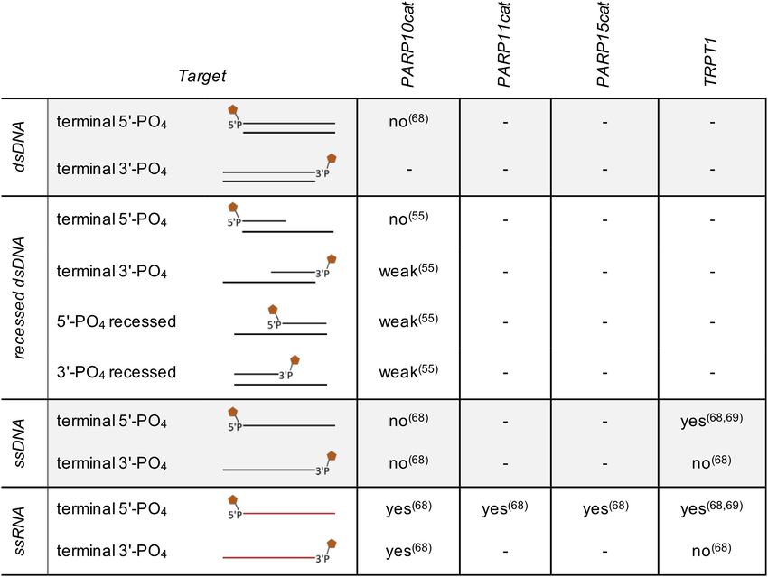
Nucleic Acids Research, 2021, Vol. 49, No. 7 3635
provide an outlook for future work, aimed at deciphering ADP-ribosyltransferases
the physiological role of nucleic acid ADP-ribosylation.
ARTD (PARPs+TNKS) ARTC (ecto-ARTs)
ADP-RIBOSYLTRANSFERASES H-Y-E R-S-E
ARTs are defined by their catalytic ART domain, a pro-
protein substrate
ADPR ADPR
tein fold that enables binding of NAD+ and transfer of
ADP-ribose from NAD+ onto a substrate under release
of nicotinamide. While the catalytic domains of individ-
ual ARTs are generally poorly conserved at the sequence many acceptors; arginine;
Downloaded from https://academic.oup.com/nar/article/49/7/3634/6163091 by Aachen University, GMB user on 05 July 2021
poly- or mono(ADP-ribose) mono(ADP-ribose)
level, the overall structure is shared among the variety of
ARTs (5). However, two distinct motifs, each comprised
PARP1–3 Pierisin-1; Scabin
of three highly conserved amino acids situated within the
ADPR
NAD+ binding sites, are used to categorize the ART su- P dsDNA
ADPR
perfamily in bacteria as well as in their eukaryotic descen- ssDNA
nucleic acid substrate
dants: ARTs with a histidine–tyrosine–glutamate triad (H–
PARP10, -11, -15, TRPT1
Y–E motif and variants thereof) are related to the diph- dsDNA
ADPR
theria toxin from Corynebacterium diphtheria and as such P ssRNA
are referred to as ADP-ribosyltransferase diphtheria-toxin
like (ARTDs), whereas the ARTs with a arginine–serine– DarT
glutamate sequence (R–S–E motif and variants thereof), ADPR
first described for cholera toxin of Vibrio cholera, are named
ssDNA
ADP-ribosyltransferase cholera-toxin like (ARTCs) (Fig-
ure 1) (10–12).
The 22 known human ARTs are thus categorized into Figure 1. ADP-ribosyltransferases and their substrates. The ADP-
two subclasses according to their NAD+ binding mode. The ribosyltransferases (ARTs) can be subdivided in two subclasses based on
key amino acids present in their catalytic domain: either H–Y–E or a
17 intracellular ADP-ribosyltransferases known as PARPs, derivate thereof for the ARTDs (including mammalian PARPs and the
as well as tRNA 2 -phosphotransferase (TRPT1), belong two tankyrases (TNKSs)), or a motif based on R–S–E for the ARTCs
to the ARTD subclass, while the ARTC subclass of ARTs (including mammalian ecto-ARTs). The PARP family is most diver-
is comprised of four ecto-ARTs (5,13). Despite divergent gent and different members modify different amino acids. Some PARPs
binding mechanisms, ARTCs and ARTDs share consid- generate poly(ADP-ribose), whereas others are limited to mono(ADP-
ribosyl)ation. The ecto-ARTs are more restricted and modify arginine with
erable similarities in the configuration of NAD+ within ADP-ribose. Several bacterial toxins of the ARTC subfamily with an R-
their binding pockets. The histidine in the H–Y–E motif of S-E motif were shown to modify DNA internally. ARTDs show a much
ARTDs binds the NAD+ at the 2 -hydroxyl of the adeno- broader spectrum of nucleic acid substrates but appear to be dependent on
sine ribose and the amine of the nicotinamide via hydro- a phosphate for modification, with the exception of DarT, as summarized
in more detail in Table 1. Examples of relevant enzymes and substrates are
gen bonds. Simultaneously, the tyrosine interacts with the shown; for PARP10, PARP11 and PARP15 only catalytic domains have
nicotinamide ribose via -stacking and the glutamate’s car- shown activity towards nucleic acids.
boxyl stabilizes the resulting ADPr-furanose intermediate
by interacting with the 2 -hydroxyl of the nicotinamide
ribose. Within the bacterial R–S–E triad, the glutamate protein modification occurring on glutamate (17). Recently
fulfils the stabilizing function and the arginine interacts serine was identified as the preferred substrate for PARP1
with the diphosphate via electrostatic interaction, whereas when HPF1 is present as co-factor (17,19). Additionally,
the serine builds hydrogen bonds with the nicotinamide ri- cysteine, histidine, threonine and tyrosine residues were
bose (5,14,15). Apart from the catalytic triad, two loop- demonstrated as sites of ADP-ribosylation, although
structures characterize the ART domain and possess im- the respective transferases and in vivo occurrence remain
portant functional roles. This is especially well documented largely unknown (18,20–22). The role of proteins as ac-
for bacterial ARTs: the acceptor loop is involved in target ceptor molecules is comprehensively discussed in several
recognition and selectivity, while the donor loop (exclusive reviews (2,9,17,23,24) and will not be further addressed
for H–Y–E motif ARTDs) participates in substrate speci- here.
ficity and in catalysis and interaction with the ADP-ribose In addition to proteins as substrate for ADP-
of NAD+ (15,16). Even though both loop structures are ribosylation, it became clear that some ARTs are capable
evolutionary highly conserved, their primary structure and of modifying oligonucleotides. The first DNA-targeting
length vary greatly among the ARTDs. ART, pierisin, was discovered in pierid butterflies (25). To
The ability of ARTs to either catalyse mono-ADP- date, six pierisins (pierisin-1, -1b and -2–5) were identified
ribosylation (MARylation) or poly-ADP-ribosylation that build together with ScARP and Scabin, found in
(PARylation) of their protein substrates is contingent on Streptomyces, the pierisin family (26). These enzymes are
the catalytic triad and the presence of certain cofactors as members of the ARTC subfamily. They ADP-ribosylate
well as additional conserved structural features. Further- the N2 position of guanine in different DNA substrates
more, ARTs exhibit wide substrate specificities in regard with varying sequence specificity. Replication of ADP-
to the acceptor amino acids (7,17,18). PARP-mediated ribosylated DNA is significantly slower than replication of
ADP-ribosylation, for example, was first considered as a non-modified DNA and appears to be perceived as DNA
3636 Nucleic Acids Research, 2021, Vol. 49, No. 7
damage, which is repaired by nucleotide excision repair the possibility that also mammalian ARTs might be able to
(NER) (27). Subsequent studies have identified the same ADP-ribosylate nucleic acids.
ADP-ribosylation activity in a variety of shellfish species
(28). These enzymes are relatively well studied and multiple Mammalian DNA ADP-ribosylation
roles for DNA modifying ARTCs have been proposed.
The pierisins are considered part of the defence system of PARP1, the first discovered and best studied PARP, is well-
cabbage butterflies against pathogens, while CARP-1 in known for its role in DNA damage repair, where it is re-
shellfish has been proposed to be part of the innate immune sponsible for the recruitment of numerous DNA repair fac-
response to DNA viruses and/or regulation of NAD+ con- tors upon activation and PAR chain synthesis (36). PARP1
centration (29). These studies provide initial evidence for is specifically linked to base excision repair (BER) and NER
where it produces PAR chains as scaffold for the DNA
Downloaded from https://academic.oup.com/nar/article/49/7/3634/6163091 by Aachen University, GMB user on 05 July 2021
ADP-ribosylation contributing to host-pathogen conflicts.
Nucleic acids have only recently been identified as sub- repair machinery. Furthermore, it is involved in homolo-
strates for mammalian ARTD-mediated ADP-ribosylation gous recombination and other cellular processes like the
and their role as potential regulator of DNA and/or RNA modulation of the chromatin structure and gene expression
functions is poorly understood. Here, we will provide an (23,37,38).
overview of the ARTs and hydrolases that modify and de- The three DNA-associated PARPs (PARP1–3) differ
modify, respectively, different DNA and RNA substrates as in their structure but commonly harbour a tryptophan–
well as hypothesize about potential functions of this novel glycine–arginine (WGR) domain, essential for DNA bind-
type of DNA/RNA modification. ing, and a C-terminal (CTD) domain with coincident three-
dimensional structure (39–41). Furthermore, the enzymatic
activities of PARP1–3 are regulated by an inducible con-
Evolutionary origins: bacterial DNA ADP-ribosylation formational change within their catalytic domains, leading
to a local stabilization or destabilization within the NAD+
Toxin-antitoxin (TA) systems regulate several biological binding sites (42,43). Upon activation these PARPs are able
processes within bacteria, including metabolism and stress to modify themselves and other proteins (44,45). PARP1
response, thereby promoting the persistence of cell popu- has a modular architecture that consists of six indepen-
lations (30). In Thermus aquaticus, the expression of DarT dent domains with diverse functions that are connected by
results in a bacteriostatic effect if the antitoxin darG is ab- flexible linkers (46,47). Activation of PARP1 is enabled by
sent (31). The induction of DarT signals DNA damage to this complex modular architecture through an allosteric
the repair machinery and triggers the SOS response (32). mechanism that induces a conformational change of the
The toxin DarT was characterized as an ARTD-like en- whole multi-domain folding upon interaction with dam-
zyme capable of DNA ADP-ribosylation (31). To identify aged DNA. Three flexible N-terminal zinc-fingers (ZF1–
potential substrates for DarT, bacterial protein extracts, to- 3) recognize the exposed bases of damaged DNA. PARP1
tal bacterial RNA or denatured bacterial DNA were in- bends the DNA duplex at the strand break via cooperative
cubated with DarT and NAD+ . Efficient incorporation of action of ZF1 and ZF2, each binding to one of the exposed
NAD+ was observed only when denatured DNA was used DNA breakage sites. ZF1, binding to the 5 -end, is required
as a substrate. Consecutive experiments demonstrated that for the activation at double strand breaks (DSB) whereas
DarT ADP-ribosylates specifically ssDNA in a sequence ZF2, binding to the 3 -end, is responsible for the recogni-
specific manner: ssDNA oligos with a minimum length of tion of single strand breaks (SSB) (48,49). In a model us-
eight bases were successfully modified at the second thymi- ing an SSB DNA mimic, bending of the DNA by ZF1 and
dine of a TNTC motif in vitro (31). As replacement of thymi- ZF2 enables the coordination of ZF3 that interacts with
dine by uridine or deoxyuridine prevents ADP-ribosylation, ZF2 and the DNA. The induced allosteric interactions re-
a strict DNA specificity is presumed. This exclusive ADP- sult in the local unfolding of an auto-inhibitory helical sub-
ribosylation of one specific base is counteracted by the anti- domain within the ART module that enables catalytic ac-
toxin DarG. The observed DNA ADP-ribosylation affects tivity of PARP1 (42,45,48–52). A more detailed discussion
replication, which might be the cause of the observed bac- about the PARP1 DNA binding modes and their functions
teriostatic effect (31). The existence of comparable TA sys- is given elsewhere (53).
tems was also shown in Mycobacterium tuberculosis and the This interaction with damaged DNA, particularly SSBs
enteropathogenic Escherichia coli (EPEC), where DarT also and DSBs, brings PARP1 into close vicinity of the 5 - and
downregulates DNA replication and specifically modifies 3 -ends of DNA that constitute potential nucleophiles able
ssDNA, which is reversed by DarG (33,34). However, this to promote ADP-ribosylation. Indeed, PARylation activ-
toxin modifies the sequences TTT or TCT in EPEC that dif- ity of PARP1 and PARP2 on phosphorylated termini of
fer from the target sequence of DarT in T. aquaticus. Ex- DNA oligonucleotide duplexes and ssDNA oligomers was
pression of RecF, which recognizes ssDNA breaks (35), is demonstrated in vitro. Depending on the structure and lo-
crucial for cell survival after DarT induction. Therefore, it calization of DNA strand breaks, PARP1 ADP-ribosylates
was suggested that cells perceive DNA ADP-ribosylation as 3 - and 5 -terminal phosphates of dsDNA and ssDNA as
DNA damage, which is antagonized by NER, similar to the well as exposed 5 -phosphates of gapped dsDNA, although
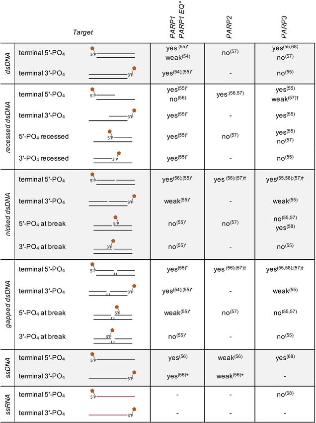
modification introduced by pierisin-1 (27). with very weak efficiency (54–56) (Table 1). PARP2 seems
These studies highlight that ARTD enzymes can mod- unable to modify termini of intact DNA duplexes, but
ify DNA in an unexpected, sequence specific manner. This prefers 5 -phosphorylated blunt ends of recessed, nicked
adds DNA, in addition to proteins, to the list of substrates and gapped dsDNA (57). ssDNA provokes weak ADP-
of ADP-ribosylation. Furthermore, these examples hint at ribosylation activity of PARP2 on both 3 - and 5 -terminalNucleic Acids Research, 2021, Vol. 49, No. 7 3637
phosphates (56). Beside these PARylating PARPs, PARP3, either as homo- or heterodimeric interaction, that modi-
which MARylates proteins, is also capable to modify DNA fies the second DNA strand break. These findings suggest
substrates. The 5 - and 3 -phosphorylated groups of dsDNA that PARP3 has strong MARylation activity towards DNA,
blunt ends were confirmed as ADPr acceptor nucleophiles while auto-modification appears to be very low. Interest-
for PARP3, whereby the 5 -end presents the preferred tar- ingly, the ADP-ribose mark deposited on DNA by PARP3
get (55,57). Additionally, exposed 5 -phosphorylated ends serves as primer for PARylation by PARP1 and PARP2. To-
of recessed dsDNA (55,57,58) and exposed 5 -phosphates gether, these findings allow to postulate that PARP3 s pri-
of nicked dsDNA (55,57,58) were examined as targets with mary function could be the modification of DNA in order
contradictory results (Table 1). The DNA substrates used to directly mark DNA lesions. This would be a novel func-
for these studies differ in terms of length and nucleic acid tion of ADP-ribose at DNA breaks, in addition to the com-
Downloaded from https://academic.oup.com/nar/article/49/7/3634/6163091 by Aachen University, GMB user on 05 July 2021
sequence. It is not known whether PARPs that are active on monly accepted model of ADP-ribosylated PARPs serving
nucleic acids possess sequence specificity or whether com- as scaffolds.
plex DNA or RNA structures are required to enable bind- DNA repair processes are often performed by ATP-
ing and/or enzymatic activity. Also the insertion of chemi- dependent polynucleotide ligases. During this reaction the
cal compounds, like cordycepin-monophosphate (an AMP ligase is activated by ATP, resulting in a ligase-AMP inter-
mimic) and phosphoribosyl-AMP, on the acceptor sites of mediate, that transfers AMP to the exposed 5 -phosphate of
the nucleic acid substrates could influence the enzymatic ac- the DNA damage site. This marks and activates the DNA
tivities and specificities. Additionally, the enzymes used in breakage and enables the attack of the 3 -hydroxyl on the
the published studies differ in their procedure of protein ex- 5 -phosphate to from a phosphodiester bond under release
pression and purification. The latter is particularly relevant of AMP (62). As PARP3 can MARylate 5 -phosphorylated
as proteins synthesized in mammalian or insect cells differ SSBs of DNA, it was tested whether the presence of ADPr
greatly in their level of posttranslational modifications com- can induce ligation of DNA strand breaks. Indeed, ADPr
pared to recombinant proteins produced in bacterial expres- seems to enable ligation of SSBs by ATP-dependent DNA
sion systems. ligases in the absence of ATP (58). Co-immunoprecipitation
PARP2 and PARP3 show tendencies to preferably mod- (co-IP) studies of PARP3 identified various proteins that are
ify DNA strand breaks over auto-modification (>5-fold involved in the non-homologous end joining (NHEJ) DNA
for PARP2 and >50-fold for PARP3) depending on the repair process (63,64). The activity of PARP3 on damaged
DNA substrates and reaction conditions (57). This ten- DNA together with the observations of the co-IP studies
dency seems to be sequence independent but contingent suggest an involvement of DNA MARylation by PARP3 in
on the configuration and localization of the strand break DNA repair pathways (58).
within the DNA duplex, as a distance of one or two helix- NHEJ mediates the direct re-ligation of DNA lesions
turns downstream of a DNA break appears to be required without the requirement of a homologous template (65).
for efficient modification. In this context, the modification of DNA SSBs by PARP3
Interestingly, an incorporated ADPr at the phosphory- could have a protective effect on DNA lesions by shield-
lated 5 -end of gapped oligonucleotides can serve as primer ing the exposed ends until repair protein complexes are
for further modification by PARP1 and PARP2. This al- recruited. As PARP1 and PARP2 can extend the initial
lows the extension of the initial MARylation to PAR ADPr added by PARP3, this hypothesis is supported by
chains via glycosidic 1 –2 linkages (58). The overall ex- the finding that PARylated DNA breakage sites are pro-
ceeding processivity of PARP1 (57,59) is countered by the tected from degradation by endonucleases (56). Evidence
surpassing activity of PARP2 on PARP3-mediated ADPr of a strong activation of PARP2 and PARP3 in response to
primed DNA substrates (58), granting speculation about 5 -phosphorylated DNA breaks further substantiates a po-
protein dimerization and/or interaction among the DNA- tential involvement of these proteins in DNA repair mech-
dependent PARPs. In this context, it is noteworthy that, anisms (43,66). The reduction of chromosomal rearrange-
depending on the DNA structure, PARP2 can indeed bind ments in cells depleted of PARP3 also supports its partici-
in two modes. As monomer it preferentially interacts with pation in the NHEJ pathway (67).
DNA that mimics SSBs, while as dimer binding to phos- Collectively, these studies demonstrate that PARP1–3 are
phorylated dsDNA is favoured (60,61). Also the distance- capable of modifying a range of DNA substrates with ei-
dependent activation of PARP3 and PARP2 on DNA with ther PAR or MAR in vitro (Table 1). However, despite the
two damage sites could be traced back to a similar dimeric- biochemical characterization of ADP-ribosylation at DNA
binding mode. However, also a divergent monomeric pro- breakage sites, the function of this novel DNA modification
tein binding mode can be suggested: monomeric PARP2 in cells remains to be unravelled.
or PARP3 modifies a single-strand break within a DNA
duplex, depending on the activation by a double-strand
ADP-ribosylation of RNA by PARPs
break in proximity (57). This could be based on the recog-
nition of one of the DNA damage sites by the WGR do- RNA, in addition to DNA, has been demonstrated to serve
main that brings the second strand-break in proximity to as substrate for ADP-ribosylation catalysed by mammalian
the catalytic domain to enable modification, while exceeded PARPs. The activities of several PARPs and hydrolases were
or reduced distance to the DSB could sterically hinder the tested on a variety of RNA substrates in vitro (68). The
interaction between the SSB and the active site. Another activity of PARP10 on phosphorylated RNA termini has
possible scenario could be a scaffold function of an ini- been demonstrated with a preference of 5 -phosphorylated
tially bound PARP that recruits a second PARP molecule, over 3 -phosphorylated ssRNA substrates (68). However,3638 Nucleic Acids Research, 2021, Vol. 49, No. 7
Table 1. Nucleic acid substrate modification by PARP1–3. The different potential substrates are schematically displayed. Illustrated are the different
oligonucleotides that are either single or double stranded, with or without nicks, gaps or single strand overhangs. Moreover, the positions of phosphate
groups are indicated. Exact information on sequence and oligonucleotide length is provided in the studies cited.
Downloaded from https://academic.oup.com/nar/article/49/7/3634/6163091 by Aachen University, GMB user on 05 July 2021
* PARP1 E998Q was used, a mutant that MARylates substrates but is unable to produce PAR; † The activity depends on the location of the nick/gap
(distance should not exceed ∼21 nt); + cordycepin 5 -[32 P]monophosphate (3 -dAM32 P) was used as ADP-ribosylation target (the 2 -hydroxyl group of
the cordycepin moiety at the 3 -end should resemble that of the ADP-ribose unit in PAR).Nucleic Acids Research, 2021, Vol. 49, No. 7 3639
PARP10 is not able to modify ssDNA, which is in con- dation by 5 →3 exonucleases, promotes the translocation
trast to PARP3 that is active on ssDNA but unable to of mature mRNA into the cytosol, and is essential for ini-
MARylate RNA (57,68). Beside PARP10, the catalytic do- tiation of translation (78). In addition, the m7Gppp cap
mains of PARP11 and PARP15 are also capable of modi- also serves as a scaffold to recruit protein factors involved
fying phosphorylated ssRNA (Table 2). Full-length PARP3 in RNA processing. On the contrary, the recently discov-
and PARP16 as well as the catalytic domains of PARP4, ered non-canonical NAD+ capping by RNA polymerase II
PARP6, PARP12, PARP13 and PARP14 were tested, how- blocks efficient translation and actively targets the RNA for
ever, these do not show activity on 5 -phosphorylated RNA degradation (79,80). In light of these observations and the
(68). Because the catalytic activity of the enzymes used in structural similarities of NAD+ and ADPr, it would be in-
this study was not verified on protein substrates, and thus a teresting to explore whether ADPr has a similar effect on
Downloaded from https://academic.oup.com/nar/article/49/7/3634/6163091 by Aachen University, GMB user on 05 July 2021
priori functionality was not documented, the results should RNA in regards to mRNA stability, localization and trans-
be interpreted with caution. lation.
Interestingly, a truncated version of PARP10, which only
contains the catalytic domain, modifies RNA more effi-
ciently than the full-length protein. It was speculated that ADP-ribosylation of RNA by TRPT1
this is caused by an auto-inhibitory mode involving an in-
TRPT1 was originally described as an essential component
teraction of N-terminal sequences with the catalytic do-
of the fungal tRNA splicing machinery. Following the ex-
main, similar to what has been described for PARP1 (68).
cision of introns, the resulting exons are joined together by
However, this hypothesis has not been further substanti-
ligases leaving a 3 –5 phosphodiester splice junction that
ated. PARP10 lacks an auto-inhibitory, helical structure in
possesses a characteristic 2 -PO4 , which has to be removed
its catalytic domain, which argues against an allosteric acti-
in order to generate mature tRNA (81). This reaction pro-
vation mechanism similar to PARP1–3, as discussed above.
ceeds in two steps, culminating in a short lived 2 -phospho-
Instead, the full-length protein with its RNA recognition
ADP-ribosylated RNA intermediate.
motif (RRM) together with the glycine-rich region may be
TRPT1 homologs are evolutionary conserved in eukarya,
regulated by binding to specific RNAs. PARP10 also con-
bacteria and archea. Its deletion in yeast is lethal, but com-
tains ubiquitin interaction motifs (UIM), which allow inter-
plementation assays with mammalian and bacterial TRPT1
action with poly-ubiquitin chains (70), and a PIP box that
homologs showed conservation of catalytic specificity in re-
enables the interaction with the DNA clamp PCNA, a pro-
moving the tRNA 2 -PO4 through a transfer to NAD+ that
cessivity factor that is essential for cell proliferation (71,72).
generates mature 2 -OH RNA (82,83).
The binding of HPF1 to PARP1 and PARP2 demonstrates
Bacterial, archeal and metazoan tRNA processing is dif-
the ability of interaction partners to alter substrate speci-
ferent from that of fungi and does not result in a 2 -PO4
ficity (61,73), which could also be the underlying concept
junction. This poses the question why enzymes that are
of the regulation of PARP10 in terms of nucleic acid modi-
capable of dephosphorylating 2 -PO4 of RNA, generating
fication.
ADP-ribose 1 –2 cyclic phosphate, exist in prokaryotes
Apart from the observation that the catalytic domains of
and metazoan. They might have an as of yet unidenti-
PARP11 and PARP15 can transfer ADP-ribose onto RNA
fied biochemical pathway enabling the production of 2 -
in vitro, no further studies have been published regarding
phosphorylated RNA. RNA modified in this way would
these enzymes. This leaves the question unanswered whether
then act as a potential substrate for TRPT1 mediated RNA
the full-length proteins possess comparable activity towards
repair. In two recent studies, prokaryotic and fungal TRPT1
RNA (68). Moreover, it will be interesting to determine
were characterized for their ADP-ribosylation activity
whether these enzymes are capable to modify RNA in cells.
(68,69). The TRPT1 homologs are able to transfer ADPr
The various PARPs, which exhibit activity towards RNA
moieties to the phosphate group of 5 -monophosphorylated
substrates, localize to different intracellular compartments
ssDNA and ssRNA in vitro. This ADP-ribosylation re-
where they could fulfil comparable biochemical reactions.
sulted in a phosphatase-resistant 5 -phospho-ADPr cap
For example, PARP10 is localized to cytoplasmic structures
structure on 5 -DNA/RNA substrates. Based on the first
containing p62 and ubiquitin and can shuttle into the nu-
reaction step of the tRNA 2 -phosphotransferase mech-
cleus and nucleolus depending on its phosphorylation sta-
anism, a potential one-step reaction via an ADPr trans-
tus (74,75), whereas PARP11 is located in the nuclear en-
ferase reaction was suggested resulting in capping of DNA
velope (76) and PARP15 in stress granules (77). Different
and RNA (69). TRPT1 was identified in humans as a
subcellular localizations provide a tenable explanation for a
phosphate-dependent mono-ART, which exclusively mod-
number of PARPs that display similar enzymatic activities,
ifies 5 -PO4 of ssDNA and ssRNA substrates, independent
complementing each other in different compartments and
of oligomer length (68). The unexpected modification of
in different cellular processes. The proteins might be dif-
RNA by TRPT1 offers a potential explanation for the ex-
ferently regulated in response to signalling cascades or by
istence of the catalytically active TRPT1 even in taxa that
interacting with distinct, localization-specific co-factors. At
lack splicing systems.
present, our knowledge is rather poor about how different
ARTs are controlled regarding activity and substrate speci-
ficity.
REVERSAL OF DNA/RNA ADP-RIBOSYLATION
Similar to DNA ADP-ribosylation, little is known about
the role of RNA ADP-ribosylation in cells. The canonical Two protein families are known that reverse ADP-
m7Gppp cap protects nascent RNA from premature degra- ribosylation. The macrodomain-containing family and3640 Nucleic Acids Research, 2021, Vol. 49, No. 7
Table 2. Overview of the oligonucleotides modified by the catalytic domains of PARP10, PARP11 and PARP15 as well as TRPT1. The different potential
substrates are schematically displayed. Exact information on sequence and oligonucleotide length is provided in the studies cited.
Downloaded from https://academic.oup.com/nar/article/49/7/3634/6163091 by Aachen University, GMB user on 05 July 2021
the ADP-ribosylhydrolase family (ARHs). Macrodomain- with the same activity, ARH1, was later identified in animal
containing proteins are conserved through all domains of cells (94). In addition to ARH1, ARH3 is also catalytically
life and are defined by the presence of a macrodomain active, while the third mammalian ARH family member,
fold, named after the non-histone region of the histone ARH2, appears to be inactive, possibly due to the lack of an
MacroH2A, which is characterized by a mixed ␣/ fold aspartate in the catalytic centre (92,95). ARH1 and ARH3
with structural resemblance to certain nucleotide hydrolases display different substrate specificities. The primary activ-
(84–88). The mammalian ARH family contains three pro- ity of ARH1 is the hydrolysis of the N-glycosidic bond be-
teins with structurally similar catalytic domains, ARH1, tween ADPr and arginine residues (94,96–99) (Figure 2A).
ARH2, and ARH3 (89–92). The three members share sim- Only weak activity against PAR and O-acetyl-ADP-ribose
ilar amino acid sequences, where ARH1 and ARH2 dis- (OAADPr) has been detected. Although the function of
play a higher degree of sequence homology (92). The follow- ARH1 is not fully understood, experiments in mouse em-
ing section addresses members of both protein families that bryonic fibroblasts deficient for ARH1 showed rapid ac-
have hydrolytic activity towards ADP-ribosylated DNA or cumulation of arginine-ADPr substrates (100), correlating
RNA substrates. with abnormal cell proliferation. So far, ARH1 is the only
mammalian enzyme that is able to cleave the N-glycosidic
ADP-ribosylhydrolase family bond between ADPr and arginine. The finding that ARH1
is not able to remove ADP-ribose from nucleic acids is in
The members of the ARH family harbour an evolution- accordance with the previously described substrate speci-
ary highly conserved catalytic domain of 290–360 residues. ficity of ARH1, since the linkage between ADP-ribose and
The first described ARH enzyme was DraG (dinitrogenase DNA/RNA is O-glycosidic.
reductase activating glycohydrolase) from Rhodospirillium ARH3 was originally described as a PAR glycohydro-
rubum, which plays an essential role in nitrogen fixation lase (Figure 2B) (92). Besides PAR, ARH3 efficiently uses
(93). Biochemical characterization has shown that DraG OAADPr as a substrate (98,101). Various inactivating mu-
specifically reverses arginine-ADP-ribosylation. An enzyme tations in the ADPRHL2 gene have been identified in in-Nucleic Acids Research, 2021, Vol. 49, No. 7 3641
A B
Downloaded from https://academic.oup.com/nar/article/49/7/3634/6163091 by Aachen University, GMB user on 05 July 2021
C
D
E
Figure 2. Substrate specificities of proteins that reverse ADP-ribosylation. (A) ARH1 cleaves the N-glycosidic bond in modified arginine residues. (B) The
O-glycosidic bond in PAR-chains is broken by PARG and ARH3 or (C) the O-glycosidic bond present in serine-ADPr is broken by ARH3, respectively. (D)
PARG, MACROD1, MACROD2, TARG1 and ARH3 have all been demonstrated to remove ADPr from DNA and RNA by cleaving the phosphoester
type O-glycosidic bond (E) ADP-ribosylation of acidic amino acids is resolved by MACROD1, MACROD2 and TARG1.
dividuals with neurodegenerative disease (102–104), hint- the recycling of mitochondrial NAD+ . In accordance with
ing at the importance of ARH3. It is not understood yet its specificity towards O-glycosidic bonds in the substrates
how loss of functional ARH3 would lead to this pheno- detected so far (OAADPr, Ser-ADPr and PAR), ARH3
type. Recent research shows that ARH3 possesses unique cleaves ADPr from DNA and RNA, the proposed linkage
activity towards MARylated serines, which seems to be being a phosphoester-type O-glycosidic bond (Figure 2D)
the predominant type of modification during the DNA (55,68).
damage response (105,106) (Figure 2C). Nuclear localiza-
tion of ARH3 supports its role in DNA damage response.
Macrodomain-containing hydrolases
However, ARH3 also localizes to the cytoplasm and mito-
chondria where serine modification has not been detected After the degradation of PAR chains via the hydrolysis
(92,107), leaving the role of ARH3, especially in mitochon- of ribose–ribose bonds was observed (109), poly(ADP-
dria, elusive. Although no ART has been identified in mito- ribose)glycohydrolase (PARG) was identified as responsible
chondria, several sirtuins are present in this organelle. Sir- enzyme (110) (Figure 2B). Structural analyses of bacterial
tuins act as deacetylases using NAD+ as cofactor resulting and protozoan PARG revealed a macrodomain fold, con-
in the formation of OAADPr (108). Consequently, a possi- taining a unique loop sequence within the ADPr binding
ble role of ARH3 could be the degradation of OAADPr in site that harbours a catalytic glutamate residue (111,112).3642 Nucleic Acids Research, 2021, Vol. 49, No. 7
PARG is encoded by a single gene in mammals (113), but (123,131). Together this suggest that TARG1 is involved in
is expressed as several isoforms due to alternative splicing the DNA-damage response and/or its nucleolar function
(114). Full-length human PARG (111 kDa) is targeted to needs to be halted in response to DNA damage. Moreover,
the nucleus, while other isoforms are located in the cyto- TARG1 binds to RNA oligomers (131), possibly through
plasm (114). Knockout of PARG results in early embryonic positively charged surface patches, as suggested for the
lethality in mice (115). Cells derived from trophoblasts of Chikungunya viral macrodomain (133). This activity of
PARG knockout embryos only survive in the presence of TARG1 might be relevant for targeting ADP-ribosylated
the broad-spectrum PARP inhibitor benzamide (16,115), RNA substrates. MACROD1 and TARG1 interactomes re-
suggesting that the lack of PAR turn-over and its accu- vealed a number of nucleic acid metabolism associated pro-
mulation is toxic, making the reversal of ADP-ribosylation teins such as helicases and nucleases (129,131). As described
Downloaded from https://academic.oup.com/nar/article/49/7/3634/6163091 by Aachen University, GMB user on 05 July 2021
by PARG in mammals essential. In contrast to the re- above, MACROD1 and TARG1 are primarily localized in
sults in mice, the absence of a catalytic active PARG in mitochondria, stress granules and nucleoli, compartments
Drosophila melanogaster results in high lethality. The sur- of intensive nucleic acid processing. Together with the in-
vived flies show an accumulation of PAR in nervous tissue teractome results, these findings are in line with a potential
along with progressive neurodegeneration (116). Accumu- role of macrodomain-containing hydrolases in nucleic acid
lation of PAR chains in the cytosol has also been described metabolism.
to be accompanied by induction of apoptosis in a process It is not well understood how PARG, MACROD1,
referred to as parthanatos (117,118). MACROD2, TARG1 and ARH3 are able to remove MAR
Besides its activity towards protein bound as well as from 5 - or 3 -phosphate groups in DNA and RNA (Fig-
free PAR chains, PARG can remove ADPr and PAR units ure 2D). PARG breaks down O-glycosidic bonds between
bound via O-glycosidic phosphoester bonds to 3 - and ribose rings, but is unable to cleave the primary ADPr of
5 -phosphates of dsDNA (55) (Figure 2D). Furthermore, amino acid acceptors (105,106,112). However, the chemi-
PARG is capable to efficiently reverse MARylation of 3 - cal structure of the bonds between ADPr and serine and
and 5 -phosphorylated ssRNA (68). The observed activities between ADPr and nucleic acids are quite similar (Figure
toward ADP-ribosylated DNA and RNA fit to the speci- 2). To understand why PARG is unable to remove MARy-
ficity of PARG for O-glycosidic linkages (Figure 2B and D). lation from serine, further structural studies will be re-
Aside from PARG, three other human macrodomain- quired. Structural constraints may prevent accommodation
containing proteins possess hydrolytic activity that is of a protein substrate within the active centre, while bind-
selective for ADPr modified substrates: MACROD1, ing of a nucleic acid substrate is possible. Indeed, PARG
MACROD2 and TARG1. These enzymes were first demon- is able to completely reverse DNA- and RNA-phosphate
strated to hydrolyse OAADPr, the by-product of sirtuin modification and even appears to be more active than
mediated deacetylation (119,120). Later it was found that MACROD2, ARH3 and TARG1 (55). As MACROD1,
MACROD1, MACROD2 and TARG1 specifically remove MACROD2 and TARG1 cleave ester-type O-glycosidic
MAR from proteins modified at acidic amino acid residues bonds between the protein proximal ribose of ADPr and
(Figure 2E) (121–124), but not PAR, making these en- side chains of acidic amino acids (121,123,124) or acetate
zymes specialized MAR hydrolases (121,125,126). Addi- groups in OAADPr (119,120), the hydrolysis of the O-
tionally, MACROD1, MACROD2 and TARG1 efficiently glycosidic phosphoester bond present in modified nucleic
reverse oligonucleotide modification by hydrolytic cleavage acid substrates seems appropriate. ARH3 shows activity to-
of ADP-ribose from the 5 or 3 terminal phosphates of ds- wards the O-glycosidic bonds in PAR chains as well as be-
DNA as well as ssRNA (Figure 2D) (55,68,127). tween serine and ADPr in modified proteins. Specific con-
Relatively little is known about the physiological function formational requirements, amino acid residue, surround-
of these macrodomain-containing hydrolases. MACROD1 ing amino acid sequences or nucleotide sequences could
is a mitochondrial protein, highly expressed in skeletal be relevant to enable the removal of ADP-ribosylation
muscle and certain breast cancers (127–130). MACROD2 from specific substrates. Whether the hydrolases share sim-
is present in the nucleus and the cytoplasm (128–130). ilar residues that carry out key catalytic steps or whether
TARG1, which is the most ubiquitously expressed pro- their reaction mechanisms are fundamentally different is
tein of these three, is mainly localized in the nucleus unknown. To define the hydrolytic mechanisms and the
(123,129,131,132). A few studies have addressed their lo- key residues responsible for the catalytic activity on ADP-
calization in response to certain stimuli and found that ribosylated nucleic acids, structural resolution of ADP-
overexpressed MACROD2 is exported from the nucleus ribosylated oligonucleotides in complex with the hydrolases
into the cytoplasm upon phosphorylation by the ataxia- will be necessary.
telangiectasia-mutated kinase, which is activated in re-
sponse to DNA damage (132). A mutation in the OARD1
Viral macrodomain-containing hydrolases
gene, which encodes the TARG1 protein, was identified
in patients with a severe neurodegenerative phenotype, al- Macrodomains are also present in various viral families, in-
though it was not clarified how loss of TARG1 leads to cluding positive single-stranded (+)ssRNA viruses such as
this illness (123). TARG1 predominantly localizes to tran- Chikungunya virus (CHIKV) and the Corona viruses (CoV).
scriptionally active nucleoli, but relocalizes to the nucleo- The function of these viral macrodomains is poorly un-
plasm when rRNA transcription is inhibited (131). Upon derstood, although it was shown that they efficiently re-
DNA damage TARG1 accumulates in the nucleoplasm and verse protein MARylation (126,134–137). Several studies
at sites of damaged DNA, depending on PAR formation found that the severe acute respiratory syndrome coro-Nucleic Acids Research, 2021, Vol. 49, No. 7 3643
navirus (SARS-CoV) macrodomain Mac1 promotes vi- of NAD+ and, consequently, it should not influence enzy-
ral replication in vivo and suppresses the interferon re- matic reactions; and second, the detection of radiolabelled
sponse, thus playing an important role in viral pathogen- substrates, including proteins and nucleic acids, is high sen-
esis (136,138,139). In addition, PARP inhibitors enhance sitivity as demonstrated in multiple in vitro studies (55,68).
the replication of SARS-CoV and decrease interferon pro- Chemical NAD+ analogues with various functional groups
duction during infection with a macrodomain mutant virus attached to the NAD+ have been generated. In contrast to
(140). Similarly, for CHIKV, the ADP-ribosylhydrolase ac- radiolabelled isotopes, the chemical and physical proper-
tivity appears essential for viral replication and virulence ties of modified NAD+ analogues are changed considerably
(141,142). Therefore, the viral macrodomains are thought compared to non-modified NAD+ . Due to the release of
to play essential roles in infectivity. nicotinamide during the reaction, NAD+ analogues com-
Downloaded from https://academic.oup.com/nar/article/49/7/3634/6163091 by Aachen University, GMB user on 05 July 2021
Uncapped mRNA in the cytoplasm can be recognized monly hold modifications at the adenosine group. Biotin-
as a non-self RNA and trigger an innate immune response labelled NAD+ has been used to label proteins with different
(143). Therefore, viruses deploy mechanisms to cap their PARPs in several studies, for example on high-density pro-
mRNA, thereby mimicking the host cell cap and avoid- tein microarrays (147–149) and as alternative to 32 P-NAD+
ing an innate immune response. The majority of (+)ssRNA in ADP-ribosylation reactions in vitro (150–153). From the
viruses synthesize the canonical m7Gppp-type RNA cap, published studies it appears that biotin-labelled NAD+ is
sometimes combined with 2 -O-me of the first nucleotide well suited as cofactor.
(cap-0 and cap-1, respectively), using their own set of cap-
ping enzymes (144,145). The canonical m7Gppp cap is es-
Detection of ADP-ribosylated substrates in cells
sential for the initiation of translation and protects nascent
RNA from premature degradation by 5 →3 exonucleases Methods to detect endogenously MARylated substrates are
(78). Although in general viral RNA capping is efficient, it scarce. Early studies on the generation of MARylated pro-
also presents a sensitive step in the viral life cycle, reflected tein specific antibodies yielded a rabbit serum with sig-
in the essential role of capping enzymes in viral replication. nificant specificity for MARylated eEF2 (154). Moreover,
Capping of the viral RNA with a structurally different cap using an ADPr-coupled carrier protein, a serum was ob-
provides the host cell with an additional strategy to recog- tained that recognized pertussis toxin modified Gα as well
nize and respond to viral RNA. The NAD+ cap blocks effi- as eEF2 (155). Then a rabbit serum (R-28) was developed
cient translation of RNAs (80) and the structurally similar specifically against ADP-ribosyl-arginine proteins, capable
ADP-ribose cap could thus impair the translation of viral of distinctly identifying arginine-specific ADP-ribosylation
RNA. A recent study showed that PARP10 overexpression products (156,157). Similarly, an anti-serum against ADP-
leads to decreased levels of processed viral non-structural ribosylated histones was generated and utilized for the in
proteins in cells (142), which could be due to impaired pro- vitro characterization of ADP-ribosylation in murine T-
cessing of the polyprotein as suggested, or due to lower pro- cells as well as for the detection of endogenous ADP-
tein translation from a potentially ADP-ribosylated viral ribosylation in rat skeletal muscle (158). Recently, a com-
RNA. Thus, viral macrodomains may have evolved to an- mercial PAR/MAR antibody has become available that en-
tagonize MARylation executed by PARPs that are respon- ables studying MARylation (159). However, this antibody,
sive to interferon signalling. Future work will have to dis- as the name suggests, cannot distinguish between MARyla-
cern whether viral macrodomains also revert MARylation tion and PARylation and thus selective antibodies to detect
of vRNA as this might be a mark by the host to interfere MARylation are still lacking.
with translation or transcription. Because the ADPr-cap is Catalytically inactive macrodomains form an alternative
likely to interfere with m7Gppp-capping, a tight coordi- to antibodies and have been used as sensing tools to de-
nation of the viral de-MARylation and capping activities tect ADP-ribosylated proteins (160). The combination of
would be expected. Moreover, the ADPr-cap could serve as affinity purification using ADPr binding macrodomains
additional signal to stimulate the innate immune response. and subsequent mass spectrometry of the macrodomain-
Key to unravel these possibilities is to follow up with stud- bound fraction enabled the discovery of both known and
ies in cells and evaluate whether and when RNA-capping novel ADP-ribosylated proteins (161–163). The majority
by ADPr occurs. For this, the ability to selectively detect of these experiments were carried out using the Af1521
MARylation in cells is essential, which has been difficult macrodomain present in Archaeoglobus fulgidus, which rec-
and a limiting factor. Recent findings have provided infor- ognizes MARylated and PARylated substrates. Unknown at
mation about newly developed tools that may help to mea- the time of the first mass spectrometry pull-downs, Af1521
sure this modification in cells, as discussed below. is also an active hydrolase. Nevertheless, the pull-down ex-
periments were successful due to their incubation at 4◦ C
DETECTION METHODS as low temperature seems to minimize its hydrolase ac-
tivity (161,162). This macrodomain has been further de-
Detection of in vitro ADP-ribosylated substrates
veloped to increase affinity (164). In contrast, the murine
The ability of ARTs to transfer ADP-ribose from the co- Parp14 macrodomains macro2/macro3 are catalytically in-
factor NAD+ to their substrates can be exploited for the in active and interact preferentially with MAR (165). There-
vitro detection and identification of ADP-ribosylated pro- fore, the Parp14 macrodomains were optimized for co-
teins by employing modified NAD+ as substrate (1,146). immunoprecipitation and co-localization experiments se-
Radiolabelled NAD+ offers two major benefits: first, the ex- lective for MARylated proteins (166). To analyse ADP-
change of isotopes does not alter the chemical properties ribosylated proteins a fusion protein of a tandem Af15213644 Nucleic Acids Research, 2021, Vol. 49, No. 7
A ribosylated RNA. No signal is observed with the Parp14-
macro2/3-Fc mutant that is defined by glycine to glutamate
mutations in the two macrodomains (166). These mutations
block the access to the ADP-ribose binding site and prevent
binding to ADPr. Despite the lower signal with Parp14-
macro2/3-Fc in this slot blot analysis, it can potentially be
employed to detect MARylated RNA as the mutant can be
used to subtract any signal derived from binding indepen-
dent of ADP-ribosylation. In addition to mere detection,
this reagent can theoretically be used to precipitate specif-
Downloaded from https://academic.oup.com/nar/article/49/7/3634/6163091 by Aachen University, GMB user on 05 July 2021
ically MARylated RNAs from a pool of cellular RNAs, to
be identified by subsequent RNA-sequencing. This makes
B macro2/3 of Parp14 a potentially promising tool for study-
ing RNA ADP-ribosylation to answer questions such as
whether and when RNA ADP-ribosylation occurs in cells.
We have not tested all available reagents, but have included
the image as proof-of-principle that reagents developed to
detect protein modification can be applied for the study of
nucleic acids.
An alternative method for the detection of ADP-
ribosylation of proteins, which does not rely on antibodies,
utilizes clickable NAD+ -analogues (168–170). After modi-
fication of targets, with for example 6-alkyne NAD+ , func-
Figure 3. Detection of ADP-ribosylated RNA by immunostaining. (A) tional tags for visualization or affinity tags for purifica-
Mono-phosphorylated ssRNA (44 nucleotides) was incubated with
PARP10(818–1025) and MACROD1 as indicated, followed by proteinase
tion can be conjugated using click chemistry. Tagging oc-
K treatment and RNA extraction. Samples were analysed on an urea– curs after the ADP-ribosylation reaction has taken place.
PAGE stained with SYBR-gold. (B) ADP-ribosylated oligonucleotides In a similar approach, a subset of PARPs have been engi-
were blotted and incubated with the indicated detection reagents, fol- neered to utilize only specific NAD+ -analogues, enabling
lowed by secondary antibodies and chemiluminescence detection. Ten the detection of ADP-ribosylation incorporated exclusively
ng ssRNA was loaded per slot. Controls: auto-modified PARP10(818–
1025) without nucleic acid; buffer alone. Detection reagents used: anti- by this specifically engineered enzyme (171–173). One draw-
PAR/MAR, Cell Signalling Technology (CST) E6F6A; murine Parp14 back of these methods is the reliance on NAD+ -analogues,
macro2/macro3 wildtype or mutant (GE) fused to an Fc-tag for detec- which are not taken up by cells. ADP-ribosylation reac-
tion (166). PARP10(818–1025) corresponds to the catalytic domain of the tions have therefore been performed in cell lysates, which
enzyme (7). Data were generated in our laboratory (L. Weixler).
may enhance the occurrence of artefacts due to disrup-
tion of cellular structures. Recently, a clickable aminooxy
probe (AO-alkyne) was developed, which together with an
macrodomain (167), as well as the macrodomains2/3 from azide reporter enables monitoring of PARP activity also
human PARP14 were generated (160). in live cells (174). This modification however relies on the
These reagents developed for detection of MARylated ADP-ribosylated glutamate/aspartate side chain undergo-
proteins have not yet been tested for their recognition of ing a 1 –2 transfer of the ADP-ribose. Considering that the
MARylated DNA or RNA. Preliminary experiments in our bond between ADP-ribose and nucleic acids differs from
laboratory showed that some MAR reagents have the po- the bond between ADP-ribose and acidic amino acids (Fig-
tential to detect ADP-ribosylated RNA. We modified ss- ure 2), it needs to be determined whether this method might
RNA by incubation with the catalytic domain of PARP10 be adapted for the detection of nucleic acid modification.
as reported before (68) (Figure 3A) and blotted the pu- Considering the fact that the NAD+ analogues can be pro-
rified oligonucleotides to enable immunostaining. Differ- cessed by the PARPs tested, it is plausible that also nu-
ent reagents that are available to read MARylation and cleic acids can be modified and studied using the mentioned
PARylation recognize the modified RNA (Figure 3B). Con- methods, although procedures need to be optimized.
sidering the chemical structure of RNA and the ability
of the macrodomain fold to bind RNA through a pos-
FUTURE OUTLOOK
itively charged groove outside of the ADP-ribose bind-
ing site, appropriate controls should be applied to ad- The modification of nucleic acids with ADP-ribose was first
dress specificity of MAR-binding reagents. The commer- observed in organisms such as clams and cabbage butter-
cially available PAR/MAR antibody recognizes ADPr- flies, where DNA ADP-ribosylation appears to be part of
RNA with high affinity. However, weak binding to non- the immune defence. Recent data highlight that also mam-
modified RNA was also observed, limiting its use in study- malian, intracellular ARTs can transfer ADP-ribose to nu-
ing RNA ADP-ribosylation in cells (Figure 3B). Com- cleic acid substrates, both RNA and DNA, if carrying either
pared to the PAR/MAR antibody, purified fusion pro- a 5 - or a 3 -phosphate. Different transferases were reported
teins containing macro2/macro3 from murine Parp14 (166) to have different affinities towards a variety of substrates,
and an Fc domain (Parp14-macro2/3-Fc) for detection, partially depending on whether the full-length enzymes or
show a lower signal overall, but higher specificity for ADP- truncations were used in the in vitro reactions (Tables 1 andNucleic Acids Research, 2021, Vol. 49, No. 7 3645
Potential biological functions of nucleic acid ADP-ribosylation
ADPR DSB (or SSB)
DarT
NNTNTCNN NNTNTCNN 3'
ssDNA DarG 5'
PARP3 PARG, ARH3
Replication
ADPR
Downloaded from https://academic.oup.com/nar/article/49/7/3634/6163091 by Aachen University, GMB user on 05 July 2021
Scabin ADPR P
Pierisin-1
dsDNA dsDNA
PARP1/2 PARG, ARH3
(Antiviral) immunity?
TRPT1 ADPR
PARP10, -11, -15 Repair
ADPR
P P factors
ssRNA MACROD1, -D2, ssRNA ADPR
TARG1, PARG
ARH3 P
Stability?
Translation?
Localisation? Transcription?
Interactions? DNA damage repair?
(Antiviral) immunity? Recruitment of PAR-binding proteins?
Figure 4. Potential in vivo functions of nucleic acid ADP-ribosylation. ADP-ribosyltransferases, hydrolases and their substrates are schematically displayed.
Possible consequences of ADP-ribosylation are indicated with a question mark. The modification of double-stranded DNA by DarT leads to inhibition of
replication, which is released by reversal of the modification by DarG. DNA modification in eukaryotes possibly play a role in the regulation of transcription,
DNA damage repair and in the recruitment of PAR-binding proteins. Mono(ADP-ribosyl)ation by PARP3 can possibly be used as primer for PARP1/2 to
generate poly(ADP-ribose). RNA modification may modify any RNA property, such as stability, translation, localization and interactome. This might not
only apply to cellular RNAs, but also to foreign nucleic acids, such as viral RNA. These are the key questions that need to be addressed by future studies.
2). The different hydrolases reverse the modification regard- cells at all? This question can now be addressed using the
less of oligonucleotide characteristics. Further research is reagents described above; even though they were developed
required to address this apparent discrepancy: why are the for protein ADP-ribosylation, they efficiently recognize in
hydrolases highly specific for either PAR or MAR, which vitro modified oligonucleotides. In an initial approach, the
is linked to certain amino acids, but promiscuous towards reagents can be used to test whether ADP-ribosylation of
modified nucleic acids? Is this caused by simple steric hin- DNA and RNA occurs under basal conditions in cells.
drance of a bulky protein blocking access of the ADPr to Then it needs to be asked in which context this modifica-
the catalytic site? tion is introduced and removed in cells by which enzymes.
Additional clarifications are required to understand the Is DNA only modified when damaged, or can DNA ADP-
stimuli that activate the transferases to ADP-ribosylate nu- ribosylation be part of other physiological processes, per-
cleic acids, and to determine whether post-translational haps as a novel epigenetic regulator? Likewise, is RNA mod-
modifications or certain co-factors regulate the activities of ified as a signal of potential errors introduced for example
different transferases. The activation mechanism of PARP1 during transcription or splicing to halt subsequent trans-
has been studied in detail, whereas this information is lack- lation, does it influence stability, or does it have other sig-
ing for the other family members. The fact that some trun- nalling functions? As a subset of PARPs is upregulated by
cated versions of PARPs, which solely harbour the catalytic interferons and interferes with viral replication, viral DNA
domain, show increased activity towards nucleic acids when and RNA are potential direct targets. This is particularly
compared to the full-length proteins (68) suggests the pres- interesting to consider in light of the role of DNA ADP-
ence of additional regulatory mechanisms. Moreover, it will ribosylation in pathogen defence and immunity in some of
be important to determine whether the activities of differ- the discussed organisms. The modification of RNA with
ent PARPs towards protein and nucleic acid substrates can NAD+ is considered as a non-conventional cap (80), which
be regulated to favour one or the other class of substrates. does not support translation of the modified transcripts.
The demonstration of ADP-ribosylation of DNA and ADP-ribose could serve similarly as non-conventional cap.
RNA in vitro has given rise to many questions concerning its When applied to a viral DNA or RNA for example, it could
relevance in cells. First of all, does it occur in mammalian form a highly effective way of inhibiting viral replication. InYou can also read