Structural and Computational Insights into a Blebbistatin-Bound Myosin ADP Complex with Characteristics of an ADP-Release Conformation along the ...
←
→
Page content transcription
If your browser does not render page correctly, please read the page content below
International Journal of
            Molecular Sciences

Article
Structural and Computational Insights into
a Blebbistatin-Bound Myosin•ADP Complex with
Characteristics of an ADP-Release Conformation
along the Two-Step Myosin Power Stoke
Wiebke Ewert 1 , Peter Franz 2 , Georgios Tsiavaliaris 2 and Matthias Preller 1,3, *
 1    Institute for Biophysical Chemistry, Structural Bioinformatics and Chemical Biology,
      Hannover Medical School, 30625 Hannover, Germany; ewert.wiebke@mh-hannover.de
 2    Institute for Biophysical Chemistry, Cellular Biophysics, Hannover Medical School,
      30625 Hannover, Germany; franz.peter@mh-hannover.de (P.F.); tsiavaliaris.georgios@mh-hannover.de (G.T.)
 3    Department of Natural Sciences, University of Applied Sciences Bonn-Rhein-Sieg,
      53359 Rheinbach, Germany
 *    Correspondence: preller.matthias@mh-hannover.de; Tel.: +49-511-532-2804

 Received: 28 September 2020; Accepted: 6 October 2020; Published: 8 October 2020

 Abstract: The motor protein myosin drives a wide range of cellular and muscular functions by
 generating directed movement and force, fueled through adenosine triphosphate (ATP) hydrolysis.
 Release of the hydrolysis product adenosine diphosphate (ADP) is a fundamental and regulatory
 process during force production. However, details about the molecular mechanism accompanying
 ADP release are scarce due to the lack of representative structures. Here we solved a novel
 blebbistatin-bound myosin conformation with critical structural elements in positions between the
 myosin pre-power stroke and rigor states. ADP in this structure is repositioned towards the surface
 by the phosphate-sensing P-loop, and stabilized in a partially unbound conformation via a salt-bridge
 between Arg131 and Glu187. A 5 Å rotation separates the mechanical converter in this conformation
 from the rigor position. The crystallized myosin structure thus resembles a conformation towards
 the end of the two-step power stroke, associated with ADP release. Computationally reconstructing
 ADP release from myosin by means of molecular dynamics simulations further supported the
 existence of an equivalent conformation along the power stroke that shows the same major
 characteristics in the myosin motor domain as the resolved blebbistatin-bound myosin-II·ADP crystal
 structure, and identified a communication hub centered on Arg232 that mediates chemomechanical
 energy transduction.

 Keywords: myosin; molecular motor; ADP release; blebbistatin; structural biology; cytoskeleton;
 molecular dynamics simulations; force generation

1. Introduction
     Myosin is a ubiquitously expressed adenosine triphosphate (ATP)-driven motor protein, producing
force and directed motion along cytoskeletal actin filament tracks by converting chemical energy from
ATP hydrolysis into mechanical work. A multitude of cellular processes is driven by these molecular
machines, ranging from cell motility and division to cargo transport, endocytosis, and contraction of
cardiac, skeletal, and smooth muscles. Despite their specialized cellular functions, all members of the
myosin superfamily share a common multistep mechanism of force generation—the chemomechanical
actomyosin motor cycle (Figure 1A) [1–3]. The states of that cycle are defined by the status of distinct
structural elements, which are allosterically interconnected and remotely affect each other. Pivotal to

Int. J. Mol. Sci. 2020, 21, 7417; doi:10.3390/ijms21197417                          www.mdpi.com/journal/ijms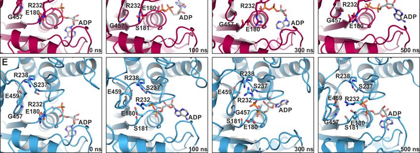
Int.
Int.J. J.Mol.
Mol.Sci. 2020,21,
Sci.2020, 21,7417
x FOR PEER REVIEW 22of
of24
23
affect each other. Pivotal to this allosteric communication is the central seven-stranded β-sheet (the
this allosteric communication is the central seven-stranded β-sheet (the transducer) (twisted in the
transducer) (twisted in the rigor state, but untwisted in the pre-power stroke state) in the myosin
rigor state, but untwisted in the pre-power stroke state) in the myosin motor domain, which couples
motor domain, which couples the large actin-binding cleft (closed while strongly attached to F-actin,
the large actin-binding cleft (closed while strongly attached to F-actin, and open in actin-detached
and open in actin-detached states) with the active site phosphate-sensing elements—the P-loop,
states) with the active site phosphate-sensing elements—the P-loop, switch-1 and switch-2—(closed in
switch-1 and switch-2—(closed in the hydrolysis-competent pre-power stroke state, but partially or
the hydrolysis-competent pre-power stroke state, but partially or fully opened in all other states) as
fully opened in all other states) as well as through the relay helix (bent in the pre-power stroke state,
well as through the relay helix (bent in the pre-power stroke state, while straight in the rigor state) and
while straight in the rigor state) and SH1/SH2-region with the mechanical converter and lever arm
SH1/SH2-region with the mechanical converter and lever arm (down position in the rigor state, and up
(down position in the rigor state, and up position in the pre-power stroke state). We and others have
position in the pre-power stroke state). We and others have shown that mutations in these key regions
shown that mutations in these key regions disturb the motor function by interfering with
disturb the motor function by interfering with chemomechanical coupling pathways [4–9].
chemomechanical coupling pathways [4–9].
Figure1.1.The
Figure Theactomyosin
actomyosinsystem.
system.(A)
(A)Scheme
Schemeofofthe theactomyosin
actomyosinmotor motorcycle.
cycle. The
Themyosin
myosin motor
motor isis
subject
subjecttotoconformational
conformationalchanges
changesofofthethesubdomains
subdomainsupon uponinteraction
interactionwithwithboth
boththe
theactin
actinfilament
filament
and
andthe thenucleotide
nucleotideadenosine
adenosinetriphosphate
triphosphate(ATP),
(ATP),asaswell
wellasasduring
duringnucleotide
nucleotidehydrolysis,
hydrolysis,and
andrelease
release
ofofitsitshydrolysis products inorganic phosphate
hydrolysis products inorganic phosphate (P (P ) and adenosine diphosphate (ADP) to eventually
i i) and adenosine diphosphate (ADP) to eventually
produce
produceforce forceinina atwo-step
two-stepmechanism.
mechanism.Color Colorcode:
code:subdomains
subdomainsN-terminal
N-terminaldomain
domain(pink),
(pink),U50
U50kDa
kDa
(blue),
(blue),L50 L50kDakDa(green),
(green),converter
converter(orange),
(orange),lever
leverarmarm(grey).
(grey).(B)
(B)Structure
Structureofofthe nucleotide-freeDd
thenucleotide-free Dd
myosin-II
myosin-IImotor motordomain,
domain,possessing
possessinga aclosed
closedactin-binding
actin-bindingcleft,
cleft,aatwisted
twistedtransducer
transducer(cyan),
(cyan),and
andthe
the
converter
converter(orange)/lever
(orange)/leverarm arm(grey)
(grey)ininthe
thedown
downposition.
position.TheTheactive
activesite
sitewith
withthethenucleotide
nucleotidesensors
sensors
P-loop
P-loop(yellow),
(yellow),switch-1
switch-1(red),
(red),and
andswitch-2
switch-2(brown)
(brown)isisempty
emptyandandprepared
preparedforforATP
ATPbinding.
binding.
The
Themyosin
myosin motor is thus
motor subject
is thus to substantial
subject conformational
to substantial changes along
conformational thatalong
changes cycle, that
traversing
cycle,
through
traversing through different actin-attached and –detached states, eventually amplifying themscale
different actin-attached and –detached states, eventually amplifying them to a large to a
rotation of the converter domain and swinging of the adjacent lever arm in the
large scale rotation of the converter domain and swinging of the adjacent lever arm in the force- force-generating power
stroke [10]. Many
generating powerofstroke
the subprocesses
[10]. Many haveof thebeen extensively
subprocesses studied
have beeninextensively
the past, and newlyinobserved
studied the past,
conformations, obtained through cryo-electron microscopy (cryoEM) [11–13]
and newly observed conformations, obtained through cryo-electron microscopy (cryoEM) [11–13] or crystallization withor
native and artificial ligands [14–17] that trapped myosin in a specific state, have been
crystallization with native and artificial ligands [14–17] that trapped myosin in a specific state, have assigned as states
ofbeen
the actomyosin
assigned as cycle
statesaccording to the status
of the actomyosin cycleofaccording
the characteristic structural
to the status of the elements in the
characteristic myosin
structural
motor domain,
elements in thethus building
myosin motorthedomain,
basis forthus
the motor
buildingfunction.
the basis Numerous studies
for the motor have significantly
function. Numerous
contributed to our understanding of the mechanism of the recovery stroke [18–23],
studies have significantly contributed to our understanding of the mechanism of the recovery stroke which primes the
myosin motor for force production while dissociated from the actin filament.
[18–23], which primes the myosin motor for force production while dissociated from the actin However, critical details
about the molecular
filament. However,eventscriticalofdetails
the actomyosin
about themotor cycle, events
molecular which directly contribute to
of the actomyosin forcecycle,
motor production
which
and product release, i.e., the myosin power stroke, are still incomplete.
directly contribute to force production and product release, i.e., the myosin power stroke, are still
The power stroke is initiated by ATP hydrolysis, followed by reattachment of the myosin motor
incomplete.
in the pre-power
The power stroke
stroke state to its actin
is initiated by ATPfilament track, coupled
hydrolysis, followedtobyconformational
reattachment of changes that finally
the myosin motor
in the pre-power stroke state to its actin filament track, coupled to conformational changes that finally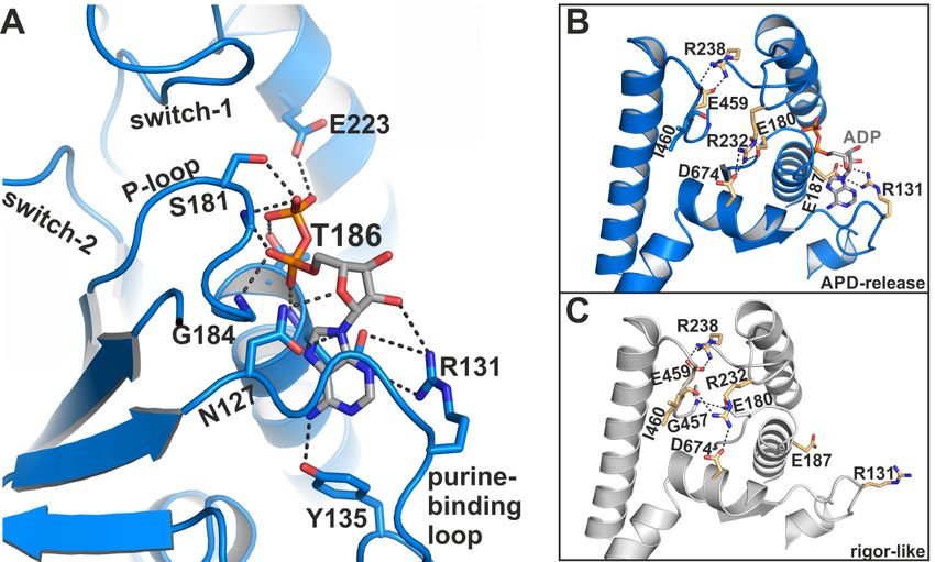
Int. J. Mol. Sci. 2020, 21, 7417                                                                     3 of 24

lead to the release of the hydrolysis products and to force generation. The exact sequence of events is
critically discussed in the field, and several models have been proposed for the conformational states
involved in the power stroke [24–27]. The conformational changes required for force production are
assumed to be driven by actin reattachment and the sequential release of inorganic phosphate (Pi ),
Mg2+ , and adenosine diphosphate (ADP). Crystal structures in the pre-power stroke state and the
nucleotide-free rigor-like state revealed that the central transducer undergoes a twisting motion of the
β-strands upon rebinding of myosin to the actin filament, linked to closure of the large cleft between
subdomains U50 and L50 kDa to allow strong actin binding [28] (Figure 1B). Kinetic analysis predicts
phosphate release from the myosin motor domain directly after ATP hydrolysis and prior to closure of
the large actin-binding cleft, which initiates the weak-to-strong actin binding [25]. Several mechanisms
for Pi release have, however, been postulated, both prior or subsequent to cleft closure, suggesting that
Pi -release occurs either through a side door [10,29], formed by repositioning of switch-1, or a backdoor
by switch-2 opening [25,30]. Comparison of the recent Mg2+ ·ADP-bound actin-attached cryoEM
models with the nucleotide-free actomyosin rigor complex suggests a two-step mechanism of the
power stroke [11–13,31]. According to these studies, the larger first step of the power stroke is linked
to actin-induced cleft closure and Pi release, while a second, smaller stroke with an approximately
10◦ rotation of the converter might occur during the release of Mg2+ ·ADP [31]. However there are
discrepancies about the degree of converter rotation in the two steps of the power stroke and due to
the resolution limitations of the cryoEM models, details regarding the molecular mechanisms for ADP
release remain speculative.
      We therefore present here high-resolution structural insights into a novel conformation of the
myosin-II motor domain that was obtained using the well-characterized small molecule myosin-II
inhibitor blebbistatin [32]. This myosin-II crystal structure reveals characteristic structural features of
the chemomechanical transducing elements—i.e., the central transducer, actin-binding cleft, active site
switches, relay helix, SH1/SH2-region, converter and lever arm—in conformations between the myosin
pre-power stroke and rigor positions, with the nucleotide partially unbound. These observations
suggest that the obtained crystal structure in the presence of blebbistatin might resemble a myosin
conformation involved in ADP release towards the end of the power stroke. ADP release is key for the
regulation of the mechanical motor function of myosin as it controls the fraction of time the motor stays
attached to the actin filament. A series of complementing molecular dynamics simulation techniques
further provided a mechanistic framework for nucleotide release from the myosin motor domain,
and corroborated that an equivalent myosin conformation plays a role during ADP release and the
second step of the power stroke.
      Blebbistatin was previously shown to inhibit myosin-II motor function by blocking the motor
domain in the pre-power stroke state [14,33,34]. Our results, however, are in line with other studies
using X-ray diffraction of muscle thick filaments [35,36] and negative stain electron microscopy together
with kinetic analysis [37], which reported blebbistatin to stabilize an additional myosin state in the
presence of ADP. These studies postulated that blebbistatin traps myosin in a state at the start of
the power stroke in the presence of ADP. In contrast, our high-resolution crystal structure reveals
a myosin-II·ADP·blebbistatin structure that shows high similarities with a putative ADP releasing
conformation of the motor cycle during the second step of the power stroke near the rigor conformation,
underpinning that the inhibitory mechanism of blebbistatin is much more complex than earlier
assumed [38].

2. Results

2.1. Structural Features of the Blebbistatin-Bound Myosin-II ADP Conformation
     In a series of crystallization trials using the myosin-II inhibitor blebbistatin, we obtained
a previously unknown conformation of myosin in the presence of ADP. We solved the crystal
structure of the Dd myosin-II motor domain in complex with blebbistatin and ADP to a resolution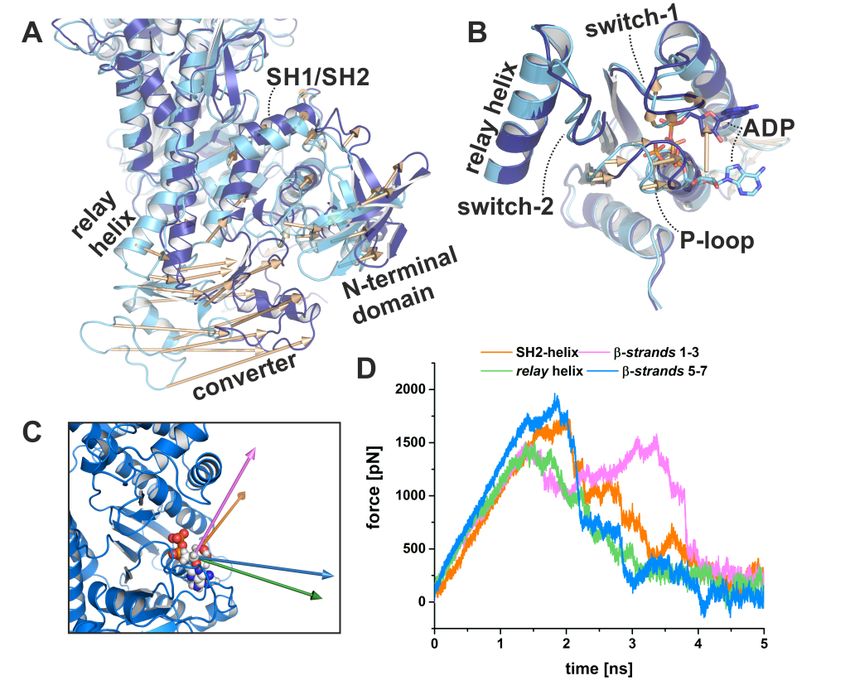
Int. J. Mol. Sci. 2020, 21, 7417 4 of 24
of 2.58
Int. J. Mol.ÅSci.
(Figure
2020, 21,2A, seePEER
x FOR Table 1 for
REVIEW data statistics). The crystals grew in the P21 21 21 space group 4 of 23
and showed unambiguous electron density for the protein residues, the nucleotide, and blebbistatin
(Figure
This 2B,C).
novel This novel
myosin myosin conformation
conformation differsfrom
differs markedly markedly from the
the current current crystallographically
crystallographically resolved
states of the actomyosin cycle. The hallmarks of the unique structure are a converter position position
resolved states of the actomyosin cycle. The hallmarks of the unique structure are a converter close to
close
the to the
rigor downrigorposition,
down position, rearrangements
rearrangements of the site
of the active active site switches
switches closer closer together,
together, and aandlargea large
shift
shift
of theofP-loop
the P-loop together
together withwith the bound
the bound ADPADP towards
towards the the protein
protein surface,
surface, whilewhile
thethe β-phosphate
β-phosphate of
of ADP lost most of its coordination with protein residues. The positions
ADP lost most of its coordination with protein residues. The positions of the active site of the active site switches
(closed with
withthe theP-loop
P-loop shifted towards
shifted the surface),
towards and the
the surface), andconverter/lever arm (near
the converter/lever down
arm (nearposition),
down
as well asasthe
position), wellstatus
as theofstatus
the actin-binding cleft (almost
of the actin-binding closed),
cleft (almost the relay
closed), helixhelix
the relay (straight), andand
(straight), the
transducer (almost fully twisted) suggest that the structure resembles a myosin
the transducer (almost fully twisted) suggest that the structure resembles a myosin conformation conformation towards
the end of
towards thetheendpower
of thestroke
power that promotes
stroke ADP release.
that promotes ADP Blebbistatin was found
release. Blebbistatin wasinfound
the same binding
in the same
binding site as observed in the crystal structure of the myosin-II pre-power stroke state [14], with
site as observed in the crystal structure of the myosin-II pre-power stroke state [14], and interacts and
residues of
interacts withtheresidues
relay andofW-helices, as well
the relay and as activeassite
W-helices, wellswitches
as activein site
close proximity
switches to β-strands
in close proximity 5 toto7
of the transducer
β-strands 5 to 7 of(Figure 2C).
the transducer (Figure 2C).
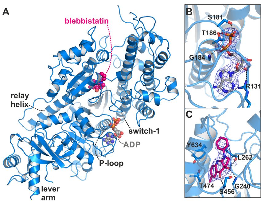
Figure 2.
Figure X-ray crystal
2. X-ray crystal structure
structure of of the Dd myosin-II
the Dd myosin-II motor
motor domain
domain in in complex
complex with
with ADPADP andand
blebbistatin. (A) Overview of the crystal structure in the blebbistatin-bound
blebbistatin. (A) Overview of the crystal structure in the blebbistatin-bound ADP-release ADP-release conformation.
The P-loop together
conformation. with thetogether
The P-loop bound ADP with(grey) is shifted
the bound ADP towards
(grey)the
is surface.
shifted Blebbistatin
towards the(magenta)
surface.
is buried in the
Blebbistatin knownisallosteric
(magenta) buried inbinding
the knownpocket at the apex
allosteric bindingof the largeatactin-binding
pocket the apex of the cleft.
large(B) The
actin-
2Fo -Fc density
binding map
cleft. (B) Theof2FADP
o-Fc in its new
density mapbinding
of ADPposition. Thebinding
in its new map was contoured
position. Theatmap
1.0 σ.
was Note that the
contoured
1.0 σ. Notehas
β-phosphate
at rotated
that away from the
the β-phosphate hasP-loop.
rotated(C) 2Fo -F
away c density
from map of(C)
the P-loop. blebbistatin at the map
2Fo-Fc density apex of
of
the actin-binding cleft and near the relay helix. The map was contoured at 1.0 σ.
blebbistatin at the apex of the actin-binding cleft and near the relay helix. The map was contoured at
1.0 σ.
Table 1. Data collection and refinement statistics.
Dd Myosion-II (apo) Dd Myosin-II·ADP·blebbistatin
Data collection
Space group P1211 P212121
Cell dimensions
a, b, c (Å) 56.68, 174.40, 100.25 47.37, 88.70, 199.89
α, β, γ (°) 90.00, 106.38, 90.00 90.00, 90.00, 90.00
Resolution (Å) 46.36–1.88 (1.95 - 1.88) * 46.10–2.58 (2.67–2.58)
Rmerge [%] 0.12 (0.88) 0.17 (1.37)
I / σI 9.46 (1.81) 14.41 (1.88)
CC1/2 0.99 (0.76) 0.99 (0.72)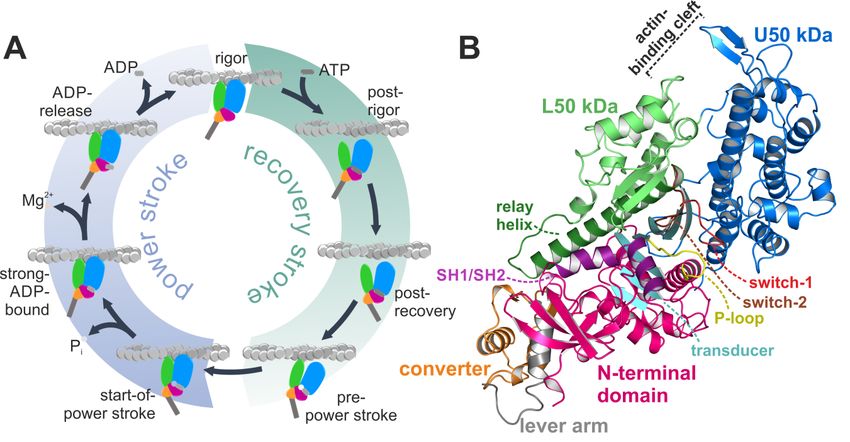
Int. J. Mol. Sci. 2020, 21, 7417                                                                                           5 of 24

                                     Table 1. Data collection and refinement statistics.

                                                      Dd Myosion-II (apo)                  Dd Myosin-II·ADP·Blebbistatin
             Data collection
              Space group                                      P121 1                                      P21 21 21
             Cell dimensions
                a, b, c (Å)                           56.68, 174.40, 100.25                        47.37, 88.70, 199.89
                α, β, γ (◦ )                           90.00, 106.38, 90.00                         90.00, 90.00, 90.00
             Resolution (Å)                          46.36–1.88 (1.95–1.88) *                     46.10–2.58 (2.67–2.58)
               Rmerge [%]                                  0.12 (0.88)                                  0.17 (1.37)
                   I / σI                                  9.46 (1.81)                                 14.41 (1.88)
                   CC1/2                                   0.99 (0.76)                                  0.99 (0.72)
            Completeness (%)                               98.70 (97.04)                                 99.92 (99.96)
              Redundancy                                        5.9                                          13.2
              Refinement
             Resolution (Å)                                 46.36–1.88                                    46.10–2.58
             No. reflections                              148,800 (14582)                                27,410 (2668)
              Rwork / Rfree                                19.10 / 21.92                                 20.02 / 23.09
               No. atoms                                       12958                                         6089
                 Protein                                       11662                                         5945
               Ligand/ion                                       161                                           85
                 Water                                         1135                                           59
                B-factors
                 Protein                                       39.96                                        52.36
               Ligand/ion                                      46.09                                        50.86
                 Water                                         39.27                                        40.13
            R.m.s. deviations
            Bond lengths (Å)                                   0.011                                        0.005
             Bond angles (◦ )                                  1.21                                          0.79
                               * Statistics for the highest-resolution shell are shown in parentheses.

2.1.1. Comparison with the Myosin-II Rigor and Pre-power Stroke States
      As a reference, we additionally crystallized the nucleotide-free apo Dd myosin-II motor domain,
and solved the structure to a resolution of 1.88 Å (Figure 1B, see Table 1 for data statistics). The overall
conformation highly resembled the previously solved structure of a myosin-II-dynamin fusion
construct [39] (root mean square deviation (rmsd) 0.54 (chain A) and 0.57 (chain B)), which was
assigned earlier as the Dd myosin-II rigor-like state, with the converter in the down position and the
relay helix straightened. A twisted transducer in our crystal structure primed the nucleotide-free
active site for binding of ATP, with an intact critical salt-bridge between switch-1 (residue Arg238)
and switch-2 (residue Glu459), which has been suggested to play a central role during Pi release [25].
The P-loop (179 GESGAKT186 ) moved away from the switches together with the N-terminal domain.
As observed with the dynamin-fusion rigor-like myosin-II structure (pdb: 2aka), the large actin-binding
cleft of our rigor-like myosin-II crystal structure was closed both in the inner (10.7 Å between the Cα
atoms of residues Ser272 and Ser465) and outer cleft (10.2 Å between the Cα atoms of residues Ser416
and Lys589) as compared to the Dd myosin-II pre-power stroke state structure (inner cleft: 12.6 Å,
outer cleft: 13.4 Å), but to a smaller extent than rigor-like structures of other myosin classes such as
myosin-V (inner cleft: 9.6 Å, outer cleft: 7.2 Å) [40]. Indeed, this is a well-documented feature of Dd
myosin-II [40].
      Comparison with our apo rigor-like myosin-II and the available pre-power stroke structure
(pdb: 1vom) reveals that the major structural features of the blebbistatin-bound myosin-II·ADP
conformation are in positions between the pre- and post-stroke states, closer to the final rigor
state at the end of the power stroke. In contrast to an earlier proposed conformation of the ternary
myosin-II·ADP·blebbistatin complex at the start of the power stroke [37], our structure shows a myosin-II
conformation in the presence of blebbistatin and ADP that, according to the characteristic structural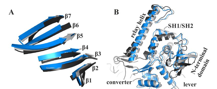
Int. J. Mol. Sci. 2020, 21, 7417 6 of 24
elements in the motor domain that were used in the past to define the states of the actomyosin motor cycle,
resembles a conformation towards the end of the power stroke, associated with the second converter
rotation. The central transducer for chemomechanical coupling in the blebbistatin-bound myosin
motor domain is partially twisted, particularly β-strands 1 to 3 (Figure 3A). During the power stroke,
the transducer mediates the straightening of the relay helix as well as the repositioning of the N-terminal
domain, which allows a continuous interaction of the N-terminal domain and the converter [28].
The N-terminal domain in the myosin-II·ADP·blebbistatin structure is pivoted by ∼2 Å relative to the
U50 kDa domain from the corresponding rigor position (Figure 3B). Consistently, the converter is
rotated by approximately 55◦ as compared to the up position in the pre-power stroke state, and would
need to undergo only a minor 4◦ to 5◦ converter rotation to transition to the down position in the rigor
state (Figure 3B). Thus, the rotation amplitude of the converter is more advanced than in the reported
strong-Mg2+ ·ADP-bound state (approximately 30◦ to 50◦ away from the up position, and 30◦ to 10◦
Int. J. Mol. Sci. 2020, 21, x FOR PEER REVIEW 6 of 23
away from the down position) [12,31]. Similarly, a transition by approximately 1 Å of both the inner and
outer actin-binding
approximately cleft
1 Å(myosin-II·ADP·blebbistatin
of both the inner and outer complex: inner
actin-binding cleft:
cleft 11.7 Å and outer cleft: 11.3 Å)
(myosin-II·ADP·blebbistatin
separatescomplex:
the blebbistatin-bound myosin-II·ADP from the fully closed actin-binding cleft as seen in the
inner cleft: 11.7 Å and outer cleft: 11.3 Å) separates the blebbistatin-bound myosin-II·ADP
apo rigorfrom
state,
the indicating a new actin cleft
fully closed actin-binding interface
as seen(Figure
in the apo3C). The
rigor status
state, of these
indicating a newimportant structural
actin interface
elements(Figure
in the3C). myosinThe status
motor of domain
these important
furtherstructural
suggestselements
that theinmyosin
the myosin motor domainasfurther
conformation, observed in
suggests that the myosin conformation, as observed in the myosin-II·ADP·blebbistatin complex,
the myosin-II·ADP·blebbistatin complex, might resemble a myosin ADP-release conformation that has
might resemble a myosin ADP-release conformation that has already transitioned through2+the first
already transitioned through the first step of the power stroke 2+and beyond the strong-Mg ·ADP-bound
step of the power stroke and beyond the strong-Mg ·ADP-bound actomyosin state. The
actomyosin state. The conformational
conformational changes required changes required
to fully close to fully close
the actin-binding thecould
cleft actin-binding
thus play acleft
rolecould
for thus
play a role for initiating
initiating ADP release ADP release
from fromdomain.
the motor the motor domain.
The blebbistatin-bound
Figure 3.Figure 3. The blebbistatin-bound myosin-II·ADP
myosin-II·ADPstructure (blue)feature
structure (blue) feature characteristics
characteristics of a myosin
of a myosin
conformation
conformation betweenbetween the pre-power
the pre-power stroke stroke (black),
(black), and theandrigor-like
the rigor-like
statesstates
(grey).(grey). (A) transducer
(A) The The
transducer is partially twisted, particularly β-strands 1 to 3. (B) The relay helix and the converter
is partially twisted, particularly β-strands 1 to 3. (B) The relay helix and the converter approached
approached the rigor-like position. The converter/lever arm must undergo a ~5° rotation to transition
the rigor-like position. The converter/lever arm must undergo a ~5◦ rotation to transition to the final
to the final rigor down position (grey). The N-terminal domain is markedly shifted towards its rigor
rigor down position
position. (C) The(grey). The N-terminal
large actin-binding domain
cleft is found in ais markedly
position shifted
between towards
pre-power strokeits rigorand
(black) position.
(C) The large actin-binding
rigor-like states (grey),cleft is found
thereby inaanew
creating position between
actin-binding pre-power
interface. stroke
(D) The active(black) andhas
site P-loop rigor-like
states (grey),
moved thereby
~9 Å creating
from itsa pre-power
new actin-binding interface.
stroke position (D) The
in the active site P-loop
blebbistatin-bound has moved ~9 Å
myosin-II·ADP
conformation,
from its pre-power but has
stroke not reached
position theblebbistatin-bound
in the rigor position. myosin-II·ADP conformation, but has not
reached the rigor position.
2.1.2. Structural Features of the Active Site in the Myosin-II·ADP·Blebbistatin Complex
In the active site of the blebbistatin-bound myosin-II·ADP structure, considerable
rearrangements took place as a consequence of the conformational changes in the actin-binding cleft,
the transducer, the N-terminal domain, and the converter. The partial twisting of the transducer
induced a shift of switch-1 (232RNNNSSR238) and switch-2 (454DISGFE459) by approximately 1 Å
towards their rigor positions (Figure 3D), with the critical salt-bridge between Arg238 (switch-1) and
Int. J. Mol. Sci. 2020, 21, 7417                                                                     7 of 24

2.1.2. Structural Features of the Active Site in the Myosin-II·ADP·Blebbistatin Complex
       In the active site of the blebbistatin-bound myosin-II·ADP structure, considerable rearrangements
took place as a consequence of the conformational changes in the actin-binding cleft, the transducer,
the N-terminal domain, and the converter. The partial twisting of the transducer induced a shift of
switch-1 (232 RNNNSSR238 ) and switch-2 (454 DISGFE459 ) by approximately 1 Å towards their rigor
positions (Figure 3D), with the critical salt-bridge between Arg238 (switch-1) and Glu459 (switch-2)
remaining intact. Recent structural studies supported a Pi release mechanism, which presumes the
salt-bridge to break in order to open the release tunnel for inorganic phosphate [25]. Our crystal
structure features a conformation of the switches in which Pi release and actin rebinding appears to
have already occurred or is unfeasible. Considering a similar active site in the myosin ADP-release
conformation during the power stroke would indicate that the salt-bridge reforms directly after release
of Pi .
       The largest deviations from the active site conformation as seen in the pre-power stroke state
were observed for the phosphate-binding P-loop, which moved ~9 Å towards the front entrance
of the active site (Figure 3D), accompanied by a rotational movement of the N-terminal domain.
Together with the P-loop, the coordinated ADP is repositioned away from switch-1 and -2 towards
the protein surface, and is now accessible to the surrounding water in a new binding position.
The adenosine base of ADP remains bound to the P-loop through hydrophobic interactions, as well
as hydrogen bonds to residues Tyr135 and Asn127 of the purine-binding loop (126 VNPFKRIPIYT136 )
(Figure 4A). In addition, the sidechain of Arg131 rearranged towards the nucleotide to facilitate the
formation of a salt-bridge with Glu187, thereby forming a binding groove for the adenosine base of the
nucleotide. This salt-bridge might therefore represent a critical feature for promoting ADP release.
Arg131 interacts furthermore with a hydroxyl group of the nucleotide ribose and thereby additionally
stabilizes the nucleotide in this new position. While the α-phosphate is involved in a tight interaction
network with residues Gly184, Thr186, and Glu187, the nucleotide β-phosphate is orientated away
from the P-loop, towards the surrounding water. As a consequence, most of the interactions of the
β-phosphate with active site residues that can be observed in known myosin structures are lost in
this ADP-bound myosin-II structure (seven interactions in the pre-power stroke state, while three
interactions are observed in our structure), suggesting a weakly bound nucleotide with a conformation
that is distinct from an earlier structure with ADP soaked into a rigor-like conformation of myosin-V [28].
A reduced number of hydrogen-bonds of the β-phosphate with residues Ser181, Thr186, and Glu223
were detected in the myosin-II·blebbistatin·ADP structure. No electron density was found for the
magnesium ion, which usually coordinates the negatively charged phosphates of the nucleotide as
well as phosphate-sensor residues Thr186 (P-loop) and Ser237 (switch-1). We assume that due to
the movement of the P-loop relative to switch-1, the coordination of Mg2+ is disturbed, causing its
dissociation from the active site. This is in good agreement with earlier studies, which showed that
Mg2+ release precedes ADP release for various myosin isoforms [41–43].
electron density was found for the magnesium ion, which usually coordinates the negatively charged
 phosphates of the nucleotide as well as phosphate-sensor residues Thr186 (P-loop) and Ser237
 (switch-1). We assume that due to the movement of the P-loop relative to switch-1, the coordination
 of Mg2+ is disturbed, causing its dissociation from the active site. This is in good agreement with
 earlier
Int. J. Mol.studies,
              Sci. 2020,which
                        21, 7417showed that Mg2+ release precedes ADP release for various myosin isoforms
                                                                                                    8 of 24
 [41–43].

      Figure 4. The myosin-II·ADP·blebbistatin crystal structure shows unique conformational features in
     Figure 4. The myosin-II·ADP·blebbistatin crystal structure shows unique conformational features in
      the active
     the  active site.
                 site. (A)
                        (A)ADP
                            ADPinteracts
                                   interactswith
                                             withitsits
                                                     phosphate  groups,
                                                        phosphate        primarily
                                                                   groups,         with
                                                                            primarily    the the
                                                                                       with  reoriented  P-loop.
                                                                                                  reoriented     The
                                                                                                             P-loop.
      β-phosphate
     The             is partially
           β-phosphate            unbound
                          is partially       and oriented
                                        unbound             towards
                                                    and oriented     the surrounding
                                                                  towards              water at
                                                                            the surrounding      the protein
                                                                                               water         surface.
                                                                                                      at the protein
      The  adenosine    base of the nucleotide  is nestled into a groove, formed  by a salt-bridge  between
     surface. The adenosine base of the nucleotide is nestled into a groove, formed by a salt-bridge between Arg131
     Arg131 and Glu187. (B) Close-up view on the active site in the myosin-II·ADP·blebbistatin structure
     shows the critical salt-bridge between switch-1 (Arg238) and switch-2 (Glu459), as well as the newly
     identified salt-bridge between Arg131 and Glu187 that stabilizes ADP in the release position. A third,
     complex salt-bridge between Arg232 (switch-1), Asp674 (SH2-helix), and Glu180 (P-loop) seems to
     be important for mediating the chemomechanical coupling between the active site, the actin-binding
     region and the mechanical converter/lever arm. (C) Close-up view on the active site in the rigor-like,
     nucleotide-free myosin-II structure. Due to slight adjustments to the active site, the interaction network
     around Arg232 is altered as compared to the myosin-II·ADP·blebbistatin structure, and Arg232 interacts
     with Asp674 (SH2-helix), and Ile460 (switch-2), while Glu180 (P-loop) formed a hydrogen-bond to
     Gly457 (switch-2) in this conformation.

2.1.3. A Potential Communication Hub Centered on Arg232 Allosterically Mediates Changes in the
Active Site to the Converter and Actin-Binding Region
     Further stabilizing interactions could be found between the three phosphate-binding loops as
a consequence of the rearrangements in the active site. Except for the critical salt-bridge between switch-1
(Arg238) and -2 (Glu459), and the identified salt-bridge between the purine-binding loop (Arg131) and
the P-loop (Glu187) that stabilizes the adenosine base of the nucleotide, a third, complex salt-bridge
has been formed in the blebbistatin-bound myosin-II·ADP structure between switch-1 (Arg232),
the P-loop (Glu180), and the SH2-helix (Asp674) (Figure 4B). This salt-bridge thus seems to couple the
active site sensors—switch-1 and P-loop—with the SH2-helix in the N-terminal domain of myosin,
indicating a critical role in communicating conformational changes in the myosin motor domain. It is
well-known that the different domains and remote sites in myosin are interconnected and highly
communicate with each other. This complex salt-bridge might therefore represent an allosteric
communication hub for mediating changes in the actin-binding cleft via switch-1 to the active site and
the N-terminal domain. The latter in turn needs to positionally dislocate in order to allow the final
rotation of the converter/lever arm during the transition to the rigor state. An equivalent complex
salt-bridge in scallop myosins has been suggested earlier to control the communication between the
actin-binding region and the active site [44].
Int. J. Mol. Sci. 2020, 21, 7417                                                                      9 of 24

     The corresponding arginine in the rigor-like Gg myosin-V [45] structure shows no interactions with
the surrounding active site motifs. In our rigor-like apo myosin-II structure, the interactions of Arg232
differ markedly (Figure 4C) from the blebbistatin-bound myosin-II·ADP structure. While Arg232 in
the rigor-like state is hydrogen-bonded to Asn674 of the SH2-helix as well, the contact with Glu180
(P-loop) is broken. Instead, Arg232 formed a hydrogen bond with the backbone of Ile460 (switch-2),
while Glu180 of switch-1 interacts with Gly457 (switch-2). Hence, both interactions in the rigor-like
state assist the maintenance of the closed conformation of the switches, highlighting the central role of
the interaction network mediated by Arg232 in chemomechanical coupling in myosin.

2.2. Blebbistatin Affects the Myosin Conformation by Binding to the Known Allosteric Binding Pocket
      The myosin-II inhibitor blebbistatin has been shown earlier to prevent cleft closure by blocking
myosin in the weakly actin-attached pre-power stroke state with the hydrolysis products Mg2+ ·ADP·Pi
bound to the active site [14,33,34]. This effect gave rise to numerous studies on cytoskeletal and muscle
function using blebbistatin as a chemical tool. More recent results suggested blebbistatin to stabilize
an additional state at the start of the power stroke with a closed actin-binding cleft but the lever arm
was still in the up position [35–37]. However, these mechanisms cannot explain all the experimental
effects observed with blebbistatin [38], and our high-resolution structure of myosin-II complexed with
ADP and blebbistatin resembled a myosin conformation towards the end of the power stroke involved
in ADP dissociation from the motor domain.
      Blebbistatin binds to the same allosteric binding pocket as reported with the pre-power stroke
state myosin-II crystal structure (pdb: 1yv3) [14] at the apex of the large actin-binding cleft and
adjacent to the active site phosphate sensors switch-1 and switch-2 (Figure 2C). In agreement with
the earlier crystal structures, binding of blebbistatin in the myosin-II·ADP·blebbistatin structure is
primarily accomplished via hydrophobic interactions with the U50 linker (the loop following β7 of
the transducer), the relay helix, and the W-helix, including residues Tyr261, Ile455, Glu467, Ile471,
Thr474, Val630, Tyr634, and Leu641. Particularly amino acids Thr474 and Tyr634 were identified earlier
to critically affect the specificity of blebbistatin towards class II myosins [14]. Moreover, as shown
before, three hydrogen bonds with residues Gly240, Leu262, and Ser456 contribute to the binding
affinity of blebbistatin. The characteristic displacement of the sidechains of residues Leu262 and Tyr634
by approximately 3 to 4 Å is clearly visible also in the blebbistatin-bound myosin-II·ADP structure,
which opened the allosteric pocket to accommodate blebbistatin. Hence, the allosteric binding pocket of
blebbistatin in our new crystal structure highly resembles the previously observed pocket conformation
with conserved protein–ligand interactions, suggesting the presence of the binding pocket in several
of the myosin states without larger structural changes of the protein around blebbistatin. In contrast
to the postulated inhibitory mechanism of blebbistatin in which switch-2 reorientation is blocked,
and thereby Pi release and converter rotation, we identified marked positional changes of switch-2 in
the presence of ADP and blebbistatin towards its rigor position (Figure 3D). This indicates a much
more complex mechanism of blebbistatin that might also affect ADP binding and release.

2.2.1. Blebbistatin Increases the Affinity of Myosin-II for ADP in the Presence and Absence of Mg2+
     In order to directly analyze the effect of blebbistatin on the bound nucleotide, we performed
binding assays using microscale thermophoresis (MST). The sequential release of the hydrolysis
products Mg2+ and ADP are assumed to drive the conformational changes required for the second
step of the mechanical power stroke. Kinetic studies showed that ADP release in distinct myosin-II
isoforms is regulated by Mg2+ [46], and for myosin-V and presumably various other myosin classes,
both the myosin·Mg2+ ·ADP and myosin·ADP states exist in equilibrium, facilitating dissociation
of Mg2+ to precede ADP release at physiological magnesium concentrations [41,43]. In accordance
with this, and assuming our crystallized structure resembles the ADP-release conformation during
the end of the myosin power stroke, we did not find electron density for the Mg2+ ion in the
myosin-II·ADP·blebbistatin structure.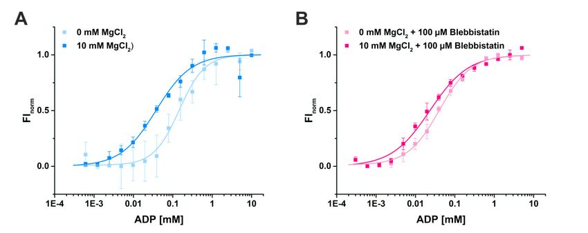
Int. J. Mol. Sci. 2020, 21, 7417                                                                                         10 of 24

      Using MST, we determined a binding constant (KD ) for ADP of 37.5 ± 7.3 µM in the presence of
10 mM Mg2+ (Figure 5A). As expected, the binding affinity of ADP to myosin-II was decreased by
a factor of 4 to a KD of 145.8 ± 19.3 µM in the absence of magnesium, consistent with a sequential
release mechanism during force generation with Mg2+ released through an as yet unknown route.
Addition of blebbistatin slightly increased the binding affinity of ADP for myosin 1.8-fold to a KD
value of 21.2 ± 3.4 µM in the presence of 10 mM Mg2+ (Figure 5B), which is in good agreement with
previous measurements of the ADP affinity to the actomyosin complex in the presence of Mg2+ and
blebbistatin that reported a KD value of 24 ± 1 µM [37]. Interestingly, without Mg2+ present, the affinity
of ADP for myosin-II (KD of 40.6 ± 3.2 µM) in the presence of blebbistatin is significantly elevated up
to a factor of 4 as compared to the KD value in the absence of the small molecule inhibitor (Figure 5B).
These results correlate well with our new myosin-II·ADP·blebbistatin structure, and we therefore
speculate that blebbistatin stabilizes the crystallographically found ADP-release-like conformation of
   Int. J. Mol.through
myosin-II       Sci. 2020, 21,
                           anxincreased ADP affinity while Mg2+ is already dissociated from the myosin. 10 of 23
                               FOR PEER REVIEW

      Figure
         Figure     Binding
                5. 5. Bindingassays
                                assaysofofADP
                                            ADPtotomyosin-II
                                                        myosin-IIusing
                                                                   usingmicroscale
                                                                          microscalethermophoresis.
                                                                                      thermophoresis. (A) The affinity of
      of ADP
         ADP for  for myosin-II
                      myosin-IIininthe
                                    theabsence
                                          absenceofof  magnesium is reduced from KD K
                                                          magnesium    is reduced  from   =D  = 37.5
                                                                                           37.5 ± 7.3 ±µM
                                                                                                        7.3(10
                                                                                                             µMmM(10MgmM2+) to
          2+
      Mg145.8) to± 145.8 ± 19.3  µM (0  mM          2+
                    19.3 µM   (0 mM   Mg  2+ ). Mg     ). (B) Addition
                                                (B) Addition   of 100 µMof 100  µM blebbistatin
                                                                            blebbistatin         increased
                                                                                         increased           the binding
                                                                                                    the binding   affinity of
      affinity
         ADP of  forADP   for myosin-II,
                      myosin-II,           bothpresence
                                 both in the      in the presence
                                                            (KD = 21.2    = 21.2
                                                                    (K±D 3.4 µM)±and
                                                                                  3.4 µM)  andof
                                                                                       absence  absence   of magnesium
                                                                                                  magnesium     (KD = 40.6 ±
      (KD3.2= µM).
              40.6 ± 3.2 µM).

2.2.2. Blebbistatin Stabilizes the Crystallographically Observed Myosin-II·ADP Conformation
   2.2.2. Blebbistatin Stabilizes the Crystallographically Observed Myosin-II·ADP Conformation
      To further support this hypothesis, we carried out 500 ns classical molecular dynamics simulations
         To further support this hypothesis, we carried out 500 ns classical molecular dynamics
(cMD) in duplicates with a total of more than 2 µs simulations in explicit solvent and in the absence
   simulations (cMD) in duplicates with a total of more than 2 µs simulations in explicit solvent and in
and presence of blebbistatin. Starting from the blebbistatin-bound myosin-II·ADP crystal structure,
   the absence and presence of blebbistatin. Starting from the blebbistatin-bound myosin-II·ADP crystal
we did not observe any larger conformational changes of the myosin motor domain along the cMD
   structure, we did not observe any larger conformational changes of the myosin motor domain along
trajectories with mean rmsd values of the protein backbone around 3 Å (Figure 6A). A minimal
   the cMD trajectories with mean rmsd values of the protein backbone around 3 Å (Figure 6A). A
shift of blebbistatin within the allosteric pocket towards the relay helix occurred, while remaining
   minimal shift of blebbistatin within the allosteric pocket towards the relay helix occurred, while
bound to the relay helix, the W-helix, and the transducer through the crystallographically determined
   remaining bound to the relay helix, the W-helix, and the transducer through the crystallographically
interactions (Figure 6B). No significant rearrangements of the relay helix together with the converter
   determined interactions (Figure 6B). No significant rearrangements of the relay helix together with
were detected along the cMD simulations (Figure 6C), and the converter stably interacted with the
   the converter were detected along the cMD simulations (Figure 6C), and the converter stably
N-terminal domain throughout the simulations. The active site switches as well as the P-loop showed
   interacted with the N-terminal domain throughout the simulations. The active site switches as well
only negligible fluctuations and remained in their unique crystallographically determined positions in
   as the P-loop showed only negligible fluctuations and remained in their unique crystallographically
the blebbistatin-bound myosin simulations (Figure 6D). These positions of the switches were spatially
   determined positions in the blebbistatin-bound myosin simulations (Figure 6D). These positions of
restrained by the stable critical salt-bridge between Arg238 (switch-1) and Glu459 (switch-2) along the
   the switches were spatially restrained by the stable critical salt-bridge between Arg238 (switch-1) and
trajectories. However, the bound ADP in its new position at the surface along the outwards shifted
   Glu459 (switch-2) along the trajectories. However, the bound ADP in its new position at the surface
P-loop showed substantial fluctuations around its crystallographic position with an average rmsd of
   along the outwards shifted P-loop showed substantial fluctuations around its crystallographic
5.4 Å. Along the trajectories, ADP seems therefore loosely coupled to the P-loop with a number of
   position with an average rmsd of 5.4 Å. Along the trajectories, ADP seems therefore loosely coupled
transient interactions, indicating a weak ADP association. These results demonstrate that blebbistatin
   to the P-loop with a number of transient interactions, indicating a weak ADP association. These
   results demonstrate that blebbistatin is capable of stabilizing a unique conformation of myosin-II with
   a rearranged P-loop towards the front entrance of the active site and a loosely bound ADP.
Int. J. Mol. Sci. 2020, 21, 7417 11 of 24
is capable of stabilizing a unique conformation of myosin-II with a rearranged P-loop towards the
front entrance of the active site and a loosely bound ADP.
Int. J. Mol. Sci. 2020, 21, x FOR PEER REVIEW 11 of 23
Figure 6. Classical molecular dynamics (cMD) simulations support the stabilizing effect of
Figure 6. Classical molecular dynamics (cMD) simulations support the stabilizing effect of blebbistatin
blebbistatin on the crystallographically determined conformation of myosin-II. (A) Root mean square
on the crystallographically determined conformation of myosin-II. (A) Root mean square deviations of
deviations of the protein backbone atoms, averaged over duplicate 500 ns cMD simulations of
the protein backbone atoms, averaged over duplicate 500 ns cMD simulations of blebbistatin-bound
blebbistatin-bound (magenta) and -unbound (blue) myosin, confirm the stabilizing effect of
(magenta) and -unbound (blue) myosin, confirm the stabilizing effect of blebbistatin on the crystallized
blebbistatin on the crystallized conformation, while in the absence of blebbistatin, larger deviations
conformation, while in the absence of blebbistatin, larger deviations from the start structure were
from the start structure were observed. (B) Blebbistatin remains embedded in its binding pocket
observed. (B) Blebbistatin remains embedded in its binding pocket throughout the simulations,
throughout the simulations, showing only negligible deviations from its crystallized binding pose.
showing only negligible deviations from its crystallized binding pose. (C) Root mean square deviations
(C) Root mean square deviations of the converter indicate no movement of the converter while
of the converter indicate no movement of the converter while blebbistatin is bound (magenta);
blebbistatin is bound (magenta); however, a smaller shift towards the pre-power stroke position of
however, a smaller shift towards the pre-power stroke position of the converter is monitored in the
the converter is monitored in the absence of blebbistatin (blue). Shown in diagrams A-C are the
absence of blebbistatin (blue). Shown in diagrams A-C are the averages of duplicate cMD simulations.
averages of duplicate cMD simulations. (D) The positions of active site phosphate sensors are
(D) The positions of active site phosphate sensors are stabilized spatiotemporally along the cMD
stabilized spatiotemporally along the cMD simulations in the presence of blebbistatin, with the critical
simulations in the presence of blebbistatin, with the critical salt-bridge formed. Binding of ADP in the
salt-bridge formed. Binding of ADP in the crystallized conformation appeared loose during the cMD
crystallized conformation appeared loose during the cMD simulations with considerable fluctuations of
simulations with considerable fluctuations of the nucleotide. (E) During cMD simulations in the
the nucleotide. (E) During cMD simulations in the absence of blebbistatin, the P-loop moved towards
absence of blebbistatin, the P-loop moved towards switch-1 and -2 in the reversed direction of the
switch-1 and -2 in the reversed direction of the power stroke. The nucleotide ADP was markedly
power stroke. The nucleotide ADP was markedly shifted together with the P-loop inside into the
shifted together with the P-loop inside into the active site.
active site.
In contrast, during the cMD simulations in the absence of blebbistatin while starting from the
In contrast, during the cMD simulations in the absence of blebbistatin while starting from the
same crystal structure, the bound ADP together with the P-loop moved linearly backwards in the
same crystal structure, the bound ADP together with the P-loop moved linearly backwards in the
direction of their pre-power stroke state positions (Figure 6E), dragging the nucleotide ~5 Å inside
direction of their pre-power stroke state positions (Figure 6E), dragging the nucleotide ~5 Å inside
the active site. Concomitantly, a slight untwisting of the transducer could be observed along the
the active site. Concomitantly, a slight untwisting of the transducer could be observed along the
trajectories, which led to the decoupling of switch-1 and switch-2 by breaking the critical salt-bridge,
trajectories, which led to the decoupling of switch-1 and switch-2 by breaking the critical salt-bridge,
and pronounced rearrangements of the switches. The entire protein appears to be subject to a partial
and pronounced rearrangements of the switches. The entire protein appears to be subject to a partial
reverse motion of the power stroke, particularly of the N-terminal domain and the adjacent converter
reverse motion of the power stroke, particularly of the N-terminal domain and the adjacent converter
domain, which drew closer to their pre-power stroke state positions. As a consequence of the loss of
the critical salt-bridge at a relatively early timestep of the cMD simulations, switch-1 forms a new
complex salt-bridge via residue Arg232 with Glu459 (switch-2) and Glu180 (P-loop), highlighting the
critical role of Arg232 as a fulcrum for mediating conformational changes between remote sites in theInt. J. Mol. Sci. 2020, 21, 7417 12 of 24
domain, which drew closer to their pre-power stroke state positions. As a consequence of the loss of the
critical salt-bridge at a relatively early timestep of the cMD simulations, switch-1 forms a new complex
salt-bridge via residue Arg232 with Glu459 (switch-2) and Glu180 (P-loop), highlighting the critical
role of Arg232 as a fulcrum for mediating conformational changes between remote sites in the myosin
motor domain. In addition, Gly457 of switch-2, usually involved in binding the γ-phosphate of ATP or
the cleaved Pi , interacts with amino acid Gly179 of the P-loop. Inside the active site, ADP rebinds to the
main chain atoms of residue Ser237 (switch-1), a critical amino acid that typically coordinates the Mg2+
ion in the active site, which in turn interacts with the β-phosphate of the nucleotide. Hence, in the
absence of blebbistatin, the nucleotide seems to bind more deeply inside the myosin active site,
and myosin-II at the end of our 500 ns cMD simulations appears to adopt a conformation resembling
the strong-ADP-bound myosin state, determined by cryoEM [12,31].
Comparing our results with these strong-ADP cryoEM models demonstrated that myosin in this
conformation would require only small rearrangements of the N-terminal domain together with the
relay helix and the converter by approximately 10◦ to reach the final rigor state. Moreover, a similar
unique coordination of ADP by the active site switch motifs was postulated earlier [31] with
directly coupled switch-1 and P-loop, as observed in our myosin-II·ADP·blebbistatin crystal structure;
however, the resolution limit of the cryoEM model in that study prevented a direct comparison of
the sidechain and nucleotide positions, and thus the final validation that the conformation at the
end of our simulations represents the strong-ADP-bound myosin state. Nevertheless, our cMD
simulations suggest that the conformation of myosin-II, stabilized by blebbistatin, can reversibly
adopt a conformation, similar to the strong-Mg2+ ·ADP state in the absence of blebbistatin, and might
therefore resemble a conformation towards the end of the power stroke with the ADP loosely bound
and involved in ADP dissociation from the motor domain.
2.3. The ADP Release Pathway Reveals a Central Role of Arg131 for Guiding ADP Dissociation
2.3.1. Computational Generation of a Strong-ADP-Bound Myosin-II Conformation, and First Step of
the Power Stroke
The positions of critical structural elements in our crystal structure of myosin-II in complex with
ADP and blebbistatin suggest a resemblance with a myosin conformation towards the end of the power
stroke that is involved in ADP release. To gain further insights into the molecular events underlying
the mechanism of the second step of the myosin-II power stroke associated with ADP release, we used
a set of unbiased and pulling molecular dynamics simulation techniques.
During the power stroke, the population of a myosin state with strongly bound Mg2+ ·ADP
after rebinding of the myosin motor to its actin filament track and Pi release was shown in recent
cryoEM studies of myosin-I, -V, and -VI [12,13,31]. We therefore computationally generated a myosin-II
conformation, resembling this strong-ADP-bound myosin state by applying an external biasing force
to the actin-binding cleft and switch-2 of the active site, in order to force cleft closure and twisting of
the β-strands of the central transducer, similar to what had been published earlier for myosin-II [24].
This external force mimics the effect exerted by the actin filament during rebinding of the myosin motor
domain. The final model of these targeted molecular dynamics (TMD) simulations possessed the same
critical features as the reported strong-ADP state, including a closed actin-binding cleft, and a partially
twisted transducer, which in turn initiated an approximately 1 /3 rotation of the converter towards
the rigor down position. Mg2+ ·ADP remained coordinated in the active site by switch-1, switch-2,
and the P-loop.
2.3.2. Computational Analysis of the Second Step of the Power Stroke
Starting from this intermediate strong-ADP-bound myosin conformation, we used subsequent
TMD simulations with pulling forces applied to the converter in order to drive the domain towards
the down position as observed in the rigor structure (Figure 7A), while at the same time positionallyInt. J. Mol. Sci. 2020, 21, 7417 13 of 24
restraining the actin-binding cleft to maintain a closed conformation. Hence, our simulations mimic
the converter rotation in an actin attached state during the power stroke, while the major part of the
myosin motor remains unbiased, particularly the active site, the N-terminal domain, and the relay
helix. Accompanying this second rotation of the converter/lever arm, the P-loop moved towards the
front entrance of the active site by up to ~6 Å (Figure 7B), pulling the nucleotide out of its pre-power
Int. J. Mol. Sci. 2020, 21, x FOR PEER REVIEW 13 of 23
stroke binding position. Neither the converter, nor the P-loop, however, reached the complete rigor
position
positionwithin
within5 5oror1010nsnsTMDTMDsimulations.
simulations.At Atthe
the end
end of
of the
the simulations,
simulations, the
the converter showed a
converter showed
a straightened ◦
straightenedrelay relayhelix
helixand
and a maximal
a maximalrotation of ~55.5
rotation (Figure
of ~55.5° 7A), 7A),
(Figure which is in good
which is in agreement with
good agreement
the conformation observed in our crystal structure in the presence of blebbistatin and
with the conformation observed in our crystal structure in the presence of blebbistatin and ADP. The ADP. The critical
salt-bridge between between
critical salt-bridge switch-1 switch-1
and switch-2
and stayed
switch-2intact
stayedover the TMD
intact trajectories,
over the with Glu459
TMD trajectories, with
interacting transiently with Arg232 in one simulation replica.
Glu459 interacting transiently with Arg232 in one simulation replica.
Figure
Figure 7. 7.Pulling
PullingMD MDsimulations
simulationsusing usingbiasing
biasingforces
forcesononthetheconverter
converter domain
domain visualize
visualizecoupled
coupled
conformational
conformational transitions
transitionsininmyosin
myosin and
andADP
ADP dissociation.
dissociation.(A) (A)Targeted
Targeted molecular
moleculardynamics
dynamics(TMD)
(TMD)
simulations
simulations of the
of converter rotation
the converter from thefrom
rotation up position
the upof position
a strong-ADP-bound myosin conformation
of a strong-ADP-bound myosin
with closed actin-binding
conformation with closedcleft and partially
actin-binding twisted
cleft transducer
and partially (lighttransducer
twisted blue) towards theblue)
(light down position
towards the
(dark blue) were coupled to rearrangements of the N-terminal domain. Structural
down position (dark blue) were coupled to rearrangements of the N-terminal domain. Structural changes are indicated
bychanges
vectors.are(B)indicated
The induced converter
by vectors. (B) rotation during
The induced the TMD
converter simulations
rotation duringled
thetoTMDa significant ~6 Å
simulations led
shift
to aofsignificant
the P-loop ~6towards
Å shift ofthethefront entrance
P-loop towards of the
the front
activeentrance
site. The bound
of the ADP
active site.moved together
The bound ADP
with the P-loop
moved togethertowards theP-loop
with the surfacetowards
and lostthe
interactions,
surface and which finally led towhich
lost interactions, larger finally
fluctuation
led tooflarger
the
nucleotide, and the population of a conformation similar to the myosin-II·ADP·blebbistatin
fluctuation of the nucleotide, and the population of a conformation similar to the myosin- crystal
structure. (C) Visualization
II·ADP·blebbistatin crystalofstructure.
the release(C)vectors used to pull
Visualization of the
the nucleotide out of used
release vectors the crystallized
to pull the
position in the myosin motor domain. (D) Pulling forces required to
nucleotide out of the crystallized position in the myosin motor domain. (D) Pulling forcesdissociate ADP fromrequired
myosinto
along the four
dissociate ADPdifferent
from vectors. Independent
myosin along the four ofdifferent
the release vector,Independent
vectors. a consistent set
of of
theinteractions was a
release vector,
involved in ADP dissociation. Shown are the averages of duplicate steered MD
consistent set of interactions was involved in ADP dissociation. Shown are the averages of duplicatesimulations.
steered MD simulations.
Moreover, our simulations further demonstrated the role of Arg232 in communicating changes
of the active site to other regions in the myosin motor domain. The arginine residue Arg232 was
Moreover, our simulations further demonstrated the role of Arg232 in communicating changes
hydrogen-bonded throughout the simulations to Glu180 of the P-loop. In addition to the transient
of the active site to other regions in the myosin motor domain. The arginine residue Arg232 was
interaction with the salt-bridge residue Glu459 of switch-2, Arg232 formed a stable interaction with the
hydrogen-bonded throughout the simulations to Glu180 of the P-loop. In addition to the transient
main chain of Gly457 (switch-2), which is broken after approximately 4.5 ns TMD simulation time.
interaction with the salt-bridge residue Glu459 of switch-2, Arg232 formed a stable interaction with
the main chain of Gly457 (switch-2), which is broken after approximately 4.5 ns TMD simulation time.
Concurrently, the P-loop seemed to have reached a critical distance to the switches, and Arg232 lost
its contacts to switch-2 by following the movement of the P-loop via a strong interaction with Glu180
(P-loop). Residue Asp674 of the SH1/SH2-region in the N-terminal domain drew closer towardsYou can also read