Motor imagery practice benefits during arm immobilization
←
→
Page content transcription
If your browser does not render page correctly, please read the page content below
www.nature.com/scientificreports
OPEN Motor imagery practice benefits
during arm immobilization
Ursula Debarnot1,2,3,4*, Aurore. A. Perrault1,2,5, Virginie Sterpenich1,2, Guillaume Legendre1,2,
Chieko Huber1,2, Aymeric Guillot3 & Sophie Schwartz1,2
Motor imagery (MI) is known to engage motor networks and is increasingly used as a relevant
strategy in functional rehabilitation following immobilization, whereas its effects when applied
during immobilization remain underexplored. Here, we hypothesized that MI practice during 11 h
of arm-immobilization prevents immobilization-related changes at the sensorimotor and cortical
representations of hand, as well as on sleep features. Fourteen participants were tested after
a normal day (without immobilization), followed by two 11-h periods of immobilization, either
with concomitant MI treatment or control tasks, one week apart. At the end of each condition,
participants were tested on a hand laterality judgment task, then underwent transcranial magnetic
stimulation to measure cortical excitability of the primary motor cortices (M1), followed by a night
of sleep during which polysomnography data was recorded. We show that MI treatment applied
during arm immobilization had beneficial effects on (1) the sensorimotor representation of hands,
(2) the cortical excitability over M1 contralateral to arm-immobilization, and (3) sleep spindles over
both M1s during the post-immobilization night. Furthermore, (4) the time spent in REM sleep was
significantly longer, following the MI treatment. Altogether, these results support that implementing
MI during immobilization may limit deleterious effects of limb disuse, at several levels of sensorimotor
functioning.
A period of immobilization does not only impair musculoskeletal motor functions, but elicits substantial modi-
fications at the brain level1. Recent experimental findings converge to suggest that short-term immobilization of
the upper-limb (10–24 h) in healthy volunteers may dampen contralateral primary motor cortex (M1) activity
and excitability2–5. Furthermore, several studies consistently reported that a brief upper-limb immobilization
(8–24 h) had a detrimental effect on the sensorimotor representation of the immobilized limb, as assessed by
response times during a hand laterality judgement t ask6–8. Conversely, research has established that ensuring a
stream of sensory inputs during immobilization may protect against motor degradation9,10. For instance, recent
data showed that proprioceptive vibrations (80 Hz) delivered onto the immobilized arm contributed to prevent
the depression of excitability over contralateral M 110. Another approach in functional rehabilitation enabling
proprioceptive signals induction, normally generated during covert movements, is motor imagery practice (MI).
Motor imagery typically involves the mental rehearsal of an action without any overt body movement11. Visual
imagery (self-visualization of the movement) and kinesthetic imagery (the ability to perceive the somatic feed-
backs that the actual movement should elicit) are the most common MI m odalities12. Several behavioral, brain
imaging and clinical studies suggest that MI and motor execution share similar characteristics. For instance,
the duration of voluntary mentally simulated movements is similar to their real e xecution13 (i.e. functional
equivalence, Holmes and Collins14). Imagined and executed actions elicit comparable physiological autonomic
responses12 and recruit largely overlapping (albeit not identical) brain networks15. In clinical context, MI may
promote the recovery of motor function by increasing the cognitive demand upon sensorimotor networks hence
boosting activity-dependent n europlasticity16–18. In case of experimental upper limb immobilization, however,
contrasting results on MI efficiency have precluded any clear-cut conclusion. On the one hand, MI practice during
arm-immobilization was found to attenuate strength loss19, and to prevent the impairment of both movement
preparation time20 and sensorimotor representation of the immobilized l imb21. On the other hand, Crew et al.22
and Bassolino et al.3 failed to show that MI was effective in compensating the maladaptive plasticity induced by
1
Department of Neuroscience, Faculty of Medicine, University of Geneva, 1211 Geneva, Switzerland. 2Swiss
Center for Affective Science, Campus Biotech, 1211 Geneva, Switzerland. 3Inter-University Laboratory of Human
Movement Biology-EA 7424, University Claude Bernard Lyon 1, Villeurbanne, France. 4Institut Universitaire de
France, Paris, France. 5Sleep, Cognition and Neuroimaging Laboratory, Department of Health, Kinesiology and
Applied Physiology, Concordia University, Montreal, Canada. *email: Ursula.debarnot@univ-lyon1.fr
Scientific Reports | (2021) 11:8928 | https://doi.org/10.1038/s41598-021-88142-6 1
Vol.:(0123456789)www.nature.com/scientificreports/
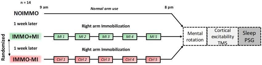
Figure 1. Experimental design. Participants underwent three experimental conditions, separated by
one week each, including one normal 11-h awake period without immobilization (NOIMMO), 11 h of
arm immobilization with MI treatment (IMMO+MI condition; including 5 MI sessions), and a similar
immobilization period without MI (IMMO−MI condition; including 5 control task sessions). Order of
IMMO+MI and IMMO-MI was randomized. After each experimental condition, we assessed changes in
sensorimotor representations (hand laterality judgment task) and cortical excitability (TMS) over both M1s.
PSG data were recorded during the night following each daytime condition.
limb disuse. One main aim of the present study was precisely to further clarify whether MI practice may alleviate
deleterious effects caused by short-term immobilization.
The effects of short-term immobilization have also been studied and observed in the sleeping brain. Nota-
bly, after one day with 12 h of immobilization, Huber et al.2 observed a decrease of motor performance and of
motor evoked potentials over the contralateral sensorimotor cortex, but also a reduction of slow wave activity
(1–4 Hz) and spindles activity (13–14 Hz) over M1 contralateral to the immobilized arm. The latter effects
predominated during the first NREM sleep period (< 1 h) immediately following the immobilization period.
The authors proposed that these local changes in sleep oscillations after immobilization may be linked to brain
plasticity processes, mirroring those induced after motor l earning2,23,24. While, it has been established that motor
learning consolidation also occurred after M I25,26, changes in plasticity-related sleep features following MI are
still unknown. Debarnot et al.27 emphasized the importance of NREM sleep for effective MI consolidation,
which in case of immobilization might compensate the reduction of NREM features over M1 contralateral to
the immobilized arm. Besides the need to confirm the immobilization-dependent neuroplasticity during sleep,
experimental limb immobilization provides a useful model to uncover the effects of MI practice on motor-related
plasticity mechanisms during wakefulness and during subsequent sleep.
In the present study, we used a randomized crossover within-subjects design to investigate whether MI train-
ing during arm-immobilization could counteract the adverse behavioral and neural effects observed after the
immobilization period, as well as during subsequent sleep. We expected that MI practice during arm-immobili-
zation (as compared to an immobilization condition without MI) would contribute to preserve (1) sensorimotor
representation of the immobilized limb, (2) cortical excitability over M1 regions, and (3) subsequent plasticity-
related sleep features over M1.
Materials and methods
General design. Fourteen healthy participants (mean age ± SD: 25.43 ± 2.56, nine women) were tested after
three different daytime conditions, each separated by 1 week (Fig. 1). All participants first spent 11 h of nor-
mal daily routine (i.e. NOIMMO condition). Then, they underwent the two remaining conditions with their
right, dominant arm immobilized during 11 h, either with concomitant MI treatment (15 min each two hours;
IMMO+MI condition), or without MI but a cognitive control task (i.e. dice game), with the same time sched-
ule (IMMO−MI condition). The order of the IMMO+MI and IMMO−MI conditions was pseudo-randomized
across participants (7 participants IMMO+MI first, 7 participants IMMO-MI first). At the end of each of the
11-h period (NOIMMO, IMMO+MI, IMMO−MI), we tested all participants for their sensorimotor representa-
tions of both hands using a hand laterality judgment task, cortical excitability for both M1s by means of transcra-
nial magnetic stimulation (TMS), and sleep features using polysomnography (PSG) recordings.
Participants. To be eligible for the study, participants were all right-handed as determined by the Edinburgh
Handedness Inventory28 (cutoff > 40 right-handedness; mean ± standard deviation [SD]: 78.73 ± 17.31), and had
no previous history of orthopedic problems with the right hand and arm. They were good sleepers as ensured
by the Pittsburgh Sleep Quality Index29 (cut-off ≤ 5; mean ± [SD]: 3.50 ± 1.91), and exhibited an intermediate
circadian-type as assessed by the Horne–Ostberg Morningness–Eveningness Questionnaire30 (range from 42 to
58; mean ± [SD]: 52.50 ± 5.47). To avoid excessive daytime sleepiness during the 11 h of each experimental condi-
tion, participants had to score ≤ 7 on the Epworth daytime sleepiness s cale31 (mean ± [SD]: 4.92 ± 1.89). Motor
imagery capacities were assessed using the Movement Imagery Questionnaire-3 (MIQ-3)32, which consists of
12 movements to perform mentally using three MI modalities (internal visual imagery, external visual imagery,
kinesthetic imagery). Participants were included with a total MI ability score ≥ 63 corresponding to 75% of the
maximum score; MIQ-332. Finally, participants had to score ≤ 9 on the Beck depression scale33 (normal range
Scientific Reports | (2021) 11:8928 | https://doi.org/10.1038/s41598-021-88142-6 2
Vol:.(1234567890)www.nature.com/scientificreports/
0–9; mean ± [SD]: 3.35 ± 2.64). Exclusion criteria included (1) the presence of ferromagnetic metallic implants or
a pacemaker, (2) previous neurosurgery, and (3) history of seizures, major head trauma, alcoholism, drug addic-
tion, or any other psychiatric, neurological disorders. All participants were instructed to be alcohol and caffeine
free for 24 h prior to and during the experimental days. The protocol was carried out in accordance with ethical
standards and was approved by the ethical committee of the Geneva University Hospital. Once the procedure
had been fully explained, participants gave written informed consent before inclusion. All participants received
an attendance fee.
Immobilization procedure. During the immobilization conditions, participants were requested to not
move the right arm (from the fingers to the shoulder) for 11 h from the morning (9 am) until the beginning
of the hand laterality judgment (8 pm). The duration of the sensorimotor deprivation was chosen based on
recent reports demonstrating modifications of M1s excitability34, with additional local changes over sensorimo-
tor regions during the subsequent night of s leep2. Here, we used a silk glove to reduce the contact between each
finger. We further put on a splint, typically used in clinical practice (FERULA, Malleable aluminum thumb-hand
right, model “OM6101D/2”), to ensure a complete immobilization of the wrist, as well as the carpometacarpal
and metacarpophalangeal joints of all fingers. In addition, a soft shoulder and elbow splint was used to support
the forearm in a comfortable way during the 11-h immobilization (DONJOY model IMMO, “DGO GLOBAL”).
To ensure that immobilization was effective, we quantitatively monitored physical activity of both arms dur-
ing the three experimental conditions, using actimetry (Actigraph GT3X-BT, Pensacola, Florida, USA) on the
participants’ left and right forearms. Data were sampled at 30 Hz. Energy cost of physical activity was calculated
in metabolic equivalent of task (MET), i.e. one MET equals the resting metabolic rate obtained during quiet
sitting35. We also verified that all participants maintained a regular sleep–wake schedule with a minimum of 7 h
of sleep per night, at least three days before each experimental day. Compliance to the schedule was assessed
using both sleep diaries and wrist actigraphy measures. Finally, every hour during the three experimental days
(i.e. 11 times for each experimental day), all participants assessed their current state of alertness by filling out
the Stanford Sleepiness S cale36.
Motor imagery sessions. During the IMMO+MI condition, participants performed 5 sessions (one ses-
sion every two hours, 15 min each) of explicit mental training with the immobilized arm, in a quiet room,
without any distracting stimuli in order to help them to focus on MI exercises. The content of MI exercises was
varied, and included monoarticular and polyarticular movements. Specifically, participants were requested to
mentally perform 15 different “monoarticular” movements (e.g. flexion/extension movements of the wrist) with
the right-immobilized arm during 1 min for each movement during the two first MI sessions. Then, during
the three next MI sessions, they performed five different and complex “polyarticular” movements (e.g. throw-
ing a ball) during 3 min each, i.e. each movement was performed 2 min first and then repeated again during
1 min in order to minimize mental fatigue resulting from maintaining focused attention. For each exercise, the
experimenter gave precise instructions about the movement to be performed and asked participants to either
sit or stand during MI in order to facilitate the postural congruency related to the movement (e.g. finger tap-
ping was performed in a seated position, performing arm circle was executed while standing up). If needed,
participants could first practice the movement with the non-immobilized arm prior to MI rehearsal with the
immobilized arm37. They were asked to imagine themselves performing movements using a combination of
visual and kinaesthetic imagery, i.e. imagining movement within one’s own body and perceiving the induced
proprioceptive sensations. They were instructed to say “go” when they started the mental simulation of move-
ment with the immobilized arm. The experimenter watched the participant continuously in order to check that
no physical movement with the immobilized arm was produced during MI, and used a stopwatch to indicate the
end of an exercise to the participant. After each MI exercise, participants were asked to rate the difficulty and the
quality of their conscious effort to produce mental images using two Likert-type scales (from 1 = very difficult/
poor mental representation to 5 = very easy/vivid mental representation).
Assessment of sensorimotor functions. Mental rotation tasks. After the 11-h immobilization or NO-
IMMO period, participants were administered two types of mental rotation tasks, i.e. a hand laterality judg-
ment task and an alphanumeric normal-mirror judgment task (control task). They sat on a chair at a distance
of 50 cm from a 17-inch computer screen with their hands on their knees. They were in a quiet room, without
any distracting stimuli, in order to help them to focus on the task. In the hand laterality judgment task, partici-
pants were asked to identify whether a hand stimulus presented visually on the screen was depicting a left or
right hand. Hand stimuli were depicting either simple, two dimensional views (dorsum or palm view with four
possible orientations: 0°, 90°, 180°, or 270°) or more complex, three dimensional views (palm from finger, dor-
sum from wrist, palm from wrist, or dorsum from finger). In the alphanumeric normal-mirror judgment task,
participants had to determine whether the letter “R” or number “2” were presented in a normal or mirror view.
Alphanumeric stimuli could be rotated by 0°, 90°, 180°, or 270°. Upright hand and alphanumeric stimuli had
13 cm in height and 7 cm in width.
At the beginning of each trial, a fixation cross was displayed (0.5 s) in the center of the screen. Then, the
stimulus was presented and remained visible during 2.5 s. Participants gave a verbal response (left/right in the
hand laterality judgment task; yes/no in the alphanumeric mental rotation task for normal or mirror image,
respectively). Verbal responses were recorded using a microphone connected to the computer. Response time was
defined by the time from stimulus onset to the beginning of the audio signal corresponding to the participants’
response. For each trial, the experimenter also collected response accuracy manually.
Scientific Reports | (2021) 11:8928 | https://doi.org/10.1038/s41598-021-88142-6 3
Vol.:(0123456789)www.nature.com/scientificreports/
During the NOIMMO condition, all participants familiarized with the hand laterality judgment task during
6 practice trials of hand stimuli. Right after, the actual test included four randomized blocks of hand laterality
judgment task (two blocks of two dimensional, and two blocks of three-dimensional items). Each block contained
16 trials with 8 left and 8 right hand pictures, presented in a random order. Then, participants familiarized with
the alphanumeric normal-mirror judgment task with 6 practice trials of alphanumeric stimuli (three letters
and three numbers) before performing two blocks of 16 trials with 8 normal and 8 mirror alphanumeric items.
The hand laterality judgment task was always performed before the alphanumeric mental rotation task, as
Toussaint and M eugnot8 demonstrated that the effects induced by arm immobilization may be reduced when
participants first perform a non-body mental rotation task. During each condition (NOIMMO, IMMO+MI and
IMMO−MI), participants thus completed a total of 64 trials of hand laterality judgment task followed by 32 trials
of alphanumeric normal-mirror judgment task. During the IMMO+MI and IMMO−MI conditions, participants
were no longer familiarized with the tests.
Stimulus presentation, data recording and analysis were handled by a homemade MATLAB program (Version
2009b. Natick, MA: The MathWorks Inc.), incorporating the P sychtoolbox38.
Transcranial magnetic stimulation. We used TMS to examine cortical excitability based on the motor evo-
cated potential (MEP) over the left M1 (contralateral to the immobilized-arm) and then right M1 (ipsilateral
to the immobilized-arm). Such as previous TMS studies conducted on arm-immobilization effect3,4,10, cortical
excitability was assessed by means of resting motor threshold (RMT) and recruitment curve (RC) after each
experimental condition. While the RMT corresponds to the excitability of a central core region of neurons,
the MEP amplitude is assumed to represent an index of both synaptic and postsynaptic a ctivity39,40. A refined
measure of cortical excitability can thus be obtained by recording stimulus–response curves of MEPs represent-
ing the input–output function of M141. The recruitment curves of MEPs are generated by plotting the amplitude
of MEPS relative to the stimulus intensity, and have been suggested as the most sensitive assessment of motor
system excitability42. A figure-of-eight coil with wing diameters of 70 mm, connected to a Magstim 200 magnetic
stimulator (Magstim, Whitland, Dyfed, UK) was placed tangentially on the scalp over M1; the handle pointed
backward and laterally at a 45° angle to the sagittal plane inducing a posteroanterior current in the brain. First,
we identified the optimal spot for eliciting motor-evoked potentials (MEPs) in the right first dorsal interosseous
(FDI) muscle and the location was marked on an cap fitted on the participant’s head to provide a reference land-
mark for each TMS session. Additionally, motor cortices marks over the cap were duplicated on the scalp of par-
ticipants to attach electroencephalographic (EEG) electrodes for subsequent sleep polysomnographic recording.
The RMT was defined as the minimal TMS intensity required to evoke MEPs of about 50 μV peak-to-peak in
the targeted muscle in 5 out of 10 consecutive trials and was expressed as a percentage of maximum stimulator
output (MSO). Once we found the RMT over left M1, RC was assessed by measuring peak-to-peak amplitude
(expressed in μV) of MEPs elicited at stimulus intensities of 5%, 10%, 15%, 20%, and 25% of MSO above individ-
ual RMT (obtained on each experimental day). Five trials were recorded at each intensity, and MEP amplitude
was then averaged. The same RMT and RC procedure was then performed over right M1.
Electromyography recording. Electromyography (EMG) was recorded and monitored continuously on-line
during each TMS session. EMG recordings used the following montage: active electrodes were attached to the
skin overlying first dorsal interosseous muscle, reference electrodes were placed over the first metacarpophalan-
geal joints and ground electrodes were placed over the wrist bone. Acknowledge 4 software along with MP36
unit (Biopac systems, Goleta, CA, USA) were used to acquire EMG signal. The EMG signals were amplified and
filtered online (20 Hz to 1 kHz), sampled at a rate of 5000 Hz and stored on a personal computer for display and
later offline data analysis.
Polysomnographic recording. Data acquisition. Polysomnography was acquired in all participants during
the night immediately following NOIMMO, IMMO+MI and IMMO−MI conditions, with a V-Amp 16 system
(Brain Products, Gilching, Germany), from 11 EEG electrodes (international 10–20 system: LM1, CZ, RM1,
F3, FZ, F4, P3, PZ, P4, A1, A2), three electrooculogram electrodes, and two chin electromyogram electrodes
(sampling rate: 250 Hz).
Sleep data analysis. Out of the 14 participants, three participants were excluded from spindles and spectral
power analysis due to poor EEG signal quality (on one of the nights), while sleep stage scoring was still possible
for these participants for all three experimental nights (n = 14).
For sleep analyses, we used the PRANA software (version 10.1. Strasbourg, France: Phitools) and custom
MATLAB scripts. Two scorers blind to the experimental conditions determined the different sleep stages (NREM
1, 2, 3, REM sleep and wake) for each recorded night of sleep on 30-s epochs according to the AASM standards43.
From the sleep scoring, we computed the total time spent (min) in each sleep stage, as well as the percentage of
each sleep stage relative to the total sleep period (TSP; from sleep onset to wake up time) and relative to the total
sleep time (TST; TSP minus intra-wake epochs). Sleep efficiency (TST/time in bed*100), and number of intra-
awakenings were also calculated. Epochs affected by artefacts were detected using a semi-automatic detection
(PRANA software), and then verified visually by the two expert scorers. We performed two independent repeated
measures analysis of variance ( ANOVARM) with condition as within-subjects factor, which did not show any dif-
ference on artefact duration during the first cycle of sleep (F(2, 26) = 0.68, p = 0.51, ηp2 = 0.05), and the whole night
of sleep (F(2, 26) = 1.68, p = 0.20, ηp2 = 0.11). The corresponding data points were removed from subsequent EEG
analyses. The EEG spectrum power average was calculated with a 0.2 Hz resolution, by applying a Fast Fourier
Transform (FFT; 50% overlapping, 5 s windows, Hanning filter) on the left and right M1 channels. Mean power
Scientific Reports | (2021) 11:8928 | https://doi.org/10.1038/s41598-021-88142-6 4
Vol:.(1234567890)www.nature.com/scientificreports/
was calculated for the following frequency bands: slow oscillations (0.4–1 Hz), delta (1–4 Hz), theta (4–8 Hz),
alpha (8–12 Hz), sigma or spindle power range (12–15 Hz), and beta (15–30 Hz). The detection of sleep spindles
was performed automatically with the PRANA software on each channel following the global standards used in
sleep research44–46: spindles were required to have a duration between 0.5 and 3 s, an inter-spindle interval no
less than 1 s, and a typical, fusiform spindle morphology (waxing and waning amplitude). An experienced scorer
visually supervised all detected spindles over the left and right M1 in each night of each participant.
Data analysis. For the hand laterality judgment task, we analyzed the percentage of correct responses and
mean response time. Only data from correct responses were used to analyze response times. Results obtained on
the hand laterality judgment for the three conditions were evaluated by means of repeated measures analysis of
variance ANOVArm with condition (NOIMMO, IMMO+MI and IMMO−MI), laterality (left, right) and
configuration (two or three dimensional) as within-subjects factors. Likewise, an ANOVArm was performed
for the alphanumeric normal-mirror judgment task, with condition (NOIMMO, IMMO+MI and IMMO−MI),
configuration (normal, mirror) and orientation (0°, 90°, 180°, or 270°) as within-subjects factors.
To evaluate the effects of immobilization on cortical excitability, we compared the resting motor threshold
(RMT) using an ANOVArm with laterality (left M1, right M1) and condition (NOIMMO, IMMO+MI and
IMMO−MI) as within-subject factors. Recruitment curve (RC) values were analyzed by means of an ANOVArm
with laterality (left M1, right M1), condition (NOIMMO, IMMO+MI and IMMO−MI) and intensity (5%,
10%, 15%, 20%, and 25% of MSO above RMT) as within-subjects factors. We then evaluated the differences
between 3 regression slopes using an ANOVArm with condition (NOIMMO, IMMO+MI and IMMO−MI)
and laterality (left M1 and right M1) as within-subjects factors.
PSG parameters (sleep latencies, N1, N2, N3 and REM data) did not distribute normally (Shapiro-Wilks W
test), and were log-transformed prior to statistical analysis. To examine whether daytime conditions (NOIMMO,
IMMO+MI and IMMO−MI) influenced sleep architecture, we performed ANOVArms with sleep stage dura-
tion (min; N1, N2, N3, REM) and condition (NOIMMO, IMMO+MI and IMMO−MI) as within-subjects
factors. Similar ANOVArms were performed with total sleep period (%), sleep latencies and efficiency (min
and % respectively). We also analysed spindle features (number, density, duration, peak amplitude and mean
frequency) by means of ANOVArms with electrode location (left M1, Right M1) and condition as within-
subjects factors. Similar analyses were performed on power spectrum measurements with electrode location
and condition as within-subjects factors. All sleep analyses (sleep architecture, spindles features and spectral
power data) were conducted separately during whole night of sleep and the first sleep cycle.
We conducted correlation analyses by computing data from the multimodal assessment obtained in each
condition to address the effect of MI treatment [IMMO+MI vs IMMO−MI] and the impact of immobilization
[NOIMMO vs IMMO−MI]. Spearman’s correlations were performed between the changes observed in sleep
architecture (i.e. REM duration) or spindles density, on the one hand, and the sensorimotor representation of
hands (i.e. RTs) on the corticospinal excitability (i.e. MEP slope), on the other hand.
Both the Stanford Sleepiness Scale and MET scores for the NOIMMO, IMMO+MI, IMMO−MI conditions
were compared using ANOVArms. We also performed ANOVArms on the individual ratings concerning the
difficulty and quality of MI during each training session.
Whenever interactions were significant, the origin of the interaction was investigated using planned com-
parisons with Bonferroni-corrected post-hoc tests. The alpha-level of all analyses was set at p < 0.05.
Results
Preliminary note. The mean score of the Stanford Sleepiness Scale assessed at 8 pm (i.e. before starting
the multimodal assessment) did not differ as a function of conditions (mean ± SD; NOIMMO: 1.92 ± 0.64,
IMMO+MI: 1.83 ± 0.55, IMMO−MI: 2.23 ± 0.89). Actigraphy data confirmed that all participants complied
with the instructions regarding the right-arm immobilization procedure. A ANOVArm on the MET (i.e. meta-
bolic equivalent of task) scores revealed main effects of condition (NOIMMO, IMMO+MI, IMMO−MI); F(2,
52) = 37.99, p < 0.001, ηp2 = 0.59) and hand (Right, Left; F(1, 26) = 135.29, p < 0.001, ηp2 = 0.83), as well as a sig-
nificant condition x hand interaction (F(2, 52) = 70.21, p < 0.001, ηp2 = 0.73) (Fig. S1). Bonferroni post-hoc
tests showed that during the NOIMMO condition, physical activity with the right arm was higher relative to
the left (p < 0.01), in line with the subjects’ right-hand dominance. Importantly, activity of the right immobi-
lized arm significantly and equivalently decreased in both IMMO+MI (1.18 ± 0.10) and IMMO−MI (1.21 ± 0.07)
conditions compared to NOIMMO (2.21 ± 0.09; all p < 0.001), and was significantly lower compared to the left
arm activity (p < 0.001 for both IMMO+MI (1.98 ± 0.18) and IMMO−MI (1.94 ± 0.22) conditions). During both
IMMO+MI and IMMO−MI conditions, activity of the left non-immobilized arm did not differ from that in
NOIMMO (2.06 ± 0.22; p ≥ 0.32 for both comparisons), suggesting that the non-immobilized arm was not more
used than usual during right-arm disuse. To note, participants were asked to avoid intense/unusual sport activity
during daytime of the first experimental condition (NOIMMO), and they spent both IMMO conditions in the
laboratory doing daily life “student” activities, such as reading, working at the computer. Therefore, and as far as
the left arm activity is concerned, it is likely that participant activity during NOIMMO condition was equivalent
to that of IMMO. Regarding MI difficulty and quality during the 5 MI sessions, participants reported that it was
rather easy for them to perform the mental exercises (4.15 ± 0.12 over a maximum score of 5) and to reach vivid
imagery (3.87 ± 0.11 over a maximum of 5; p > 0.1 for all comparisons between MI sessions).
Mental rotation tasks. Hand laterality judgment. A ANOVArm performed on the percentage of correct
responses did not yield a main effect of condition (F(2,104) = 0.85, p = 0.43, ηp2 = 0.01), but showed a main effect
of configuration (two or three dimensional hand stimuli; F(1,52) = 10.21, p < 0.01, ηp2 = 0.16), a main effect of
Scientific Reports | (2021) 11:8928 | https://doi.org/10.1038/s41598-021-88142-6 5
Vol.:(0123456789)www.nature.com/scientificreports/
1.45
***
1.40
1.35
Response Time (s)
1.30
1.25
1.20
1.15
1.10
1.05
1.00
NOIMMO
Baseline IMMO–MI
Ctrl IMMO+MI
MI
Figure 2. Response times during the hand laterality judgment task. Independently of hand laterality, response
times after the IMMO+MI condition was significantly faster compared to NOIMMO and IMMO−MI
conditions. Error bars indicate the standard error of the mean. ***p < .001.
laterality (right or left; F(1,52) = 11.87, p < 0.01, ηp2 = 0.18), and a significant configuration × laterality
interaction (F(1,52) = 4.32, p < 0.05, ηp2 = 0.07; Table S1). Post-hoc comparisons revealed that performance was
better for the 3D (95.31% ± 1.81) compared to the 2D (92.11% ± 1.99) configurations, and that left hand stimuli
(96.14% ± 1.71) were more correctly identified compared to the right hand ones (91.96% ± 2.09).
A similar ANOVArm on response times yielded main effects configuration (F(1,52) = 4.54, p < 0.05,
ηp2 = 0.08) and condition (F(2,104) = 24.05, p < 0.001, ηp2 = 0.32), but no effect of laterality (F(1,52) = 0.45,
p = 0.50, ηp2 = 0.01), and no interaction. Bonferroni post-hoc tests revealed that participants were faster at rec-
ognizing 3D (vs. 2D; p < 0.05) configurations, and also both left and right hand stimuli after the IMMO+MI
condition compared to NOIMMO and IMMO−MI conditions (p < 0.001 for each condition comparison with
IMMO+MI; Fig. 2), while there was no difference between NOIMMO and IMMO−MI (p = 0.69). In other words,
we expected that if MI training would be effective in preserving sensorimotor functions, we would observe a
reduction of RTs, as compared to the NOIMMO condition, because the latter was always performed before any
of the immobilization conditions. Therefore, the present results support that MI training (compared to no MI)
during immobilization induced a preservation of sensorimotor representation of the immobilized limb.
Alphanumeric normal‑mirror judgment task (control task). A ANOVArm on the percentage of correct responses
for the control mental rotation task revealed a significant effect of configuration (F(1,26) = 48.71, p < 0.0001,
ηp2 = 0.65) because participants were more accurate for the normal (94%) rather than the mirror (72%) con-
figuration of stimuli. The ANOVA also yielded a condition x configuration interaction (F(2,52) = 3.88.71,
p < 0.0001, ηp2 = 0.65), and post-hoc analyses showed that normal stimuli were better identified after the
IMMO+MI compared to the NOIMMO (p < 0.05), but not compared to the IMMO−MI condition (p = 0.20),
while performance on mirror stimuli did not show such a modulation by IMMO+MI condition (p < 0.08 vs.
NOIMMO and IMMO−MI conditions).
A similar ANOVArm on response times showed a main effect of configuration (F(1,26) = 4.55, p < 0.05,
ηp2 = 0.15), as well as a main effect of condition (F(2,52) = 4.36, p < 0.01, ηp2 = 0.14), but no condition x con-
figuration interaction (F(2,52) = 0.28, p = 0.75, ηp2 = 0.01). Post-hoc tests indicated faster responses for normal
(vs. mirror) configurations in both IMMO+MI and IMMO−MI conditions compared to the NOIMMO (p < 0.01
and p < 0.05, respectively).
Transcranial magnetic stimulation. One participant was excluded from TMS analysis due to very high
resting motor threshold (RMT > 80% machine output).
ANOVArm on the resting motor threshold (RMT) in both M1s showed a main condition effect
(F(2,48) = 35.90, p < 0.01, ηp2 = 0.15), and post-hoc analyses revealed a lower RMT in the NOIMMO condi-
tion over the left M1 compared to the IMMO+MI (p < 0.05; Table S2). Comparing the motor evoked potential
(MEP), obtained by means of recruitment curve, between conditions (Fig. 3A,B) revealed a main effect of
intensity (F(4,120) = 49.73, p < 0.001, ηp2 = 0.62), as well as a significant condition x laterality interac-
tion (F(2,240) = 21.49, p < 0.001, ηp2 = 0.15). Post-hoc analyses showed that MEP amplitude recorded after the
IMMO−MI condition was significantly lower than after NOIMMO (p < 0.001) and IMMO+MI (p < 0.01) condi-
tions. Most importantly, ANOVArm performed on MEP slope for both M1s showed a condition x laterality
interaction (F(2,48) = 3.05, p = 0.05, ηp2 = 0.11). Post-hoc tests indicated that immobilization reduced the slope
of left M1 after the IMMO−MI condition relative to NOIMMO (p < 0.05), without difference when comparing
the IMMO+MI and IMMO−MI conditions (Fig. 3C,D).
We used spearman’s correlation analyses to assess the possible relationship between changes in the corti-
cospinal excitability (i.e. MEP slope) with those at the sensorimotor representation level (i.e. RTs), but found no
significant correlation regarding the influence of MI treatment [IMMO+MI – IMMO−MI], or of immobilization
[NOIMMO – IMMO−MI].
Scientific Reports | (2021) 11:8928 | https://doi.org/10.1038/s41598-021-88142-6 6
Vol:.(1234567890)www.nature.com/scientificreports/
AA 500
RC
leC3M1
RCRCle M1 B 500
RC C4
RC right M1
(µV)
(µV)
400 400
(µv)
amplitude (µv)
amplitude(µV)
amplitude
amplitude
MEPamplitude
300 300
200 Baseline
NOIMMO 200 Baseline
NOIMMO
MEPMEP
MEPMEP
CTRL immo
IMMO‒MI CTRL immo
IMMO‒MI
100 100
MI immo
IMMO+MI MIIMMO+MI
immo
0 0
5% 10% 15% 20% 25% 5% 10% 15% 20% 25%
Smulus intensity
Smulus
Smulus (%(%
intensity(%
intensity MSO
MSOMSO above RMT)
aboveRMT)
above RMT) Smulus intensity (% MSO above RMT)
Smulus intensity (% MSO above RMT)
C le
C3 M1 Slope
Slope
D C4 Slope
right M1 Slope
0,09
O.09 * 0,09
O.09
0,08
O.08 0,08
O.08
RCslope
slope
RCRCslope
slope
0,07
O.07 0,07
O.07
RC
0,06
O.06 0,06
O.06
MEP
MEP
MEP
0,05 0,05
O.05
MEP
O.05
0,04
O.04 0,04
O.04
0,03
O.03 0,03
O.03
NOIMMO
Baseline IMMO‒MI
NoMI IMMO+MI
MI NOIMMO
Baseline IMMO‒MI
NoMI IMMO+MI
MI
Figure 3. Cortical excitability over left and right M1 after NOIMMO, IMMO−MI and IMMO+MI conditions.
(A) MEP amplitude from the left M1 at increasing strength of MSO intensity for the three conditions. (B) Slope
data showed a significant reduction in the cortical excitability over left M1 after immobilization alone (IMMO−
MI) relative to the NOIMMO condition, while MI practice limited the deleterious effect of immobilization. (C)
MEP amplitudes over right M1. (D) Slope data showed no significant differences over the right M1 as a function
of conditions. MEP, motor evoked potential; MSO, maximum stimulator output; M1, primary motor cortex; RC,
recruitment curve; RMT, resting motor threshold.
Polysomographic data. Sleep architecture. Participants showed overall normal sleep patterns across the
three PSG nights (Fig. 4A and Table S3). We first tested whether experimental conditions affected the sleep ar-
chitecture of the whole night recordings. An ANOVArm on stage duration (min) with sleep stage (N1, N2, N3,
REM) and condition (NOIMMO, IMMO+MI, IMMO−MI) as within-subjects factors showed an main effect
of sleep stage (F(3, 117) = 329.92 ; p < 0.001, ηp2 = 0.89), no overall effect of condition (F(2, 39) = 0.73 ; p = 0.48,
ηp2 = 0.03), or interaction (F(6, 117) = 0.75; p = 60). A similar pattern of results was obtained using percentages of
the total sleep period (TSP), while no other change in sleep latencies, total sleep time [TST], and sleep efficiency,
was found when comparing experimental conditions (all p > 0.05).
Because studies investigating the impact of learning on subsequent sleep reported effects predominating early
in the night sleep, most often during the first sleep cycle2,24, we performed the same analyses on the first sleep
cycle (Fig. 4B). Regarding stage duration (min), we again found the expected main effect of sleep stage (F(3,
117) = 130.38; p < 0.001, ηp2 = 0.77), no effect of condition (F(2, 39) = 0.99; p = 0.37, ηp2 = 0.04), but a significant
interaction between sleep stage and condition (F(6, 117) = 2.35; p = 0.03). This effect was due to an increase
in the time spent in REM after MI treatment (17 ± 2.97 min), as compared to NOIMMO (10.75 ± 1.80 min,
p = 0.01) and to IMMO−MI (10.57 ± 0.96 min, p = 0.01). No modulation by MI conditions arose for other sleep
parameters (see above, all p > 0.05).
We further examined the possible links between changes in the time spent in REM (min) during the first
sleep cycle with the corticospinal excitability (i.e. MEP slope), and the sensorimotor representation of hands (i.e.
RTs), and found no significant correlation regarding the effect of MI treatment [IMMO+MI – IMMO−MI], or
of immobilization [NOIMMO – IMMO−MI].
Sleep spindles during NREM sleep. Because sleep spindles are known to be modulated by prior learning and
experience45,47, including local effects after motor adaptation and limb immobilization2,48, we tested whether
spindles may also be affected in our experiment. We first looked at the total number of spindles detected over left
and right M1 during NREM sleep (N2+N3) from the whole sleep night (Table S4). An ANOVArm with elec-
trode location (left M1, right M1) and condition (NOIMMO, IMMO+MI, IMMO−MI) as within-subjects
factors revealed a main effect of condition (F(2, 40) = 18.73; p < 0.001, ηp2 = 0.48), but no effect of electrode
location (F(1, 20) = 0.29; p = 0.59, ηp2 = 0.01), or interaction (F(2, 40) = 0.51; p = 0.60, ηp2 = 0.02). To clarify the
Scientific Reports | (2021) 11:8928 | https://doi.org/10.1038/s41598-021-88142-6 7
Vol.:(0123456789)www.nature.com/scientificreports/
WHOLE NIGHT FIRST SLEEP CYCLE
A B 60
200
NOIMMO NOIMMO
IMMO–MI 50 IMMO–MI
Sleep stage (min)
Sleep stage (min)
150 IMMO+MI IMMO+MI
40
100 30
**
20 **
50
10
0 0
Wake N1 N2 N3 REM Wake N1 N2 N3 REM
C 3
D 3
M1s spindle density (nbr/min)
M1s spindle density (nbr/min)
** **
2
*** *** 2
1 1
0 0
NOIMMO
Baseline IMMO‒MI
NoMI IMMO+MI
MI NOIMMO
Baseline IMMO‒MI
NoMI IMMO+MI
MI
Figure 4. Sleep architecture and spindles density over both M1s during the whole night and the first sleep cycle
after each condition (NOIMMO, IMMO−MI, IMMO+MI). (A) Sleep stage duration (min) for each condition
during the whole night, and (B) during the first sleep stage. REM sleep duration increased in the IMMO+MI
condition relative to NOIMMO and IMMO−MI. (C) Reduced spindle density (nbr/min) over the left and right
M1 in the IMMO−MI compared to NOIMMO and IMMO+MI conditions during the whole night, and (D)
during the first sleep cycle.
main effect of condition, we performed Bonferroni post-hoc comparisons and showed that compared to the
number of spindles was lower during IMMO−MI condition (372.79 ± 24.32; p < 0.001) compared to NOIMMO
(519.66 ± 17.61) and IMMO+MI conditions (431.17; p < 0.01). Likewise, an ANOVArm on spindle density meas-
ures revealed a main effect of condition (F(2, 40) = 15.86; p < 0.001, ηp2 = 0.44), no effect of electrode loca-
tion (F(1, 20) = 0.23; p = 0.63, ηp2 = 0.01) and no interaction, F(2, 40) = 1.02; p = 0.36, ηp2 = 0.04). Bonferroni post-
hoc tests showed a decreased density of spindles after the IMMO−MI condition relative to the NOIMMO and
IMMO+MI conditions (all comparison p < 0.001; Fig. 4C). Noteworthy, there was no difference in the density of
spindles between NOIMMO and IMMO+MI conditions over both M1s (p = 0.70).
Huber et al.2 reported that spindle activity was locally modulated (here decreased) following 12 h of arm-
immobilization and, in line with several studies looking at the effects of prior learning on sleep spindles45,49, they
found that this modulation occurred at the beginning of the sleep period. Thus, and like we did for the sleep
architecture above, we analyzed the number and density of sleep spindles during the first NREM period, again
distinguishing between electrodes over the left and right M1 (Table S5). For the total number of sleep spindles
(during N2+N3), we found a significant main effect of condition (F(2, 40) = 17.07; p < 0.001, ηp2 = 0.46), no effect
of electrode location (F(1, 20) = 0.31; p = 0.58, ηp2 = 0.01) or interaction (F(2, 40) = 0.66; p = 0.52, ηp2 = 0.03).
We observed similar effects for density measures (condition, F(2, 40) = 12.30; p < 0.001, ηp2 = 0.38; electrode
location, F(1, 20) = 0.21; p = 0.64, ηp2 = 0.01; interaction, F(2, 40) = 1.20; p = 0.31, ηp2 = 0.05). Bonferroni post-
hoc tests revealed a decreased number of spindles after the IMMO−MI condition relative to the NOIMMO and
IMMO+MI conditions for both the number and density of spindles (all comparison p < 0.01; Fig. 4D). Impor-
tantly, there was no difference in the number or density of sleep spindles over both M1s between NOIMMO
and IMMO+MI conditions (p = 0.17 and p = 0.85, respectively). We further analysed the potential relationship
between changes in the density of sleep spindles (whole night and first sleep cycle) with the corticospinal excit-
ability (i.e. MEP slope), and the sensorimotor representation of hands (i.e. RTs). No significant correlations
arose from these analyses.
EEG spectral analysis. Two separate ANOVArms on the whole night and first sleep cycle were performed with
frequency bands data (slow oscillations, delta, theta, alpha, sigma, spindle power range, and beta), electrode
location (left M1, right M1) and condition (NOIMMO, IMMO+MI, IMMO−MI) as within-subjects factors.
All analyses yielded a main effect of frequency bands (all p < 0.001, as expected for different sleep stages), but
no effect of condition (all p ≥ 0.26), electrode location (all p ≥ 0.57), or interaction (all p ≥ 0.82).
Scientific Reports | (2021) 11:8928 | https://doi.org/10.1038/s41598-021-88142-6 8
Vol:.(1234567890)www.nature.com/scientificreports/
Discussion
We designed the present study to investigate the effects of MI practice administered during 11 h of arm-immo-
bilization on the sensorimotor and cortical representations of both hands, as well as on sleep features. We found
that MI practice during unilateral immobilization contributed to preserve sensorimotor representation and
cortical excitability over left M1 contralateral to the arm-immobilization. Furthermore, the time spent in REM
sleep was significantly longer particularly during the first cycle of sleep following the IMMO+MI condition
compared to that after NOIMMO and IMMO−MI conditions. Finally, data revealed that, while immobilization
decreased the number of sleep spindles compared to the condition without immobilization, this decrease was
no longer significant after the IMMO+MI condition over both M1s.
Effect of MI practice on the sensorimotor representation of hands. The first important finding
of the present study is that hand laterality judgment performance improved after the IMMO+MI but not after
the IMMO−MI condition, for both right-and-left hand stimuli. The lack of task-repetition benefit after the
IMMO−MI condition for both right and left hand stimuli confirms and extends previous findings by Toussaint
et al.8 obtained after 48 h of non-dominant hand immobilization. By contrast, the same authors further reported
that only 24 h of non-dominant left hand immobilization induced an effector-dependent effect as reflected by
task-repetition benefit for right hand stimuli (non-immobilized hand), but not for left hand stimuli. Here, we
found an effector-independent effect of immobilization following only 11 h of right (dominant) arm immobi-
lization, which might be explained by differences in the immobilization paradigms. Here, we choose dominant
arm immobilization as higher effect on sensorimotor representation has been observed rather than with non-
dominant limb immobilization50. Moreover, immobilization of the entire upper limb, from the shoulder to the
fingers (only the wrist and three fingers were immobilized in Toussaint et al., 2013) might have extended the
somatotopic regions impacted by immobilization. Therefore, the dominance of handedness as well as the extent
of upper-limb immobilization might have elicited more profound changes to organising features of the body
map, which may explain why we could observe such effector-independent effect on the sensorimotor represen-
tation after a relatively short period of immobilization. Importantly, performance gains observed following the
IMMO+MI condition further support previous data showing that 15 min of kinesthetic MI (i.e. hand and finger
movements using bodily information) performed right before splint removal following 24 h of left hand immo-
bilization, contributed to faster discrimination of left hand s timuli21. In our study, participants were involved in
more intensive MI practice (75 min vs. 15 min), with the last MI session performed 1 h before the splint removal,
thus supporting that the present MI regime may have not only transiently activated sensorimotor processing, but
also promoted its maintenance at a normal functioning level. Interestingly, a recent neuroimaging study showed
that 24 h of hand immobilization, without MI practice sessions, decreased the neural activation underpinning
MI, specifically over the sensorimotor areas contralateral to the limb d isused5. Taken together, these obser-
vations suggest that reactivation of the sensorimotor system by means of MI practice during immobilization
contributed to prevent the maladaptive functional consequences of immobilization. This issue is of particular
importance in the clinical context where MI is increasingly used to reactivate injured sensorimotor networks to
assist the recovery of lost motor functions16,51,52.
Effect of MI practice on corticomotor excitability. A second main result of the present study is that
MI prevented the decrease in cortical excitability of left M1 caused by the contralateral arm-immobilization. We
found a significant reduction in the slope of RC data after the IMMO−MI condition, plausibly reflecting local
synaptic depression induced by unilateral short-term immobilization2,4,10,34. Such downregulation of left M1
excitability was not observed after the IMMO+MI condition. This finding challenges data by Bassolino et al.3
and Crew et al.22 who reported that MI training could not prevent corticomotor depression after upper-limb
immobilization. Such discrepancy may be due to differences in the nature and dosage of the MI intervention.
Unlike the simple arm/hand movements used in previous studies, here the experimenter guided participants
to mentally rehearse movements using different upper-limb joints, i.e., wrist, elbow and shoulder, and the MI
program was structured with a regular increment in task complexity, from simple unilateral movements up
to bilateral upper-limb movements. Thus, this thorough and progressive MI training design emphasized the
voluntary and effortful engagement of complex motor representations. Moreover, instead of an interactive and
tailored guidance, previous studies used audiotaped guidelines or visual stimuli displayed on a computer screen.
Crucially, the overall time spent to practice MI was significantly longer in the present study (i.e., 75 min vs. 40
min3 and 3 × 30 min across 3 successive days22. We thus speculate that a relatively intensive MI intervention
might be needed to promote significant M1s activation, hence protecting against loss of sensorimotor functions
during limb immobilization. This latter assumption is also supported by reports in the motor learning domain
showing that MI increases M1 excitability and that such increase is specific to the representation of the limb
involved during imagery53,54.
Effect of MI practice on sleep. A third original aim of the present study was to investigate whether and
how MI practice during immobilization influenced the following sleep period. We first observed an increase
in the time spent in REM during the first sleep cycle, consistent with sleep changes occurring predominantly
early in the night sleep2,23,24. Motor circuits are known to be activated during REM sleep55–57; this activation may
support MI in dreams and the reactivation of motor skills learning58–66, although a causal role of REM sleep for
motor learning is still debated67. Following this line of reasoning, here we suggest that practicing MI exercises
during arm-immobilization might have potentiated the demand for REM sleep related to motor rehearsal and
consolidation, yielding longer REM duration. However, whether increased REM sleep after MI induced a better
Scientific Reports | (2021) 11:8928 | https://doi.org/10.1038/s41598-021-88142-6 9
Vol.:(0123456789)www.nature.com/scientificreports/
recovery of motor skills (i.e. motor test after the night sleep) was not tested in the present study and would thus
require further investigation.
As expected, we found that 11 h of immobilization decreased the density of spindles, not only over the
contralateral M1, but over both M1s after the IMMO−MI c ondition2. Importantly, spindle density over motor
regions after the IMMO+MI condition was unchanged as compared to NOIMMO, hence supporting that MI
practice might have protected or maintained spindles activity following day-time immobilization. In line with
this hypothesis, Debarnot et al. (2011) reported similar MI performance gains after both a short (20 min NREM)
and long (NREM and REM) intervening naps (compared to wake period), suggesting that NREM sleep, and
particularly spindles activity may have contributed to the MI consolidation process. Altogether, these findings
are in accordance with the critical role of sleep spindles in procedural memory reactivation and consolidation
during NREM s leep45,47.
To conclude, the present results provide strong evidence that MI practice can alleviate the impact of arm-
immobilization on sensorimotor representations, and motor cortex excitability. Our findings also reveal lasting
effects of MI practice on subsequent REM sleep (longer REM duration) and NREM sleep (limitation of the
reduction in spindle density). Overall, MI contributed to maintain sensorimotor networks activation during
immobilization, hence protecting from the maladaptive neuroplasticity occurring during and after immobiliza-
tion. Lately, Newbold et al. (2020) reported that spontaneous pulses of activity propagate through the contralat-
eral somatomotor subcircuit, following 12 h arm-immobilization, while a loss of strength and fine motor skill
abilities was observed after 14 days of disuse68. The authors suggested that spontaneous pulses might play a role
in maintaining neuronal functional integrity early after immobilization-induced plasticity. Based on this observa-
tion, we may speculate that the early reactivation of the motor network with MI practice during immobilization
might delay or prevent the occurrence of such spontaneous pulses of activity, and/or subsequent detrimental
long-term effects. Furthermore, Clark et al. (2014) reported that MI strength training (i.e. maximal contractions
of the wrist muscles during two weeks) during wrist immobilization, contributed to limit the loss of w eakness19.
Thus, the type of MI exercises (here functional vs. Clark et al. strength) likely influences which dimension of
motor functioning in long-term immobilization. Future studies should examine the effect of MI training that
includes both strength and functional exercises, on motor skill capacities following long-term immobilization.
Finally, one of the main findings in our experiment is the effectiveness of MI on early neuroplasticity, i.e. when
delivered soon after immobilization. The optimal timing for MI delivery is still debated in the context of post-
stroke rehabilitation69, while another issue is the inclusion of stroke patients who may benefit from multiple
brain computer interface-based interventions (BCI)70. Based on our results, we suggest that it is plausible that
the sooner the patient gets MI practice (in post-stroke subacute state), the greater are chances to reactivate and
preserve motor functions, which consequently could enable patients to benefit most from BCI procedures.
Received: 1 December 2020; Accepted: 30 March 2021
References
1. Furlan, L., Conforto, A. B., Cohen, L. G. & Sterr, A. Upper limb immobilisation: A Neural plasticity model with relevance to
poststroke motor rehabilitation. Neural. Plast. 2016, 8176217. https://doi.org/10.1155/2016/8176217 (2016).
2. Huber, R. et al. Arm immobilization causes cortical plastic changes and locally decreases sleep slow wave activity. Nat. Neurosci.
9, 1169–1176. https://doi.org/10.1038/nn1758 (2006).
3. Bassolino, M., Campanella, M., Bove, M., Pozzo, T. & Fadiga, L. Training the motor cortex by observing the actions of others
during immobilization. Cereb. Cortex 24, 3268–3276. https://doi.org/10.1093/cercor/bht190 (2014).
4. Avanzino, L., Bassolino, M., Pozzo, T. & Bove, M. Use-dependent hemispheric balance. J. Neurosci. 31, 3423–3428. https://doi.org/
10.1523/jneurosci.4893-10.2011 (2011).
5. Burianova, H. et al. Adaptive motor imagery: A multimodal study of immobilization-induced brain plasticity. Cereb. Cortex 26,
1072–1080. https://doi.org/10.1093/cercor/bhu287 (2016).
6. Meugnot, A., Almecija, Y. & Toussaint, L. The embodied nature of motor imagery processes highlighted by short-term limb
immobilization. Exp. Psychol. 61, 180–186. https://doi.org/10.1027/1618-3169/a000237 (2014).
7. Debarnot, U., Huber, C., Guillot, A. & Schwartz, S. Sensorimotor representation and functional motor changes following short-
term arm immobilization. Behav. Neurosci. 132, 595–603. https://doi.org/10.1037/bne0000274 (2018).
8. Toussaint, L. & Meugnot, A. Short-term limb immobilization affects cognitive motor processes. J. Exp. Psychol. Learn. Mem. Cogn.
39, 623–632. https://doi.org/10.1037/a0028942 (2013).
9. Lissek, S. et al. Immobilization impairs tactile perception and shrinks somatosensory cortical maps. Curr. Biol. 19, 837–842. https://
doi.org/10.1016/j.cub.2009.03.065 (2009).
10. Avanzino, L. et al. Shaping motor cortex plasticity through proprioception. Cereb. Cortex 24, 2807–2814. https://doi.org/10.1093/
cercor/bht139 (2014).
11. Jeannerod, M. & Decety, J. Mental motor imagery: a window into the representational stages of action. Curr. Opin. Neurobiol. 5,
727–732 (1995).
12. Guillot, A. in Imagery-Based Forms of the Imagination (ed A. Abraham) 207–226 (2020).
13. Decety, J. & Jeannerod, M. Mentally simulated movements in virtual reality: does Fitts’s law hold in motor imagery?. Behav. Brain
Res. 72, 127–134. https://doi.org/10.1016/0166-4328(96)00141-6 (1995).
14. Holmes, P. S. & Collins, D. J. The PETTLEP Approach to Motor Imagery: A Functional Equivalence Model for Sport Psychologists.
J. Appl. Sport Psychol. 13, 60–83 (2001).
15. Hetu, S. et al. The neural network of motor imagery: an ALE meta-analysis. Neurosci. Biobehav. Rev. 37, 930–949. https://doi.org/
10.1016/j.neubiorev.2013.03.017 (2013).
16. Di Rienzo, F., Collet, C., Hoyek, N. & Guillot, A. Impact of neurologic deficits on motor imagery: a systematic review of clinical
evaluations. Neuropsychol. Rev. 24, 116–147. https://doi.org/10.1007/s11065-014-9257-6 (2014).
17. Lotze, M. & Halsband, U. Motor imagery. J. Physiol. Paris 99, 386–395. https://doi.org/10.1016/j.jphysparis.2006.03.012 (2006).
18. Sharma, N., Pomeroy, V. M. & Baron, J. C. Motor imagery: a backdoor to the motor system after stroke?. Stroke 37, 1941–1952.
https://doi.org/10.1161/01.STR.0000226902.43357.fc (2006).
Scientific Reports | (2021) 11:8928 | https://doi.org/10.1038/s41598-021-88142-6 10
Vol:.(1234567890)You can also read