Functional Alterations in Cerebellar Functional Connectivity in Anxiety Disorders
←
→
Page content transcription
If your browser does not render page correctly, please read the page content below
The Cerebellum (2021) 20:392–401
https://doi.org/10.1007/s12311-020-01213-8

    ORIGINAL ARTICLE

Functional Alterations in Cerebellar Functional Connectivity
in Anxiety Disorders
Yoon Ji Lee 1 & Xavier Guell 2 & Nicholas A. Hubbard 3,4 & Viviana Siless 5 & Isabelle R. Frosch 4 & Mathias Goncalves 4 &
Nicole Lo 4 & Atira Nair 1 & Satrajit S. Ghosh 5,6 & Stefan G. Hofmann 7 & Randy P. Auerbach 8 & Diego A. Pizzagalli 9 &
Anastasia Yendiki 5,6 & John D.E. Gabrieli 4 & Susan Whitfield-Gabrieli 1,4 & Sheeba Arnold Anteraper 1

Accepted: 8 November 2020 / Published online: 18 November 2020
# The Author(s) 2020

Abstract
Adolescents with anxiety disorders exhibit excessive emotional and somatic arousal. Neuroimaging studies have shown
abnormal cerebral cortical activation and connectivity in this patient population. The specific role of cerebellar output
circuitry, specifically the dentate nuclei (DN), in adolescent anxiety disorders remains largely unexplored. Resting-state
functional connectivity analyses have parcellated the DN, the major output nuclei of the cerebellum, into three functional
territories (FTs) that include default-mode, salience-motor, and visual networks. The objective of this study was to
understand whether FTs of the DN are implicated in adolescent anxiety disorders. Forty-one adolescents (mean age
15.19 ± 0.82, 26 females) with one or more anxiety disorders and 55 age- and gender-matched healthy controls com-
pleted resting-state fMRI scans and a self-report survey on anxiety symptoms. Seed-to-voxel functional connectivity
analyses were performed using the FTs from DN parcellation. Brain connectivity metrics were then correlated with
State-Trait Anxiety Inventory (STAI) measures within each group. Adolescents with an anxiety disorder showed sig-
nificant hyperconnectivity between salience-motor DN FT and cerebral cortical salience-motor regions compared to
controls. Salience-motor FT connectivity with cerebral cortical sensorimotor regions was significantly correlated with
STAI-trait scores in HC (R2 = 0.41). Here, we report DN functional connectivity differences in adolescents diagnosed
with anxiety, as well as in HC with variable degrees of anxiety traits. These observations highlight the relevance of DN
as a potential clinical and sub-clinical marker of anxiety.

Keywords Anxiety . Cerebellum . Dentate nucleus . Salience network . Motor network

                                                                     Introduction

                                                                     Anxiety disorders are the most common mental illnesses
* Sheeba Arnold Anteraper
  s.anteraper@northeastern.edu
                                                                     among adolescents, with the median age of onset at 11 years
                                                                     and a lifetime prevalence rate of 31.9% during adolescence [1,
1
     Department of Psychology, ISEC 672D, Northeastern University,   2]. Adolescence is a unique developmental period during
     Boston, MA 02115, USA                                           which sensitivity to affective information peaks and emotional
2
     Massachusetts General Hospital, Boston, MA, USA                 responses to stimuli are particularly intense [3]. Adolescent
3
     University of Nebraska-Lincoln, Lincoln, NE, USA
                                                                     anxiety disorders may thus lead to prolonged psychosocial
4
                                                                     problems and be a precursor to other psychiatric disorders
     Massachusetts Institute of Technology, Cambridge, MA, USA
                                                                     such as major depressive disorders [4]. Neurocognitive studies
5
     Athinoula A. Martinos Center for Biomedical Imaging,            on adolescent anxiety have mostly focused on amygdala and
     Massachusetts General Hospital, Boston, MA, USA
                                                                     prefrontal cortex due to their roles in emotion- and cognitive
6
     Harvard Medical School, Boston, MA, USA                         control-related behaviors [5–7]. However, anxiety disorders
7
     Boston University, Boston, MA, USA                              are associated with widespread network disruption [8].
8
     Columbia University, New York, NY, USA                             Anxiety disorders are characterized by the exaggerated
9
     McLean Hospital, Belmont, MA, USA
                                                                     aversive response to actual or perceived threatening
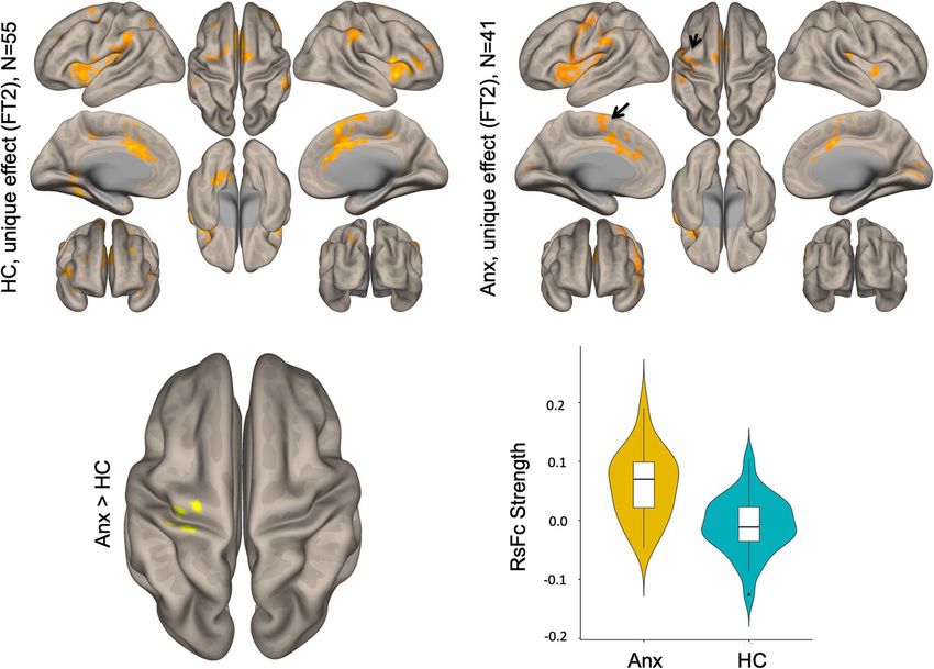
Cerebellum (2021) 20:392–401                                                                                              393

stimuli, often accompanied by emotional, cognitive, and        and parietal cortices, allowing the cerebellum to contrib-
somatic arousal [9]. One region implicated in motor, cog-      ute to virtually all streams of information processing in
nitive, and emotional processes involved in anxiety disor-     the cerebral cortex [22, 23]. As the cerebellum serves a
ders is the cerebellum. Neuroimaging studies have              wide range of functions and is suggested to be composed
highlighted cerebellar changes associated with anxiety         of discrete regions dedicated to unique functions [24, 25],
disorders. Specifically, increased cerebellar gray matter      we aimed to identify whether alterations in cerebellar
volume has been reported in social anxiety disorder [10]       functional connectivity in adolescent anxiety are predom-
and specific phobia [11]. Relative to healthy participants,    inantly located within a specific functional territory within
patients with anxiety disorders have shown enhanced cer-       the DN or whether these alterations are present in all
ebellar activity when presented with angry faces [12], so-     aspects of the DN. Here, we employed the functional
cial tasks [13], and at rest [14]. Patients with anxiety       parcellations of DN, the major cerebellar output nuclei,
disorders also display changes in cerebellar functional        from Guell et al.’s [26] research, which parcellated the
connectivity with anxiety-related regions in the cerebral      DN into three territories with specific functional connec-
cortex, such as the limbic system and prefrontal cortical      tions to default-mode, salience-motor, and visual net-
areas. For example, enhanced connectivity between cere-        works in the cerebral cortex using resting-state fMRI
bellum and amygdala, a core structure implicated in emo-       scans in a healthy population. We used these three DN
tion processing, has been reported in multiple studies of      functional territories (FTs) as seed regions of interest
generalized anxiety disorder [5, 7, 15]. Furthermore, in       (Fig. 1) to newly examine the intrinsic functional connec-
patients with anxiety disorders, the cerebellum has shown      tivity of cortico-cerebellar network in adolescent anxiety
aberrant intrinsic connectivity with the salience network      disorders and investigate its relationship with individual
[16, 17], default-mode network (DMN) [18], and central-        differences in anxious symptoms.
executive networks (Hilber et al.) [19] during resting-state
functional magnetic resonance imaging (fMRI). Such al-
terations in cerebellar connectivity are also evident in       Methods
non-clinical populations with high state or trait anxiety
[20, 21], suggesting that the cerebellum has a role in anx-    Study Participants and Procedure
iety susceptibility.
   Although a cerebellar role in anxiety disorders has         Adolescents aged 14–17 diagnosed with anxiety disorders
been evidenced in the literature, prior investigation has      (Anx = 41; mean age = 15.19) and matched healthy controls
rarely examined abnormalities in the cortico-cerebellar        (HC = 55, mean age = 15.31) were enrolled in the Boston
functional networks of adolescents with anxiety disorders.     Adolescent Neuroimaging of Depression and Anxiety
The cerebellum communicates with cortical regions by           (BANDA) project. Clinical characteristics, diagnostic criteria,
projecting to the deep cerebellar nuclei, the largest of       and demographics information have been previously reported
which is the dentate nuclei (DN). DN projects first syn-       (see [27, 28]). Sample clinical characteristics are summarized
apse in the thalamus, and then projects to frontal, motor,     in Table 1.

Fig. 1 Structural location and FTs
of the dentate nuclei as reported in
[26]). Red, FT1 = default-mode
network FT. Blue, FT2 =
salience-motor FT. Green, FT3 =
visual FT
394                                                                                                           Cerebellum (2021) 20:392–401

Table 1 Demographics
                                                                                Anx                    HC                        p value

                                Demographics
                                  Total participants                            41                     55
                                  Mean age (years)                              15.19 ± 0.82           15.31 ± 0.86              p = 0.51
                                  Sex: female                                   26 (63)                31 (56)
                                  Handedness: right-handed                      39 (95)                46 (83)
                                  Full-scale WASI score                         115.80 ± 16.40         118.13 ± 14.11            p = 0.47
                                  STAI: trait score                             43.98 ± 9.67           30.45 ± 7.44              p < 0.001
                                  STAI: state score                             39.4 ± 10.49           29.18 ± 8.04              p < 0.001
                                  Psychotropic medication treatment             18 (44)                0
                                Quality assurance
                                 No. of invalid scans                           123.88                 109.11                    p = 0.45
                                 Max motion                                     0.69                   0.53                      p = 0.37
                                 Mean motion                                    0.06                   0.06                      p = 0.98
                                Anxiety disorders
                                  Generalized anxiety disorder                  23 (56)                0
                                  Social anxiety disorder                       18 (44)                0
                                  Overanxious disorder                          17 (41)                0
                                  ADHD                                          11 (27)                0
                                  Specific phobia                               9 (22)                 0
                                  Oppositional defiant disorder                 5 (12)                 0
                                  OCD/excoriation disorder                      5 (12)                 0
                                  Separation anxiety                            4 (10)                 0
                                  Avoidant personality disorder                 4 (10)                 0
                                  Panic disorder                                4 (10)                 0
                                  Substance use disorder                        1 (2.4)                0
                                  PTSD                                          1 (2.4)                0
                                  Others                                        4 (10)                 0
                                Types of medication treatment
                                  Serotonin reuptake inhibitor (SSRI)           14 (78)                0
                                  Stimulant                                     4 (22)                 0
                                  Antipsychotic drug                            1 (6)                  0
                                  Benzodiazepine                                1 (6)                  0
                                  Tricyclic antidepressant                      1 (6)                  0
                                  Anxiolytic agent                              1 (6)                  0
                                  Alpha-agonist agent                           1 (6)                  0
                                  Anticonvulsant                                1 (6)                  0

                                Values expressed as n (%) or mean ± standard deviation; WASI, Wechsler Abbreviated Scale of Intelligence;
                                ADHD, attention-deficit/hyperactivity disorder; OCD, obsessive-compulsive disorder; PTSD, post-traumatic
                                stress disorder; Others include Tourette syndrome, enuresis, misophonia, and unspecified anxiety disorder

   Informed consent was obtained from legal guardians and               Structural and Resting-State Functional MRI
assent was obtained from the adolescent. Adolescent-parent              Acquisition Parameters
dyads were administered the KSADS to assess adolescent
lifetime mental disorders, and the State-Trait Anxiety                  Imaging data were collected on a Siemens 3T Prisma whole-
Inventory (STAI; [29]) was used to assess continuous anxiety.           body scanner with vendor-provided 64-channel head coil
The STAI is a commonly used self-report questionnaire that              (Siemens Healthcare, Erlangen, Germany). High-resolution
measures both anxiety levels rooted in the personality (STAI-           structural data (0.8-mm isotropic voxels) were acquired using
trait) and anxiety as a transitional emotional state (STAI-             a T1-weighted MPRAGE sequence with a duration of 7 min
state); each subscale ranges from 20 to 80, and higher scores           50 s (in-plane acceleration factor of 2). Scan parameters for
indicate a greater level of anxiety [30].                               TR, TE, TI, and flip angle were 2.4 s, 2.18 ms, 1.04 s, and 8°.
Cerebellum (2021) 20:392–401 395
Anatomical scans were immediately followed by resting-state Whole-brain Pearson’s correlation maps derived from
scans, during which subjects were asked to stay awake and denoised time series from whole DN and the three DN
keep their eyes fixated on a crosshair. Four resting-state ses- functional territories were converted to z-scores using
sions per participant were acquired, with two scans of diffu- Fisher’s r to z transformation to carry out second-level
sion MRI in AP-PA directions in the middle. Scan parameters general linear model (GLM) analyses.
(T2*-weighted EPI sequence) for TR, TE, flip angle, echo
spacing, and bandwidth were 800 ms, 37 ms, 85°, 0.58 ms,
and 2290 Hz per pixel. Seventy-two interleaved (ascending/ Data Processing: Second-Level GLM Analysis
foot-head) slices were collected in the AC-PC plane using an
auto-align procedure to minimize inter-subject variability in Seed-to-voxel analysis was carried out using the whole DN as
data acquisition. Combination of 64ch array coil and simulta- a seed, as well as using the unique effect of each of the three
neous multi-slice (SMS) acquisition (multiband factor of 8) functional territories (DMN, salience-motor, and visual). The
provided high temporal sampling (420 time points during an unique effect of each functional territory [35] was calculated
acquisition window of 5 min and 46 s; four runs) and spatial using a previously described method [26]. Specifically, the
resolution (2 mm isotropic) while maintaining whole-brain DMN unique effect (FT1) was calculated as DMN > (sa-
coverage (including the entire cerebellum). lience-motor and visual), salience-motor unique effect (FT2)
was calculated as salience-motor > (DMN and visual), and
Data Processing: Seed-to-Voxel Functional visual unique effect (FT3) was calculated as visual > (DMN
Connectivity Analysis and salience-motor). Statistical significance thresholding for
between-group effects included p < 0.001 (two-sided) at the
Resting-state data were realigned and spatially normalized voxel level and p < 0.05 false discovery rate (FDR) correction
to the MNI template using SPM12 (Wellcome Department at the cluster level.
of Imaging Neuroscience; www.fil.ion.ucl.ac.uk/spm).
Structural images were segmented into white matter
(WM), gray matter, and cerebrospinal fluid (CSF) using Symptom Correlation Analyses
SPM12. The CONN Toolbox [31] was used to compute
whole-brain correlation maps from the seed regions of For all three functional territories, correlations between
interest (ROIs). ROIs included the whole DN (as defined resting-state fMRI correlations and STAI scores were calcu-
using the SUIT DN mask [32]), and three functional sub- lated in both HC and Anx groups, with a voxel threshold of
territories of DN that were defined in a previous study by p < 0.001 and a cluster-forming threshold of p < 0.05 (FDR
our group [26], including default-mode network, motor- corrected).
salience, and visual functional regions (see Fig. 1). The
CONN Toolbox uses an anatomical component-based cor-
rection method (aCompCor [33]) for denoising BOLD
time series and integrates quality assurance (QA) methods Results
to address the deleterious effects of motion artifacts
(Artifact Detection Tools, www.nitrc.org/projects/ Second-Level GLM Analysis
artifact_detect). There was no between-group difference
in the number of motion outliers and maximum and mean Within functional sub-regions of DN (see Fig. 1), statistically
head motion. Band-pass filtering was carried out at 0. significant differences were detected only for the salience-
008–0.09 Hz. Time points with a mean signal intensity motor territory (FT2), revealing increased functional connec-
beyond three standard deviations from the global mean tivity with pre- and postcentral cerebral cortices (peak coordi-
signal and 0.4-mm scan-to-scan motion (about one-fifth nate at (− 34, − 28, 70)) (Fig. 2). For both HC and Anx groups,
the acquisition voxel size) were flagged as problematic within-group seed-to-voxel functional connectivity analyses
scans and were regressed out along with six realignment for FT2 (Fig. 1, top panel) showed functional connectivity to
parameters (along with derivatives) and physiological cerebral cortical salience-motor regions including the primary
sources of noise (three principal components of WM, motor cortex and supplementary motor area, as well as the
and three principal components of CSF segments, using bilateral insula, dorsal anterior cingulate cortex, anterior
aCompCor [33]). WM and CSF segments were derived supramarginal gyrus, and rostral middle frontal gyrus. The
from the structural images using the segmentation routine results were significant after covarying for medication use.
in SPM12. Because of the small size of the DN, un- Using the whole DN as a seed did not reveal any statistically
smoothed data were used for data analysis to minimize significant differences between HC and Anx, highlighting the
partial volume effects from structures close to DN [34]. relevance for investigating the functional parcellations of DN.396 Cerebellum (2021) 20:392–401
Fig. 2 Top: Within-group results
(overlaid on surface maps in
CONN) using functional connec-
tivity calculated from the
salience-motor FT of the DN
(FT2), at voxel-level height
threshold of p < 0.001 (two-sided)
and cluster size FDR correction of
p < 0.05. Bottom: Between-group
results after controlling for medi-
cation use (Anx > HC) at voxel-
level height threshold of p < 0.001
(two-sided) and cluster size FDR
correction of p < 0.05, T = 5.15.
Bar plots provide data for the
significant cluster (precentral and
postcentral cerebral cortex) in
Anx and HC
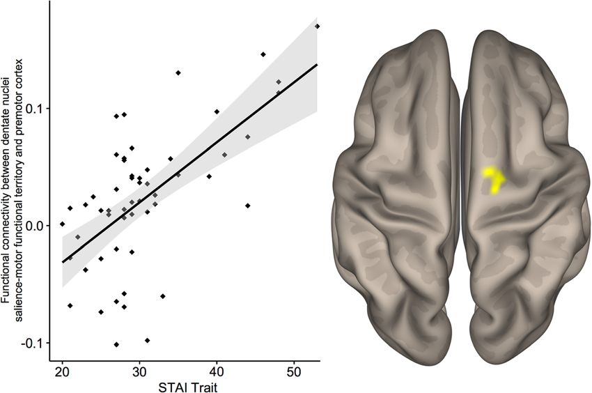
Symptom Correlation Analyses STAI-trait scores within the Anx group. STAI-state scores
did not reveal any significant difference in either group.
To examine individual differences within each group, corre-
lation analyses between functional connectivity and STAI
scores were conducted separately for HC and Anx groups. Discussion
Within HC, STAI-trait scores and functional connectivity be-
tween the salience-motor DN FC and cerebral motor/ We show for the first time that functional connectivity alter-
somatosensory cortex (20, − 8, 70) showed a significant cor- ations between cerebellar output structures and cerebral corti-
relation, linking higher trait anxiety level to stronger connec- cal areas are associated with adolescent anxiety. Specifically,
tivity between salience-motor DN FT and cerebral cortical hyperconnectivity was detected between DN salience-motor
motor areas (Fig. 3). There was no significant cluster with FT and primary motor and somatosensory cortices in
Fig. 3 Whole-brain correlation
between STAI-trait scores in
healthy controls and salience-
motor DN FT-motor/somatosen-
sory cortex functional connectiv-
ity, thresholded at a height
threshold of p < 0.001, cluster-
corrected at p < 0.05 FDR. R2 =
0.41Cerebellum (2021) 20:392–401 397
adolescents with anxiety disorders compared to HC. No system that false internal representation of external stimuli
anxiety-related differences were observed with default mode would generate unsuited sensorimotor response, which leads
or visual territories or all combined territories of the DN. to physiological stress and anxiety state [49].
Within HC, stronger functional connectivity of salience- Besides anatomical investigations, other studies of cerebel-
motor DN to motor/somatosensory cortex correlated with lar function also support a role of the cerebellum in fear and
higher trait anxiety. Taken together, this new evidence illus- anxiety in animals and humans. In rodent models, manipula-
trates the use of DN functional sub-divisions as a relevant tions on cerebellar functioning impaired anxiety-related be-
structure to detect functional differences in psychiatric disease haviors [50, 51]. Others have shown a cerebellar role in sa-
and highlights the role of the DN as a potential target for lience attribution to avoid harmful stimuli [14, 16, 52].
disease prediction or prevention of affective disorders. Patients with anxiety disorders also show changes in cerebel-
lar activity and connectivity with anxiety-related brain areas.
Cerebellar-Cortical Salience-Motor Network in Anxiety A few studies observed increased cerebellar activation in so-
cial anxiety disorder [12, 13] and specific phobia [53, 54].
Evidence from both human and animal studies has established Enhanced connectivity between the cerebellum and amygdala
that the cerebellum is connected with various parts of anxiety has been replicated several times across age range and symp-
circuitry, including the insula, basal ganglia, and ventral teg- tom severity [5, 7, 15].
mental area (VTA) (see [8]) for review). Cerebellar connec- Functional connectivity differences between adolescents
tions with cortical and subcortical areas relevant in sensorimo- with anxiety disorders and HC were observed specifically in
tor perception and anticipation of stimulus underpin the non- the salience-motor territory of the DN; the salience network
motor role of the cerebellum in anxiety. The cerebellar vermis, has clear associations with fear and anxiety processing and is
which contains the fastigial and dentate nuclei, is referred to as connected to sensorimotor processing systems in the brain as
the “limbic cerebellum” for its connections to the mesolimbic discussed earlier. The salience network is implicated in detect-
dopaminergic pathway that originate from the VTA to the ing emotional salience and triggering cognitive regulation
nucleus accumbens [36]. The dentate nucleus forms direct [38]. Individuals with high trait anxiety or anxiety disorders
monosynaptic excitatory connections with neurons in the have shown aberrance in the salience network. Weaker
VTA [37], which is one of the key subcortical structures that within-network salience connectivity has been associated with
is activated as part of the salience network [38]. Then, the adolescents with higher trait anxiety and patients with social
dopaminergic fibers of the meso-cortico-limbic system project anxiety disorder, suggesting impaired ability in emotion reg-
to motor cortical fields, mainly to the pre- and postcentral ulation and overreaction to threatening stimuli [20, 55, 56].
gyrus [39]. Hypoactivations in the cerebellar vermis and sub- One study on healthy young adults suggested that differential
cortical regions in the dopaminergic pathways have been as- resting-state connectivity between the cerebellum and execu-
sociated with disturbed predictive motor timing paradigm tive and salience cortical regions correlated with behavioral
[36]. inhibition, which may have a mediating role in anxiety vul-
Also, the insula sends efferent projections to the sensori- nerability [19].
motor cortex from which it receives reciprocal afferent pro-
jections, forming the salience-motor network [40, 41]. Symptom Correlation
Neuroimaging and invasive stimulation studies in humans
support a physiological coupling between sensorimotor sys- Within the group of HC, STAI-trait scores correlated with
tems and stimulus-driven attentional processes [38, 40–44]. functional connectivity between the premotor cortex and
The cerebellum also receives inputs from the subthalamic the salience-motor FT of the DN (Fig. 2). The somatosen-
nucleus of the basal ganglia, indicating that the cerebellum sory cortex is involved in emotional processing through
may be a key substrate for reward-related signals during learn- its connection to the amygdala through the insula [57, 58].
ing [45]. Reward-based communication of the basal ganglia After early evaluations of emotional significance are con-
and the cerebellum has been highlighted during predictive ducted through amygdala interaction with thalamus and
motor timing tasks [46]. The salience-motor network plays limbic regions, somatosensory and related cerebral corti-
an important role in detecting behaviorally relevant stimuli cal areas take in for higher order re-evaluation of emo-
by mediating the switch between the DMN and task-positive tional perception to establish emotional salience [59] and
central-executive network and permitting response to the stim- generate fear memory [60]. Thus, people with higher
uli [47]. The hyperconnectivity that we report in the DN alertness and sensitivity to stress stimuli may bring in
salience-motor FT points to the possibility that there is a mal- more involvement of the somatosensory cortex displayed
adaptive attribution of salience to internal and environmental as heightened activity. It has also been reported that peo-
stimuli in anxious individuals [48]. It can also be interpreted as ple with specific phobia show higher activation in the
the discrepancy in the “internal model” of the cerebellar somatosensory cortex [61], and heightened activity in this398 Cerebellum (2021) 20:392–401
area may contribute to difficulties in emotion regulation establish a causal link between anxiety and alterations in DN
and lower self-control [62, 63]. Trait anxiety does not functional connectivity.
necessarily predict conversion to anxiety disorders but is
an index of vulnerability to anxiety disorders. Individuals
with high trait anxiety and those diagnosed with anxiety
Conclusion
disorders share common disruptions in brain activation
and connectivity [64, 65]. Individual differences in state
These findings advance our understanding of the
anxiety, although not detected in this study, have also
cerebellar-cortical salience-motor network in anxiety dis-
been correlated with increased fluctuations of activation
orders by identifying abnormal functional connectivity of
in the right postcentral gyrus and right precentral gyrus
salience-motor territories within DN, a major cerebellar
and with connectivity between the postcentral gyrus and
output to the cerebral cortex, in adolescents with anxiety
left cerebellum gyrus [21, 66]. The relationship between
disorders. In addition, our study identified a relationship
DN salience-motor FT connectivity and anxious symp-
between trait anxiety level and DN salience-motor FT
toms may be shown in other populations with anxious
functional connectivity in adolescents without a diagnosis
symptoms [67].
of anxiety disorder, suggesting DN salience-motor FT as a
potential biomarker for abnormal emotional and attentional
Limitations and Future Directions processing in sub-clinical anxiety. With recent advances in
the field of neuromodulation and neurostimulation, these
First, our findings are based on exploratory analyses of findings also illustrate the idea that the salience-motor ter-
cerebellar output structure that suggest overarching alter-
ritories in DN may be used as an experimental target region
ations in the cortical-cerebellar salience-motor network
for non-invasive neuromodulation for treatment or preven-
but do not specify the direction of connectivity. While tion of anxiety disorders in the near future.
DN functional connectivity may not uniquely correspond
to unidirectional communication between the cerebellar Funding This project was supported by the National Institute of Mental
cortex and extracerebellar structures, DN is the largest Health, U01MH108168 (JDEG, SWG), and F32MH114525 (NAH).
location of synapses for anatomical connections linking NAH was partially supported by the Brain and Behavior Research
Foundation. AY was partially supported by R01EB021265 and
the cerebellar cortex to the thalamus and ultimately to
U01EB026996. DAP was partially supported by R37MH068376 and
the cerebral cortex. It is therefore reasonable to consider R01MH101521. RPA was partially supported by R01MH119771 and
that DN functional connectivity is predominantly a mea- R56MH121426. SSG was partially supported by R01EB020740 and
sure of cerebellar functional output. Since we have under- P41EB019936. SGH was partly supported by R01AT007257,
R01MH099021, and the James S. McDonnell Foundation. This project
standing of anatomical connections between the cerebel-
was made possible by the resources provided by Shared Instrumentation
lum and cortical and subcortical regions of the salience- Grants 1S10RR023401, 1S10RR019307, and 1S10RR023043.
motor network, future studies can use causal modeling to
characterize the effective connectivity between those re- Compliance with Ethical Standards
gions and examine the directionality of the communica-
tion within the cortical-subcortical-cerebellar network. Conflict of Interest Over the past 3 years, Dr. Pizzagalli has received
Second, 44% of our Anx group were taking psychotropic consulting fees from Akili Interactive Labs, BlackThorn Therapeutics,
medications. To overcome this limitation, we covaried for Boehringer Ingelheim, Compass Pathway, Otsuka Pharmaceuticals, and
Takeda Pharmaceuticals; one honorarium from Alkermes; and research
medication use in the between-group analyses. The number funding from NIMH, Dana Foundation, Brain and Behavior Research
of adolescents not on medication use was small (n = 18), and Foundation, Millennium Pharmaceuticals. In addition, he has received
STAI score correlation calculations within this subsample stock options from BlackThorn Therapeutics.
would have resulted in a substantial loss of statistical power
making false-negative findings very likely; STAI score corre- Disclaimer The content is solely the responsibility of the authors and
does not necessarily represent the official views of the National Institutes
lation analyses were thus performed on the full sample of the of Health or of any other sponsor.
Anx group. We did not have information on behavioral ther-
apy and thus could not control for this variable. The impact of Open Access This article is licensed under a Creative Commons
Attribution 4.0 International License, which permits use, sharing, adap-
behavioral therapy on the brain’s functional connectivity
tation, distribution and reproduction in any medium or format, as long as
could have blurred individual differences in correlation be- you give appropriate credit to the original author(s) and the source, pro-
tween brain networks and anxiety symptoms within Anx as vide a link to the Creative Commons licence, and indicate if changes were
well. More generally, our findings provide only correlational made. The images or other third party material in this article are included
in the article's Creative Commons licence, unless indicated otherwise in a
evidence to support altered DN functional connectivity in anx-
credit line to the material. If material is not included in the article's
iety disorders. Interventional studies such as non-invasive Creative Commons licence and your intended use is not permitted by
stimulation experiments or lesion-based analyses may statutory regulation or exceeds the permitted use, you will need to obtainCerebellum (2021) 20:392–401 399
permission directly from the copyright holder. To view a copy of this 14. Warwick JM, Carey P, Jordaan GP, Dupont P, Stein DJ. Resting
licence, visit http://creativecommons.org/licenses/by/4.0/. brain perfusion in social anxiety disorder: a voxel-wise whole brain
comparison with healthy control subjects. Prog Neuro-
Psychopharmacol Biol Psychiatry. 2008;32(5):1251–6. https://doi.
org/10.1016/j.pnpbp.2008.03.017.
15. Etkin A, Prater KE, Schatzberg AF, Menon V, Greicius MD.
References Disrupted amygdalar subregion functional connectivity and evi-
dence of a compensatory network in generalized anxiety disorder.
1. Kessler RC, Berglund P, Demler O, Jin R, Merikangas KR, Walters Arch Gen Psychiatry. 2009;66(12):1361–72. https://doi.org/10.
EE. Lifetime prevalence and age-of-onset distributions of DSM-IV 1001/archgenpsychiatry.2009.104.
disorders in the National Comorbidity Survey Replication. Arch 16. Habas C, Kamdar N, Nguyen D, Prater K, Beckmann CF, Menon
Gen Psychiatry. 2005;62(6):593–602. https://doi.org/10.1001/ V, et al. Distinct cerebellar contributions to intrinsic connectivity
archpsyc.62.6.593. networks. J Neurosci. 2009;29(26):8586–94. https://doi.org/10.
2. Merikangas KR, He JP, Burstein M, Swanson SA, Avenevoli S, 1523/jneurosci.1868-09.2009.
Cui L, et al. Lifetime prevalence of mental disorders in U.S. ado- 17. Sang L, Qin W, Liu Y, Han W, Zhang Y, Jiang T, et al. Resting-
lescents: results from the National Comorbidity Survey state functional connectivity of the vermal and hemispheric subre-
Replication–Adolescent Supplement (NCS-A). J Am Acad Child gions of the cerebellum with both the cerebral cortical networks and
Adolesc Psychiatry. 2010;49(10):980–9. https://doi.org/10.1016/j. subcortical structures. Neuroimage. 2012;61(4):1213–25. https://
jaac.2010.05.017. doi.org/10.1016/j.neuroimage.2012.04.011.
3. McLaughlin KA, Garrad MC, Somerville LH. What develops dur- 18. Yuan M, Zhu H, Qiu C, Meng Y, Zhang Y, Shang J, et al. Group
ing emotional development? A component process approach to cognitive behavioral therapy modulates the resting-state functional
identifying sources of psychopathology risk in adolescence. connectivity of amygdala-related network in patients with general-
Dialogues Clin Neurosci. 2015;17(4):403–10. ized social anxiety disorder. BMC Psychiatry. 2016;16:198. https://
4. Essau CA, Lewinsohn PM, Olaya B, Seeley JR. Anxiety disorders doi.org/10.1186/s12888-016-0904-8.
in adolescents and psychosocial outcomes at age 30. J Affect 19. Caulfield MD, Zhu DC, McAuley JD, Servatius RJ. Individual
Disord. 2014;163:125–32. https://doi.org/10.1016/j.jad.2013.12. differences in resting-state functional connectivity with the execu-
033. tive network: support for a cerebellar role in anxiety vulnerability.
5. Liu WJ, Yin DZ, Cheng WH, Fan MX, You MN, Men WW, et al. Brain Struct Funct. 2016;221(6):3081–93. https://doi.org/10.1007/
Abnormal functional connectivity of the amygdala-based network s00429-015-1088-6.
in resting-state FMRI in adolescents with generalized anxiety dis- 20. Geng H, Li X, Chen J, Li X, Gu R. Decreased intra- and inter-
order. Med Sci Monit. 2015;21:459–67. https://doi.org/10.12659/ salience network functional connectivity is related to trait anxiety
msm.893373. in adolescents. Front Behav Neurosci. 2015;9:350. https://doi.org/
6. Monk CS, Telzer EH, Mogg K, Bradley BP, Mai X, Louro HM, 10.3389/fnbeh.2015.00350.
et al. Amygdala and ventrolateral prefrontal cortex activation to 21. Li X, Zhang M, Li K, Zou F, Wang Y, Wu X, et al. The altered
masked angry faces in children and adolescents with generalized somatic brain network in state anxiety. Front Psychiatry. 2019;10:
anxiety disorder. Arch Gen Psychiatry. 2008;65(5):568–76. https:// 465. https://doi.org/10.3389/fpsyt.2019.00465.
doi.org/10.1001/archpsyc.65.5.568. 22. Middleton FA, Strick PL. Anatomical evidence for cerebellar and
7. Roy AK, Fudge JL, Kelly C, Perry JS, Daniele T, Carlisi C, et al. basal ganglia involvement in higher cognitive function. Science.
Intrinsic functional connectivity of amygdala-based networks in 1994;266(5184):458–61. https://doi.org/10.1126/science.7939688.
adolescent generalized anxiety disorder. J Am Acad Child 23. Schmahmann JD, Pandya DN. The cerebrocerebellar system. Int
Adolesc Psychiatry. 2013;52(3):290–299.e292. https://doi.org/10. Rev Neurobiol. 1997;41:31–60. https://doi.org/10.1016/s0074-
1016/j.jaac.2012.12.010. 7742(08)60346-3.
8. Moreno-Rius J. The cerebellum in fear and anxiety-related disor- 24. Buckner RL, Krienen FM, Castellanos A, Diaz JC, Yeo BTT. The
ders. Prog Neuro-Psychopharmacol Biol Psychiatry. 2018;85:23– organization of the human cerebellum estimated by intrinsic func-
32. https://doi.org/10.1016/j.pnpbp.2018.04.002. tional connectivity. J Neurophysiol. 2011;106(5):2322–45. https://
9. Perusini JN, Fanselow MS. Neurobehavioral perspectives on the doi.org/10.1152/jn.00339.2011.
distinction between fear and anxiety. Learn Mem. 2015;22(9): 25. Guell X, Schmahmann JD, Gabrieli JDE, Ghosh SS. Functional
417–25. https://doi.org/10.1101/lm.039180.115. gradients of the cerebellum. eLife. 2018;7:e36652. https://doi.org/
10. Talati A, Pantazatos SP, Schneier FR, Weissman MM, Hirsch J. 10.7554/eLife.36652.
Gray matter abnormalities in social anxiety disorder: primary, rep- 26. Guell X, D’Mello AM, Hubbard NA, Romeo RR, Gabrieli JDE,
lication, and specificity studies. Biol Psychiatry. 2013;73(1):75–84. Whitfield-Gabrieli S, et al. Functional territories of human dentate
https://doi.org/10.1016/j.biopsych.2012.05.022. nucleus. Cereb Cortex. 2020;30(4):2401–17. https://doi.org/10.
11. Hilbert K, Evens R, Maslowski NI, Wittchen HU, Lueken U. 1093/cercor/bhz247.
Neurostructural correlates of two subtypes of specific phobia: a 27. Hubbard NA, Siless V, Frosch IR, Goncalves M, Lo N, Wang J,
voxel-based morphometry study. Psychiatry Res. 2015;231(2): et al. Brain function and clinical characterization in the Boston
168–75. https://doi.org/10.1016/j.pscychresns.2014.12.003. adolescent neuroimaging of depression and anxiety study.
12. Evans KC, Wright CI, Wedig MM, Gold AL, Pollack MH, Rauch Neuroimage Clin. 2020;27:102240. https://doi.org/10.1016/j.nicl.
SL. A functional MRI study of amygdala responses to angry sche- 2020.102240.
matic faces in social anxiety disorder. Depress Anxiety. 2008;25(6): 28. Siless V, Hubbard NA, Jones R, Wang J, Lo N, Bauer CCC, et al.
496–505. https://doi.org/10.1002/da.20347. Image acquisition and quality assurance in the Boston Adolescent
13. Nakao T, Sanematsu H, Yoshiura T, Togao O, Murayama K, Neuroimaging of Depression and Anxiety study. Neuroimage Clin.
Tomita M, et al. fMRI of patients with social anxiety disorder 2020;26:102242. https://doi.org/10.1016/j.nicl.2020.102242.
during a social situation task. Neurosci Res. 2011;69(1):67–72. 29. Spielberger CD, Sydeman SJ. State-Trait Anxiety Inventory and
https://doi.org/10.1016/j.neures.2010.09.008. State-Trait Anger Expression Inventory. In: The use of400 Cerebellum (2021) 20:392–401
psychological testing for treatment planning and outcome assess- 46. Lungu OV, Bares M, Liu T, Gomez CM, Cechova I, Ashe J. Trial-
ment. Hillsdale: Lawrence Erlbaum Associates, Inc.; 1994. p. 292– to-trial adaptation: parsing out the roles of cerebellum and BG in
321. predictive motor timing. J Cogn Neurosci. 2016;28(7):920–34.
30. Julian LJ. Measures of anxiety: State-Trait Anxiety Inventory https://doi.org/10.1162/jocn_a_00943.
(STAI), Beck Anxiety Inventory (BAI), and Hospital Anxiety and 47. Sridharan D, Levitin DJ, Menon V. A critical role for the right
Depression Scale-Anxiety (HADS-A). Arthritis Care Res. 2011;63 fronto-insular cortex in switching between central-executive and
Suppl 11:S467–72. https://doi.org/10.1002/acr.20561. default-mode networks. Proc Natl Acad Sci U S A. 2008;105(34):
31. Whitfield-Gabrieli S, Nieto-Castanon A. Conn: a functional con- 12569–74. https://doi.org/10.1073/pnas.0800005105.
nectivity toolbox for correlated and anticorrelated brain networks. 48. Robinson OJ, Vytal K, Cornwell BR, Grillon C. The impact of
Brain Connect. 2012;2(3):125–41. https://doi.org/10.1089/brain. anxiety upon cognition: perspectives from human threat of shock
2012.0073. studies. Front Hum Neurosci. 2013;7:203. https://doi.org/10.3389/
32. Diedrichsen J, Maderwald S, Kuper M, Thurling M, Rabe K, fnhum.2013.00203.
Gizewski ER, et al. Imaging the deep cerebellar nuclei: a probabi- 49. Hilber P, Cendelin J, Le Gall A, Machado ML, Tuma J, Besnard S.
listic atlas and normalization procedure. Neuroimage. 2011;54(3): Cooperation of the vestibular and cerebellar networks in anxiety
1786–94. https://doi.org/10.1016/j.neuroimage.2010.10.035. disorders and depression. Prog Neuro-Psychopharmacol Biol
33. Behzadi Y, Restom K, Liau J, Liu TT. A component based noise Psychiatry. 2019;89:310–21. https://doi.org/10.1016/j.pnpbp.
correction method (CompCor) for BOLD and perfusion based 2018.10.004.
fMRI. Neuroimage. 2007;37(1):90–101. https://doi.org/10.1016/j. 50. Otsuka S, Konno K, Abe M, Motohashi J, Kohda K, Sakimura K,
neuroimage.2007.04.042. et al. Roles of Cbln1 in non-motor functions of mice. J Neurosci.
34. Bernard JA, Peltier SJ, Benson BL, Wiggins JL, Jaeggi SM, 2016;36(46):11801–16. https://doi.org/10.1523/jneurosci.0322-16.
Buschkuehl M, et al. Dissociable functional networks of the human 2016.
dentate nucleus. Cereb Cortex. 2014;24(8):2151–9. https://doi.org/ 51. Sacchetti B, Scelfo B, Tempia F, Strata P. Long-term synaptic
10.1093/cercor/bht065. changes induced in the cerebellar cortex by fear conditioning.
35. McClure EB, Monk CS, Nelson EE, Parrish JM, Adler A, Blair RJ, Neuron. 2004;42(6):973–82. https://doi.org/10.1016/j.neuron.
et al. Abnormal attention modulation of fear circuit function in 2004.05.012.
pediatric generalized anxiety disorder. Arch Gen Psychiatry. 52. Shinn AK, Baker JT, Lewandowski KE, Ongur D, Cohen BM.
2007;64(1):97–106. https://doi.org/10.1001/archpsyc.64.1.97. Aberrant cerebellar connectivity in motor and association networks
36. Lošák J, Hüttlová J, Lipová P, Marecek R, Bareš M, Filip P, et al. in schizophrenia. Front Hum Neurosci. 2015;9:134. https://doi.org/
Predictive motor timing and the cerebellar vermis in schizophrenia: 10.3389/fnhum.2015.00134.
an fMRI study. Schizophr Bull. 2016;42(6):1517–27. https://doi. 53. Ahs F, Pissiota A, Michelgard A, Frans O, Furmark T, Appel L,
org/10.1093/schbul/sbw065. et al. Disentangling the web of fear: amygdala reactivity and func-
tional connectivity in spider and snake phobia. Psychiatry Res.
37. Carta I, Chen CH, Schott AL, Dorizan S, Khodakhah K. Cerebellar
modulation of the reward circuitry and social behavior. Science. 2009;172(2):103–8. https://doi.org/10.1016/j.pscychresns.2008.
2019;363(6424):eaav0581. https://doi.org/10.1126/science. 11.004.
aav0581. 54. Goossens L, Schruers K, Peeters R, Griez E, Sunaert S. Visual
presentation of phobic stimuli: amygdala activation via an
38. Seeley WW, Menon V, Schatzberg AF, Keller J, Glover GH,
extrageniculostriate pathway? Psychiatry Res. 2007;155(2):113–
Kenna H, et al. Dissociable intrinsic connectivity networks for sa-
20. https://doi.org/10.1016/j.pscychresns.2006.12.005.
lience processing and executive control. J Neurosci. 2007;27(9):
2349–56. https://doi.org/10.1523/jneurosci.5587-06.2007. 55. Gaspar JM, McDonald JJ. High level of trait anxiety leads to
salience-driven distraction and compensation. Psychol Sci.
39. Hosp JA, Coenen VA, Rijntjes M, Egger K, Urbach H, Weiller C,
2018;956797618807166:2020–30. https://doi.org/10.1177/
et al. Ventral tegmental area connections to motor and sensory
0956797618807166.
cortical fields in humans. Brain Struct Funct. 2019;224(8):2839–
56. Klumpp H, Angstadt M, Phan KL. Insula reactivity and connectiv-
55. https://doi.org/10.1007/s00429-019-01939-0.
ity to anterior cingulate cortex when processing threat in general-
40. Mesulam MM, Mufson EJ. Insula of the old world monkey. III:
ized social anxiety disorder. Biol Psychol. 2012;89(1):273–6.
efferent cortical output and comments on function. J Comp Neurol.
https://doi.org/10.1016/j.biopsycho.2011.10.010.
1982;212(1):38–52. https://doi.org/10.1002/cne.902120104.
57. Hoistad M, Barbas H. Sequence of information processing for emo-
41. Mufson EJ, Mesulam MM. Insula of the old world monkey. II: tions through pathways linking temporal and insular cortices with
afferent cortical input and comments on the claustrum. J Comp the amygdala. Neuroimage. 2008;40(3):1016–33. https://doi.org/
Neurol. 1982;212(1):23–37. https://doi.org/10.1002/cne. 10.1016/j.neuroimage.2007.12.043.
902120103.
58. Nummenmaa L, Glerean E, Viinikainen M, Jaaskelainen IP, Hari
42. Deen B, Pitskel NB, Pelphrey KA. Three systems of insular func- R, Sams M. Emotions promote social interaction by synchronizing
tional connectivity identified with cluster analysis. Cereb Cortex. brain activity across individuals. Proc Natl Acad Sci U S A.
2011;21(7):1498–506. https://doi.org/10.1093/cercor/bhq186. 2012;109(24):9599–604. https://doi.org/10.1073/pnas.
43. Stephani C, Fernandez-Baca Vaca G, Maciunas R, Koubeissi M, 1206095109.
Luders HO. Functional neuroanatomy of the insular lobe. Brain 59. Cunningham WA, Zelazo PD. Attitudes and evaluations: a social
Struct Funct. 2011;216(2):137–49. https://doi.org/10.1007/ cognitive neuroscience perspective. Trends Cogn Sci. 2007;11(3):
s00429-010-0296-3. 97–104. https://doi.org/10.1016/j.tics.2006.12.005.
44. Uddin LQ. Salience processing and insular cortical function and 60. Damasio AR, Grabowski TJ, Bechara A, Damasio H, Ponto LL,
dysfunction. Nat Rev Neurosci. 2015;16(1):55–61. https://doi.org/ Parvizi J, et al. Subcortical and cortical brain activity during the
10.1038/nrn3857. feeling of self-generated emotions. Nat Neurosci. 2000;3(10):
45. Bostan AC, Dum RP, Strick PL. The basal ganglia communicate 1049–56. https://doi.org/10.1038/79871.
with the cerebellum. Proc Natl Acad Sci U S A. 2010;107(18): 61. Rauch SL, Savage CR, Alpert NM, Miguel EC, Baer L, Breiter HC,
8452–6. https://doi.org/10.1073/pnas.1000496107. et al. A positron emission tomographic study of simple phobicCerebellum (2021) 20:392–401 401
symptom provocation. Arch Gen Psychiatry. 1995;52(1):20–8. and anxiety disorders. Trends Neurosci. 2012;35(9):527–35.
https://doi.org/10.1001/archpsyc.1995.03950130020003. https://doi.org/10.1016/j.tins.2012.04.012.
62. Volkow ND, Wang GJ, Fowler JS, Tomasi D, Baler R. Food and 66. Geng H, Wang Y, Gu R, Luo YJ, Xu P, Huang Y, et al. Altered
drug reward: overlapping circuits in human obesity and addiction. brain activation and connectivity during anticipation of uncertain
Curr Top Behav Neurosci. 2012;11:1–24. https://doi.org/10.1007/ threat in trait anxiety. Hum Brain Mapp. 2018;39(10):3898–914.
7854_2011_169. https://doi.org/10.1002/hbm.24219.
63. Wang GJ, Volkow ND, Thanos PK, Fowler JS. Similarity between 67. Roy B, Ehlert L, Mullur R, Freeby MJ, Woo MA, Kumar R, et al.
obesity and drug addiction as assessed by neurofunctional imaging: Regional brain gray matter changes in patients with type 2 diabetes
a concept review. J Addict Dis. 2004;23(3):39–53. https://doi.org/ mellitus. Sci Rep. 2020;10(1):9925. https://doi.org/10.1038/
10.1300/J069v23n03_04. s41598-020-67022-5 (Accession No. 32555374).
64. Etkin A, Wager TD. Functional neuroimaging of anxiety: a meta-
analysis of emotional processing in PTSD, social anxiety disorder,
Publisher’s Note Springer Nature remains neutral with regard to jurisdic-
and specific phobia. Am J Psychiatry. 2007;164(10):1476–88.
tional claims in published maps and institutional affiliations.
https://doi.org/10.1176/appi.ajp.2007.07030504.
65. Sylvester CM, Corbetta M, Raichle ME, Rodebaugh TL, Schlaggar
BL, Sheline YI, et al. Functional network dysfunction in anxietyYou can also read