Genetic interaction screen for severe neurodevelopmental disorders reveals a functional link between Ube3a and Mef2 in Drosophila melanogaster
←
→
Page content transcription
If your browser does not render page correctly, please read the page content below
www.nature.com/scientificreports

            OPEN           Genetic interaction screen for
                           severe neurodevelopmental
                           disorders reveals a functional
                           link between Ube3a and Mef2 in
                           Drosophila melanogaster
                           Jonas Straub1,2, Anne Gregor1,2, Tatjana Sauerer1, Anna Fliedner1, Laila Distel1,
                           Christine Suchy1, Arif B. Ekici 1, Fulvia Ferrazzi 1 & Christiane Zweier1*
                           Neurodevelopmental disorders (NDDs) are clinically and genetically extremely heterogeneous with
                           shared phenotypes often associated with genes from the same networks. Mutations in TCF4, MEF2C,
                           UBE3A, ZEB2 or ATRX cause phenotypically overlapping, syndromic forms of NDDs with severe
                           intellectual disability, epilepsy and microcephaly. To characterize potential functional links between
                           these genes/proteins, we screened for genetic interactions in Drosophila melanogaster. We induced
                           ubiquitous or tissue specific knockdown or overexpression of each single orthologous gene (Da, Mef2,
                           Ube3a, Zfh1, XNP) and in pairwise combinations. Subsequently, we assessed parameters such as
                           lethality, wing and eye morphology, neuromuscular junction morphology, bang sensitivity and climbing
                           behaviour in comparison between single and pairwise dosage manipulations. We found most stringent
                           evidence for genetic interaction between Ube3a and Mef2 as simultaneous dosage manipulation
                           in different tissues including glia, wing and eye resulted in multiple phenotype modifications. We
                           subsequently found evidence for physical interaction between UBE3A and MEF2C also in human cells.
                           Systematic pairwise assessment of the Drosophila orthologues of five genes implicated in clinically
                           overlapping, severe NDDs and subsequent confirmation in a human cell line revealed interactions
                           between UBE3A/Ube3a and MEF2C/Mef2, thus contributing to the characterization of the underlying
                           molecular commonalities.

                           Neurodevelopmental disorders (NDDs) are clinically and genetically extremely heterogeneous, and currently
                           more than 1,000 genes have been implicated (SysID database1). Within the last years, phenotypically and biolog-
                           ically coherent modules within this large and heterogeneous group have been increasingly delineated, indicating
                           that overlapping phenotypes are often caused by mutations in genes involved in the same molecular networks1–5.
                           This has been demonstrated mainly for well-defined pathways or complexes such as the RAS-MAPK-pathway4 or
                           the SWI/SNF chromatin remodelling complex6, but is less characterized for disease genes involved in a broader
                           biological context or process such as transcriptional regulation. Identifying and characterizing such connections
                           and correlations contributes to a better understanding of the complex mechanisms and pathomechanisms in
                           neurodevelopment and its associated disorders.
                               Pitt-Hopkins syndrome (PTHS, MIM# 610954), MEF2C-related intellectual disability (MRD20; MIM#
                           613443), Mowat-Wilson syndrome (MOWS, MIM# 235730), ATRX-related intellectual disability (ATRX,
                           MIM#301040; MRXHF1, MIM#309580) and Angelman syndrome (AS, MIM#105830) represent a particular
                           group of syndromic NDDs. They are important differential diagnoses towards each other and share phenotypic
                           characteristics such as severe intellectual disability, epilepsy, postnatal microcephaly and some facial features7
                           (Fig. 1a). Angelman syndrome is caused by loss of the maternal allele of UBE3A, encoding an ubiquitin-protein
                           ligase8,9. PTHS, MRD20 and MOWS are caused by haploinsufficiency of TCF4, MEF2C or ZEB2, respectively, all

                           1
                            Institute of Human Genetics, Friedrich-Alexander-Universität Erlangen-Nürnberg (FAU), 91054, Erlangen, Germany.
                           2
                            These authors contributed equally: Jonas Straub and Anne Gregor. *email: christiane.zweier@uk-erlangen.de

Scientific Reports |   (2020) 10:1204 | https://doi.org/10.1038/s41598-020-58182-5                                                         1
www.nature.com/scientificreports/                                                             www.nature.com/scientificreports

                           Figure 1. Genes of interest and study outline. (a) Five syndromic NDDs with considerable phenotypic overlap
                           were selected. AS, Angelman syndrome; MOWS, Mowat-Wilson syndrome; MRD20, mental retardation,
                           autosomal dominant 20 (MEF2C haploinsufficiency syndrome); MRHXF1, mental retardation-hypotonic
                           facies syndrome/ATRX, alpha-thalassemia-mental retardation syndrome; PTHS, Pitt-Hopkins syndrome (b)
                           Schematic drawing of the study outline and the work flow. (c) Overview of results from genetic interaction
                           studies in Drosophila. Black lines indicate gene-gene connections investigated in this study by one (thin line),
                           two (middle line) or four (thick line) different combinations. Magenta lines indicate genetic interactions
                           between two genes, respectively. Genetic interaction was defined here as the observation of multiple phenotypic
                           modifications upon pairwise dosage manipulation in several tissues. Solid magenta line indicates strongest
                           evidence for genetic interaction with several consistent phenotypic modifications observed in different
                           tissues. Dashed line in magenta indicates evidence for a possible genetic interaction with some phenotypic
                           modifications observed. Single or inconsistent phenotypic modifications are not indicated.

                           encoding transcription factors10–12. Also X-chromosomal ATRX, implicated in ATRX-related intellectual disabil-
                           ity, encodes a transcriptional regulator13.
                                Drosophila melanogaster has been demonstrated to be a powerful model to investigate functional relationships
                           between NDD-associated genes/proteins, given a high conservation of genes, pathways and regulatory networks
                           between flies and humans14–17. Drosophila screens for eye, wing and neuronal phenotypes upon RNAi-based
                           knockdown of NDD-associated gene orthologues revealed robust correlations between fly and human phenotype
                           groups1,18 in terms of phenologs19, thus indicating conservation of functional modules. More specific functional
                           relationships between individual genes can be investigated by genetic interaction studies. Genetic interaction
                           is defined as the observation that a double mutant’s phenotype deviates from what is expected from each indi-
                           vidual mutant20. Such approaches, based on quantifiable phenotypes in Drosophila, were utilized to investigate
                           multiple-hit and copy number variant (CNV) models as done e.g. for autism spectrum disorders21,22 or to validate
                           newly identified NDD-associated candidate genes by establishing biological links to phenotypically overlapping,
                           known NDD-associated genes in terms of a chromatin-modification module5.
                                By utilizing Drosophila melanogaster as a model to screen for genetic interactions, we identified specific
                           functional links between several genes in the fly, most stringent between Ube3a and Mef2. This interaction was
                           furthermore confirmed in a human cell line using co-immunoprecipitation experiments. These molecular com-
                           monalities might contribute to the clinically overlapping features of the investigated disorders.

                           Results
                           Ubiquitous and tissue specific dosage manipulation in Drosophila melanogaster.                        To system-
                           atically investigate functional links between these genes of interest, we used Drosophila melanogaster as an in
                           vivo model system and tested genetic interaction between the fly orthologues Mef2 (MEF2C), Zfh1 (ZEB2),
                           Daughterless (Da) (TCF4), XNP (ATRX) and Ube3a (UBE3A).
                               Quantitative reverse transcriptase PCR (RT-PCR) confirmed knockdown (KD) to 35–70% residual levels and
                           3 to 8.5fold overexpression (OE) for all used lines (Supplementary Table S1) except for Zfh1 (Supplementary
                           Fig. S1). Overexpression of Zfh1 resulted in early lethality, preventing quantitative RT-PCR, and knockdown
                           could not be shown (Supplementary Fig. S1), leading to exclusion of the KD_Zfh1-line from subsequent experi-
                           ments. To evaluate the possibility of overlapping phenotypes resulting from dosage manipulation of Ube3a, Mef2,
                           Da, XNP or Zfh1 and to identify quantifiable phenotypes for subsequent genetic interaction experiments, we
                           induced knockdown (four genes) or overexpression (five genes) either ubiquitously or in several different tissues
                           and evaluated parameters such as viability, morphological alterations, synapse development and gross neurolog-
                           ical behaviour (Fig. 1b).
                               Ubiquitous knockdown of two of the four tested genes (Mef2 and Da) and overexpression of all five tested
                           genes resulted in lethality. Wing specific knockdown resulted in morphological phenotypes for two genes (Mef2,
                           Da), and wing specific overexpression resulted in (male) lethality or morphological wing phenotypes for all five
                           genes (Table 1, Figs. 2, S2). Eye-specific knockdown resulted in morphological phenotypes for two genes (Mef2,
                           Da), and eye-specific overexpression resulted in lethality or morphological phenotypes for four of the genes
                           (Ube3a, Mef2, Da, Zfh1) (Table 1, Figs. 3, S3).
                               Nervous system specific knockdown either pan-neuronally or in glia cells did not result in reduced viability.
                           However, pan-neuronal overexpression of either Zfh1, Da or Ube3a or glial overexpression of either Zfh1, Da,
                           XNP or Mef2 resulted in variable lethality phenotypes (Tables 1 and S2). Evaluation of several parameters of larval

Scientific Reports |   (2020) 10:1204 | https://doi.org/10.1038/s41598-020-58182-5                                                          2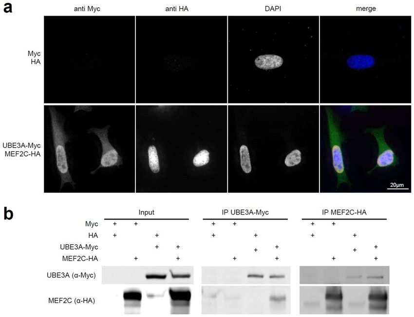
www.nature.com/scientificreports/                                                                      www.nature.com/scientificreports

                            condition                 ubi.   wing                  eye                 neur.     glia
                            single knockdown
                            KD_Ube3a_1                N      N                     N18                 N         N
                            KD_Ube3a_2                N      N                     N                   N         N
                                                             P (curled, alt.
                            KD_Mef2                   L                            P (bubbles)         N         N
                                                             veins)62
                                                                                   P (red.
                            KD_Da                     (L)    P1                                        N         N
                                                                                   bristles),18
                            KD_XNP                    N      N1                    N18                 N         N
                            single overexpression
                            OE_Ube3a_1                L      mL,P (curled)         P (rough)           (L)       N
                            OE_Ube3a_2                L      (mL),P                P (rough)           N         N
                            OE_Mef2                   L      P33                   P (red. bristles)   N         mL, (L)
                            OE_Da                     L      mL,P                  L                   (mL)      L
                                                             P (crumbled,
                            OE_XNP                    L                            N                   N         L
                                                             curled, alt. veins)
                            OE_Zfh1                   L      L                     L                   L         L
                            pairwise manipulation
                            KD_Ube3a_1;KD_Mef2        L      P↑                    P↑                  N         N
                            OE_Ube3a_1;OE_Mef2        L      mL, P↓                P↓                  (L)       L↓
                            KD_Ube3a_1;OE_Mef2        L      P                     P                   N         L↓
                            OE_Ube3a_1;KD_Mef2        L      mL,P↓                 (mL)↓, P↓           L↓        L↓
                            KD_Ube3a;KD_XNP           N      N                     N                   N         N
                            OE_Ube3a_1;OE_XNP         L      mL,P                  P                   (L)       L
                            KD_Ube3a_1;OE_XNP         L      P↓                    N                   N         (L)
                            OE_Ube3a_1;KD_XNP         L      (mL)↑, P              P                   mL ↓      N
                            KD_Ube3a_1; KD_Da         (L)    P                     P                   N         N
                            OE_Da;OE_Ube3a_2          L      mL,P                  L                   (mL)      L
                            OE_Da;KD_Ube3a_2          L      (L)↓                  L                   (mL)      L
                            OE_Ube3a_1;KD_Da          L      (mL),P                P                   (L)↓      N
                            OE_Da;OE_Mef2             L      (L)↓                  (L)                 N(↑)      L
                            OE_Da;KD_Mef2             L      mL,P                  L                   (mL)      L
                            OE_Da;OE_XNP              L      mL,P                  mL↑,P               (mL)      L
                            OE_Da;KD_XNP              L      mL,P                  L                   (mL)(↓)   L
                            OE _Zfh1;OE_Ube3a_2       L      L                     L                   L         L
                            OE_Zfh1;KD_Ube3a_2        (L)    L                     L                   L         L
                            OE_Zfh1;OE_Mef2           L      L                     L                   L         L
                            OE_Zfh1;KD_Mef2           L      L                     L                   L         L
                            OE_Zfh1;OE_XNP            L      L                     L                   L         L
                            OE _Zfh1;KD_XNP           L      L                     L                   L         L
                            OE_Zfh1;KD_Da             L      L                     L                   L         L

                           Table 1. Viability and morphology upon tissue-specific dosage manipulation. Ubi., ubiquitous; neur., pan-
                           neuronal; N, normal viability and normal morphology; L, lethality; mL, male lethality; P, morphological
                           phenotype; ↑, milder phenotype compared to single KD or OE; ↓, more severe phenotype compared to single
                           KD or OE; (), incomplete penetrance or borderline phenotype/modification; some of the phenotypes have
                           been reported previously for the same line or condition; References indicate reports with similar phenotypic
                           observations (absence of phenotype or presence of identical phenotype) in previous studies.

                           neuromuscular junctions (NMJs) such as NMJ area, NMJ length, number of islands, branches, boutons or active
                           zones did not reveal any consistent alterations in synaptic morphology upon either pan-neuronal knockdown or
                           overexpression of any of the genes (Supplementary Fig. S4). We did not observe a satellite bouton phenotype (data
                           not shown) as previously described in an Ube3a mutant23. This might be related to a weaker knockdown of the
                           RNAi line. Furthermore, gross neurological functioning was tested using the climbing assay upon pan-neuronal
                           knockdown or overexpression. We found that overexpression of Da or XNP resulted in a mild climbing defect,
                           respectively, and that overexpression of Ube3a led to a severe climbing deficit (Supplementary Fig. S5). Additional
                           testing of seizure susceptibility with the bang sensitivity assay (Supplementary Table S3) did not reveal consistent
                           phenotypes for any of the tested KD and OE lines.

                           Genetic interaction screen.       Based on the location of RNAi- or UAS-elements on chromosomes 2 or 3,
                           we could create three lines combining two KD elements each, eight lines combining two OE elements each,
                           and twelve lines combining KD with OE elements each (Table 1, Fig. 1c). By quantitative RT-PCR we found no

Scientific Reports |   (2020) 10:1204 | https://doi.org/10.1038/s41598-020-58182-5                                                           3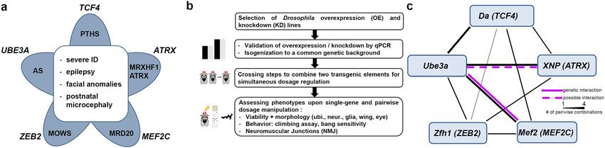
www.nature.com/scientificreports/                                                              www.nature.com/scientificreports

                           Figure 2. Genetic interaction of Ube3a and Mef2 or XNP in the Drosophila wing (ms1096-GAL4). (a)
                           Knockdown (KD) of Ube3a does not cause a wing phenotype, KD of Mef2 causes abnormally curled wings
                           in male flies with additional cross vein defects, such as missing anterior cross veins and/or ectopic cross
                           veins (marked with an arrow). Simultaneous KD of Ube3a and Mef2 results in a milder phenotype with
                           significantly more flies with both cross veins present and fewer flies with ectopic cross veins, quantified in (b).
                           (c) Overexpression (OE) of Ube3a causes abnormally curled wings in females, while KD of Mef2 does not cause
                           a phenotype in female flies (male phenotype see above). Simultaneous OE of Ube3a and KD of Mef2 results in
                           male lethality and in a more severe disorganization of wing architecture in about 75% of females, as quantified
                           in (d). (e) OE of XNP causes abnormally curled wings in female flies with additional cross vein defects, such
                           as missing anterior cross veins and/or ectopic cross veins (marked with an arrow). Simultaneous KD of Ube3a
                           (normal) and OE of XNP results in a more severe phenotype with more flies with ectopic cross veins and fewer
                           flies with both cross veins intact, as quantified in (f). Statistical analysis was performed using Fisher’s Exact
                           test, **p ≤ 0.001; ***p ≤ 0.0001). Flies are counted towards the more severe phenotype if at least one wing was
                           affected. These results are from an independent experiment than in Supplementary Table S2, thus numbers are
                           different.

                           indication that distribution of GAL4 between two UAS-elements would dilute its effect (Supplementary Fig. S1).
                           We tested all combinations for lethality and wing and eye morphology phenotypes. NMJ morphology, bang sen-
                           sitivity and climbing behaviour were assessed for the three KD combination constructs and for the eight OE
                           combinations.
                               Phenotypic modifications were considered as a) additive, when the combined phenotype represented the sum
                           of the two single phenotypes, b) as antagonistic, when the combined phenotype was milder than the more severe
                           of the two single phenotypes, and c) as synergistic, when the combined phenotype was different or more severe
                           than what was expected from the sum of the two single phenotypes.
                               In the evaluation of viability and gross morphologic eye and wing phenotypes upon ubiquitous and tissues
                           specific dosage manipulation we observed several modified phenotypes in the pairwise combinations (Table 1).
                           We considered those most indicative of a true genetic interaction, where modified phenotypes occurred at least
                           in three different KD/OE combinations and tissues and were markedly milder (antagonistic) or more severe or
                           different than expected from adding the single phenotypes (synergistic) (Fig. 1c).

Scientific Reports |   (2020) 10:1204 | https://doi.org/10.1038/s41598-020-58182-5                                                           4
www.nature.com/scientificreports/ www.nature.com/scientificreports
Figure 3. Genetic interaction of Mef2 and Ube3a in the Drosophila eye (GMR-GAL4). (a) Knockdown (KD)
of Ube3a does not cause a phenotype, KD of Mef2 causes rough eyes with more severely affected flies also
displaying a bubble-like appearance. Simultaneous KD of Ube3a and Mef2 results in a milder phenotype with
significantly fewer eyes with bubble-like appearance, quantified in (b) (***: p ≤ 0.001, Fisher’s Exact test). (c)
Overexpression (OE) of Ube3a causes rough eyes, OE of Mef2 results in a mildly reduced number of bristles but
grossly intact ommatidial structure. Simultaneous OE of both results in a severe phenotype with reduced eye
size and dissolved ommatidia structure in all eyes as quantified in (d). (e) Simultaneous OE of Ube3a (rough
eye) and KD of Mef2 (rough eyes and occasionally bubble-like appearance) results in a different and more severe
phenotype with disorganized ommatidia structure and progressive necrosis as quantified for male and female
flies in (f). Pictures and quantifications are from male flies if not indicated otherwise.
The most robust interaction was observed between Ube3a and Mef2. Combined knockdown in either wing
(Fig. 2a,b) or eye (Fig. 3a,b) resulted in an amelioration of the phenotype observed in the single conditions. Wing
venation defects as well as the rough eye with loss of ommatidia integrity, resulting in bubble-like appearance of
the eye surface, improved. Pairwise overexpression of Mef2 and Ube3a in either wing or eye led to a worsening
of phenotypes. In the wing, the only mildly abnormal, curled wings in females from each of the single condi-
tions were severely disorganized and not fully unfolded in the combined overexpression (Supplementary Fig. S1).
While each of the single overexpression conditions was associated with a mild eye phenotype only, their com-
bination resulted in a severe eye malformation with markedly reduced size and dissolved ommatidia structure
(Fig. 3c,d). Overexpression of Ube3a and simultaneous knockdown of Mef2 in either wing or eye also resulted in
increased phenotypic severity, compared to the single conditions. Wings showed a severe disorganization in 75%
of the females, which was not present in the single manipulations (Fig. 2c,d, Table 1). In the eye, the same com-
bination resulted in partial male lethality and progressive necrotic patches in eyes of females (Fig. 3e,f, Tables 1
and S2). While pan-neuronal knockdown of Mef2 did not affect viability and while pan-neuronal overexpres-
sion of Ube3a only resulted in partial lethality, a combination of these conditions resulted in complete lethality
(Tables 1, S2). A similar observation was made for glial deregulation. There, simultaneous overexpression of both
and simultaneous overexpression of Mef2 with knockdown of Ube3a and vice versa resulted in complete lethality,
while the single conditions were viable or only lethal in males (Tables 1, S2).
In summary, we have identified multiple phenotypic modifications upon pairwise dosage manipulation of
Ube3a and Mef2. The synergistic or antagonistic direction of an interaction was dependent on the combination of
KD and OE conditions and consistent across multiple tissues for each specific combination.
Also for combined dosage manipulation of Ube3a and XNP, phenotypic modifications were observed for
several combinations and tissues. Simultaneous overexpression of Ube3a and knockdown of XNP in the wing
resulted in increased viability of male flies (6% in OE_Ube3a alone and 39% in OE_Ube3a; KD_XNP, p < 0.001,
Scientific Reports | (2020) 10:1204 | https://doi.org/10.1038/s41598-020-58182-5 5www.nature.com/scientificreports/ www.nature.com/scientificreports
Figure 4. Physical interaction of human MEF2C and UBE3A in cells. (a) Co-localization studies of
overexpressed human tagged MEF2C (HA, red) and UBE3A (Myc, green) in HeLa cells show diffuse
nuclear localization of both proteins. (b) Co-immunoprecipitation experiments show physical interaction of
overexpressed human MEF2C and UBE3A in HEK293 cells. Immunoprecipitation of Myc-tagged UBE3A also
precipitates MEF2C-HA and vice versa. Please note, that the figure panels are cropped from two different blots
with different exposure times. For full blots see Supplementary Fig. S6.
Fisher’s exact test) and therefore in an improvement of the phenotype. In contrast, simultaneous knockdown of
Ube3a and overexpression of XNP in the wing led to a more severe phenotype in the females, with stronger dis-
organization of cross veins compared to the phenotype in the single lines (Fig. 2e,f, Tables 1, S2). Pan-neuronal
overexpression of Ube3a in combination with knockdown of XNP resulted in worsening of incompletely pen-
etrant lethality in females (20% of balancer flies in OE_Ube3a_1 alone and 10% in OE_Ube3a_1;KD_XNP,
p < 0.05, Fisher’s exact test) and complete lethality in males (76% vs. 0%, compared to females, p < 0.05, and 23%
vs. 0%, compared to balancer flies, p < 0.0001, Fisher’s exact test).
Pairwise manipulations of other tested genes did not result in multiple or consistent phenotypic modifications.
When we evaluated the climbing assay upon pairwise dosage manipulations, we observed a significantly increased
climbing impairment of the OE_Da;OE_XNP construct compared to the respective single conditions, which,
however, might represent an additive effect of both single gene conditions. No other consistent modifications of
the observed single phenotypes were observed (Supplementary Fig. S5). Analysis of NMJ morphology and bang
sensitivity upon pairwise dosage manipulations did not reveal any phenotypic modifications (Supplementary
Fig. S4, Supplementary Table S3).
Protein co-localization and interaction. To investigate whether UBE3A and MEF2C also physically
interact, we performed co-localization and co-immunoprecipitation studies in human cell lines. Firstly, we
could confirm that UBE3A and MEF2C both localize to the nucleus as described previously24 when simulta-
neously overexpressed in HeLa cells, indicating that a physical interaction is possible (Fig. 4a). Subsequently,
we could show that UBE3A and MEF2C can be co-immunoprecipitated from HEK293 cells co-transfected
with Myc-tagged UBE3A and HA-tagged MEF2C. This was true for immunoprecipitation of either Myc-tagged
UBE3A or HA-tagged MEF2C, suggesting that they indeed form a direct or indirect interaction (Figs. 4b, S6).
Using quantitative RT-PCR, we tested if UBE3A/Ube3a or MEF2C/Mef2 might regulate transcriptional levels of
each other. We did not find evidence for transcriptional effects as in fly larvae Mef2 levels were unaltered upon
Ube3a knockdown and vice versa (Supplementary Fig. S1). Furthermore, also in human cells (HEK293) MEF2C
expression levels were unaltered upon UBE3A knockdown using siRNAs (Supplementary Fig. S7).
Discussion
Though it has increasingly been acknowledged that similar clinical neurodevelopmental phenotypes are caused
by mutations in genes/proteins connected in common networks and processes, this has mostly been characterized
for specific complexes or pathways (e.g2,4.) or on a more systematic, large scale but consequently less detailed1,18
level. We now investigated possible functional links between TCF4, MEF2C, ZEB2, UBE3A and ATRX which
are all implicated in clinically overlapping, severe human neurodevelopmental disorders. To do this, we utilized
Scientific Reports | (2020) 10:1204 | https://doi.org/10.1038/s41598-020-58182-5 6www.nature.com/scientificreports/ www.nature.com/scientificreports
Drosophila melanogaster as a model system to systematically investigate functional links between the orthologues
of these genes/proteins (Da, Mef2, Zfh1, Ube3a, XNP) in vivo.
Drosophila lethality and morphological phenotypes point to developmental and glial roles.
Our initial screen for lethality and gross morphological phenotypes upon ubiquitous and tissue specific gene dos-
age manipulation in the fruit fly identified a large number of quantifiable lethality and morphological phenotypes
utilizable for the subsequent genetic interaction experiments.
In general, a high frequency of lethality upon ubiquitous but also upon tissue specific dosage manipulation
underlines both the dosage sensitivity of these genes and their role for developmental processes. This has been
shown in mouse models before. Ubiquitous knockout of either Zeb225, Tcf426, Mef2c27 or Atrx28 resulted in early
lethality, while Ube3a brain-specific maternal-deficient mice displayed neurological deficits29. Interestingly, not
only the loss of the maternal UBE3A copy in Angelman syndrome but also duplication of UBE3A in terms of
duplication 15q syndrome is associated with a neurodevelopmental and epilepsy phenotype30, thus demonstrating
bi-directional dosage sensitivity. We observed more frequent and more severe phenotypes upon overexpression
than upon knockdown. Though knockdown might be a more suitable model for loss-of-function mechanisms,
it might not always be phenotypically penetrant, particularly with RNA interference approaches as these usually
leave a residual expression level of 30–50%. Therefore, overexpression screens provide a valuable additional tool
to investigate gene function31–33 and to generate quantifiable phenotypes.
Contrary to our expectations for NDD-relevant genes, we did not observe consistent synaptic or behavioural
phenotypes upon pan-neuronal manipulation in the utilized assays. Although all of the investigated genes are
associated with epilepsy in humans, neither pan-neuronal knockdown nor overexpression of any single condition
or combination resulted in bang sensitivity, a Drosophila model for seizure susceptibility, where mechanical shock
can induce hyperactivity, spasms and paralysis34. This might suggest that seizures associated with haploinsuffi-
ciency of these transcriptional regulators might be related to different pathomechanisms from seizure-associated
ion channel dysfunction which is typically reflected in bang sensitivity in flies35. Interestingly, in accordance
with our findings, a previous report did not observe bang sensitivity upon neuronal overexpression of Ube3a as
a model for epilepsy-associated duplications 15q11.2, either, but instead upon glial overexpression concomitant
with down regulation of an ion pump36. Correlating a glial role of Ube3a between Drosophila and vertebrates
is, however, difficult due to a discordant imprinting status of Ube3a in Drosophila neurons37, and as in a mouse
model, Ube3a has shown to be expressed in glia but not to be imprinted there38. However, a possible, so far
underestimated glial role of Ube3a and other genes investigated in this study would be in accordance with our
observations that glial overexpression of either Zfh1, Da, XNP or Mef2 resulted in lethality while this was only
the case for pan-neuronal overexpression of Zfh1 and with reduced penetrance for pan-neuronal overexpres-
sion of Ube3a. Although a relevance of Zfh1/ZEB2, XNP/ATRX or Da for (mainly peripheral) glia development
and maintenance has been reported or discussed39–43, it has not yet been characterized in detail. Transcriptome
analysis on cell populations from mouse brain and on human brain tissues summarized in the Brain RNA-Seq
database indicates expression of all five genes not only in neurons but also in astrocytes, oligodendrocytes and
microglia, with a higher expression of TCF4, UBE3A and ATRX in fetal compared to mature astrocytes44,45. Our
observations therefore might suggest not only a role of neuronal but also of glial defects that might contribute to
the neurodevelopmental phenotypes in humans with mutations in either of these genes.
Genetic interaction between Ube3a and Mef2. Though the phenotypic overlap between the neurode-
velopmental disorders investigated in this study is widely appreciated and discussed in the literature7,46, corre-
sponding experimental follow-up of possibly underlying molecular commonalities is largely lacking. According
to the mouse brain atlas47, there is overlapping expression of orthologues of UBE3A, MEF2C, ATRX, ZEB2 and
TCF4 in several neuronal subtypes including excitatory neurons of the cerebral cortex and various cell types of
the hippocampus. This would be in line with molecular commonalities in the pathomechanisms of the associated
NDDs. However, according to the STRING database48 (status July 2019), there is no experimental evidence on
interaction between the human proteins so far, and available data is restricted to co-expression or interaction in
other species. In our genetic interaction screen, we observed modification of phenotypes upon combination of
several manipulated genes. Severe phenotypes such as severe and fully penetrant lethality upon Zfh1 overexpres-
sion precluded further evaluation for genetic interaction. For all other combinations, we observed modified phe-
notypes which might point to a functional link between these proteins in terms of genetic interaction (Table 1).
Often, only two tissues and/or combinations were involved. More evidence was there for genetic interaction
between Ube3a and XNP with modified phenotypes in two tissues and three different combinations. Further
experimental follow-up would be necessary to confirm these potential interactions.
The most stringent evidence for a functional interaction was detected between Ube3a and Mef2. Several lines of
evidence point to a true genetic interaction: a) we observed modified phenotypes in several tissues and in various
knockdown/overexpression combinations, b) additive effects only can be excluded as some phenotypes occurred
only upon pairwise manipulation (e.g. neuronal lethality) or were different or much more severe than expected from
both single conditions (e.g. eye phenotypes), and c) we observed both antagonistic (milder phenotypes in eyes and
wings upon simultaneous knockdown of Ube3a and Mef2) and synergistic effects (more severe phenotypes upon all
other combinations). Additionally, we confirmed a physical interaction between human UBE3A and MEF2C in an
independent cell-based system. To our knowledge, this is the first evidence for a functional link between UBE3A/
Ube3a and MEF2C/Mef2 that might contribute to the phenotypic overlap between Angelman syndrome and
MEF2C-associated intellectual disability. Apart from that, variants in either UBE3A or MEF2C might also represent
modifiers for the phenotypic expression/severity of the respective other condition as discussed for CNV models21,22.
Taking into account the diverse functional roles of UBE3A and MEF2C, there are different possibilities con-
ceivable how their interaction or mutual regulation might work. Observations from the genetic interaction and
Scientific Reports | (2020) 10:1204 | https://doi.org/10.1038/s41598-020-58182-5 7www.nature.com/scientificreports/ www.nature.com/scientificreports
expression studies might already provide some insights into the possible nature of these interactions. The UBE3A
gene encodes a member of the large family of E3 ubiquitin ligase proteins, initially termed E6-associated protein
(E6-AP), and contributing to protein homeostasis by being involved in tagging substrate proteins with ubiquitin
which are then degraded in the proteasome9. MEF2C (myocyte enhancer factor 2) belongs to the subfamily of
MADS (MCM1-agamous-deficiens-serum response factor) transcription factors, whose transcriptional activ-
ity relies on the recruitment of and cooperation with other transcription factors as well as on translational and
posttranslational modifications49. MEF2C might therefore be a transcriptional regulator of UBE3A expression.
This might be supported by the identification of MEF2 binding sites in the Ube3a promoter50. Vice versa, also
for UBE3A a transcriptional co-activation function has been reported51. However, a transcriptional regulation
mechanism appears unlikely as in flies expression levels of Mef2 were unaltered upon Ube3a knockdown and vice
versa, and as in a human cell line UBE3A knockdown did not change MEF2C expression levels. Additionally, stem
cell-derived neurons modelling Angelman syndrome, did not show significant transcriptional changes of other
genes (compared to 15q duplication neurons)52. As the most likely hypothesis, we suggest that UBE3A might reg-
ulate MEF2C activity and levels by ubiquitination, leading to subsequent degradation in the proteasome. UBE3A
has been found to be located in the neuronal nuclei in discrete hotspots over euchromatin, thus well-positioned
to regulate active genes24, and such a ubiquitin-ligase-dependent regulation has been reported for other transcrip-
tions factors before in mouse models, e.g. for SOD153. Interestingly, only very recently, the critical importance
of the nuclear isoform of UBE3A for the Angelman syndrome phenotype was characterized. Mice lacking the
nuclear isoform but not mice lacking the cytosolic isoform displayed all major behavioural phenotypes and syn-
aptic deficits also seen upon complete UBE3A knockout in the previous Angelman syndrome mouse model54. The
hypothesis of ubiquitin-ligase dependent regulation of a putative nuclear substrate such as MEF2C by UBE3A is
also supported by our genetic interaction findings. Pairwise knockdown of Ube3a and Mef2 in the fly resulted in
antagonistic genetic interaction with ameliorated eye and wing phenotypes. This might be explained by knock-
down of Ube3a leading to decreased ubiquitination and degradation of Mef2. Subsequently increased Mef2 levels
might result in a partial compensation of the Mef2 knockdown phenotype. All other combinations resulted in
synergistic genetic interaction, i.e. more severe phenotypes. This would be in line with a) overexpression of Ube3a
resulting in increased ubiquitination and degradation of Mef2 and thus in a further decrease of its already low
knockdown levels and b) knockdown of Ube3a resulting in decreased ubiquitination and degradation of Mef2
and thus in increased Mef2 abundance even above its overexpression levels. The observed interaction of MEF2C
and UBE3A in co-immunoprecipitation experiments supports such a link. However, specific experimental proof
of UBE3A regulating MEF2C in an ubiquitin-dependent fashion is still required.
By using Drosophila melanogaster as a model to screen for genetic interactions and by subsequent
co-immunoprecipitation in a human cell line, we identified a robust interaction between UBE3A/Ube3a and
MEF2C/Mef2. These molecular commonalities might contribute to the clinically overlapping features of the asso-
ciated disorders.
Material and Methods
Drosophila lines and conditions. Drosophila orthologues of TCF4 (daughterless [CG5102]), ZEB2 (Zfh1
[CG1322]), MEF2C (Mef2 [CG1429]), ATRX (XNP [CG4548]), and UBE3A (Ube3a [CG6190]) were identi-
fied with DIOPT55. Manipulation of Da/TCF4 in Drosophila has been established as a specific model for PTHS
previously56.
All RNAi lines and the respective control (VDRC no. 60000) were obtained from the Vienna Drosophila
Resource Center (VDRC)57. GAL4-driver stocks and transgenic lines for overexpression of Zfh1 and XNP,
respectively, were obtained from the Bloomington Drosophila Stock Center. UAS-Mef2 was obtained from
the Zurich ORFeome Project (FlyORF) 31, and the UAS-Da line was a gift from Pascal Heitzler (IBMP
Strasbourg). For generation of the UAS-Ube3a lines, the gene was amplified from fly cDNA (forward:
5’-GTAAAGTGCGCAGATTTCAGC-3’, reverse: 5’-GGTATCAGTTCCAGATGACAGAC-3’) and cloned into
a pUAST fly expression vector58. After verification of the sequence, the vector was sent to BestGene Inc. for the
creation of a stable transgenic line. For a complete list of used Drosophila lines see Supplementary Table S1. All
overexpression lines were isogenized to the VDRC 60000 control by backcrossing for at least seven generations.
Double-transgenic fly lines were generated using a double balancer line (Kr/CyO;D/TM6C) (Supplementary
Table S2). Overexpression or RNAi-mediated knockdown was induced with the UAS-Gal4 system58 and con-
firmed by quantitative RT-PCR (Supplementary Fig. S1). In the text and figures, RNAi-lines are referred to as
KD_gene, and UAS-lines as OE_gene. Flies were maintained on standard food, containing cornmeal, sugar, agar
and yeast at 25 °C and bred at 28 °C because of temperature sensitivity of the UAS-GAL4 system59.
Lethality and morphology analysis. RNAi- and transgenic overexpression lines were crossed to five dif-
ferent driver lines: GMR-GAL4 (eye), ms1096-GAL4 (wing), repo-GAL4 (glia cells), elav-GAL4 (pan-neuronal)
™
and Actin-GAL4 (II, ubiquitous). Resulting offspring were counted and analysed with a Carl Zeiss 2000C stereo
microscope for gross morphological abnormalities. If the driver line contained a balancer chromosome, offspring
with balancers were counted, too. In crosses with double OE/KD constructs retaining balancers, the expected
ratio of balancer to non-balancer offspring may deviate from 50%. Wings and eyes were analysed per fly, and
a phenotype was counted when it occurred in at least one wing or eye per fly, respectively. Wings were visually
evaluated for parameters such as shape, degree of unfolding and wing vein structure. Eyes were visually evaluated
for parameters such as ommatidial structure, bristles and gross size and shape. If applicable, p-values were deter-
mined using Fisher’s Exact test.
Climbing and bang sensitivity assays. Climbing behaviour and bang sensitivity was performed as
described elsewhere60 upon pan-neuronal manipulation (elav-GAL4). In brief, offspring were collected within
Scientific Reports | (2020) 10:1204 | https://doi.org/10.1038/s41598-020-58182-5 8www.nature.com/scientificreports/ www.nature.com/scientificreports
72 h of eclosion under CO2 anaesthesia in groups of ten (5 males, 5 females, at least 40 flies tested per genotype).
After 24 h, flies were transfered to the testing vial, tapped to the bottom and filmed for 30 s while climbing up.
Time until 70% of the flies had crossed line at 8.8 cm height was measured from the videos. If less than 70% of flies
from a vial managed to cross the line within the videotaped interval, time was considered to be 30 s. P-values were
determined using the Wilcoxon-Mann-Whitney test, and Bonferroni correction was applied for multiple testing.
For testing bang sensitivity, flies were vortexed for 10 s and filmed while recovering. The fraction of flies within a
vial displaying spasms 5 s after vortexing was determined from the videotapes.
Analysis of neuromuscular junctions (NMJs) from L3 larvae. Analysis of type 1b neuromus-
cular junctions (NMJs) of muscle 4 was performed as previously described60. Male L3 non-GFP larvae upon
pan-neuronal manipulation (elav-GAL4/CyO-GFP;elav-GAL4) were dissected in PBST, fixated in 4% paraform-
aldehyde and stained with nc82 and anti-discs large antibodies (both from the Developmental Studies Hybridoma
Bank, Iowa City, IA). Secondary antibodies used were Alexa 488 labeled anti-mouse antibody and the Zenon ™
™
Alexa Fluor 546 Mouse IgG1 Labeling Kit (ThermoFisher Scientific). Images were taken with a Zeiss Axio
Imager Z2 microscope in z-stacks and analysed in ImageJ61. NMJ area and length, as well as the number of
active zones, synaptic islands, branches, and synaptic boutons were determined from the image stacks. Per gen-
otype, at least 11 NMJs from at least four independent larvae were analysed. P-Values were determined using the
Wilcoxon-Mann-Whitney test, and Bonferroni correction was applied for multiple-testing. Upon Mef2 over-
expression, we observed an additional signal in cross-segmental neurons with Dlg staining (red channel). This
signal was also present in the parental Mef2 line, in another line from the same background, and was also present
without Dlg staining (Supplementary Fig. S8). It therefore most likely represents a background signal from the
RFP gene under control of the artificial 3xP3 promoter present in the Fly ORF lines.
RNA samples. For RNA sampling from flies, whole larvae (~5), adult flies (~4), heads (~10) or larval brains
(~40) were collected and frozen at −80 °C for at least one hour. Total RNA was isolated with the RNeasy Lipid
™
Tissue Mini Kit (Qiagen) using TRIzol (ThermoFisher Scientific) instead of QIAzol and QIAshredder col-
umns (Qiagen) for homogenization. DNAse digestion was performed on-column with the RNAse-free DNAse
™
kit (Qiagen). Reverse transcription of RNA into cDNA was performed using the SuperScript II reverse tran-
scriptase (ThermoFisher Scientific). RNA from HEK293 cells was isolated using the RNeasy Minikit. DNAse
digestion and reverse transcription was performed as described above.
Quantitative reverse transcriptase PCR (quantitative RT-PCR). Expression analysis was performed
using the ABsolute QPCR SYBR Green ROX Mix (ThermoFisher Scientific) and (whenever possible) exon
™
spanning primers (sequences available on request) on a QuantStudio 12 K Flex System (Life Technologies).
Reactions were performed in quadruplicates, and Ct values were normalized to those of the endogenous controls
Actin or Tubulin for Drosophila experiments or B2M for experiments on human cells. Relative expression levels
were obtained using the ∆∆Ct method with isogenic background lines (Drosophila) or cells transfected with
scrambled siRNA as references. Results were confirmed in at least a second biological replicate.
Immunofluorescence. Expression plasmids expressing human MEF2C and UBE3A and respective negative
control plasmids were obtained from Sino Biologicals (MEF2C-HA: HG12320-CY, UBE3A-Myc:HG11130-CM,
pCMV-Myc:CV014 and pCMV-HA:CV013) and used for transient transfection. Transfected HeLa cells were
grown on poly-lysine coated coverslips, fixated with 4% paraformaldehyde in PBS for 10 minutes and stained
with anti-Myc (M4439, Sigma-Aldrich) and anti-HA (H6908, Sigma-Aldrich) and with Alexa Fluor 488 goat ™
™
anti–mouse and Alexa Fluor 488 donkey anti–rabbit antibodies (A11001 and A10040, Thermo Fisher). Nuclei
were counterstained with DAPI (Serva). Images were taken with a Zeiss Axio Imager Z2 Apotome microscope
with a 63x objective and analyzed in ImageJ.
Co-Immunoprecipitation. HEK293 cells were transiently transfected with a combination of UBE3A-Myc
and MEF2C-HA or the respective negative controls and harvested 48 h post transfection. Cells were scraped
from the culture dish in lysis buffer (100 mM TRIS-HCl pH8, 150 mM NaCl, 1 mM EDTA, 1% Triton X-100).
Immunoprecipitation was performed with Protein A Mag Sepharose bead suspension (GE Healthcare), incu-
bated with the sample and anti-Myc or anti-HA antibodies (M4439 or H6908, Sigma-Aldrich) at 4 °C overnight.
Subsequently, beads were washed in lysis buffer, and samples were eluted with 1x Lämmli buffer.
® ™
Proteins were separated in stain-free 4–20% Mini-PROTEAN TGX Precast Protein Gels (Bio-Rad),
™
blots were stained with anti-Myc and anti-HA antibodies and imaged on a ChemiDoc Touch Imaging System
(Bio-Rad).
siRNA knockdown. Two different siRNAs against UBE3A (Qiagen) were transiently transfected into
HEK293 cells using jetPrime (Polyplus) according to the manufacturer’s instructions. 48 h post transfection, RNA
for expression analysis (see description above), was harvested.
Data availability
The datasets generated and/or analyzed during the current study are available from the corresponding author on
reasonable request.
Received: 8 September 2019; Accepted: 13 January 2020;
Published: xx xx xxxx
Scientific Reports | (2020) 10:1204 | https://doi.org/10.1038/s41598-020-58182-5 9www.nature.com/scientificreports/ www.nature.com/scientificreports
References
1. Kochinke, K. et al. Systematic Phenomics Analysis Deconvolutes Genes Mutated in Intellectual Disability into Biologically Coherent
Modules. American journal of human genetics 98, 149–164, https://doi.org/10.1016/j.ajhg.2015.11.024 (2016).
2. Bahi-Buisson, N. et al. The wide spectrum of tubulinopathies: what are the key features for the diagnosis? Brain 137, 1676–1700,
https://doi.org/10.1093/brain/awu082 (2014).
3. Zaghloul, N. A. & Katsanis, N. Functional modules, mutational load and human genetic disease. Trends in genetics: TIG 26, 168–176,
https://doi.org/10.1016/j.tig.2010.01.006 (2010).
4. Zenker, M. Clinical manifestations of mutations in RAS and related intracellular signal transduction factors. Curr Opin Pediatr 23,
443–451, https://doi.org/10.1097/MOP.0b013e32834881dd (2011).
5. Kleefstra, T. et al. Disruption of an EHMT1-associated chromatin-modification module causes intellectual disability. American
journal of human genetics 91, 73–82, https://doi.org/10.1016/j.ajhg.2012.05.003 (2012).
6. Bogershausen, N. & Wollnik, B. Mutational Landscapes and Phenotypic Spectrum of SWI/SNF-Related Intellectual Disability
Disorders. Front Mol Neurosci 11, 252, https://doi.org/10.3389/fnmol.2018.00252 (2018).
7. Tan, W. H., Bird, L. M., Thibert, R. L. & Williams, C. A. If not Angelman, what is it? A review of Angelman-like syndromes.
American journal of medical genetics. Part A 164A, 975–992 (2014).
8. Kishino, T., Lalande, M. & Wagstaff, J. UBE3A/E6-AP mutations cause Angelman syndrome. Nat Genet 15, 70–73, https://doi.
org/10.1038/ng0197-70 (1997).
9. Matsuura, T. et al. De novo truncating mutations in E6-AP ubiquitin-protein ligase gene (UBE3A) in Angelman syndrome. Nat
Genet 15, 74–77, https://doi.org/10.1038/ng0197-74 (1997).
10. Wakamatsu, N. et al. Mutations in SIP1, encoding Smad interacting protein-1, cause a form of Hirschsprung disease. Nat Genet 27,
369–370, https://doi.org/10.1038/86860 (2001).
11. Zweier, C. et al. Haploinsufficiency of TCF4 causes syndromal mental retardation with intermittent hyperventilation (Pitt-Hopkins
syndrome). American journal of human genetics 80, 994–1001, https://doi.org/10.1086/515583 (2007).
12. Zweier, M. et al. Mutations in MEF2C from the 5q14.3q15 microdeletion syndrome region are a frequent cause of severe mental
retardation and diminish MECP2 and CDKL5 expression. Hum Mutat 31, 722–733, https://doi.org/10.1002/humu.21253 (2010).
13. Villard, L. et al. Splicing mutation in the ATR-X gene can lead to a dysmorphic mental retardation phenotype without alpha-
thalassemia. American journal of human genetics 58, 499–505 (1996).
14. Bellen, H. J., Tong, C. & Tsuda, H. 100 years of Drosophila research and its impact on vertebrate neuroscience: a history lesson for
the future. Nat Rev Neurosci 11, 514–522, https://doi.org/10.1038/nrn2839 (2010).
15. Bier, E. Drosophila, the golden bug, emerges as a tool for human genetics. Nature reviews. Genetics 6, 9–23, https://doi.org/10.1038/
nrg1503 (2005).
16. Coll-Tane, M., Krebbers, A., Castells-Nobau, A., Zweier, C. & Schenck, A. Intellectual disability and autism spectrum disorders ‘on
the fly’: insights from Drosophila. Dis Model Mech 12, https://doi.org/10.1242/dmm.039180 (2019).
17. van der Voet, M., Nijhof, B., Oortveld, M. A. & Schenck, A. Drosophila models of early onset cognitive disorders and their clinical
applications. Neurosci Biobehav Rev 46(Pt 2), 326–342, https://doi.org/10.1016/j.neubiorev.2014.01.013 (2014).
18. Oortveld, M. A. et al. Human intellectual disability genes form conserved functional modules in Drosophila. PLoS genetics 9,
e1003911, https://doi.org/10.1371/journal.pgen.1003911 (2013).
19. McGary, K. L. et al. Systematic discovery of nonobvious human disease models through orthologous phenotypes. Proceedings of the
National Academy of Sciences of the United States of America 107, 6544–6549, https://doi.org/10.1073/pnas.0910200107 (2010).
20. Mani, R., St Onge, R. P., Hartman, J. L. T., Giaever, G. & Roth, F. P. Defining genetic interaction. Proceedings of the National Academy
of Sciences of the United States of America 105, 3461–3466, https://doi.org/10.1073/pnas.0712255105 (2008).
21. Grice, S. J., Liu, J. L. & Webber, C. Synergistic Interactions between Drosophila Orthologues of Genes Spanned by De Novo Human
CNVs Support Multiple-Hit Models of Autism. Plos Genet 11, https://doi.org/10.1371/journal.pgen.1004998 (2015).
22. Iyer, J. et al. Pervasive genetic interactions modulate neurodevelopmental defects of the autism-associated 16p11.2 deletion in
Drosophila melanogaster. Nat Commun 9, 2548, https://doi.org/10.1038/s41467-018-04882-6 (2018).
23. Li, W. et al. Angelman Syndrome Protein Ube3a Regulates Synaptic Growth and Endocytosis by Inhibiting BMP Signaling in
Drosophila. PLoS genetics 12, e1006062, https://doi.org/10.1371/journal.pgen.1006062 (2016).
24. Burette, A. C. et al. Subcellular organization of UBE3A in neurons. J Comp Neurol 525, 233–251, https://doi.org/10.1002/cne.24063
(2017).
25. Van de Putte, T. et al. Mice lacking ZFHX1B, the gene that codes for Smad-interacting protein-1, reveal a role for multiple neural
crest cell defects in the etiology of Hirschsprung disease-mental retardation syndrome. American journal of human genetics 72,
465–470, https://doi.org/10.1086/346092 (2003).
26. Bergqvist, I. et al. The basic helix-loop-helix transcription factor E2-2 is involved in T lymphocyte development. Eur J Immunol 30,
2857–2863, 10.1002/1521-4141(200010)30:103.0.CO;2-G (2000).
27. Edmondson, D. G., Lyons, G. E., Martin, J. F. & Olson, E. N. Mef2 gene expression marks the cardiac and skeletal muscle lineages
during mouse embryogenesis. Development 120, 1251–1263 (1994).
28. Garrick, D. et al. Loss of Atrx affects trophoblast development and the pattern of X-inactivation in extraembryonic tissues. PLoS
genetics 2, e58, https://doi.org/10.1371/journal.pgen.0020058 (2006).
29. Miura, K. et al. Neurobehavioral and electroencephalographic abnormalities in Ube3a maternal-deficient mice. Neurobiol Dis 9,
149–159, https://doi.org/10.1006/nbdi.2001.0463 (2002).
30. Conant, K. D. et al. A survey of seizures and current treatments in 15q duplication syndrome. Epilepsia 55, 396–402, https://doi.
org/10.1111/epi.12530 (2014).
31. Bischof, J. et al. A versatile platform for creating a comprehensive UAS-ORFeome library in Drosophila. Development 140,
2434–2442, https://doi.org/10.1242/dev.088757 (2013).
32. Prelich, G. Gene overexpression: uses, mechanisms, and interpretation. Genetics 190, 841–854, https://doi.org/10.1534/
genetics.111.136911 (2012).
33. Schertel, C. et al. A large-scale, in vivo transcription factor screen defines bivalent chromatin as a key property of regulatory factors
mediating Drosophila wing development. Genome Res 25, 514–523, https://doi.org/10.1101/gr.181305.114 (2015).
34. Pavlidis, P. & Tanouye, M. A. Seizures and failures in the giant fiber pathway of Drosophila bang-sensitive paralytic mutants. J
Neurosci 15, 5810–5819 (1995).
35. Lee, J. & Wu, C. F. Genetic modifications of seizure susceptibility and expression by altered excitability in Drosophila Na(+) and
K(+) channel mutants. Journal of neurophysiology 96, 2465–2478, https://doi.org/10.1152/jn.00499.2006 (2006).
36. Hope, K. A., LeDoux, M. S. & Reiter, L. T. Glial overexpression of Dube3a causes seizures and synaptic impairments in Drosophila
concomitant with down regulation of the Na(+)/K(+) pump ATPalpha. Neurobiol Dis 108, 238–248, https://doi.org/10.1016/j.
nbd.2017.09.003 (2017).
37. Hope, K. A., LeDoux, M. S. & Reiter, L. T. The Drosophila melanogaster homolog of UBE3A is not imprinted in neurons. Epigenetics
11, 637–642, https://doi.org/10.1080/15592294.2016.1214783 (2016).
38. Grier, M. D., Carson, R. P. & Lagrange, A. H. Toward a Broader View of Ube3a in a Mouse Model of Angelman Syndrome:
Expression in Brain, Spinal Cord, Sciatic Nerve and Glial Cells. PloS one 10, e0124649, https://doi.org/10.1371/journal.pone.0124649
(2015).
Scientific Reports | (2020) 10:1204 | https://doi.org/10.1038/s41598-020-58182-5 10www.nature.com/scientificreports/ www.nature.com/scientificreports
39. Ohayon, D. et al. Zfh1 promotes survival of a peripheral glia subtype by antagonizing a Jun N-terminal kinase-dependent apoptotic
pathway. EMBO J 28, 3228–3243, https://doi.org/10.1038/emboj.2009.247 (2009).
40. He, L. et al. Transcriptional Regulator ZEB2 Is Essential for Bergmann Glia Development. J Neurosci 38, 1575–1587, https://doi.
org/10.1523/JNEUROSCI.2674-17.2018 (2018).
41. Sun, X., Morozova, T. & Sonnenfeld, M. Glial and neuronal functions of the Drosophila homolog of the human SWI/SNF gene
ATR-X (DATR-X) and the jing zinc-finger gene specify the lateral positioning of longitudinal glia and axons. Genetics 173,
1397–1415, https://doi.org/10.1534/genetics.106.057893 (2006).
42. Wada, T. et al. Neuroradiologic features in X-linked alpha-thalassemia/mental retardation syndrome. AJNR Am J Neuroradiol 34,
2034–2038, https://doi.org/10.3174/ajnr.A3560 (2013).
43. Nelson, H. B. & Laughon, A. Drosophila glial development is regulated by genes involved in the control of neuronal cell fate. Roux
Arch Dev Biol 204, 118–125, https://doi.org/10.1007/BF00361106 (1994).
44. Zhang, Y. et al. An RNA-sequencing transcriptome and splicing database of glia, neurons, and vascular cells of the cerebral cortex. J
Neurosci 34, 11929–11947, https://doi.org/10.1523/JNEUROSCI.1860-14.2014 (2014).
45. Zhang, Y. et al. Purification and Characterization of Progenitor and Mature Human Astrocytes Reveals Transcriptional and
Functional Differences with Mouse. Neuron 89, 37–53, https://doi.org/10.1016/j.neuron.2015.11.013 (2016).
46. Vrecar, I. et al. Further Clinical Delineation of the MEF2C Haploinsufficiency Syndrome: Report on New Cases and Literature
Review of Severe Neurodevelopmental Disorders Presenting with Seizures, Absent Speech, and Involuntary Movements. Journal of
pediatric genetics 6, 129–141, https://doi.org/10.1055/s-0037-1601335 (2017).
47. Zeisel, A. et al. Molecular Architecture of the Mouse Nervous System. Cell 174, 999–1014 e1022, https://doi.org/10.1016/j.
cell.2018.06.021 (2018).
48. Szklarczyk, D. et al. STRING v10: protein-protein interaction networks, integrated over the tree of life. Nucleic Acids Res 43,
D447–452, https://doi.org/10.1093/nar/gku1003 (2015).
49. Potthoff, M. J. et al. Regulation of skeletal muscle sarcomere integrity and postnatal muscle function by Mef2c. Mol Cell Biol 27,
8143–8151, https://doi.org/10.1128/MCB.01187-07 (2007).
50. Greer, P. L. et al. The Angelman Syndrome protein Ube3A regulates synapse development by ubiquitinating arc. Cell 140, 704–716,
https://doi.org/10.1016/j.cell.2010.01.026 (2010).
51. El Hokayem, J., Weeber, E. & Nawaz, Z. Loss of Angelman Syndrome Protein E6AP Disrupts a Novel Antagonistic Estrogen-Retinoic
Acid Transcriptional Crosstalk in Neurons. Mol Neurobiol 55, 7187–7200, https://doi.org/10.1007/s12035-018-0871-9 (2018).
52. Urraca, N. et al. Significant transcriptional changes in 15q duplication but not Angelman syndrome deletion stem cell-derived
neurons. Mol Autism 9, 6, https://doi.org/10.1186/s13229-018-0191-y (2018).
53. Mishra, A. et al. E6-AP association promotes SOD1 aggresomes degradation and suppresses toxicity. Neurobiol Aging 34, 1310
e1311–1323, https://doi.org/10.1016/j.neurobiolaging.2012.08.016 (2013).
54. Avagliano Trezza, R. et al. Loss of nuclear UBE3A causes electrophysiological and behavioral deficits in mice and is associated with
Angelman syndrome. Nat Neurosci 22, 1235–1247, https://doi.org/10.1038/s41593-019-0425-0 (2019).
55. Hu, Y. et al. An integrative approach to ortholog prediction for disease-focused and other functional studies. BMC Bioinformatics
12, 357, https://doi.org/10.1186/1471-2105-12-357 (2011).
56. Tamberg, L., Sepp, M., Timmusk, T. & Palgi, M. Introducing Pitt-Hopkins syndrome-associated mutations of TCF4 to Drosophila
daughterless. Biol Open 4, 1762–1771, https://doi.org/10.1242/bio.014696 (2015).
57. Dietzl, G. et al. A genome-wide transgenic RNAi library for conditional gene inactivation in Drosophila. Nature 448, 151–156,
https://doi.org/10.1038/nature05954 (2007).
58. Brand, A. H. & Perrimon, N. Targeted gene expression as a means of altering cell fates and generating dominant phenotypes.
Development 118, 401–415 (1993).
59. Duffy, J. B. GAL4 system in Drosophila: a fly geneticist’s Swiss army knife. Genesis 34, 1–15, https://doi.org/10.1002/gene.10150
(2002).
60. Straub, J. et al. Missense Variants in RHOBTB2 Cause a Developmental and Epileptic Encephalopathy in Humans, and Altered
Levels Cause Neurological Defects in Drosophila. American journal of human genetics 102, 44–57, https://doi.org/10.1016/j.
ajhg.2017.11.008 (2018).
61. Schneider, C. A., Rasband, W. S. & Eliceiri, K. W. NIH Image to ImageJ: 25 years of image analysis. Nat Methods 9, 671–675 (2012).
62. Crittenden, J. R., Skoulakis, E. M. C., Goldstein, E. S. & Davis, R. L. Drosophila mef2 is essential for normal mushroom body and
wing development. Biol Open 7, https://doi.org/10.1242/bio.035618 (2018).
Acknowledgements
We thank Annette Schenck and Pascal Heitzler for providing fly lines. We also thank André Reis for continuous
support. Fly stocks were obtained from the Bloomington Drosophila Stock Center (NIH P40OD018537) and
the Vienna Drosophila Resource Center (http://www.vdrc.at). Antibodies from the Developmental Studies
Hybridoma Bank (created by the NIH National Institute of Child Health and Human Development) were used
in this study. C.Z. is supported by grants from the German Research Foundation (ZW184/1-2, ZW184/3-1, and
270949263/GRK2162) and by the Interdisciplinary Center for Clinical Research in Erlangen (E26, E31).
Author contributions
J.S., T.S., L.D., C.S. and A.G. performed the fly experiments. A.G. performed cell-based experiments. A.B.E., A.F.
and F.F. contributed to data analysis. J.S. and A.G. prepared the figures, and C.Z. initiated and supervised the
project and with help of A.G. wrote the manuscript. All authors reviewed the manuscript.
Competing interests
The authors declare no competing interests.
Additional information
Supplementary information is available for this paper at https://doi.org/10.1038/s41598-020-58182-5.
Correspondence and requests for materials should be addressed to C.Z.
Reprints and permissions information is available at www.nature.com/reprints.
Publisher’s note Springer Nature remains neutral with regard to jurisdictional claims in published maps and
institutional affiliations.
Scientific Reports | (2020) 10:1204 | https://doi.org/10.1038/s41598-020-58182-5 11You can also read