Crystal structure of P. falciparum Cpn60 bound to ATP reveals an open dynamic conformation before substrate binding
←
→
Page content transcription
If your browser does not render page correctly, please read the page content below
www.nature.com/scientificreports
OPEN Crystal structure of P. falciparum
Cpn60 bound to ATP reveals
an open dynamic conformation
before substrate binding
Brian Nguyen, Rui Ma, Wai Kwan Tang, Dashuang Shi & Niraj H. Tolia*
Plasmodium falciparum harbors group 1 and group 2 chaperonin systems to mediate the folding
of cellular proteins in different cellular locations. Two distinct group 1 chaperonins operate in the
organelles of mitochondria and apicoplasts, while group 2 chaperonins function in the cytosol. No
structural information has been reported for any chaperonin from plasmodium. In this study, we
describe the crystal structure of a double heptameric ring Plasmodium falciparum mitochondrial
chaperonin 60 (Cpn60) bound with ATP, which differs significantly from any known crystal structure
of chaperonin 60. The structure likely represents a unique intermediate state during conformational
conversion from the closed state to the opened state. Three of the seven apical domains are highly
dynamic while the equatorial domains form a stable ring. The structure implies large movements
of the apical domain in the solution play a role in nucleotide-dependent regulation of substrate
binding and folding. A unique 26–27 residue insertion in the equatorial domain of Plasmodium
falciparum mitochondrial chaperonin greatly increases both inter-ring and intra-ring subunit–subunit
interactions. The present structure provides new insights into the mechanism of Cpn60 in chaperonin
assembly and function.
Abbreviations
Cpn10 Chaperonin with molecular weight about 10 kDa
Cpn60 Chaperonin with molecular weight about 60 kDa
Hsp60 Heat shock protein with molecular weight of ~ 60 kDa
NCS Non-crystallographic symmetry
SP Substrate protein
Group 1 and group 2 chaperonins are two distinct classes of chaperones that exist in diverse organisms to assist
with protein folding in an ATP-dependent manner1,2. Group 1 chaperonins are found in the cytosol of bacteria,
such as GroEL from Escherichia coli or Cpn60 from Thermus Thermophilus, while in eukaryotic organisms, they
are located in the chloroplast or mitochondria3. Group 2 chaperonins include the archaeal thermosome, the
eukaryote tailless complex polypeptide 1 (CCT), and the eukaryotic tailless complex polypeptide 1 ring complex
(TRiC)4. In both chaperone groups, each monomer is comprised of three domains: an equatorial domain, an
intermediate domain, and an apical domain. Seven or eight monomers assemble together to form a heptameric
or octameric ring, and two rings interact back-to-back through the equatorial domains to form tetradecameric
or hexadecameric double-ring s tructures4.
Most of the group 1 chaperonins are homo-oligomers. In contrast, the hexadecamer of group 2 chaperonins
are composed of eight different monomers despite having similar three-dimensional s tructures2. The equatorial
domains contain the ATP binding site, the intermediate domain serves as a hinge connecting the equatorial to the
apical domain, and the apical domain is involved in substrate binding. Group 1 chaperonins require the binding
of a co-chaperone Cpn10 to the apical domain to encapsulate the substrate protein (SP), while the apical domain
of group 2 chaperonins utilizes a built-in lid. The binding of Cpn10 to group 1 chaperonins is critical for the
folding mechanism that forms bullet-shaped (Cpn6014Cpn107), American football-shaped (Cpn6014Cpn1014), or
American half-football-shaped (Cpn607Cpn107) complexes at distinct stages of the folding mechanism (Fig. 1).
Host‑Pathogen Interactions and Structural Vaccinology Section, Laboratory of Malaria Immunology and Vaccinology,
Division of Intramural Research, National Institute of Allergy and Infectious Disease, National Institutes of Health,
Rm 4NN08, Building 29B, 9000 Rockville Pike, Bethesda, MD 20892, USA. *email: niraj.tolia@nih.gov
Scientific Reports | (2021) 11:5930 | https://doi.org/10.1038/s41598-021-85197-3 1
Vol.:(0123456789)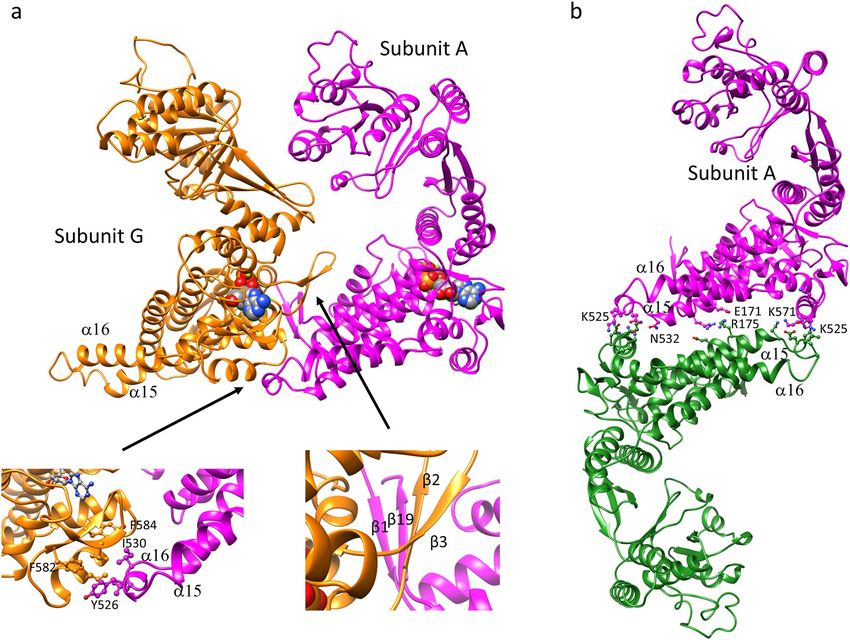
www.nature.com/scientificreports/
Figure 1. Reaction mechanism of GroEL/GroES. (a) Asymmetric cycle of GroEL. Adapted and modified from
Hayer-Hartl et al.1 and Yan et al.16. (b) Symmetric cycle of GroEL. Adapted and modified from Lizuka and
Funatsu10. Violet semi-circle represents a Cpn10 heptamer, blue or green rectangles/trapezoids represent the
closed/open conformation of two distinct Cpn60 heptamers, tan elongated/condensed line represents unfolded
or folded SP.
Since the first GroEL structure from E. coli5, multiple structures of group 1 chaperonin from 6 different
organisms have been deposited into the Protein Data Bank (PDB) (Supplementary Table S1). A vast major-
ity of structures are from E. coli, making E. coli GroEL and its partner GroES the best-characterized group 1
chaperonin. GroEL consists of two heptameric rings of identical monomers, each with a molecular weight of
approximately 60 kDa (also termed Cpn60 or Hsp60 in other organisms). The partner co-chaperonin, GroES, is
a dome-shaped heptameric ring of identical monomers with a monomeric molecular weight of approximately
10 kDa (also termed Cpn10 or Hsp10 in other organisms). GroEL requires ATP binding in order to shift from
a closed conformational state to an open conformational state that allows for SP and GroES binding6. In this
open conformation, the intermediate and apical domains of GroEL rotate and move upward to form a complex
Scientific Reports | (2021) 11:5930 | https://doi.org/10.1038/s41598-021-85197-3 2
Vol:.(1234567890)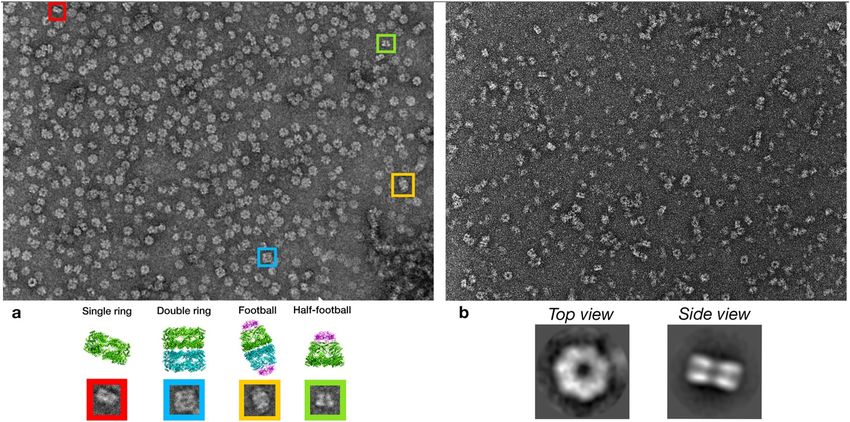
www.nature.com/scientificreports/
with GroES. Two cycles have been suggested for the function of GroEL. The prevailing asymmetric cycle which
(Fig. 1a) states that only one heptameric ring can partake in folding at a time6–9 and the symmetric cycle10–15
(Fig. 1b) where both rings can fold simultaneously. Recent research has also determined another mechanism of
GroEL within the asymmetric cycle, inter-ring separation, where ATP hydrolysis and binding causes disassocia-
tion between the two heptameric rings16.
However, there is evidence that chaperonins from organisms other than E. coli may have slightly different
mechanisms. It is therefore imperative to study group 1 chaperonins from diverse organisms to holistically define
the mechanism of chaperonin action. For example, the structure determination of a human mitochondrial chap-
eronin demonstrated that a novel intermediate exists with subunit asymmetry within the rings (different confor-
mations for intra-ring subunits) and nucleotide symmetry between the rings (ATP or ADP binds both rings)17.
Furthermore, the human chaperonin forms football-shaped complexes with a different inter-ring arrangement
from that of GroEL17. Additionally, the crystal structure of the football complex with both rings bound to ADP
suggests that human Cpn60 follows a symmetrical cycle, where both rings can undergo hydrolysis at the same
time. Cryo-electron microscopy recently revealed that in addition to the full football-shaped C pn6014Cpn1014
(PDB: 6MRC), the half-football-shaped complex consisting of C pn607Cpn107 (PDB: 6MRD) can independently
exist in solution, with both forms active in assisting the folding of imported mitochondrial p roteins18; this sug-
19
gests ring separation may be a possible step during the chaperonin cycle . Similarly, the bullet-shaped structure
from T. thermophilus exhibited a significant deviation from the sevenfold symmetry for the apical domain
around the cis-cavity20. Studies on the chaperonin from Chlamydomonas chloroplast demonstrated a significantly
different mechanism for chaperonin to capture, encapsulate, fold, and release substrate proteins. The binding
and hydrolysis of ATP induce not only both positive (intra-ring) and negative (inter-ring) cooperative actions,
but also promotes the partial disassembly of chaperonin into monomer, a phenomenon unique to chloroplast
chaperonin21,22. Research on Cpn60 from Paracocus denitrificans indicated that the inter-ring interaction in this
Cpn60 is weakened compared to E. coli GroEL, thus both single ring and double ring forms were observed23. In
summary, due to structural and functional variations between Cpn60 orthologs, studies on Cpn60 from diverse
organisms are warranted to better define the overall mechanism of Cpn60.
Malaria remains a major public health problem worldwide resulting in nearly half a million deaths and
200 million clinic illnesses annually24. Cpn60 is a ubiquitous protein that exists in nearly all domains of life
including bacteriophage25 and plays an essential role in maintaining protein homeostasis26. In yeast, deletion of
mitochondrial Cpn60 is lethal27. In mice, the inactivation of its homolog causes embryonic m ortality28. Heat-
shock proteins such as Cpn60 are often upregulated in response to stressful conditions. PfCpn60 must cope with
two radically different host environments, mosquito and human, making it an interesting ortholog to study. In
order to understand its mechanism in P. falciparum, we report the first crystal structure of P. falciparum Cpn60
(PfCpn60) bound with ATP, which reveals the large conformational dynamic of the apical domain relative to
the intermediate and equatorial domains.
Results
Negative stain electron microscopy of WT‑PfCpn60 and D474A‑PfCpn60. Both group 1 and 2
chaperonins can be identified in the genome of the malaria parasite P. falciparum. Two group 1 Cpn60 genes
exist, with one located on chromosome 12 that expresses a Cpn60 believed to localize to m itochondria29,30, and
a second located on chromosome 10 with an encoded Cpn60 that is expected to function specifically inside the
apicoplast30,31. Compared to known structures of Cpn60, the mitochondria-specific P. falciparum Cpn60 con-
tains insertions including a 26–27 residue insert in the equatorial domain and a 10 residue insert in the apical
domain (Supplementary Fig. S1), as well as N- and C-terminal extensions.
After nickel-his affinity and size-exclusion chromatography purification, both WT-PfCpn60 and ATP hydrol-
ysis-deficient mutant D472A-PfCpn60 were submitted to negative stain electron microscopy. In the WT-PfCpn60
sample, several forms of structures including single-ring, double-ring, half-football, and whole football coex-
ist (Fig. 2a). In contrast, most structures in the D474A-PfCpn60 sample are single-ring structures (Fig. 2b).
Consistent with this observation, crystals were grown only from the D474A-PfCpn60 sample. No crystals were
grown from WT-PfCpn60 probably due to the structural heterogeneity caused by the fast hydrolysis of ATP in
WT-PfCpn60 which occurs in the matter of s econds14. Comparatively, it is estimated that the D474A mutation
slows hydrolysis to a rate of around 40 min. Since only the D474A-PfCpn60 crystal structure was determined,
the following results and discussion of the PfCpn60 structure is based on D474A-PfCpn60.
Structure determination of D474A‑PfCpn60. Matthew’s coefficient calculation estimated that there
are seven subunits in an asymmetric unit in the crystal structure of PfCpn60, with a coefficient of 3.9 and an
estimated solvent content of 68%32, a significantly higher solvent content than most other proteins due to the
large cavity inside the chaperonin structure. A blast search of the PDB database indicated the closest model of
PfCpn60 was the Cpn60 structure from T. thermophilus (TtCpn60, PDB: 4V4O) with a sequence identity of
39%20. The structure of 4V4O is a bullet-shaped complex, C pn6014Cpn107, with one heptameric ring in closed
conformation with no nucleotide bound, and the other heptameric ring in opened conformation with ADP and
Cpn10 binding20. Since PfCpn60 was co-purified with PfCpn10, the opened conformation of TtCpn60 was used
as the search model first. However, no molecular replacement solution was found.
Interestingly, no molecular replacement solution was found using the closed conformation of TtCpn60 as
well, implying the conformation of PfCpn60 in the current structure may differ from both conformations of
TtCpn60 in 4V4O. We hypothesized that the flexibility within the intermediate hinge region may have caused
the positions of the equatorial and apical domains to differ from the search model. Therefore, in order to iden-
tify the molecular replacement solution, the individual domains were used as the search model. The molecular
Scientific Reports | (2021) 11:5930 | https://doi.org/10.1038/s41598-021-85197-3 3
Vol.:(0123456789)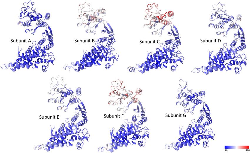
www.nature.com/scientificreports/
Figure 2. Negative stain electron microscopy analysis of PfCpn60. (a) Wild-type Cpn60 co-purified with Cpn10
with 1 mM ATP. Bottom panel shows the represented view of single ring, double ring, football and half-football
structures of WT-Cpn60. (b) ATP hydrolysis deficient D474A mutant co-purified with Cpn10 with 1 mM ATP.
Bottom panel represents the 2D classification of the negative stains, demonstrating the top view and side view of
D474A PfCpn60 mutant, which is in a single ring conformation.
replacement solution was identified using the equatorial domain alone from both conformations of the TtCpn60
structure. Furthermore, the equatorial domain from the closed conformation ring performed better as a search
model than the opened domain, indicating that the current structure is closer to the closed conformation. Care-
ful inspection of molecular packing for the molecular replacement solution indicated that enough empty space
was available for the packing of the intermediate plus apical domains, but not enough for the additional Cpn10
binding to the apical domain, suggesting that Cpn10 may not be present in the current structure. Eventually, the
molecular replacement solution was successfully found by searching one copy of the heptamer of the equatorial
domain alone and seven copies of the intermediate plus apical domain. The final solution gave a total likelihood
gain of 7360.
Inspection of the molecular replacement solution indicated that Cpn60 is well-packed in the crystal struc-
ture, confirming the absence of Cpn10. A reason for Cpn10 not co-crystallizing with Cpn60 may be due to weak
interaction between Cpn10 and Cpn60 resulting in the loss of Cpn10 during purification, due to specific condi-
tions during crystallization, or due to the ATP binding state. We performed a negative stain electron microscopy
analysis which revealed that the major species existing in solution is a heptamer single ring structure of Cpn60
without the presence of Cpn10 (Fig. 2b), even though both Cpn60 and Cpn10 were present on the SDS-PAGE
gel following gel filtration purification (Supplementary Fig. S2). In addition, the low pH of 4.2 in the crystalliza-
tion buffer may have further prevented the formation of the Cpn10 and Cpn60 complex. Finally, the eventual
shift from open to closed conformation by ATP hydrolysis may have hampered Cpn10 binding and retention.
One heptameric ring was found per asymmetric unit of the PfCpn60 structure. The sevenfold NCS axis was
almost parallel with the crystallographic C-axis. A second heptameric ring in an adjacent asymmetric unit that
is related by twofold crystallographic symmetry results in a tetradecameric complex with the rings stacked back-
to-back, as observed in the other Cpn60 structures. The seven subunits in the asymmetric unit are in slightly
different conformations and differ from the closed and opened conformations typically observed for other Cpn60
molecules (Fig. 3a,b, Table 1 and Supplementary Table S2). The apical domain in PfCpn60 has a relative angle
movement of as large as − 42° and 92° compared to the closed and opened conformations (subunit A and subunit
H in 4V4O), respectively, which were calculated by first superimposing the equatorial domain as the reference,
then determining the angle of rotation required to superimpose the apical domain using the secondary structure
superimpose tool in COOT (Fig. 3b). This large movement explained why the search models from both closed
and opened conformations were unsuccessful in molecular replacement. A search on the PDBeFold server33
(https: //www.ebi.ac.uk/ssm) with the final refined structure revealed that the current PfCpn60 structure is closest
to that of ATP-bound EcGroEL structure (PDB ID: 2C7E)34, with a root mean square deviation (RMSD) of 2.3 Å.
The electron density in the equatorial domain is of very high quality with strong backbone density and clearly
identifiable side chains. In particular, the electron density for the 26–27 extra amino residues between K509
and E536, compared to E. coli and T. thermophilus Cpn60 sequence (Supplementary Fig. S1), is traceable and
a model can be built (Supplementary Fig. S3). The diverse conformations and flexibility of the apical domain
conformations hampered structure determination of these segments due to poor electron density, particularly
Scientific Reports | (2021) 11:5930 | https://doi.org/10.1038/s41598-021-85197-3 4
Vol:.(1234567890)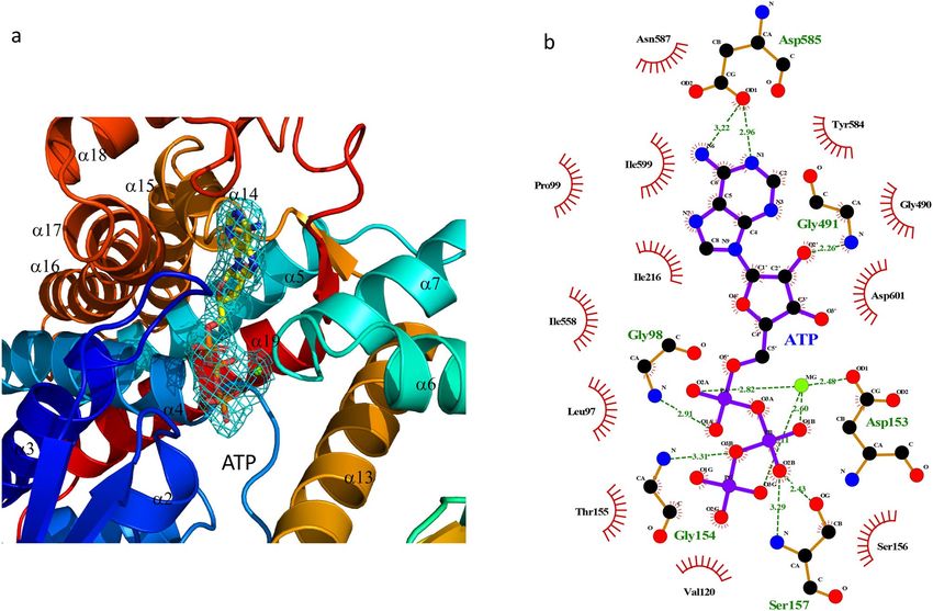
www.nature.com/scientificreports/

                                  Figure 3.  Structure of PfCpn60 and comparison with TtCpn60. (a) Superimposition of seven subunits, shown
                                  in different color ribbons, in the asymmetric unit using equatorial domain showing the variations of the apical
                                  and intermediate domains. Subunit G (shown in purple ribbons) is in the most closed conformation while
                                  subunit D (shown in red ribbons) is in the most opened conformation. (b) The conformational differences
                                  of PfCpn60 (subunit A, shown in brown ribbons) with the opened conformation of TtCpn60 (4V4O, subunit
                                  A, shown in light-blue ribbons) and the closed conformation (4V4O, subunit H, shown in pink ribbons). The
                                  superimposition was carried on using their equatorial domain. (c) Superimposition of PfCpn60 (subunit A,
                                  shown in magenta ribbons) and TtCpn60 (closed form shown in light-blue ribbons) for the equatorial domain
                                  only. The 27-residue insert is highlighted. (d) Superimposition of PfCpn60 (shown in magenta ribbons) and
                                  TtCpn60 (closed form shown in light-blue ribbons) for the apical domain only. (e) Superimposition of PfCpn60
                                  (shown in magenta ribbons) and TtCpn60 (closed form shown in light-blue ribbons) for the intermediate
                                  domain only. The 10-residue insert is highlighted.

                                   Subunit   Apical domain   Intermediate domain
                                   A         0               0
                                   B         0.7a            1.2
                                   C         1.3a            1.3
                                   D         2.4             1.4
                                   E         1.9             1.4
                                   F         1.1a            1.8
                                   G         − 7.3           − 1.0

                                  Table 1.  Relative rotation angle (°) of apical and intermediate domains. a The electron density of apical
                                  domains in subunit B, C and F is weak. Thus, the values are not reliable.

                                  in the apical domains of subunit B, C and F. However, the electron density of the apical domains of subunits A,
                                  D, E, and G allowed for structural modeling, particularly with chain A having good electron density and model-
                                  building properties.
                                      The flexibility observed in the PfCpn60 structure may have functional significance in the binding of Cpn10
                                  and substrates. Although the electron density of the apical domains for subunit B, C, and F are weak, the signifi-
                                  cant residual electron density in their corresponding regions indicated their presence and dynamic nature. In
                                  order to maintain the completeness of the model, the models of the apical domains in subunits B, C, and F were
                                  created through NCS operation of the apical domain of subunit A as a template.

                                  Structure of P. falciparum mitochondrial Cpn60. The overall structure of PfCpn60 represents a typical
                                  group 1 chaperonin, with a sevenfold symmetrical cylinder consisting of two back-to-back stacked rings com-
                                  posed of seven subunits (Fig. 4a,b). The N- and C-termini are angled towards the inner cavity while the α11–α12
                                  loop of the apical domain is located on the outermost surface of the cylinder. The two rings were exactly related
                                  to each other by the two-fold crystallographic symmetry. Similar to the structures of other Cpn60 orthologs,
                                  each subunit consisted of an equatorial, intermediate, and apical domain (Fig. 3a).
                                      The equatorial domain consists of 11 α-helices in the core and four anti-parallel two-stranded β sheets
                                  on the surface. The ATP binding-site is located on the edge of the equatorial domain with a phosphate group
                                  binding to the N-terminal end of α4 helix, and adenosine and sugar groups surrounded by β16, α17, β18, and
                                  β15–α14, α2–β2 loops (Fig. 3C). The intermediate domain is composed of three α helix bundle (α6, α7, and
                                  α13) and one three-stranded β sheets (β4↑β14↑β5↓) (Fig. 3E). The apical domain is formed by double layers
                                  of β-sheets, which consist of one four-stranded β-sheet (β11↓β8↑β9↑β10↑) and one four-stranded antiparallel

Scientific Reports |   (2021) 11:5930 |                https://doi.org/10.1038/s41598-021-85197-3                                                   5

                                                                                                                                               Vol.:(0123456789)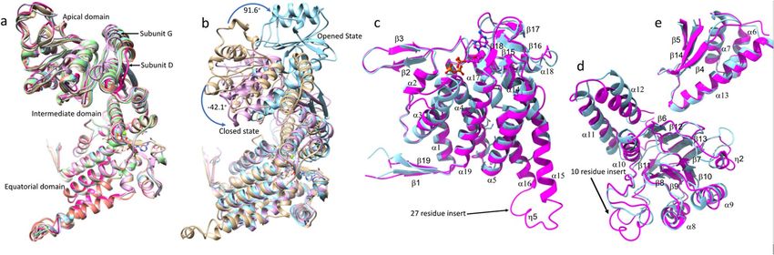
www.nature.com/scientificreports/
Figure 4. Structure of PfCpn60 oligomer. (a) The top view of PfCpn60 tetradecamer. The protein was shown in
rainbow ribbon colored from blue (N-termini) to red (C-termini). Two termini form an anti-parallel β-strand
arrangement towards to the inner cavity. (b) The side-view of PfCpn60 tetradecamer. The top heptameric ring
interacts with the bottom heptameric ring in a back-to-back manner and are related to each other by twofold
crystallographic symmetry. The protein is shown in ribbon with subunits G and A in orange and magenta,
respectively. The interacting subunit in the opposite ring is shown in forest green. Bound ATP is shown as filled
spheres.
β-sheet (β6↓β12↑β13↓β7↑), as a core with a three α helix bundle (α10, α11, and α12) hanging on one side of the
edge and other three helices (α8, α9 and η2) on the other side of the edge (Fig. 3d). In the open conformation of
TtCpn60, the residues from equivalent helix α8 and α9 interact with residues from the mobile loop of C pn1020.
Although structures of diverse Cpn60 orthologs with double heptameric rings have been determined (Sup-
plementary Table S1), a vast majority of them are closed-conformations of apo-Cpn60 without ATP or ADP
bound. ATP-bound symmetric double-ring structures have only been determined at low resolution by cryo-
electron microscopy34,35. No crystal structure for an ATP-bound double-ring lidless complex ( Cpn6014) of group
1 chaperonin currently exists, possibly due to the high flexibility of the apical domain in this conformation.
There are several unique features in the current PfCpn60 structure. In contrast to most other known Cpn60
structures, which are either in opened or closed conformation, the present PfCpn60 structure represents a unique
ATP-bound conformation. The superimposition of the current structure with the closed and opened conforma-
tion of TtCpn60 (using the equatorial domain) confirmed that the conformation of the PfCpn60 structure is a
unique ATP-induced conformation distinct from both the closed and open conformation (Fig. 3b). The whole
PfCpn60 structure cannot be superimposed to either opened or closed conformation complexes, but each domain
can be superimposed with those of the closed conformation of TtCpn60, with rsmd of 1.0, 1.1, and 2.0 Å for
equatorial, intermediate, and apical domains, respectively (Fig. 3c–e).
In comparison with the sequences of other Cpn60 orthologs with known structures (Supplementary Fig. S1),
PfCpn60 has two long insertions: an extra 10 amino residues between L378 and N387, and an extra 27 amino
residues between K510 and S536. The residues L378–N387, which form an extended loop between helix α10
and β11, are located at the edge of the apical domain close to helix α8, which is involved in the binding of co-
chaperone Cpn10 (Fig. 3d). The residues E513–E538 form two extended α-helices (α15 and α16) and a loop
with a short η5 helix to interact with an adjacent subunit in the intra-ring (Fig. 4a). Furthermore, the extended
α-helices are part of an inter-ring interface and may enhance inter-ring interactions (Fig. 4b).
Intra‑ring subunit–subunit interactions. The intra-ring subunit-subunit interactions consist of two
parts: inter-equatorial domain interactions, and intermediate domain and apical domain interaction (Fig. 5a).
The interactions among intermediate and apical domains vary with conformational change, but the interactions
between equatorial domains are consistent across all monomers. One general feature for intra-ring subunit-sub-
unit interactions is that an antiparallel β-loop (residues 103–117, β2↑β3↓) projects from the body of the equa-
torial domain towards the inner surface of its right-handed adjacent subunit, where it forms a parallel β-sheet
structure with its C-terminal segment (residues 624–629, β19↑). This C-terminal segment further interacts with
the N-terminal segment (residues 70–74, β1↓) in an anti-parallel β-strand arrangement to form a four-stranded
β-sheet (β1↓β19↑β2↑β3↓) that glues the two adjacent subunits together (Fig. 5a). The four-stranded β-sheet
Scientific Reports | (2021) 11:5930 | https://doi.org/10.1038/s41598-021-85197-3 6
Vol:.(1234567890)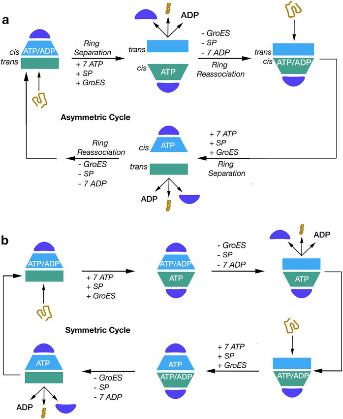
www.nature.com/scientificreports/
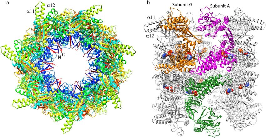
Figure 5. Intra-ring (a) and inter-ring (b) subunit–subunit interactions. Subunit A is shown in magenta ribbon
while the adjacent subunit G is shown in orange ribbon. The opposite subunit in the opposite ring is shown
in forest-green ribbon. ATP is shown as filled spheres. The selected side chains involved in the interaction are
shown in balls-sticks.
interaction across the intra-ring adjacent subunits is likely to be essential for Cpn60 to form a heptameric ring
since this feature is conserved in all Cpn60 structures in different conformational forms (1KP8, 1PCQ, and
5OPX) and from different organisms (4V4O, 1IOK, and 6MRC). Deletion mutagenesis of the C-terminal seg-
ment of EcGroEL impaired the EcGroEL assembly supporting this notion36.
One unique feature of PfCpn60 that impacts intra-ring subunit-subunit interactions is the extra 27 residue
insertion (residues 513–539, α15–α16) that forms an extended α-helix loop and contacts the left-handed intra-
ring subunit (Fig. 5a). This results in a much larger intra-ring subunit-subunit interaction area of 1828 Å2 in
PfCpn60 as compared to other Cpn60 structures (Table 2). Specifically, residues Y526, L529, and I530 from the
extended α-helix loop make hydrophobic interactions with residues I572, F582, Y584, and F591 from adjacent
intra-ring subunits (Fig. 5a).
Inter‑ring subunit‑subunit interactions. As revealed in the other Cpn60 structures, the two hepta-
meric rings contact each other primarily at two sites with two different opposite ring subunits, termed left and
right sites in the equatorial domain5. In the bullet-shaped EcGroEL structure (PDB: 1PCQ), two salt-bridges,
R452-E461 and van der Waals contacts among S463, V 464, and N467, form the right site contact. The van der
Waals contacts among K105, A108, and A109 in the cis-ring, and A109, G110, and M111 in the trans-ring,
contribute to the left site interactions. However, these residues are not conserved in PfCpn60 and the specific
inter-ring interactions are not directly comparable (Supplementary Fig. S1).
PDBePISA web s erver37 was used to analyze the inter-ring subunit–subunit interaction and the results are
listed in Table 2. The right site contacts of Cpn60 are better retained across different conformations than the
left site contacts due to the relative movement of inter-ring subunits. This relative movement was calculated by
superimposing the equatorial domain of one subunit in one of the heptameric rings, then determining the angle
of rotation required to superimpose the equatorial domain of the subunit in the opposite heptamer ring. With the
structure of PfCpn60 subunit A as the reference, the relative rotation angles are listed in Table 3 for Cpn60 from
different organisms. Nucleotide binding (1OEL vs. 1KP8) caused a slight relative equatorial domain movement
Scientific Reports | (2021) 11:5930 | https://doi.org/10.1038/s41598-021-85197-3 7
Vol.:(0123456789)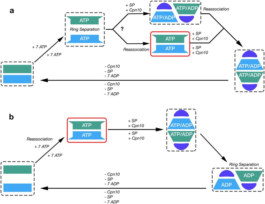
www.nature.com/scientificreports/

                                                                                             Inter-ring buried
                                                                                             area (Å2)
                                             PDB               Intra-ring buried area (Å2)   1         2         3
                                             This study        1828                          683.0     62.8      55.2
                                             1KP8              1486                          180.9     131.7
                                             1OEL              1542                          189.1     188.0
                                             1PCQcis           1581                          198.4     197.8
                                             1PCQtrans         1557
                                             5OPX              1697                          197.1     0.0
                                             1IOK              1193                          198.5     161.4
                                             5DA8              1284                          234.8     152.1
                                             5CDI              1442                          209.0     167.8
                                             6MRC              1808                          165.1     73.9

                                            Table 2.  Comparison of subunit-subunit interactions among different Cpn60s.

                                             PDB          Inter-ring relative rotation angle (°)     Composition
                                             1KP8         6                                          EcCpn6014AGS14
                                             1OEL         6                                          EcCpn6014
                                             1PCQ         14                                         EcCpn6014Cpn107ADP7
                                             5OPX         26                                         EcCpn6014Cpn1014ADP14
                                             1IOK         8                                          PdCpn6014
                                             5DA8         7                                          CtCpn6014
                                             5CDI         4                                          CrCpn6014
                                             6MRC         29                                         HsCpn6014Cpn1014ADP14

                                            Table 3.  Relative movement of inter-ring subunit of other Cpn60 to PfCpn60.

                                            (< 1°). The formation of a bullet-shaped complex induced about 7° of the relative movement; as a result, the
                                            interactions of the left inter-ring contacts became weaker (1PCQ vs. 1OEL). The formation of football-shaped
                                            complexes (5OPX and 6MRC) causes a larger relative movement of ~ 20° (5OPX vs. 1OEL) for inter-ring subunits
                                            and weakens the left site interaction. Different forms of structures, which are caused by the binding of different
                                            nucleotides and substrate protein, appear to affect inter-ring interactions significantly.
                                                The inter-ring subunit-subunit interface buried area of 683 Å2 in PfCpn60 is much larger than the buried area
                                            of ~ 200 Å2 in other Cpn60 structures, due to the extra 27 residue insertion in PfCpn60 (Table 2). Approximately
                                            18–24 residues from both rings form the inter-ring interface. Specifically, five hydrogen bonding interactions
                                            of R175–E171, K571–E528, K577–E528, K571–N532, and K525–N575 and two salt bridges of R175–E171 and
                                            K577–E528 may be involved in forming this interface (Figs. 4a, 5b). L529 N565, N568, K571, N575 interact
                                            with residues K528, L529, N532 from an adjacent inter-ring subunit. In comparison to the EcGroEL structure
                                            with bound ATP analog (1KP8), a 6° relative angle movement was observed in the current PfCpn60 structure
                                            (Table 3), calculated by superimposing the equatorial domain of subunit A as a reference, then determining the
                                            angle of rotation required to superimpose the equatorial domain of the opposing subunit using the secondary
                                            structure superimpose tool in COOT (Fig. 5b). Different inter-ring interactions in PfCpn60 may affect ring
                                            association-disassociation and inter-ring cooperativity.

                                            The ATP binding site of Cpn60.             Inspection of the electron density indicated that the ATP is present in
                                            the conserved binding site (Fig. 6a and Supplementary Fig. S4b). ATP binds to PfCpn60 in a similar fashion as
                                            observed in EcGroEL (PDB: 2C7E and 1KP9) and TtCpn60 (PDB:4V4O). The side chains of D153, S156, and
                                            mainchain N of G154 are involved in hydrogen bonding to the phosphate group, the side chains of D691 are
                                            involved in hydrogen bonding to the sugar group, and sidechains of I599, P99, V586, I216, and I558 are involved
                                            in hydrophobic interaction to the adenosine ring. The hydrogen bond interactions of the side chains of N587
                                            and D585 help anchor the position of the adenosine group (Fig. 6b). The hydrogen bond interactions of the side
                                            chains of N587 and D585 also help anchor the position of the adenosine group. The phosphate group binding
                                            motif, A151–G152–D153–G154–T155, is absolutely conserved across known Cpn60 (Supplementary Fig. S1).
                                            Although the sequence of PfCpn60 aligns well with the sequences of other group 1 chaperonins, the residues
                                            between T144 and G154 between helix α3 and α4, the loop structure of PfCpn60 in which part of C-terminal ⍺3
                                            helix melts, is significantly different from other group 1 chaperonins (Supplementary Fig. S5). The flexibility of
                                            this loop appears to be important for the entry of ATP and the exit of ADP.

          Scientific Reports |   (2021) 11:5930 |                        https://doi.org/10.1038/s41598-021-85197-3                                         8

Vol:.(1234567890)
www.nature.com/scientificreports/
Figure 6. ATP binding site of PfCpn60. (a) The electron density map (2Fo–Fc) around bound ATP and Mg2+
with ATP are shown as ball-and-sticks, and the electron density is shown in sky-blue mesh cage. The figure was
generated using Pymol. (b) The interactions of ATP with surrounding residues. The ATP and Mg2+ are shown
as ball-and-sticks. Hydrogen bonds are shown as green dotted lines, while the spoked arcs represent protein
residues making nonbonded contacts with the ligand. The figure was made using L igplot+38.
Highly dynamic apical domains. The overall average B factor of the model was 163.5 Å2. In the model,
the equatorial domain had a relatively low B factor of 132.5 Å2, while the intermediate and apical domains
had higher B factors of 155.8 and 206.7 Å2, respectively, reflecting the different mobility of different domains
with the lowest being the equatorial domain and the highest being the apical domain (Table 4). This phenom-
enon has been observed in many other Cpn60 s tructures23,39. Furthermore, it was interesting to observe that the
temperature factors of the equatorial domains were similar for each intra-ring subunit, while the temperature
factors of the intermediate and apical domains were very different for each intra-ring subunit (Fig. 7, Supple-
mentary Table S3). Inspection of the molecular packing in the crystal revealed that a strong correlation exists
between the high-temperature factor and the amount of crystal lattice constraints. This correlation implies that
the intermediate domain, particularly the apical domain of PfCpn60, is extremely mobile in the solution and
can be ordered by packing in the crystal. In the current structure, the apical domains of subunit B, C, and F are
much more mobile (weak electron density and high-temperature factors) than those of subunit A, D, E, and G,
due to less interaction with other symmetry-related molecules. Crystallographic constraints may explain why
apical domains in different conformations were observed in individual intra-ring subunits in many other group
1 chaperonin structures17,20,40,41. The conformational variation for each subunit may also reflect chaperonin’s
functional requirement for the chaperonin to bind various protein substrates to facilitate their folding, rather
than a single protein.
Discussion
This structure is the first group 1 chaperonin structure from P. falciparum, the most prevalent parasite on the
African continent responsible for nearly half a million deaths and 200 million clinical illnesses each y ear24.
The availability of the PfCpn60 structure provides a new group 1 chaperonin from a different organism that
will help to better understand the overall function and mechanism of group 1 chaperonins. EcGroEL is the
best-characterized chaperonin and several different forms of structures have been determined (Supplementary
Table S1): (1) Apo or ADP or phosphothiophosphoric acid adenylate ester (AGS) bound double-ring C pn6014
closed form (1OEL, 4KI8, 1KP8)40–42; (2) ATP bound lidless double-ring C pn6014 partially opened form (2C7E
and 4AAQ)34,35; (3) ADP or ATP bound single lid bullet-shaped form, Cpn6014Cpn107 (1PCQ), with ADP or
ATP binding only to the cis ring43; (4) ATP or ADP + Be3F bound double lidded football-shaped opened form,
Scientific Reports | (2021) 11:5930 | https://doi.org/10.1038/s41598-021-85197-3 9
Vol.:(0123456789)www.nature.com/scientificreports/
Data collection
Space group P622
Cell dimensions
a, b, c (Å) 281.77, 281.77, 299.32
Resolution (Å) 63.0–3.69 (3.79–3.69)a
Total reflections 667,450
Unique reflections 74,184 (5,368)
Rmeas 0.133 (1.50)
I/σI 12.9 (1.7)
Completeness (%) 98.8 (98.3)
CC1/2 99.9 (60.7)
Redundancy 9.1 (8.1)
Wilson B factor (Å2) 117.9
Refinement
Resolution (Å) 48.2–3.69 (3.80–3.69)
No. of reflections 74,077 (7262)
Rwork/Rfree 0.227/0.277
No. of atoms 30,938
Protein 30,714
Ligand/ion 224
B-factors (Å2)
Protein 163.5
Equatorial domain 132.5
Intermediate domain 155.8
Apical domain 206.7
ATP 126.5
rmsd
Bond lengths (Å) 0.003
Bond angles (°) 0.803
Ramachandran plot (%)
Favored 92.60
Outliers 0.0
Table 4. Data collection and refinement statistics. a Values in parentheses are for highest-resolution shell.
Cpn6014Cpn1014 (3WVL and 4PKO), with ADP + Be3F or ATP binding to both rings44,45. The PfCpn60 D474A
mutant crystal structure revealed a symmetric double-ring complex with all subunits occupied by ATP that is
similar to pre-open ATP-bound GroEL Cryo-EM structures 4AAU and 4 AB235. Negative stain images reveal
varying forms of PfCpn60 for the wildtype (Fig. 2a) compared to the single ring form seen in the D474A mutant
(Fig. 2b). It is likely that the slowed hydrolysis in the mutant staggers the particles into a similar stage and allows
for the crystallization of intermediate conformations. Based on conformational studies on GroEL by Clare et al.,
this pre-open ATP-bound conformation occurs between the closed and open conformations, to prepare for SP
and Cpn10 b inding35. The most apparent feature shared among pre-open conformation structures is the outward
position of α11 and α12 and an inward shift of the intermediate d omain35.
In the presence of Cpn10 and ATP, Cpn60 from diverse organisms can be found to exist as in both single-
and double-ring f orms18,23. Furthermore, it was found that the single-ring assembly is enough for productive
chaperonin-mediated protein folding18,46 and human Cpn60 (also termed Hsp60) does not display any nega-
tive inter-ring cooperativity at any point of the reaction cycle. PfCpn60 has been observed in the single ring
conformation (Fig. 2b) suggesting the occurrence of inter-ring separation. This study is consistent with the two
prevailing models of Cpn60 a ction18,47, and the pre-open conformation described here can be readily placed in
both cycles (Fig. 8).
As in all other Cpn60 structures, both N- and C-termini are oriented inward towards the inner cavity. The
structure is based on PfCpn60 sequence (XP_001350715) from 69 to 631 only. After cleavage of the first 23
residues of the mitochondrial signal peptide, predicted based on the SignalP 3.0 S erver48, the mature PfCpn60
should have 46 extra residues at the N-terminus before G69, and 87 extra residues at the C-terminus after E631.
It is interesting to notice that there are 51 negatively charged residues of Asp + Glu at its C-terminus. The extra
residues protruding from the equatorial domain towards the inner cavity in the wild-type PfCpn60 certainly
block free passage between the cavities in both rings. The negative charges inside the equatorial ring in wild-type
PfCpn60 may also prevent double-ring formation. Further study of PfCpn60 in the native state may be essential
to fully understand the structure and mechanism of PfCpn60.
Scientific Reports | (2021) 11:5930 | https://doi.org/10.1038/s41598-021-85197-3 10
Vol:.(1234567890)www.nature.com/scientificreports/
Figure 7. Dynamic of the equatorial, the intermediate and the apical domains for individual subunit. The
ribbons are colored according to the B factors from low (blue) to high (red).
Although PfCpn60 was co-purified with Cpn10, it is interesting to note that Cpn60 did not crystallize with
Cpn10. The dissociation of Cpn10 from Cpn60 should not be due to the crystallization buffer because negative
stain electron microscopy did not observe the presence of Cpn10-bound complexes comprising the football or
bullet. The interactions between Cpn10 and Cpn60 are mainly through the interactions of the residues from the
mobile loop of Cpn10 and the residues from α8 and α9 of Cpn60, which include the critical triplet residues of
the mobile loop16,18,45. The triplet residues in PfCpn10 and L31-F32-L33 are comparable to the corresponding
triplets I31-M32-L33 in human mitochondrial Cpn10 and I25-V26-L27 in G roES30. The corresponding interact-
ing residues such as L303, E304, and L328 in Cpn60 are also conserved (Supplementary Fig. S1). Therefore, in
the presence of ATP and Cpn10, PfCpn60 should likely form a football-shaped or half-football-shaped complex
with Cpn10. The absence of Cpn10 in the current structure is likely due to the poor binding to Cpn60 in the
ATP-bound open form in the absence of SP.
The tetradecameric assembly of PfCpn60 is mainly held together by the subunit-subunit interaction of the
equatorial domains. The apical domain is highly dynamic; its position is affected easily by nucleotide binding,
protein substrate binding, and potentially the crystal packing environment. Asymmetry with subunits in a differ-
ing apical domain conformation has been observed in several Cpn60 crystal s tructures17,20. It is plausible that this
asymmetric phenomenon may reflect the extreme conformational flexibility of the Cpn60 apical domain in solu-
tion. Examples of subunit asymmetry are well-demonstrated by human Cpn60 structures, which were recently
determined by both X-ray crystallography and single-particle cryo-EM (PDB: 4PJ1 and 6MRC). Both structures
are American football-shaped Cpn6014Cpn1010 structures bound with ADP. In the X-ray crystallographic struc-
ture (4PJ1), the apical domains in two of the subunits (subunit G in one ring and subunit N in the opposite ring)
are very different from the conformations of the apical domains in other intra-ring subunits, exhibiting as large
as 100° of counterclockwise rigid body movement of the apical domain compared to those in other s ubunits17.
However, in the structure (6MRC) determined by cryo-EM, no such asymmetry phenomenon was observed, and
all subunits are in a similar conformation. The breakage of perfect seven-fold symmetry for intra-ring subunits
was reported previously but to a milder degree. In the structures of apo GroEL and ATP-bound GroEL (PDB:
1OEL and 1KP8), the conformational variations of the apical domains relative to the equatorial domains among
the intra-ring subunits were apparent, with spreads of 5° and 7° for apo and ATP-bound states, respectively40,41.
Because no distinct gap between these two states was observed, it was realized the variations were likely induced
by different packings due to different lattices ( C2221 for 1OEL and P
21 for 1KP8). Similarly, the apical domain
in the cis-ring of the bullet-shaped TtCpn6014Cpn107 complex exhibited large deviation from the seven-fold
symmetry20. The conformational variations of the apical domain in group 1 chaperonins are likely to be intrinsic
to encapsulate different SPs rather than a single SP.
Scientific Reports | (2021) 11:5930 | https://doi.org/10.1038/s41598-021-85197-3 11
Vol.:(0123456789)www.nature.com/scientificreports/
Figure 8. Possible models of the PfCpn60/Cpn10 reaction cycle, based on previous research, incorporating
the crystal structure (solid red box) and the possible states observed with negative stain electron microscopy in
Fig. 2 (dashed red box). Schematic drawing shows a simplified reaction cycle indicating ATP-bound as a distinct
state during the conversion from apo-closed state to Cpn10 + SP + ATP-bound opened state. In addition, the ring
dissociation due to ATP binding is shown with model a demonstrating the mechanism adapted from Yan et al.16.
and model b demonstrating the mechanism adapted from Gomez-Llorente et al.18. Violet semi-circle represents
a Cpn10 heptamer, blue or green rectangles/trapezoids represent the closed/open conformation of two distinct
Cpn60 heptamers, tan elongated/condensed line represents unfolded or folded SP.
Materials and methods
Plasmid construction, protein expression, and purification. The full sequence of Plasmodium fal-
ciparum (isolate FCR-3/Gambia) mitochondria Cpn60 was retrieved from Uniprot entry number P34940 and
then aligned to the crystal structure sequence of E. coli GroEL (PDB: 1AON). The flanking N and C terminal
residues of P. falciparum Cpn60 were truncated to the alignment of E. coli GroEL. A D474A mutation was made
in accordance to the ATP hydrolysis-deficient GroEL mutant (PDB: 3WVL)45 to inhibit ATP hydrolysis of Cpn60
and stabilize the complex for purification. Normal ATP binding is retained but ATP hydrolysis occurs at a 0.1%
rate14. The mutant follows the same cycle as the wild-type but at a much slower rate, allowing for the observation
of more transient intermediates. The overall structure of GroEL appears largely unaffected by the ATP hydrol-
ysis-deficient mutation when compared to the wild-type football complex (PDB: 4PKO) with a rmsd value
between the superimposed structures of 1.79 Å. The sequence was then synthesized by GenScript and cloned
into a pET-28a vector with kanamycin antibiotic resistance genes. The sequence of mitochondria Cpn10 from
P. falciparum was retrieved from Uniprot entry Q50JA6, synthesized by GenScript, and cloned into a pET-28a
vector with the same antibiotic genes and a six-histidine tag. Expression of both constructs was accomplished
by transformation of the plasmid into BL21 Rosetta (DE3) E. coli competent cells followed by initial culture
growth of the transformed cells into a 50 mL flask with LB buffer and antibiotics. 20 mL of the initial culture was
transferred into 1L LB flasks, incubated for 3 h at 37 °C, and induced with the addition of 1.0 mM isopropyl β-d-
1-thiogalactopyranoside (IPTG). After overnight induction at 18 °C, the cell culture was spun down at 4000g
for 15 min. The pellet was resuspended with 25 mL of buffer solution A containing 50 mM Tris pH 8.0, 100 mM
NaCl, 10 mM KCl, and 10 mM MgCl2. A tablet of Pierce Protease inhibitor cocktail, 0.1 mM of PMSF, 0.1% (v/v)
Scientific Reports | (2021) 11:5930 | https://doi.org/10.1038/s41598-021-85197-3 12
Vol:.(1234567890)www.nature.com/scientificreports/
β-mercaptoethanol, and 5 mg of lysozyme was also added to the lysis buffer. Following sonication, the lysed cells
were pelleted by centrifugation at 55,000g for 20 min. The supernatant of Cpn10 was purified using nickel-his
affinity chromatography with two 10 mL washes containing buffer solution A and 10 mM imidazole. The protein
was eluted from the nickel resin with 15 mL of buffer solution A containing 500 mM imidazole. The purified
protein was concentrated to 1 mL using a 3 kDa cutoff Centricon and further purified with size-exclusion chro-
matography into buffer solution A. An average of 25 mg of Cpn10 protein was yielded from 1 L of culture. 1 mM
of ATP was added to the supernatant of Cpn60 and incubated for an hour at 4 °C. 25 mg of purified Cpn10 was
then mixed with the supernatant of Cpn60 and incubated for 1 h at 4 °C for complex formation. To purify the
complex, a nickel-his pulldown was performed with the same procedure as the nickel-his affinity purification
for Cpn10. The eluted complex was concentrated to 1 mL in a 10 kDa cutoff Centricon and further purified
using size exclusion chromatography in buffer A. Fractions containing the complex were pooled and then con-
centrated to 20 mg/mL in a 100 kDa cutoff Centricon to remove unbound Cpn10 and used for crystallization.
Negative stain electron microscopy. The sample quality and structural features were assessed by nega-
tive stain transmission electron microscopy in a Thermo Scientific Tecnai T20 microscope equipped with a
charge-coupled device (CCD) camera. The protein samples (100 ng/mL) were applied to fresh plasma-cleaned
carbon-coated grids (Quantifoil), followed by negative staining with 2% (w/v) uranyl acetate. Particles were
auto-picked using Gautomatch (http://www.mrc-lmb.cam.ac.uk/kzhang/) and 2D classes were generated using
RELION49.
Crystallization and data collection. Initial screening of Cpn60 was performed using Qiagen’s PEG Suite
and JCSG Core Suite I–IV. 96-well screening trays were laid with a mosquito crystal robot with 70 µl of well
buffer and a 1:1 ratio of 100 nL of protein to well buffer. After one week, crystals were produced in a condition
containing 0.2 M lithium sulfate, 0.1 M phosphate citrate pH 4.2, and 10% (v/v) isopropanol. Optimization of
conditions was then performed in 24-well hanging drop trays with a 1:1 ratio of 1 µl of protein to well buffer.
After optimization of crystallization conditions 0.2 mm hexagonal prism shaped crystals grew in phosphate cit-
rate buffer pH 4.4, 13% (v/v) isopropanol, and 0.21 mM of lithium sulfate. Crystals were cryoprotected with 30%
(v/v) glycerol and then immediately submerged in liquid nitrogen for shipment to Argonne National Laboratory
for data collection. X-ray diffraction data were collected at 100 K on beamline SERCAT 22-ID at the Argonne
National Laboratory, Argonne, IL, USA through remote access mode. The data was recorded with an Eiger 16 M
detector, processed, and scaled with the XDS package50. The data statistics are listed in Table 4.
Structural solution and refinement. The structure of Cpn60mt-D474A was determined by molecu-
lar replacement with the program P haser51 in Phenix p ackage34 using the Cpn60 structure from T. thermophi-
lus (PDB ID: 4V4O, TtCpn60) as the search m odel20. The structure was manually built using graphic package
COOT52 and refined using Phenix package53. During refinement, sevenfold NCS torsional-angle restraints with
three NCS groups consisting of the equatorial domain (residues 69–203 and 486–631), intermediate domain
(residues 204–255 and 452–485), and apical domain (residues 256–451) were applied. Secondary structure
restraints with Ramachandran restraints and planar peptide restraints were also applied. In the final cycle of
refinement, the TLS refinement with three TLS groups (equatorial, intermediate, and apical domains) per chain,
for a total of 21 TLS groups, was performed. The final Rwork and R free were 22.7% and 27.7%, respectively, with
reasonably good geometry (Table 4). The atomic coordinates and structure factors have been deposited in the
Protein Data Bank under accession number 7K3Z.
Received: 2 October 2020; Accepted: 22 February 2021
References
1. Hayer-Hartl, M., Bracher, A. & Hartl, F. U. The GroEL-GroES chaperonin machine: A nano-cage for protein folding. Trends Bio-
chem. Sci. 41, 62–76. https://doi.org/10.1016/j.tibs.2015.07.009 (2016).
2. Grantham, J. The molecular chaperone CCT/TRiC: An essential component of proteostasis and a potential modulator of protein
aggregation. Front. Genet. 11, 172. https://doi.org/10.3389/fgene.2020.00172 (2020).
3. Zhao, Q. et al. Hetero-oligomeric CPN60 resembles highly symmetric group-I chaperonin structure revealed by Cryo-EM. Plant
J. 98, 798–812. https://doi.org/10.1111/tpj.14273 (2019).
4. Gutsche, I., Essen, L. O. & Baumeister, W. Group II chaperonins: New TRiC(k)s and turns of a protein folding machine. J. Mol.
Biol. 293, 295–312. https://doi.org/10.1006/jmbi.1999.3008 (1999).
5. Braig, K. et al. The crystal structure of the bacterial chaperonin GroEL at 2.8 A. Nature 371, 578–586. https: //doi.org/10.1038/37157
8a0 (1994).
6. Xu, Z., Horwich, A. L. & Sigler, P. B. The crystal structure of the asymmetric GroEL-GroES-(ADP)7 chaperonin complex. Nature
388, 741–750. https://doi.org/10.1038/41944 (1997).
7. Hartl, F. U. Molecular chaperones in cellular protein folding. Nature 381, 571–579. https://doi.org/10.1038/381571a0 (1996).
8. Rye, H. S. et al. Distinct actions of cis and trans ATP within the double ring of the chaperonin GroEL. Nature 388, 792–798. https
://doi.org/10.1038/42047(1997).
9. Rye, H. S. et al. GroEL-GroES cycling: ATP and nonnative polypeptide direct alternation of folding-active rings. Cell 97, 325–338.
https://doi.org/10.1016/s0092-8674(00)80742-4 (1999).
10. Iizuka, R. & Funatsu, T. Chaperonin GroEL uses asymmetric and symmetric reaction cycles in response to the concentration of
non-native substrate proteins. Biophys. Physicobiol. 13, 63–69. https://doi.org/10.2142/biophysico.13.0_63 (2016).
11. Koike-Takeshita, A., Yoshida, M. & Taguchi, H. Revisiting the GroEL-GroES reaction cycle via the symmetric intermediate implied
by novel aspects of the GroEL(D398A) mutant. J. Biol. Chem. 283, 23774–23781. https://doi.org/10.1074/jbc.M802542200 (2008).
Scientific Reports | (2021) 11:5930 | https://doi.org/10.1038/s41598-021-85197-3 13
Vol.:(0123456789)www.nature.com/scientificreports/
12. Sameshima, T., Iizuka, R., Ueno, T. & Funatsu, T. Denatured proteins facilitate the formation of the football-shaped GroEL-
(GroES)2 complex. Biochem. J. 427, 247–254. https://doi.org/10.1042/BJ20091845 (2010).
13. Sameshima, T. et al. Football- and bullet-shaped GroEL-GroES complexes coexist during the reaction cycle. J. Biol. Chem. 283,
23765–23773. https://doi.org/10.1074/jbc.M802541200 (2008).
14. Yang, D., Ye, X. & Lorimer, G. H. Symmetric GroEL:GroES2 complexes are the protein-folding functional form of the chaperonin
nanomachine. Proc. Natl. Acad. Sci. U. S. A. 110, E4298-4305. https://doi.org/10.1073/pnas.1318862110 (2013).
15. Ye, X. & Lorimer, G. H. Substrate protein switches GroE chaperonins from asymmetric to symmetric cycling by catalyzing nucleo-
tide exchange. Proc. Natl. Acad. Sci. U. S. A. 110, E4289-4297. https://doi.org/10.1073/pnas.1317702110 (2013).
16. Yan, X. et al. GroEL ring separation and exchange in the chaperonin reaction. Cell 172, 605–617. https://doi.org/10.1016/j.
cell.2017.12.010 (2018).
17. Nisemblat, S., Yaniv, O., Parnas, A., Frolow, F. & Azem, A. Crystal structure of the human mitochondrial chaperonin symmetrical
football complex. Proc. Natl. Acad. Sci. U. S. A. 112, 6044–6049. https://doi.org/10.1073/pnas.1411718112 (2015).
18. Gomez-Llorente, Y. et al. Structural basis for active single and double ring complexes in human mitochondrial Hsp60–Hsp10
chaperonin. Nat. Commun. 11, 1916. https://doi.org/10.1038/s41467-020-15698-8 (2020).
19. Yang, X. et al. Chaperonin-containing Tcomplex protein 1 subunit 8 promotes cell migration and invasion in human esopha-
geal squamous cell carcinoma by regulating alpha-actin and beta-tubulin expression. Int. J. Oncol. 52, 2021–2030. https://doi.
org/10.3892/ijo.2018.4335 (2018).
20. Shimamura, T. et al. Crystal structure of the native chaperonin complex from Thermus thermophilus revealed unexpected asym-
metry at the cis-cavity. Structure 12, 1471–1480. https://doi.org/10.1016/j.str.2004.05.020 (2004).
21. Viitanen, P. V. et al. Functional characterization of the higher plant chloroplast chaperonins. J. Biol. Chem. 270, 18158–18164.
https://doi.org/10.1074/jbc.270.30.18158 (1995).
22. Hemmingsen, S. M. et al. Homologous plant and bacterial proteins chaperone oligomeric protein assembly. Nature 333, 330–334.
https://doi.org/10.1038/333330a0 (1988).
23. Fukami, T. A., Yohda, M., Taguchi, H., Yoshida, M. & Miki, K. Crystal structure of chaperonin-60 from Paracoccus denitrificans.
J. Mol. Biol. 312, 501–509. https://doi.org/10.1006/jmbi.2001.4961 (2001).
24. Hoffman, S. L., Vekemans, J., Richie, T. L. & Duffy, P. E. The march toward malaria vaccines. Am. J. Prev. Med. 49, S319-333. https
://doi.org/10.1016/j.amepre.2015.09.011 (2015).
25. Bracher, A. et al. Structure and conformational cycle of a bacteriophage-encoded chaperonin. PLoS ONE 15, e0230090. https://
doi.org/10.1371/journal.pone.0230090 (2020).
26. Tiroli-Cepeda, A. O. & Ramos, C. H. An overview of the role of molecular chaperones in protein homeostasis. Protein Pept. Lett.
18, 101–109. https://doi.org/10.2174/092986611794475093 (2011).
27. Cheng, M. Y. et al. Mitochondrial heat-shock protein hsp60 is essential for assembly of proteins imported into yeast mitochondria.
Nature 337, 620–625. https://doi.org/10.1038/337620a0 (1989).
28. Christensen, J. H. et al. Inactivation of the hereditary spastic paraplegia-associated Hspd1 gene encoding the Hsp60 chaperone
results in early embryonic lethality in mice. Cell Stress Chaperones 15, 851–863. https: //doi.org/10.1007/s12192 -010-0194-x (2010).
29. Holloway, S. P., Min, W. & Inselburg, J. W. Isolation and characterization of a chaperonin-60 gene of the human malaria parasite
Plasmodium falciparum. Mol. Biochem. Parasitol. 64, 25–32. https://doi.org/10.1016/0166-6851(94)90131-7 (1994).
30. Sato, S. & Wilson, R. J. Organelle-specific cochaperonins in apicomplexan parasites. Mol. Biochem. Parasitol. 141, 133–143. https
://doi.org/10.1016/j.molbiopara.2005.01.010 (2005).
31. Syin, C. & Goldman, N. D. Cloning of a Plasmodium falciparum gene related to the human 60-kDa heat shock protein. Mol. Bio-
chem. Parasitol. 79, 13–19. https://doi.org/10.1016/0166-6851(96)02633-3 (1996).
32. Matthews, B. W. Solvent content of protein crystals. J. Mol. Biol. 33, 491–497. https://doi.org/10.1016/0022-2836(68)90205-2
(1968).
33. Krissinel, E. & Henrick, K. Secondary-structure matching (SSM), a new tool for fast protein structure alignment in three dimen-
sions. Acta Crystallogr. D Biol. Crystallogr. 60, 2256–2268. https://doi.org/10.1107/S0907444904026460 (2004).
34. Ranson, N. A. et al. ATP-bound states of GroEL captured by cryo-electron microscopy. Cell 107, 869–879. https: //doi.org/10.1016/
s0092-8674(01)00617-1 (2001).
35. Clare, D. K. et al. ATP-triggered conformational changes delineate substrate-binding and -folding mechanics of the GroEL chap-
eronin. Cell 149, 113–123. https://doi.org/10.1016/j.cell.2012.02.047 (2012).
36. Burnett, B. P., Horwich, A. L. & Low, K. B. A carboxy-terminal deletion impairs the assembly of GroEL and confers a pleiotropic
phenotype in Escherichia coli K-12. J. Bacteriol. 176, 6980–6985. https://doi.org/10.1128/jb.176.22.6980-6985.1994 (1994).
37. Krissinel, E. & Henrick, K. Inference of macromolecular assemblies from crystalline state. J. Mol. Biol. 372, 774–797. https://doi.
org/10.1016/j.jmb.2007.05.022 (2007).
38. Laskowski, R. A. & Swindells, M. B. LigPlot+: Multiple ligand–protein interaction diagrams for drug discovery. J. Chem. Inf. Model
51, 2778–2786. https://doi.org/10.1021/ci200227u (2011).
39. Chaudhry, C., Horwich, A. L., Brunger, A. T. & Adams, P. D. Exploring the structural dynamics of the E. coli chaperonin GroEL
using translation-libration-screw crystallographic refinement of intermediate states. J. Mol. Biol. 342, 229–245. https://doi.
org/10.1016/j.jmb.2004.07.015 (2004).
40. Braig, K., Adams, P. D. & Brunger, A. T. Conformational variability in the refined structure of the chaperonin GroEL at 2.8 A
resolution. Nat. Struct. Biol. 2, 1083–1094. https://doi.org/10.1038/nsb1295-1083 (1995).
41. Wang, J. & Boisvert, D. C. Structural basis for GroEL-assisted protein folding from the crystal structure of (GroEL-KMgATP)14
at 2.0A resolution. J. Mol. Biol. 327, 843–855. https://doi.org/10.1016/s0022-2836(03)00184-0 (2003).
42. Fei, X., Yang, D., LaRonde-LeBlanc, N. & Lorimer, G. H. Crystal structure of a GroEL-ADP complex in the relaxed allosteric state
at 2.7 A resolution. Proc. Natl. Acad. Sci. U. S. A. 110, E2958-2966. https://doi.org/10.1073/pnas.1311996110 (2013).
43. Chaudhry, C. et al. Role of the gamma-phosphate of ATP in triggering protein folding by GroEL-GroES: Function, structure and
energetics. EMBO J. 22, 4877–4887. https://doi.org/10.1093/emboj/cdg477 (2003).
44. Fei, X., Ye, X., LaRonde, N. A. & Lorimer, G. H. Formation and structures of GroEL:GroES2 chaperonin footballs, the protein-
folding functional form. Proc. Natl. Acad. Sci. U. S. A. 111, 12775–12780. https://doi.org/10.1073/pnas.1412922111 (2014).
45. Koike-Takeshita, A., Arakawa, T., Taguchi, H. & Shimamura, T. Crystal structure of a symmetric football-shaped GroEL:GroES2-
ATP14 complex determined at 3.8A reveals rearrangement between two GroEL rings. J. Mol. Biol. 426, 3634–3641. https://doi.
org/10.1016/j.jmb.2014.08.017 (2014).
46. Nielsen, K. L. & Cowan, N. J. A single ring is sufficient for productive chaperonin-mediated folding in vivo. Mol. Cell 2, 93–99.
https://doi.org/10.1016/s1097-2765(00)80117-3 (1998).
47. Weiss, C., Jebara, F., Nisemblat, S. & Azem, A. Dynamic complexes in the chaperonin-mediated protein folding cycle. Front. Mol.
Biosci. 3, 80. https://doi.org/10.3389/fmolb.2016.00080 (2016).
48. Bendtsen, J. D., Nielsen, H., von Heijne, G. & Brunak, S. Improved prediction of signal peptides: SignalP 3.0. J. Mol. Biol. 340,
783–795. https://doi.org/10.1016/j.jmb.2004.05.028 (2004).
49. Scheres, S. H. RELION: Implementation of a Bayesian approach to cryo-EM structure determination. J. Struct. Biol. 180, 519–530.
https://doi.org/10.1016/j.jsb.2012.09.006 (2012).
50. Kabsch, W. Xds. Acta Crystallogr. D Biol. Crystallogr. 66, 125–132. https://doi.org/10.1107/S0907444909047337 (2010).
Scientific Reports | (2021) 11:5930 | https://doi.org/10.1038/s41598-021-85197-3 14
Vol:.(1234567890)You can also read