Captivity Influences Gut Microbiota in Crocodile Lizards (Shinisaurus crocodilurus)
←
→
Page content transcription
If your browser does not render page correctly, please read the page content below
ORIGINAL RESEARCH
                                                                                                                                                published: 23 April 2020
                                                                                                                                        doi: 10.3389/fmicb.2020.00550

                                             Captivity Influences Gut Microbiota
                                             in Crocodile Lizards (Shinisaurus
                                             crocodilurus)
                                             Guo-Shuai Tang 1,2† , Xi-Xi Liang 1† , Meng-Yuan Yang 1,2 , Ting-Ting Wang 1,3 , Jin-Ping Chen 4 ,
                                             Wei-Guo Du 1 , Huan Li 5* and Bao-Jun Sun 1*
                                             1
                                              Key Laboratory of Animal Ecology and Conservation Biology, Institute of Zoology, Chinese Academy of Sciences, Beijing,
                                             China, 2 University of Chinese Academy of Sciences, Beijing, China, 3 State Key Laboratory of Cardiovascular Disease, Fuwai
                                             Hospital, National Center of Cardiovascular Disease, Chinese Academy of Medical Sciences and Peking Union Medical
                                             College, Beijing, China, 4 Guangdong Key Laboratory of Animal Conservation and Resource Utilization, Guangdong Public
                                             Laboratory of Wild Animal Conservation and Utilization, Guangdong Institute of Applied Biological Resources, Guangzhou,
                                             China, 5 Institute of Occupational Health and Environmental Health, School of Public Health, Lanzhou University, Lanzhou,
                                             China

                                             Captivity is an important measure for conservation of an endangered species, and
                         Edited by:
                                             it is becoming a hot topic in conservation biology, which integrates gut microbiota
                         Lifeng Zhu,         and endangered species management in captivity. As an ancient reptile, the crocodile
     Nanjing Normal University, China
                                             lizard (Shinisaurus crocodilurus) is facing extreme danger of extinction, resulting in great
                       Reviewed by:
                                             significance to species conservation in the reserve. Thus, it is critical to understand
                        Zhisong Yang,
 China West Normal University, China         the differences in gut microbiota composition between captive and wild populations,
               Muniyandi Nagarajan,          as it could provide fundamental information for conservative management of crocodile
    Central University of Kerala, India
                                             lizards. Here, fecal samples of crocodile lizards were collected from two wild and one
                   *Correspondence:
                               Huan Li
                                             captive populations with different ages (i.e., juveniles and adults) and were analyzed
                     lihuan@lzu.edu.cn       for microbiota composition by 16S ribosomal RNA (rRNA) gene amplicon sequencing.
                          Bao-Jun Sun
                                             This study showed that the lizard gut microbiota was mainly composed of Firmicutes
                  sunbaojun@ioz.ac.cn
     † These
                                             and Proteobacteria. The gut microbiota composition of crocodile lizard did not differ
               authors have contributed
                    equally to this work     between juveniles and adults, as well as between two wild populations. Interestingly,
                                             captivity increased community richness and influenced community structures of gut
                   Specialty section:
                                             microbiota in crocodile lizards, compared with wild congeners. This was indicated by
         This article was submitted to
                 Microbial Symbioses,        higher abundances of the genera Epulopiscium and Glutamicibacter. These increases
               a section of the journal      might be induced by complex integration of simple food resources or human contact
             Frontiers in Microbiology
                                             in captivity. The gut microbiota functions of crocodile lizards are primarily enriched
          Received: 03 January 2020
           Accepted: 13 March 2020           in metabolism, environmental information processing, genetic information processing,
            Published: 23 April 2020         and cellular processes based on the Kyoto Encyclopedia of Genes and Genomes
                              Citation:      (KEGG) database. This study provides fundamental information about the gut microbiota
       Tang G-S, Liang X-X, Yang M-Y,
                                             of crocodile lizards in wild and captive populations. In the future, exploring the
     Wang T-T, Chen J-P, Du W-G, Li H
          and Sun B-J (2020) Captivity       relationship among diet, gut microbiota, and host health is necessary for providing
Influences Gut Microbiota in Crocodile       animal conservation strategies.
     Lizards (Shinisaurus crocodilurus).
               Front. Microbiol. 11:550.     Keywords: Shinisaurus crocodilurus, gut microbiota, age, captive population, wild population, wild animal
       doi: 10.3389/fmicb.2020.00550         conservation

Frontiers in Microbiology | www.frontiersin.org                                     1                                                April 2020 | Volume 11 | Article 550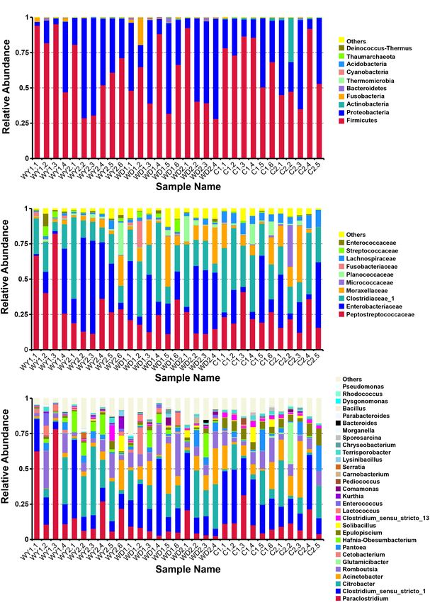
Tang et al.                                                                                                     Gut Microbiota in Crocodile Lizards

INTRODUCTION                                                             the gut microbial composition of animals. Meanwhile, the
                                                                         composition of the gut microbiome in an animal can affect
Bringing animals into captivity and maintaining breeding                 its health status (Clemente et al., 2012; Martín et al., 2014),
populations in natural reserves is an important measure                  metabolism (Ramakrishna, 2013), immunity (Thaiss et al., 2016),
undertaken to protect the declining biodiversity of endangered           and coevolution of the host (Ley et al., 2008; Moeller et al., 2016).
species (Redford and Mcaloose, 2012). For example, the crested           Thus, promoting the conservation of endangered species by
ibis Nipponia nippon was once thought extinct before seven               studying gut microbiota has been receiving increasing attention
individuals were rediscovered in 1981. After captive breeding,           and has become a hot topic of conservation biology (e.g., Zhu
the individual number increased to more than 200, including              et al., 2011; Wu et al., 2017). With limited studies conducted on
130 in captivity by 2000 (Xi et al., 2002). Meta-analysis                the lizard gut microbiota, factors such as diet (Hong et al., 2011;
of marine reserves indicates that there are 3.7 times more               Jiang et al., 2017), gender (Martin et al., 2010), adaptation (Ren
fish populations inside the reserves than outside (Mosquera              et al., 2016), captive breeding (Kohl and Dearing, 2014; Kohl
et al., 2000). Furthermore, the panda reserve system in China            et al., 2017), and even climate change (Bestion et al., 2017) have
provides one of the highest biodiversity among temperate                 been demonstrated to affect the gut microbiota.
regions worldwide (Mackinnon, 2008; Li and Pimm, 2016).                      As the gut microbiota is tightly associated with host health
Given the control of fundamental information of species and              and physiology, it is critical to understand the differences in
scientific management by the scientific community, capacity              gut microbiota composition in crocodile lizards between captive
and breeding populations in natural reserves can effectively             and wild populations during the processes of conservation. It
manage and conserve endangered species and their biodiversity            remains unknown whether captivity can influence gut microbiota
(Ebenhard, 1995).                                                        and thus influence animal health. This comparison is not only
    The crocodile lizard (Shinisaurus crocodilurus Ahl, 1930) is         important to understanding the gut microbiota variation but also
a monotypic species in the genus Shinisaurus and monotypic               critical to providing conservation insight into endangered species
family Shinisauridae, which is remnant of an ancient lineage             conservation in captivity. In addition, captive conservation
from the Pleistocene with around 200 million years of history            should be related to multiple stages of life history, including
(Zhao et al., 1999). Because of their narrow distribution, small         adults and juveniles. In particular, juveniles are more vulnerable
population, being heavily hunted, and environmental changes,             to challenges currently confronting captive crocodile lizards (i.e.,
it is listed as a class I protected species in China. The                diseases and nutritional deficiency). It has been known that
International Union for Conservation of Nature (IUCN) Red List           age-dependent gut microbiota is important to digestibility and
of Threatened Species also list S. crocodilurus as an endangered         consequently to conservation efforts (Redford and Mcaloose,
species (Nguyen et al., 2014). What is more, it was listed as            2012; Jian et al., 2015). Therefore, in order to explore whether
appendix I species (CITES I) by the Convention on International          the gut microbiota composition of the crocodile lizard varies
Trade in Endangered Species of Wild Fauna and Flora (Schingen            along ages and captive environment, it is necessary to analyze
et al., 2016). Consequently, the current captive reserve is one          its composition of gut microbiota between captive and wild
of the most effective protection strategies for crocodile lizards        environments, as well as between juveniles and adults.
(Huang et al., 2008; Van Schingen et al., 2015).                             Here, fecal samples of crocodile lizards with different ages
    During capacity, the fundamental information of crocodile            were collected from captive and wild populations. We aimed
lizard, including its genetic classification (Huang et al.,              to determine variations in gut microbiota of crocodile lizards
2014, 2015), morphological structure (Conrad, 2006), habit               between wild and captive environments, as well as between
distribution (Wu et al., 2012; Huang et al., 2014), and artificial       juveniles and adults, using 16S ribosomal RNA (rRNA) gene
breeding (Wang et al., 2008; Yu et al., 2009), has been revealed         sequencing of gut microbiota. In addition to promoting the
gradually. These studies have provided great information about           conservation of this endangered species, it provides further
crocodile lizards for captive breeding and conservation. However,        insight into the ecological and evolutionary relationship between
like other captive species, some serious challenges are posed            reptiles and their gut microbiota.
by the crocodile lizards during capacity in the nature reserves
(Snyder et al., 1996). For example, the captive population
has been plagued by various unknown diseases, nutritional                MATERIALS AND METHODS
deficiency, and low reproductive rates (Jiang et al., 2017).
    In recent years, with rapid development of high-throughput           Sample Collection
sequencing, an increasing number of studies interpreted the              Fecal samples of 31 crocodile lizards were collected from Guangxi
health and nutritional utilization of animals by integrating the         Daguishan S. crocodilurus National Nature Reserve, Guangxi
relationships between bacteria in gastrointestinal tracts and the        Province, China. These 31 crocodile lizards were from the Yusan
animals themselves (Mcfall-Ngai et al., 2013). For instance, the         stream (N = 10), Dachai stream (N = 10), and captive populations
host’s genotype (Kovacs et al., 2011; Goodrich et al., 2014), age        (N = 11), respectively (Figure 1). For each population, fecal
(Elena et al., 2010; Yatsunenko et al., 2012; Jian et al., 2015),        samples from both juveniles and adults were collected according
health (Dethlefsen et al., 2007), dietary composition (Castillo          to body sizes, respectively. The snout-vent lengths (SVLs) were
and Martín, 2007; David et al., 2013; Zhang et al., 2014),               161.46 ± 1.98 (151–173) and 106.70 ± 1.69 (98–117) mm,
and even social interaction (Lombardo, 2008) can determine               and body masses (BMs) were 90.45 ± 3.89 (63.5–112.7) and

Frontiers in Microbiology | www.frontiersin.org                      2                                          April 2020 | Volume 11 | Article 550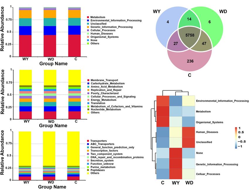
Tang et al. Gut Microbiota in Crocodile Lizards
the V3–V4 region of the bacteria 16S rRNA gene. Thermal
cycling conditions of the PCR assay were as follows: 1 min
initial denaturation at 98◦ C, 30 cycles of 10 s denaturation at
98◦ C, 30 s annealing at 50◦ C, finally 30 s elongation at 72◦ C,
and a final extension at 72◦ C for 5 min. A 30-µL reaction
system was used for PCR products, which contained 10 ng
template DNA, forward and reverse primers (0.2 µM), and
15 µL Phusion High-Fidelity PCR Master Mix (New England
R
Biolabs, United Kingdom). The GeneJETTM Gel Extraction
Kit (Thermo Scientific, United States) was used for sufficient
mixture and purification of the obtained amplification products.
Then, with Ion Plus Fragment Library Kit (Thermo Scientific,
United States), the sequencing libraries were established
according to published protocols of the kit. After establishment,
the libraries were measured on the Qubit 2.0 Fluorometer
R
(Thermo Scientific, United States). After, an Ion S5TM XL
platform was used to sequence the library, with 400/600 bp
single-end reads generated. Obtained raw sequences were
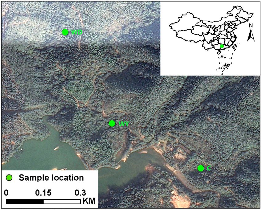
FIGURE 1 | Location of Yusan stream population (WY), Dachai stream submitted to the National Center for Biotechnology Information
population (WD), and captive population (C) in the Crocodile Lizard National
Nature Reserve.
(NCBI) Bioproject database (accession number PRJNA594801)
(See details in Supplementary Data Sheet 1).
29.89 ± 1.41 (20.7–36.9) g for adults and juveniles, respectively.
Data Analysis
According to the location and age of the crocodile lizards, Clean Raw Data
the fecal samples were from one of six groups: adults in the The tags of raw sequences were filtered using Cutadapt (V1.9.11 )
wild population of Yusan stream (WY1, N = 4), juveniles in (Martin, 2011). All sequences were compared on UCHIME
the wild population of Yusan stream (WY2, N = 6), adults in algorithm to find out chimera sequences (UCHIME Algorithm2 )
the wild population of Dachai stream (WD1, N = 6), juveniles (Edgar et al., 2011), with Silva database as reference (Silva
in the wild population of Dachai stream (WD2, N = 4), adults database3 ) (Quast et al., 2013). After filtering out all low-quality
in the captive population (C1, N = 6), and juveniles in the and chimera sequences, the remaining clean reads were obtained.
captive population (C2, N = 5) (see details in Supplementary
OTU Production
Table S1). The Yusan and Dachai streams are two independent
Sequences were assigned with similarity no less than 97% (i.e.,
wild ravine streams in the Crocodile Lizard National Nature
≥97%) to the same operational taxonomic units (OTUs) using
Reserve (Figure 1). It is plausible that the crocodile lizards
Uparse (Uparse v7.0.10014 ) (Edgar, 2013). For each OTU, we
of Yusan and Dachai streams are independent from each
searched the Silva Database5 to annotate screened representative
other without population communication because of the limited
sequence with threshold 0.8 using RDP Classifier 2.2 (Wang et al.,
dispersal ability and small home range of crocodile lizards
2007; Quast et al., 2013).
and the isolation of the two streams. During collection, the
diet type of two wild populations was randomly investigated. Data Normalization
All fecal samples were collected directly without touching In order to compare different samples, the number of the
anything (Wang et al., 2016a). After collection, the fecal samples samples with the lowest counts was used to normalize the
were transported back to the laboratory in Beijing with sterile OTU abundance information. The rarefaction curves of observed
containers. The fecal samples were stored in a −80◦ C refrigerator species were calculated to assess the sufficiency of current
before DNA extraction. depth of sequencing, in yielding a stable estimate of the species
richness. Whether the bacterial diversity in the 31 fecal samples
Extracting DNA, PCR Amplification, and represents the overall bacterial diversity in the gastrointestinal
Sequencing tract of the crocodile lizard was determined with a species
All DNA extraction and sequencing were conducted by accumulation box plot.
Novogene Corporation (Beijing, China) with established
Alpha and Beta Diversity Estimation
protocols. In brief, cetyltrimethyl ammonium bromide
The observed-species index and Simpson index was calculated
(CTAB)/sodium dodecyl sulfate (SDS) method was employed
with QIIME V1.7.0 to estimate alpha diversity for each fecal
for total DNA extraction from the lizard fecal samples. Then,
1% agarose gels was used for concentration and purification of 1
http://cutadapt.readthedocs.io/en/stable/
DNA. After, DNA were diluted to 1 ng/µL with bacteria-free 2
http://www.drive5.com/usearch/manual/uchime_algo.html
water before bacteria 16S rRNA amplification. Barcodes of 341F 3
https://www.arb-silva.de/
(50 -CCTAYGGGRBGCASCAG-30 ) and 806R (50 -GGACTACNN 4
http://drive5.com/uparse/
GGGTATCTAAT-30 ) were the primers for amplification of 5
https://www.arb-silva.de/
Frontiers in Microbiology | www.frontiersin.org 3 April 2020 | Volume 11 | Article 550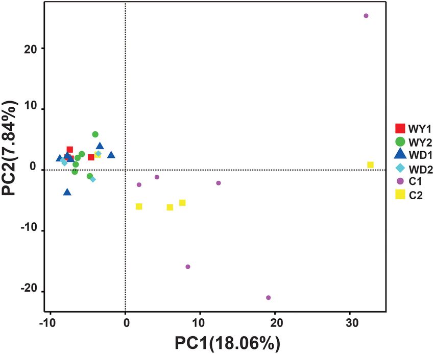
Tang et al.                                                                                                                 Gut Microbiota in Crocodile Lizards

sample of crocodile lizard (Caporaso et al., 2010), which were                         General Analyses of the Gut Microbial
indicators in community richness and community evenness                                Community Structure
identifications, respectively. Then, the Mann–Whitney U-test
                                                                                       The bacterial composition of 31 crocodile lizard fecal samples
was performed to detect differences in alpha diversity indices
                                                                                       was analyzed (Supplementary Table S1). The average effective
between two independent groups.
                                                                                       sequences of 31 samples were 52,342 (Supplementary Figure S1).
   For the beta diversity metrics, principal component analysis
                                                                                       The estimates of species richness were stable and unbiased
(PCA) and analysis of similarities (ANOSIM) were conducted to
                                                                                       according to the rarefaction curves (Supplementary Figure S2).
determine the communities and structure of the gut microbiota
                                                                                       The species accumulation boxplot indicated that the sample size
among groups. PCA, which is based on the OTU level, can
                                                                                       was sufficient and greatly saturated the bacterial diversity found
intuitively present the differences among groups on a two-
                                                                                       under this condition (Supplementary Figure S3).
dimensional graph. Notably, ANOSIM based on the Bray–
                                                                                           The total sequences of crocodile lizards were classified into
Curtis distances, considered both flora types and the relative
                                                                                       five major phyla (Figure 2A), Firmicutes, with the relative
abundance of microbes.
                                                                                       abundance of 61.2%, holding the overwhelming predominance.
   The differential abundances were compared at family and
                                                                                       Proteobacteria (35.8%), Actinobacteria (1.4%), Fusobacteria
genus levels of bacteria among groups using LEfSe analysis to
                                                                                       (1.0%), and Bacteroidetes (0.5%) were the other four major
identify microbes accounting for the effect of captivity or age.
                                                                                       phyla. Totally, these five most dominant phyla contributed
Thereafter, a set of pairwise tests was used to investigate biological
                                                                                       more than 99% abundance across all the samples. At the
consistency among subgroups. The linear discriminatory analysis
                                                                                       family level, the top 10 families are listed (Figure 2B). The
(LDA) was also performed to evaluate the effect size of each
                                                                                       most abundant taxa were Peptostreptococcaceae (25.5%),
selected classification. In this study, only bacterial taxa with a log
                                                                                       Clostridiaceae_1 (25.3%), Enterobacteriaceae (25.0%), and
LDA score more than 4 (more than four orders of magnitude)
                                                                                       Moraxellaceae (9.3%). In addition, the top 30 genera are also
were used (Segata et al., 2011).
                                                                                       listed (Figure 2C). The gut microbiota of all these crocodile
                                                                                       lizards was dominated by Clostridium sensu_stricto 1 (21.0%),
Functional Classification                                                              Citrobacter (14.8%), Paraclostridium (14.3%), Acinetobacter
Functional prediction of the sequences among groups was                                (9.3%), and Romboutsia (9.1%).
conducted for classification. In brief, PICRUSt was utilized to
search the protein sequences of the predicted genes in the Kyoto
Encyclopedia of Genes and Genomes (KEGG) database with E                               Comparison of Gut Microbial Community
value < 1E-5. These genes were assigned to KEGG pathways                               Structure Between Age or Populations
(Langille et al., 2013). Then, relative abundance in each group was                    First, the gut microbial diversity was compared between
counted. The unique and shared genes between populations were                          adult and juvenile crocodile lizards within each population,
also plotted by Venn diagram. A heatmap was used to show genes                         respectively. No significant difference between the adult and
with high expression.                                                                  juvenile individuals was identified in terms of community
                                                                                       richness (Figure 3A), community evenness (Figure 3B), or
                                                                                       community composition (Figure 4) (all P > 0.05). ANOSIM
                                                                                       also indicated similarity between adult and juvenile individuals
RESULTS                                                                                in each population (all P > 0.05) (Figure 5). Integrated in the
                                                                                       results of alpha and beta diversity analyses, the gut microbiota
Food Composition of Wild and Captive                                                   of adults and juveniles within each population were highly
Populations                                                                            similar, respectively. Therefore, adults and juveniles from each
The primary food types of Yusan and Dachai stream populations                          population were combined as available individual candidates to
were similar, mainly consisting of earthworm, centipede, and                           compare the variation in gut microbiota at the population level.
larvae of lepidopteran, which comprised around 70% of the food                         Accordingly, data analysis was reconducted and recalculated to
availability. In contrast, the earthworm is the only food type for                     elucidate the difference in alpha diversity and beta diversity
captive crocodile lizards during breeding (Table 1).                                   using population as main factor. The results indicated that
                                                                                       the community richness of the captive population was clearly
                                                                                       higher than wild populations of Yusan stream (Z = −3.170,
TABLE 1 | Primary food types of wild and captive crocodile lizards.                    P < 0.05) and Dachai stream (Z = −3.239, P < 0.05), but no
                                                                                       significant difference was detected between two wild populations
Proportion of                     Captive                 Wild lizards
                                                                                       (Z = −1.362, P = 0.173) (Figure 3A). After combination of two
composition                       lizards
                                                                                       wild populations, a significant difference was detected between
Most                              Earthworm               Earthworm                    wild and captive populations in community richness (Z = 2.412,
Secondary                                                 Centipede                    P = 0.016) (Figure 6A). However, no significant difference was
Tertiary                                                  Larva of Lepidoptera         detected between wild and captive populations in the community
Other                                                     Larva of other insects       evenness (Z = 0.949, P = 0.343) (Figure 6B). With regard to
Food types are shown at category levels, and rough proportion of food                  beta diversity, the results of the PCA plot and ANOSIM showed
compositions is shown in sequence.                                                     significant differences between the captive population and two

Frontiers in Microbiology | www.frontiersin.org                                    4                                        April 2020 | Volume 11 | Article 550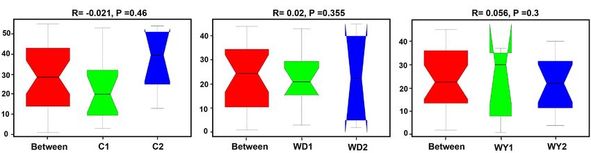
Tang et al.                                                                                                                             Gut Microbiota in Crocodile Lizards

  FIGURE 2 | Composition of the gut microbiota of each sample at the (A) phylum, (B) family, and (C) genus levels. Different colors in the figures indicate the different
  groups, and details are shown on the right sides of each figure, respectively. Details of sample names are shown in Supplementary Table S1. In each panel,
  “Others” represented the sum of the relative abundances of all other phylum (A), families (B), and genus (C) except the items listed in the figure.

Frontiers in Microbiology | www.frontiersin.org                                      5                                                  April 2020 | Volume 11 | Article 550
Tang et al. Gut Microbiota in Crocodile Lizards
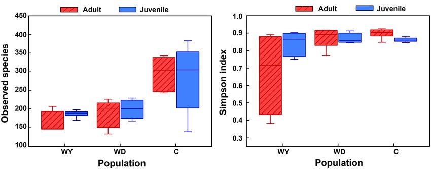
FIGURE 3 | The alpha diversity of the gut microbial composition, shown by observed species index (A) and Simpson index (B) among populations. WY indicates
wild population of Yusan stream, WD indicates wild population of Dachai stream, and C indicates captive population. Data are expressed as mean ± SEM.
wild populations, respectively (C-WY, R = 0.2935, P < 0.05; (9.7%), and Acinetobacter (8.0%)at the genus level. In the
C-WD, R = 0.2929, P < 0.05), with similarity between two wild captive population, the composition of the gut microbiota
populations (R = 0.08122, P = 0.128) (Figure 4). mainly include Firmicutes (64.2%), Proteobacteria (31.6%),
A comparison of the gut microbiota between the wild Actinobacteria (3.9%), and Bacteroidetes (0.2%) at the phyla
and the captive populations showed in wild populations, level; Clostridiaceae_1 (29.3%), Peptostreptococcaceae (22.7%),
the composition of the gut microbiota mainly includes Enterobacteriaceae (19.0%), and Moraxellaceae (11.6) at
Firmicutes (60.1%), Proteobacteria (37.6%), Fusobacteria the family level; and Clostridium_sensu_stricto_1 (24.0%),
(1.4%), Bacteroidetes (0.7%), and Actinobacteria (0.2%) at the Paraclostridium (11.7%), Citrobacter (11.7%), Acinetobacter
phyla level; Peptostreptococcaceae (28.3%), Enterobacteriaceae (11.6%), and Romboutsia (7.9%) at the genus level.
(27.9%), Clostridiaceae_1 (22.4%), and Moraxellaceae (8.0%)
at the family level; and Clostridium_sensu_stricto_1 (18.7%), LEfSe Analysis of the Differential
Paraclostridium (17.2%), Citrobacter (16.4%), Romboutsia
Microbes Between Captive and Wild
Populations
The LEfSe analysis indicated that five genera and three families
were enriched differently in captive and wild populations. In
contrast to wild populations, the gut microbiota of captive
crocodile lizards showed significantly higher abundances in
genera Epulopiscium and Glutamicibacter, and in families
Lachnospiraceae and Micrococcaceae (Figure 7).
Functional Predictions of Gut Microbiota
Between Captive and Wild Populations
16S RNA of gut microbiota from 31 fecal samples were predicted
into three levels in functional categories. At the top level,
metabolism, environmental information processing, genetic
information processing, and cellular processes were four primary
categories (Figure 8A); at the second level, membrane transport,
carbohydrate metabolism, amino acid metabolism, replication
and repair, cellular processes and signaling, energy metabolism,
translation, metabolism of cofactors and vitamins, and nucleotide
metabolism were the primary functions (Figure 8B); while at
FIGURE 4 | The beta diversity of the gut microbiota composition of two wild
the third level, transporters, ATP-binding cassette (ABC)
populations and captive population. Principal component analysis (PCA) was
performed. The variation explanation is indicated on each axis, respectively.
transporters, and transcription factors were the primary
functions (Figure 8C).
Frontiers in Microbiology | www.frontiersin.org 6 April 2020 | Volume 11 | Article 550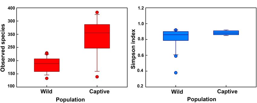
Tang et al.                                                                                                                        Gut Microbiota in Crocodile Lizards

  FIGURE 5 | Analysis of similarity between adults and juveniles of (A) captive, (B) WD, and (C) WY populations. (A) C1 and C2 indicate the adults and juveniles of
  captive population, (B) WD1 and WD2 indicate the adults and juveniles of wild Dachai population, and WY1 and WY2 indicate the adults and juveniles of wild Yusan
  population, respectively. Data are expressed as mean ± SEM. Statistical significance is defined as α < 0.05.

  FIGURE 6 | The alpha diversity of the gut microbial composition, shown by observed (A) species index and (B) Simpson index between wild and captive populations.
  “Wild” indicates combination of wild populations from Yusan and Dachai streams, and “Captive” indicates captive population. Data are expressed as mean ± SEM.

   Venn diagram of shared genes indicated that most of the                            Firmicutes (33.2–73%) have been documented as the dominant
knockouts (KOs) were common in captive and two wild                                   gut microbiota in lizards, while Proteobacteria (5.7–62.3%)
populations, while 236 KOs were exclusive to the captive                              and Bacteroidetes (6.2–45.7%) were varied among host species
population (Figure 8D). Heatmap of the cluster indicated                              (Nelson et al., 2010; Hong et al., 2011; Ren et al., 2016; Kohl
that at the top level, the KOs of captive population were                             et al., 2017). In addition, the gut microbiota is similar to lizards in
enriched in environmental information processing, metabolism,                         other reptile categories (Costello et al., 2010; Colston et al., 2015;
and organismal systems (Figure 8E). However, no significant                           Yuan et al., 2015). Interestingly, previous study on gut microbiota
differences among groups were found after statistical analysis                        of crocodile lizards in wild and captive populations reported
(minimum P = 0.270).                                                                  that Proteobacteria and Bacteroidetes were primary, while the
                                                                                      proportion of Firmicutes is lower than was detected in this study
                                                                                      (Jiang et al., 2017). The potential reason for the discrepancy
DISCUSSION                                                                            between the two studies could be the sampling methods used.
                                                                                      Jiang et al. (2017) used cloacal swabs for sampling, whereas
Firmicutes and Proteobacteria were two major gut microbiotas                          fecal sampling was utilized in the present study. It has been
in crocodile lizard, while Actinobacteria, Fusobacteria, and                          demonstrated that the fecal communities were largely similar
Bacteroidetes were minor gut microbiotas. Like other studies,                         to hindgut microbial communities in lizards, thus becoming an
Firmicutes and Proteobacteria are two of the most important                           acceptable indicator in the gut region for microbial diversity
types of gut microbiota in numerous vertebrate species (Xenoulis                      (Kohl et al., 2017). The communities of cloacal swabs have
et al., 2010; Waite et al., 2012; Wang et al., 2016b). Phylum                         clear microbial community characteristics, especially in terms of

Frontiers in Microbiology | www.frontiersin.org                                   7                                                April 2020 | Volume 11 | Article 550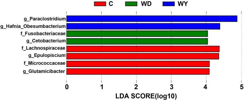
Tang et al.                                                                                                                            Gut Microbiota in Crocodile Lizards

  FIGURE 7 | Differences in bacterial taxa among populations determined by linear discriminative analysis of effect size (LEfSe). The highlighted taxa were significantly
  enriched in the group that corresponds to each color. Linear discriminatory analysis (LDA) scores can be interpreted as the degree of difference in relative
  abundance. The letters “g” and “f” indicate genus and family, respectively.

  FIGURE 8 | Functional classifications of 16s RNA in microbiota at (A) top level, (B) second level, and (C) third levels of relative abundance, and (D) Venn and (E)
  clusters analysis of functions between captive and wild populations. C indicates captive population, WD indicates wild Dachai population, and WY indicates wild
  Yusan population, respectively.

Frontiers in Microbiology | www.frontiersin.org                                      8                                                 April 2020 | Volume 11 | Article 550
Tang et al.                                                                                                       Gut Microbiota in Crocodile Lizards

community members, from the communities of large intestine                 may contribute to the increased community richness in the
(Colston et al., 2015). Therefore, different sampling methods              gut microbiota of the captive animals (Nelson et al., 2013).
may lead to variation in gut microbiota. Given that, based on              In this study, the captive lizards had higher abundances
the previous study (Jiang et al., 2017), we provided further               of families Lachnospiraceae and Micrococcaceae and genera
understanding of gut microbiota in the crocodile lizards.                  Epulopiscium and Glutamicibacter than wild lizards. Among
    The effects of age in crocodile lizards on the gut microbiota          them, Lachnospiraceae (phylum Firmicutes, order Clostridiales)
were revealed to be trivial, either in the captive or in the wild          is typically abundant in the digestive tracts of humans, ruminants,
environments (Figure 5). This may be largely due to the fact               and many other mammals (e.g., Gosalbes et al., 2011; Sandra
that adult and juvenile crocodile lizards of each population               et al., 2013). Lachnospiraceae has been demonstrated to be
live in the same environmental conditions, and food intake is              related with the production of butyrate, which is necessary to
identical accordingly. In the wild, earthworm is a conservatively          sustain the health of colonic epithelial tissue (Duncan et al.,
primary food resource for crocodile lizards (Ning, 2007). In               2002). The captive population of crocodile lizard has more
contrast, comparing with the microbial communities of adults,              opportunities in contacting with humans, by frequent feeding,
juveniles are usually different, as they are greatly dependent on          cleaning of the breeding pond, examination of diseases, etc.,
environments and resources. The difference in environmental                which may result in colonization of the Lachnospiraceae bacteria
dependence of gut microbiota along ontogeny implies more                   from human. However, whether the increase in Lachnospiraceae
self-governing in adults after maturation (Trosvik et al., 2010;           in abundance has a positive impact on the captive population
Burns et al., 2016). The crocodile lizard survives independently           is still unclear, even though some functional categories for
after birth, so the gut microbiota of juvenile individuals may             genes of gut microbiota were found in this study in wild and
be similar to adults. In addition to the neutral effect of age on          captive populations. More exclusive KOs were found in captive
gut microbiota, the gut microbiota did not differ in two wild              population (Figure 8D), but no significant difference in functions
populations. This similarity is accompanied with homologous                was found. In the future, whole genome sequencing of gut
food composition between two wild populations (Table 1);                   microbiota in crocodile lizards may be helpful at revealing the
however, they are isolated. In the field, diet may be one of               functions underlying gut microbiota difference between captive
the most important factors affecting the composition of the gut            and wild populations.
microbiota of wild animals (Wu et al., 2011; David et al., 2013). In           These captivity-related changes in gut microbial communities
the future, it would be interesting to reveal the effect of ontogeny       may have implications for the health of the captive animal and
on gut microbiota variation in crocodile lizard, which could               thus determining the success of species conservation (Redford
provide more insight.                                                      and Mcaloose, 2012). Thus, understanding the effect of the
    Most interestingly, the captive population was found to                captivity on the composition of gut microbiota is important to
modify the community structure and had higher community                    provide breeding environments for the health management of
richness than the two wild populations (Figure 6A). This study             the endangered species. This is important, as the composition
found a contrasting pattern to those studies that demonstrated             of the gut microbiome of animals could have long-term effect
lower microbial diversity in animals under captivity (Kohl and             on their health status and immunity (e.g., Clemente et al., 2012;
Dearing, 2014; Kohl et al., 2014) or those that found similar              Martín et al., 2014; Thaiss et al., 2016). It was speculated that the
gut microbiota between wild and captive lizards (Wang et al.,              increase in the abundance of these specific bacteria in the captive
2016c; Kohl et al., 2017). Although a number of studies have               population may be one of the reasons that affect the survival
demonstrated that the gut microbiota of animals in captive                 status of the crocodile lizards. However, the actual relationship
are different from congeners in the wild (Villers et al., 2010;            in Lachnospiraceae by contacting between lizards and humans,
Xenoulis et al., 2010; Wienemann et al., 2011; Nelson et al., 2013),       and potential function of Lachnospiraceae on lizards’ conditions
captive population is seldom detected to have higher community             have not been determined to date. Prospectively, the necessity
richness than wild populations. It might be led by different food          to take actions is recommended in order to minimize the
composition between captive and wild populations (Table 1). The            direct contact between human managers and crocodile lizards,
food types are more diverse in wild environments than in captive           including wearing gloves and protection suits during operation
environments for crocodile lizards. Diet is one of most important          on lizards, or sterilizing the equipment used for lizards breeding
factors that affect the assembly of gut microbiota (Muegge et al.,         before operations. Future studies should be centered on the
2011; Carmody et al., 2015; Pérez-Cobas et al., 2015). However,            functional interaction between gut microbiota and animals to
most of the studies indicated positive relationships between food          reveal the functional significance of different richness, as well as
and gut microbiota diversities (e.g., Laparra and Sanz, 2010; Li           the effects of human contact.
et al., 2016). In contrast, captive crocodile lizard has an opposite
pattern. The underlying mechanisms are still largely unknown.
Future studies with food type manipulations would be helpful to            CONCLUSION
reveal their relationships.
    In captivity, constant cohabitation, social interaction, and           This study revealed the similarity of gut microbiota between
interaction with human keepers provide increased opportunities             adult and juvenile crocodile lizards, both in the captive and
for transmission of microbiota from host-associated sources,               wild environments as well as between two wild populations.
which are capable of colonizing the animals. This, in turn,                Interestingly, a significant effect of captivity was found on the

Frontiers in Microbiology | www.frontiersin.org                        9                                          April 2020 | Volume 11 | Article 550
Tang et al. Gut Microbiota in Crocodile Lizards
composition of gut microbiota of the crocodile lizard, mainly G-ST, M-YY, and B-JS analyzed the data and led the writing of
reflected in the increase in community richness and community the manuscript. All authors contributed critically to the drafts
structure change. After comparison, it was speculated that the and gave final approval for publication.
gut microbiota variation in captive population might be from
human contact. Although the functions are unclear, it was
recommended that minimal direct contact was crucial for the FUNDING
health of wild animals between crocodile lizards and human
managers in captive environment. The study was supported by the National Key Research
and Development Program of China (2016YFC0503200) and
National Natural Science Foundation of China (No. 31821001,
DATA AVAILABILITY STATEMENT 31901223, 31870391). B-JS was supported by Youth Innovation
The datasets generated for this study can be found in the NCBI Promotion Association CAS (No. 2019085).
Bioproject database (PRJNA594801).
ACKNOWLEDGMENTS
ETHICS STATEMENT
We thank Yu-Jie Yang, Hong-Xin Xie, and Xing-Zhi
The animal study was reviewed and approved by the Animal Han for assistance.
Ethics Committee at the Institute of Zoology, Chinese Academy
of Sciences (IOZ14001).
SUPPLEMENTARY MATERIAL
AUTHOR CONTRIBUTIONS
The Supplementary Material for this article can be found
B-JS, G-ST, and W-GD conceived the ideas and designed online at: https://www.frontiersin.org/articles/10.3389/fmicb.
methodology. G-ST, X-XL, and T-TW collected the data. HL, 2020.00550/full#supplementary-material
REFERENCES Dethlefsen, L., Mcfall-Ngai, M., and Relman, D. A. (2007). An ecological and
evolutionary perspective on human-microbe mutualism and disease. Nature
Bestion, E., Jacob, S., Zinger, L., Di, G. L., Richard, M., White, J., et al. (2017). 449, 811–818. doi: 10.1038/nature06245
Climate warming reduces gut microbiota diversity in a vertebrate ectotherm. Duncan, S. H., Adela, B., Stewart, C. S., Pryde, S. E., and Flint, H. J. (2002). Acetate
Nat. Ecol. Evol. 1:161. doi: 10.1038/s41559-017-0161 utilization and butyryl coenzyme A (CoA): acetate-CoA transferase in butyrate-
Burns, A. R., Stephens, W. Z., Stagaman, K., Wong, S., Rawls, J. F., Guillemin, K., producing bacteria from the human large intestine. Appl. Environ. Microbiol. 8,
et al. (2016). Contribution of neutral processes to the assembly of gut microbial 5186–5190. doi: 10.1128/aem.68.10.5186-5190.2002
communities in the zebrafish over host development. ISME J. 10, 655–664. Ebenhard, T. (1995). Conservation breeding as a to for saving animal species
doi: 10.1038/ismej.2015.142 from extinction. Trends Ecol. Evol. 10, 438–443. doi: 10.1016/s0169-5347(00)89
Caporaso, J. G., Kuczynski, J., Stombaugh, J., Bittinger, K., Bushman, F. D., 176-4
Costello, E. K., et al. (2010). QIIME allows analysis of high-throughput Edgar, R. C. (2013). UPARSE: highly accurate OTU sequences from microbial
community sequencing data. Nat. Methods 7, 335–336. amplicon reads. Nat. Methods 10:996. doi: 10.1038/nmeth.2604
Carmody, R. N., Gerber, G. K., Luevano, J. M. Jr., Gatti, D. M., Somes, L., Edgar, R. C., Haas, B. J., Clemente, J. C., Quince, C., and Knight, R. (2011).
Svenson, K. L., et al. (2015). Diet dominates host genotype in shaping the UCHIME improves sensitivity and speed of chimera detection. Bioinformatics
murine gut microbiota. Cell Host Microbe 17, 72–84. doi: 10.1016/j.chom.2014. 27, 2194–2200. doi: 10.1093/bioinformatics/btr381
11.010 Elena, B., Lotta, N., Marco, C., Rita, O., Laura, B., Elisa, P., et al. (2010). Through
Castillo, M., and Martín, S. M. (2007). Adaptation of gut microbiota to corn ageing, and beyond: gut microbiota and inflammatory status in seniors and
physical structure and different types of dietary fibre. Livest. Sci. 109, 149–152. centenarians. PLoS One 5:e10667. doi: 10.1371/journal.pone.0010667
doi: 10.1016/j.livsci.2007.01.129 Goodrich, J. K., Waters, J. L., Poole, A. C., Sutter, J. L., Koren, O., Blekhman, R.,
Clemente, J. C., Ursell, L. K., Parfrey, L. W., and Knight, R. (2012). The impact of et al. (2014). Human genetics shape the gut microbiome. Cell 159, 789–799.
the gut microbiota on human health: an integrative view. Cell 148, 1258–1270. doi: 10.1016/j.cell.2014.09.053
doi: 10.1016/j.cell.2012.01.035 Gosalbes, M. J., Durbán, A., Pignatelli, M., Abellan, J. J., Jiménez-Hernández, N.,
Colston, T. J., Noonan, B. P., and Jackson, C. R. (2015). Phylogenetic Analysis and Pérez-Cobas, A. E. (2011). Metatranscriptomic approach to analyze the
of bacterial communities in different regions of the gastrointestinal tract of functional human gut microbiota. PLoS One 6:e17447. doi: 10.1371/journal.
Agkistrodon piscivorus, the cottonmouth snake. PLoS One 10:e0128793. doi: pone.0017447
10.1371/journal.pone.0128793 Hong, P. Y., Wheeler, E., Cann, I. K. O., and Mackie, R. I. (2011). Phylogenetic
Conrad, J. L. (2006). Postcranial skeleton of Shinisaurus crocodilurus (Squamata: analysis of the fecal microbial community in herbivorous land and marine
Anguimorpha). J. Morphol. 267, 759–775. doi: 10.1002/jmor.10291 iguanas of the Galápagos Islands using 16S rRNA-based pyrosequencing. ISME
Costello, E. K., Gordon, J. I., Secor, S. M., and Knight, R. (2010). Postprandial J. 5:1461. doi: 10.1038/ismej.2011.33
remodeling of the gut microbiota in Burmese pythons. ISME J. 4:1375. doi: Huang, C. M., Yu, H., Wu, Z. J., Li, Y. B., Wei, F. W., and Gong, M. H.
10.1038/ismej.2010.71 (2008). Population and conservation srategies for the Chinese crocodile lizard
David, L. A., Maurice, C. F., Carmody, R. N., Gootenberg, D. B., and Turnbaugh, (Shinisaurus crocodilurus) in China. Anim. Biodivers. Conserv. 31, 63–70.
P. J. (2013). Diet rapidly and reproducibly alters the gut microbiome. Nature Huang, H. H., Wang, H., Li, L. M., Wu, Z. J., and Chen, J. P. (2014).
505, 559–563. doi: 10.1038/nature12820 Genetic diversity and population demography of the Chinese crocodile lizard
Frontiers in Microbiology | www.frontiersin.org 10 April 2020 | Volume 11 | Article 550Tang et al. Gut Microbiota in Crocodile Lizards
(Shinisaurus crocodilurus) in China. PLoS ONE 9:e91570. doi: 10.1371/journal. Nelson, D. M., Cann, I. K. O., Eric, A., and Mackie, R. I. (2010). Phylogenetic
pone.0091570 evidence for lateral gene transfer in the intestine of marine iguanas. PLoS One
Huang, H. Y., Zhuo, C., Tang, Z. J., and Chen, J. P. (2015). Genetic analysis 5:e10785. doi: 10.1371/journal.pone.0010785
of multiple paternity in an endangered ovoviviparous lizard Shinisaurus Nelson, T. M., Rogers, T. L., Carlini, A. R., and Brown, M. V. (2013). Diet and
crocodilurus. Asian Herpetol. Res. 6, 150–155. phylogeny shape the gut microbiota of Antarctic seals: a comparison of wild and
Jian, P., Wang, Q., Wang, J., Niu, L., Zhu, H., Zeng, Y., et al. (2015). Difference captive animals. Environ. Microbiol. 15, 1132–1145. doi: 10.1111/1462-2920.
analysis of gut microbiome of Rhinopithecus roxellana in different ages. Chin. J. 12022
Anim. Nutr. 27, 1302–1309. Nguyen, T. Q., Hamilton, P., and Ziegler, T. (2014). Shinisaurus crocodilurus. The
Jiang, H. Y., Ma, J. E., Li, J., Zhang, X. J., Li, L. M., He, N., et al. (2017). Diets IUCN Red List of Threatened Species 2014. Available online at: http://dx.doi.org/
alter the gut microbiome of crocodile lizards. Front. Microbiol. 8:2073. doi: 10.2305/IUCN.UK.2014-1.RLTS.T57287221A57287235.en (accessed March 20,
10.3389/fmicb.2017.02073 2014).
Kohl, K. D., Brun, A., Magallanes, M., Brinkerhoff, J., Laspiur, A., Acosta, J. C., Ning, J. J. (2007). Behavioral Time Budget and Diet of the Chinese Crocodile Lizard
et al. (2017). Gut microbial ecology of lizards: insights into diversity in the wild, (Shinisaurus crocodilurus) in the Luokeng Nature Reserve, Guangdong. Master
effects of captivity, variation across gut regions and transmission. Mol. Ecol. 26, thesis, Guangxi Normal University, Guangxi.
1175–1189. doi: 10.1111/mec.13921 Pérez-Cobas, A. E., Maiques, E., Angelova, A., Carrasco, P., Moya, A., and
Kohl, K. D., and Dearing, M. D. (2014). Wild-caught rodents retain a majority of Latorre, A. (2015). Diet shapes the gut microbiota of the omnivorous cockroach
their natural gut microbiota upon entrance into captivity. Environ. Microbiol. Blattella germanica. FEMS Microbiol. Ecol. 91:fiv022. doi: 10.1093/femsec/fi
Rep. 6, 191–195. doi: 10.1111/1758-2229.12118 v022
Kohl, K. D., Skopec, M. M., and Dearing, M. D. (2014). Captivity results in Quast, C., Pruesse, E., Yilmaz, P., Gerken, J., Schweer, T., Yarza, P., et al. (2013). The
disparate loss of gut microbial diversity in closely related hosts. Conserv. Physiol. SILVA ribosomal RNA gene database project: improved data processing and
2:cou009. doi: 10.1093/conphys/cou009 web-based tools. Nucleic Acids Res. 41, D590–D596. doi: 10.1093/nar/gks1219
Kovacs, A., Ben-Jacob, N., Tayem, H., Halperin, E., Iraqi, F. A., and Gophna, Ramakrishna, B. S. (2013). Role of the gut microbiota in human nutrition and
U. (2011). Genotype is a stronger determinant than sex of the mouse gut metabolism. J. Gastroenterol. Hepatol. 28, 9–17. doi: 10.1111/jgh.12294
microbiota. Microb. Ecol. 61, 423–428. doi: 10.1007/s00248-010-9787-2 Redford, K. H., and Mcaloose, D. (2012). Conservation and the microbiome.
Langille, M. G. I., Zaneveld, J., and Caporaso, J. G. (2013). Predictive functional Conserv. Biol. 26, 195–197. doi: 10.1111/j.1523-1739.2012.01829.x
profiling of microbial communities using 16S rRNA marker gene sequences. Ren, T., Kahrl, A. F., Wu, M., and Cox, R. M. (2016). Does adaptive radiation
Nat. Biotechnol. 31, 814–821. doi: 10.1038/nbt.2676 of a host lineage promote ecological diversity of its bacterial communities?
Laparra, J. M., and Sanz, Y. (2010). Interactions of gut microbiota with functional A test using gut microbiota of Anolis lizards. Mol. Ecol. 25, 4793–4804. doi:
food components and nutraceuticals. Pharmacol. Res. 61, 219–225. doi: 10. 10.1111/mec.13796
1016/j.phrs.2009.11.001 Sandra, K., Henning, S., Walters, W. A., Clemente, J. C., Rob, K., Gordon, J. I., et al.
Ley, R. E., Hamady, M., Lozupone, C., Turnbaugh, P. J., Ramey, R. R., Bircher, (2013). Simultaneous amplicon sequencing to explore co-occurrence patterns
J. S., et al. (2008). Evolution of mammals and their gut microbes. Science 320, of bacterial, archaeal and eukaryotic microorganisms in rumen microbial
1647–1651. doi: 10.1126/science.1155725 communities. PLoS One 8:e47879. doi: 10.1371/journal.pone.0047879
Li, B. V., and Pimm, S. L. (2016). China’s endemic vertebrates sheltering under Schingen, M. V., Le, M. D., Ngo, H. T., Pham, C. T., Ha, Q. Q., Nguyen,
the protective umbrella of the giant panda. Conserv. Biol. 30, 329–339. doi: T. Q., et al. (2016). Is there more than one Crocodile Lizard? An integrative
10.1111/cobi.12618 taxonomic approach reveals Vietnamese and Chinese Shinisaurus crocodilurus.
Li, H., Li, T. T., Beasley, D. E., Hedenec, P., Xiao, Z. S., Zhang, S. H., et al. Represent separate conservation and taxonomic units. Der Zoologische Garten
(2016). Diet diversity is associated with beta but not alpha diversity of pika gut 85, 240–260. doi: 10.1016/j.zoolgart.2016.06.001
microbiota. Front. Microbiol. 7:1169. doi: 10.3389/fmicb.2016.01169 Segata, N., Izard, J., Waldron, L., Gevers, D., Miropolsky, L., Garrett, W. S.,
Lombardo, M. P. (2008). Access to mutualistic endosymbiotic microbes: an et al. (2011). Metagenomic biomarker discovery and explanation. Genome Biol.
underappreciated benefit of group living. Behav. Ecol. Sociobiol. 62, 479–497. 12:R60. doi: 10.1186/gb-2011-12-6-r60
doi: 10.1007/s00265-007-0428-9 Snyder, N. F. R., Derrickson, S. R., Beissinger, S. R., Wiley, J. W., Toone, W. D.,
Mackinnon, J. (2008). Species richness and adaptive capacity in animal and Miller, B. (1996). Limitations of captive breeding in endangered species
communities: lessons from China. Integr. Zool. 3, 95–100. doi: 10.1111/j.1749- recovery. Conserv. Biol. 10, 338–348. doi: 10.1046/j.1523-1739.1996.10020338.x
4877.2008.00081.x Thaiss, C. A., Zmora, N., Levy, M., and Elinav, E. (2016). The microbiome and
Martin, M. (2011). Cutadapt removes adapter sequences from high-throughput innate immunity. Nature 535:65. doi: 10.1038/nature18847
sequencing reads. EMBnet J. 17, 10–12. Trosvik, P., Stenseth, N. C., and Rudi, K. (2010). Convergent temporal dynamics
Martin, M. O., Gilman, F. R., and Weiss, S. L. (2010). Sex-specific asymmetry of the human infant gut microbiota. ISME J. 4, 151–158. doi: 10.1038/ismej.20
within the cloacal microbiota of the striped plateau lizard, Sceloporus virgatus. 09.96
Symbiosis 51, 97–105. doi: 10.1007/s13199-010-0078-y Van Schingen, M., Schepp, U., Cuong The, P., Truong Quang, N., and Ziegler, T.
Martín, R., Miquel, S., Langella, P., and Bermúdezhumarán, L. G. (2014). The (2015). Last Chance to See? A review of the threats to and use of the crocodile
role of metagenomics in understanding the human microbiome in health and lizard. Traffic Bull. 27, 19–26.
disease. Virulence 5, 413–423. doi: 10.4161/viru.27864 Villers, L. M., Jang, S. S., Lent, C. L., Lewin-Koh, S. C., and Norosoarinaivo, J. A.
Mcfall-Ngai, M., Hadfield, M. G., Bosch, T. C. G., Carey, H. V., and Wernegreen, (2010). Survey and comparison of major intestinal flora in captive and wild
J. J. (2013). Animals in a bacterial world, a new imperative for the life ring-tailed lemur (Lemur catta) populations. Am. J. Primatol. 70, 175–184.
sciences. Proc. Natl. Acad. Sci. U.S.A. 110, 3229–3236. doi: 10.1073/pnas.121852 doi: 10.1002/ajp.20482
5110 Waite, D. W., Deines, P., and Taylor, M. W. (2012). Gut microbiome of the critically
Moeller, A. H., Caro-Quintero, A., Mjungu, D., Georgiev, A. V., Lonsdorf, E. V., endangered New Zealand parrot, the kakapo (Strigops habroptilus). PLoS One
Muller, M. N., et al. (2016). Cospeciation of gut microbiota with hominids. 7:e35803. doi: 10.1371/journal.pone.0035803
Science 353, 380–382. doi: 10.1126/science.aaf3951 Wang, Q., Garrity, G. M., Tiedje, J. M., and Cole, J. R. (2007). Naive Bayesian
Mosquera, I., Côté, I. M., Jennings, S., and Reynolds, J. D. (2000). Conservation classifier for rapid assignment of rRNA sequences into the new bacterial
benefits of marine reserves for fish populations. Anim. Conserv. 3, 321–332. taxonomy. Appl. Environ. Microbiol. 73, 5261–5267. doi: 10.1128/aem.000
doi: 10.1111/j.1469-1795.2000.tb00117.x 62-07
Muegge, B., Kuczynski, J., Knights, D., Clemente, J., González, A., Fontana, L., Wang, W., Cao, J., Li, J. R., Yang, F., Li, Z., and Li, L. X. (2016a). Comparative
et al. (2011). Diet drives convergence in gut microbiome functions across analysis of the gastrointestinal microbial communities of bar-headed goose
mammalian phylogeny and within humans. Science 332, 970–974. doi: 10.1126/ (Anser indicus) in different breeding patterns by high-throughput sequencing.
science.1198719 Microbiol. Res. 182, 59–67. doi: 10.1016/j.micres.2015.10.003
Frontiers in Microbiology | www.frontiersin.org 11 April 2020 | Volume 11 | Article 550Tang et al. Gut Microbiota in Crocodile Lizards Wang, W., Cao, J., Yang, F., Wang, X., Zheng, S., Sharshov, K., et al. (2016b). High- Xi, Y. M., Lu, B. Z., Zhang, Y. M., and Fujihara, N. (2002). Restoration of the throughput sequencing reveals the core gut microbiome of Bar-headed goose Crested Ibis, Nipponia nippon. J Appl. Anim. Res. 22, 193–200. doi: 10.1080/ (Anser indicus) in different wintering areas in Tibet. Microbiol. Open 5, 287–295. 09712119.2002.9706397 doi: 10.1002/mbo3.327 Yatsunenko, T., Rey, F. E., Manary, M. J., Trehan, I., Dominguez-Bello, M. G., Wang, W., Zheng, S., Sharshov, K., Cao, J., Sun, H., Yang, F., et al. (2016c). Contrera, M., et al. (2012). Human gut microbiome viewed across age and Distinctive gut microbial community structure in both the wild and farmed geography. Nature 486, 222–227. doi: 10.1038/nature11053 Swan goose (Anser cygnoides). J. Basic Microbiol. 56, 1299–1307. doi: 10.1002/ Yu, S., Wu, Z. J., Wang, Z. X., Chen, L., Huang, C. M., and Yu, H. (2009). jobm.201600155 Courtship and mating behavior of Shinisaurus crocodilurus bred in Luokeng Wang, Z. X., Wu, Z. J., Yu, H., Huang, C. M., and Zhong, Y. M. (2008). Nature Reserve, Guangdong. Chin. J. Zool. 44, 38–44. Thermoregulatory and thermal dependence of resting metabolic Yuan, M. L., Dean, S. H., Longo, A. V., Rothermel, B. B., Tuberville, T. D., and rates in the Chinese crocodile lizard Shinisaurus crocodilurus in Zamudio, K. R. (2015). Kinship, inbreeding and fine-scale spatial structure the Luokeng Nature Reserve, Guangdong. Acta Zool. Sin. 54, influence gut microbiota in a hindgut-fermenting tortoise. Mol. Ecol. 24, 2521– 964–971. 2536. doi: 10.1111/mec.13169 Wienemann, T., Schmitt-Wagner, D., Meuser, K., Segelbacher, G., Schink, Zhang, J. C., Guo, Z., Lim, A. A., Zheng, Y., Koh, E. Y., Ho, D., et al. (2014). B., Brune, A., et al. (2011). The bacterial microbiota in the ceca Mongolians core gut microbiota and its correlation with seasonal dietary of Capercaillie (Tetrao urogallus) differs between wild and captive changes. Sci. Rep. 4:5001. doi: 10.1038/srep05001 birds. Syst. Appl. Microbiol. 34, 542–551. doi: 10.1016/j.syapm.2011. Zhao, E. M., Zhao, K. T., and Zhou, K. Y. (1999). Fauna Sinica Reptilia, Vol. 2 06.003 Squamata. Beijing: Chinese Science Press. Wu, G., Chen, J., Hoffmann, C., Bittinger, K., Chen, Y., Keilbaugh, S., et al. (2011). Zhu, L. F., Wu, Q., Dai, J. Y., Zhang, S. N., and Wei, F. W. (2011). Evidence of Linking long-term dietary patterns with gut microbial enterotypes. Science 334, cellulose metabolism by the giant panda gut microbiome. Proc. Natl. Acad. Sci. 105–108. doi: 10.1126/science.1208344 U.S.A 108, 17714–17719. doi: 10.1073/pnas.1017956108 Wu, Q., Wang, X., Ding, Y., Hu, Y. B., Nie, Y. G., Wei, W., et al. (2017). Seasonal variation in nutrient utilization shapes gut microbiome structure and function Conflict of Interest: The authors declare that the research was conducted in the in wild giant pandas. Proc. R. Soc. B Biol. Sci. 284:20170955. doi: 10.1098/rspb. absence of any commercial or financial relationships that could be construed as a 2017.0955 potential conflict of interest. Wu, Z. J., Dai, D. L., NinG, J. J., Huang, C. M., and Yu, H. (2012). Seasonal differences in habitat selection of the Crocodile lizard (Shinisaurus crocodilurus) Copyright © 2020 Tang, Liang, Yang, Wang, Chen, Du, Li and Sun. This is an in Luokeng Nature Reserve, Guangdong. Acta Ecol. Sin. 32, 4691–4699. doi: open-access article distributed under the terms of the Creative Commons Attribution 10.5846/stxb201105030579 License (CC BY). The use, distribution or reproduction in other forums is permitted, Xenoulis, P. G., Gray, P. L., Brightsmith, D., Palculict, B., Hoppes, S., Steiner, provided the original author(s) and the copyright owner(s) are credited and that the J. M., et al. (2010). Molecular characterization of the cloacal microbiota of wild original publication in this journal is cited, in accordance with accepted academic and captive parrots. Vet. Microbiol. 146, 320–325. doi: 10.1016/j.vetmic.2010. practice. No use, distribution or reproduction is permitted which does not comply 05.024 with these terms. Frontiers in Microbiology | www.frontiersin.org 12 April 2020 | Volume 11 | Article 550
You can also read