MEFLOQUINE, A POTENT ANTI-SEVERE ACUTE RESPIRATORY SYNDROME-RELATED CORONAVIRUS 2 (SARS-COV-2) DRUG AS AN ENTRY INHIBITOR IN VITRO - FRONTIERS
←
→
Page content transcription
If your browser does not render page correctly, please read the page content below
ORIGINAL RESEARCH
                                                                                                                                                      published: 30 April 2021
                                                                                                                                             doi: 10.3389/fmicb.2021.651403

                                             Mefloquine, a Potent Anti-severe
                                             Acute Respiratory Syndrome-Related
                                             Coronavirus 2 (SARS-CoV-2) Drug as
                                             an Entry Inhibitor in vitro
                                             Kaho Shionoya 1,2 , Masako Yamasaki 1,2 , Shoya Iwanami 3,4 , Yusuke Ito 4 , Shuetsu Fukushi 5 ,
                                             Hirofumi Ohashi 1,2 , Wakana Saso 1,6,7 , Tomohiro Tanaka 8 , Shin Aoki 9 , Kouji Kuramochi 2 ,
                                             Shingo Iwami 3,4,10,11,12,13,14 , Yoshimasa Takahashi 15,16 , Tadaki Suzuki 17 ,
                                             Masamichi Muramatsu 1 , Makoto Takeda 18 , Takaji Wakita 1 and Koichi Watashi 1,2,10,16,19*
                                             1
                                               Department of Virology II, National Institute of Infectious Diseases, Tokyo, Japan, 2 Department of Applied Biological
                                             Science, Tokyo University of Science, Tokyo, Japan, 3 Interdisciplinary Biology Laboratory (iBLab), Division of Biological
                                             Science, Graduate School of Science, Nagoya University, Nagoya, Japan, 4 Department of Biology, Faculty of Sciences,
                                             Kyushu University, Fukuoka, Japan, 5 Department of Virology I, National Institute of Infectious Diseases, Tokyo, Japan, 6 The
                                             Institute of Medical Science, The University of Tokyo, Tokyo, Japan, 7 AIDS Research Center, National Institute of Infectious
                                             Diseases, Tokyo, Japan, 8 Faculty of Pharmaceutical Sciences, Tokyo University of Science, Tokyo, Japan, 9 Research
                                             Institute for Science and Technology, Tokyo University of Science, Tokyo, Japan, 10 MIRAI, JST, Saitama, Japan, 11 Institute
                                             of Mathematics for Industry, Kyushu University, Fukuoka, Japan, 12 Institute for the Advanced Study of Human Biology
                                             (ASHBi), Kyoto University, Kyoto, Japan, 13 NEXT-Ganken Program, Japanese Foundation for Cancer Research (JFCR),
                         Edited by:
                                             Tokyo, Japan, 14 Science Groove Inc., Fukuoka, Japan, 15 Department of Immunology, National Institute of Infectious
                 Masako Nomaguchi,
                                             Diseases, Tokyo, Japan, 16 Research Center for Drug and Vaccine Development, National Institute of Infectious Diseases,
          Tokushima University, Japan
                                             Tokyo, Japan, 17 Department of Pathology, National Institute of Infectious Diseases, Tokyo, Japan, 18 Department
                         Reviewed by:        of Virology III, National Institute of Infectious Diseases, Tokyo, Japan, 19 Institute for Frontier Life and Medical Sciences, Kyoto
                     Eiichi N. Kodama,       University, Kyoto, Japan
             Tohoku University, Japan
                   Atsushi Kawaguchi,
          University of Tsukuba, Japan       Coronavirus disease 2019 (COVID-19) has caused serious public health, social, and
                   *Correspondence:          economic damage worldwide and effective drugs that prevent or cure COVID-19
                        Koichi Watashi       are urgently needed. Approved drugs including Hydroxychloroquine, Remdesivir or
                    kwatashi@nih.go.jp
                                             Interferon were reported to inhibit the infection or propagation of severe acute respiratory
                    Specialty section:       syndrome-related coronavirus 2 (SARS-CoV-2), however, their clinical efficacies have
          This article was submitted to      not yet been well demonstrated. To identify drugs with higher antiviral potency,
                               Virology,
                a section of the journal     we screened approved anti-parasitic/anti-protozoal drugs and identified an anti-
              Frontiers in Microbiology      malarial drug, Mefloquine, which showed the highest anti-SARS-CoV-2 activity among
          Received: 09 January 2021          the tested compounds. Mefloquine showed higher anti-SARS-CoV-2 activity than
            Accepted: 06 April 2021
                                             Hydroxychloroquine in VeroE6/TMPRSS2 and Calu-3 cells, with IC50 = 1.28 µM,
            Published: 30 April 2021
                                             IC90 = 2.31 µM, and IC99 = 4.39 µM in VeroE6/TMPRSS2 cells. Mefloquine
                              Citation:
             Shionoya K, Yamasaki M,         inhibited viral entry after viral attachment to the target cell. Combined treatment with
Iwanami S, Ito Y, Fukushi S, Ohashi H,       Mefloquine and Nelfinavir, a replication inhibitor, showed synergistic antiviral activity.
             Saso W, Tanaka T, Aoki S,
  Kuramochi K, Iwami S, Takahashi Y,         Our mathematical modeling based on the drug concentration in the lung predicted that
   Suzuki T, Muramatsu M, Takeda M,          Mefloquine administration at a standard treatment dosage could decline viral dynamics
       Wakita T and Watashi K (2021)
                                             in patients, reduce cumulative viral load to 7% and shorten the time until virus elimination
     Mefloquine, a Potent Anti-severe
 Acute Respiratory Syndrome-Related          by 6.1 days. These data cumulatively underscore Mefloquine as an anti-SARS-CoV-2
 Coronavirus 2 (SARS-CoV-2) Drug as          entry inhibitor.
              an Entry Inhibitor in vitro.
         Front. Microbiol. 12:651403.        Keywords: COVID-19, severe acute respiratory syndrome-related coronavirus 2, SARS-CoV-2, repurposing,
    doi: 10.3389/fmicb.2021.651403           malaria, mefloquine, coronavirus

Frontiers in Microbiology | www.frontiersin.org                                       1                                                April 2021 | Volume 12 | Article 651403
Shionoya et al.                                                                                                     Mefloquine Inhibits SARS-CoV-2 Entry

INTRODUCTION                                                                     MATERIALS AND METHODS
Coronavirus disease 2019 (COVID-19), caused by infection of                      Cell Culture
severe acute respiratory syndrome-related coronavirus 2 (SARS-                   VeroE6/TMPRSS2 cells, a VeroE6 cell clone overexpressing the
CoV-2), has spread into a worldwide since it was first reported                  TMPRSS2 from Japanese Collection of Research Bioresources
in Wuhan, China in December 2019, and caused severe damage                       (JCRB) cell bank (Nao et al., 2019; Matsuyama et al., 2020),
to public health, the economy, and society in many countries                     were cultured in Dulbecco’s modified Eagle’s medium (D-
and areas. Several therapeutic drug candidates, including                        MEM; Wako) supplemented with 10% fetal bovine serum (FBS;
Remdesivir (RDV), Hydroxychloroquine (HCQ), Lopinavir and                        SIGMA), 100 units/mL penicillin, 100 µg/mL streptomycin,
Interferon, have been undergone clinical trials with drug-                       10 mM HEPES (pH 7.4) and 1 mg/mL G418 (Nacalai) at 37◦ C
repurposing approaches (Touret et al., 2020), of which treatment                 in 5% CO2 . During the infection assay, G418 was removed and
efficacies have yet been fully demonstrated. New drug choices                    10% FBS was replaced with 2% FBS. Calu-3 cells, a human
for both therapeutic and prophylactic use against COVID-19                       lung epithelial cell line, were cultured in the above medium
are urgent needs.                                                                without G418 through the assay. Human hepatoma cell line,
    Chloroquine and its derivative, HCQ, are used clinically as                  Huh-7 cells, were cultured in D-MEM supplemented with 10%
anti-malarial drugs (Sinha et al., 2014). These drugs (particularly              FBS (SIGMA), 100 units/mL penicillin, 100 µg/mL streptomycin,
the less toxic HCQ) were expected to be COVID-19 drug                            10 mM HEPEPS (pH 7.4), 0.1 mM nonessential amino acids
candidates from the early days of the COVID-19 pandemic                          (Invitrogen) and 1 mM sodium pyruvate.
(Cortegiani et al., 2020), given their anti-SARS-CoV-2 activity
in vitro and the ability to reduce pathogenesis caused by the
                                                                                 Reagents
related coronaviruses, SARS-CoV and human coronavirus OC43
                                                                                 All the reagents were purchased from ChemScene, Selleck,
in vivo (Keyaerts et al., 2009; Liu et al., 2020; Wang et al.,
                                                                                 Cayman Chemical, Tokyo Chemical Industry (TCI), Sigma and
2020; Weston et al., 2020). However, despite over 30 randomized
                                                                                 Mochida pharmaceutical cooperation.
controlled trials or observational studies in different countries,
no consensus demonstrates a sufficient anti-COVID-19 effect of
these drugs (Geleris et al., 2020; Rosenberg et al., 2020; Tang                  Infection Assay
et al., 2020; Yu B. et al., 2020). Therefore, the FDA revoked                    SARS-CoV-2 was handled in a biosafety level 3 (BSL3) facility.
the emergency use of chloroquine and HCQ for COVID-19                            We used the SARS-CoV-2 Wk-521 strain, a clinical isolate from
treatment in June 2020. The discrepancy between in vitro and                     a COVID-19 patient, that was propagated in VeroE6/TMPRSS2
in vivo experimental data and the clinical outcomes reported                     cells and amplified (Matsuyama et al., 2020). Virus infectious
to date is not well understood. Possibilities include differences                titer (TCID50 /mL) was measured by observing the cytopathic
in drug sensitivities among cell types used in experiments (see                  effect of cells inoculated with 10-fold serial dilution of the
section “Discussion”) and the insufficient potential of anti-SARS-               virus (Matsuyama et al., 2020). For the infection assay using
CoV-2 activity of these drugs: The concentrations of HCQ                         VeroE6/TMPRSS2 cells, SARS-CoV-2 was inoculated at a
required for 50 and 90% virus reduction (IC50 , IC90 ), determined               multiplicity of infection (MOI) of 0.001 for 1 h, and the unbound
in vitro (i.e., several µM), is higher than an achievable in plasma              virus was removed by washing (Figures 1B–E, 2B, 3B,C, 4A,
value in clinical settings (1–2 µM at the maximum) (McLachlan                    left). Cells were cultured for 24 h to measure extracellular viral
et al., 1993; Hattori et al., 2020; Liu et al., 2020; Touret et al., 2020)       RNA or to detect viral N protein, or for 48 h to observe virus-
(see section “Discussion”). Thus, identifying another drug with                  induced cytopathic effect (CPE). Compounds were added during
a higher antiviral potential at the maximum drug concentration                   virus inoculation (1 h) and after inoculation (24 or 48 h),
based on clinical data is a probable approach to improving the                   except the time-of-addition assay (Figure 3B) and the assay
treatment efficacy.                                                              evaluating the post-attachment phase from membrane fusion to
    In this study, from a cell-based functional screening of                     virus secretion (Figure 3D).
FDA/EMA/PMDA-approved anti-parasitic/anti-protozoal drugs,                           The Calu-3 cell infection assay was performed by virus
we identified Mefloquine (MFQ), a derivative of HCQ originally                   inoculation [100 (Figure 1F) and 1,000 (Supplementary
used for anti-malarial therapy and prophylaxis (Sinha et al.,                    Figure 2A) times higher amount of virus inoculation compared
2014), that has a higher anti-SARS-CoV-2 activity than HCQ                       with that in VeroE6/TMPRSS2-based assay] for 3 h and
in both tansmembrane protease, serine 2 gene (TMPRSS2)-                          incubation for an additional 72 h to detect viral N protein
overexpressed VeroE6 cells and human lung-derived Calu-3 cells.                  (Figure 1F) or quantify viral RNA in the culture supernatant
MFQ inhibited viral entry process after attachment of the virus                  (Supplementary Figure 2A).
to the cell. Importantly, our mathematical modeling predicted
that MFQ administration (1,000 mg, once) could decline viral                     Compound Screening
dynamics in patients to significantly reducing the cumulative                    We screened 27 approved anti-parasitic and anti-protozoal
viral load and shortening the period until virus elimination in                  drugs (Selleck). VeroE6/TMPRSS2 cells were treated with 5 µM
clinical concentration ranges. Our data provide foundational                     of each drug for 1 h during virus inoculation at an MOI
evidence that proposes MFQ as an alternative drug for anti-                      of 0.001. After removing the unbound virus, the cells were
COVID-19 treatment.                                                              incubated with the drugs for an additional 48 h and were

Frontiers in Microbiology | www.frontiersin.org                              2                                     April 2021 | Volume 12 | Article 651403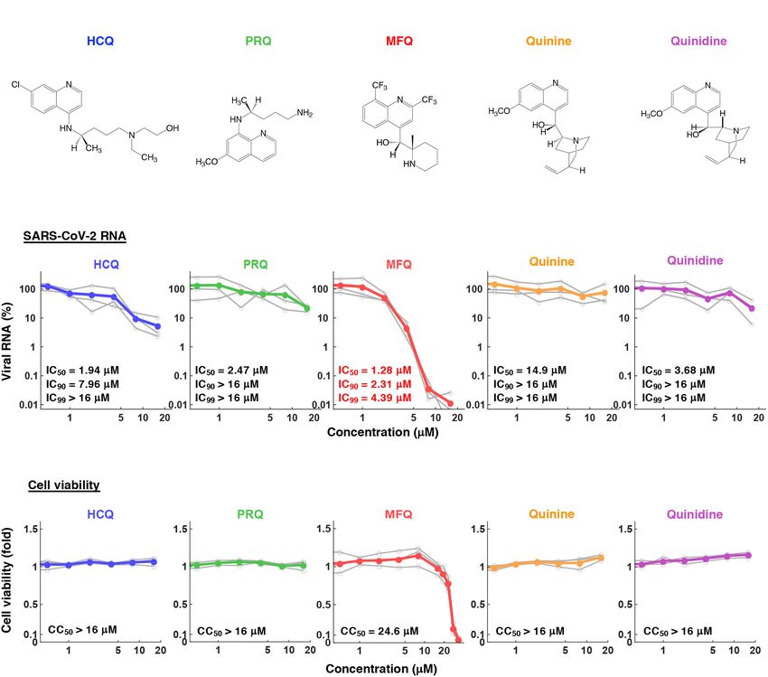
Shionoya et al.                                                                                                                   Mefloquine Inhibits SARS-CoV-2 Entry

  FIGURE 1 | Mefloquine (MFQ) inhibits Severe Acute Respiratory Syndrome-related coronavirus 2 (SARS-CoV-2) propagation. (A) Schematic representation of the
  SARS-CoV-2 infection assay. VeroE6/TMPRSS2 cells were inoculated with SARS-CoV-2 (Wk-521 strain) at an MOI of 0.001 for 1 h. After removing the unbound
  virus, cells were cultured for 24 h to detect virus-encoding N protein by immunofluorescence assay (IFA) and immunoblot (IB) or to detect viral RNA in the culture
  supernatant by RT-qPCR, or for 48 h to observe virus-induced cytopathic effect (CPE). Compounds were treated given throughout the assay. (B) Dose dependency
  of Hydroxychloroquine (HCQ) on CPE suppression. VeroE6/TMPRSS2 cells were inoculated with the virus for 1 h. Removing the unbound virus, cells were cultured
  with a medium containing the indicated compounds for 48 h. CPE was observed by microscopy. (C) Screening of anti-parasitic/protozoal drugs in the cell-based
  infection assay. Compounds were administrated at 5 µM, at which hydroxychloroquine showed little effect on CPE. The viability of infected cells was quantified via a
  high content imaging analyzer by setting the value for the sample treated with DMSO solvent as 1. MFQ showed more than 57-fold higher cell viability than DMSO
  controls. (D,E) SARS-CoV-2-induced CPE and viral N protein expression upon compound treatments [DMSO at 0.08%; hydroxychloroquine (HCQ), mefloquine
  (MFQ), and primaquine (PRQ) at 8 µM]. Red and blue signals of merged images indicate viral N protein and nucleus, respectively (D, lower). Viral N protein and actin,
  an internal control, were detected by immunoblot (E). (F) The anti-SARS-CoV-2 activity of the indicated compounds in Calu-3 cells, a human lung epithelial
  cell-derived line.

Frontiers in Microbiology | www.frontiersin.org                                    3                                             April 2021 | Volume 12 | Article 651403
Shionoya et al.                                                                                             Mefloquine Inhibits SARS-CoV-2 Entry

recovered, fixed in 4% paraformaldehyde and stained with 0.02%          allow virus-cell attachment. After removing unbound virus by
40 ,6-diamidino-2-phenylindole (DAPI). The number of surviving          washing, attached viral RNA was extracted with RNeasy Mini
cells was quantified with a high content imaging analyzer. The          Kit (QIAGEN) and measured by real time RT-qPCR. The same
survival cell numbers treated with each drug are presented as           assay was done without cells to measure the background level.
a fold value relative to the cells treated with DMSO solvent            Heparin was used as a positive control that inhibits SARS-CoV-
(Figure 1C and Supplementary Figure 1). Drugs that protected            2 attachment to cells. The specific cell-attached SARS-CoV2
cells from virus-induced CPE to more than 10-fold of the infected       RNA was calculated by subtracting viral RNA levels without
cells treated with DMSO were selected as hits.                          cells from those with VeroE6/TMPRSS2 cells and are shown in
                                                                        Figure 3C.
Immunofluorescence and Immunoblot
Analysis
Viral encoded N protein expression was detected using a rabbit
                                                                        Post-attachment Assay
anti-SARS-CoV N antibody (Mizutani et al., 2004) as a primary           The assay examining the steps from Spike cleavage/membrane
antibody with anti-rabbit AlexaFlour 568 or anti-rabbit IgG-HRP         fusion through viral secretion, was conducted by initiating
(Thermo Fisher Scientific) as a secondary antibody by indirect          compound treatment after viral attachment to already
immunofluorescence or immunoblot analysis (Figures 1D–F) as             highly infected cells and detecting the secreted virus
previously reported (Ohashi et al., 2018). Anti-actin (Sigma) was       for a short duration (Figure 3D): Cells were incubated
used as an internal control for the immunoblot analysis. For            with the virus at an MOI of 1.5 at 4◦ C for 1 h, then
immunofluorescence, nuclei were stained with DAPI (blue).               removing the unattached virus. These virus-attached
                                                                        cells were incubated at 37◦ C for 6 h in the presence
                                                                        of compounds to allow viral entry through replication
Quantification of Viral RNA                                             and secretion. The culture supernatant was recovered
Viral RNA was extracted with QIAamp Viral RNA Mini Kit
                                                                        to detect extracellular viral RNA. E-64d (12.5 µM),
(QIAGEN), RNeasy Mini Kit (QIAGEN) and MagMAXTM
                                                                        lysosomal/cytosolic cysteine protease inhibitor, was used as
Viral/Pathogen II Nucleic Acid Isolation Kit (Thermo Fisher
                                                                        a positive control.
Scientific). We quantified viral RNA by real time RT-PCR
analysis with a one-step RT-qPCR kit (THUNDERBIRD
Probe One-step RT-qPCR kit, TOYOBO) using 50 -
                                                                        Pseudovirus Infection Assay
ACAGGTACGTTAATAGTTAATAGCGT-30 for forward primer
                                                                        SARS-CoV-2 pseudotype virus was produced using the vesicular
and 50 -ATATTGCAGCAGTACGCACACA-30 for reverse primer,
                                                                        stomatitis virus (VSV)-pseudotype system essentially as
and a 50 -FAM-ACACTAGCCATCCTTACTGCGCTTCG-30
                                                                        described previously (Fukushi et al., 2005; Tani et al., 2010)
probe, as described (Corman et al., 2020). The detection limit of
                                                                        using the expression plasmid encoding the SARS-CoV-2 Spike
SARS-CoV-2 RNA in this study was 39 cycle (Ct value).
                                                                        protein and G-deficient VSV, which contains the luciferase
                                                                        gene instead of the VSV-G gene. HCV pseudotype virus was
Cell Viability                                                          prepared from the retrovirus pseudoparticle system using the
Cell viability was examined by MTT assay as previously reported         expression plasmid for murine leukemia virus Gag-Pol, luciferase
(Ohashi et al., 2018; Figure 2C) or by quantification of survival       protein and HCV E1E2 envelope protein (kindly provided by
cell numbers fixed with 4% paraformaldehyde and stained with            Dr. Francois-Loic Cosset at University of Lyon) as described
0.02% DAPI with a high content imaging analyzer ImageXpress             (Bartosch et al., 2003).
Micro Confocal (MOLECULAR DEVICES) (Figures 1C, 4A,                        The pseudovirus for SARS-CoV-2 was inoculated to
right and Supplementary Figure 2B).                                     VeroE6/TMPRSS2 cells in the presence or absence of compounds
                                                                        and the intracellular luciferase activity was measured at 24 h
Time-of-Addition Analysis                                               post-inoculation. Camostat (TCI) 50 µM and E-64d (Cayman)
VeroE6/TMPRSS2 cells were inoculated with the virus at an               50 µM were used as a positive control to inhibit SARS-
MOI of 0.001 for 1 h, and the free virus was removed by                 CoV-2 entry (Figure 3E, left). HCV pseudotype virus was
washing. Compounds were added at three different times before           inoculated to Huh-7 cells (kindly provided by Dr. Francis
measuring extracellular viral RNA (Figure 3B): (a) throughout           Chisari at The Scripps Research Institute) for 4 h, followed
the entire assay covering viral lifecycle (whole: 1 h + 24 h            by washing, culturing for 72 h and measuring luciferase
after virus inoculation), (b) only the early phase of the assay         activity. Compounds were treated for 1 h prior to infection
covering viral entry steps (entry: initial 1 h + 2 h after virus        and for 4 h during the virus inoculation. Bafilomycin A1 at
inoculation), (c) during the late phase of the assay, from              10 nM was used as a positive control for inhibiting HCV entry
viral replication to viral secretion (post-entry: last 22 h after       (Figure 3E, right).
virus inoculation).

Virus-Cell Attachment Assay                                             Statistical Analysis
Virus and compounds were preincubated at 4◦ C for 1 h and               Statistical significance was analyzed using the two-tailed Student’s
then exposed to VeroE6/TMPRSS2 cells at 4◦ C for 5 min to               t-test (∗ p < 0.05; ∗∗ p < 0.01; N.S., not significant).

Frontiers in Microbiology | www.frontiersin.org                     4                                      April 2021 | Volume 12 | Article 651403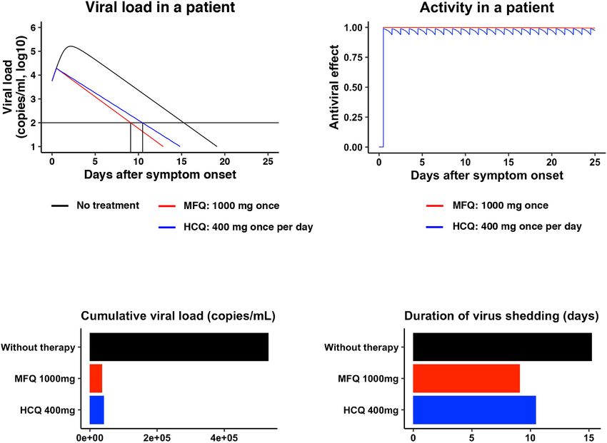
Shionoya et al. Mefloquine Inhibits SARS-CoV-2 Entry
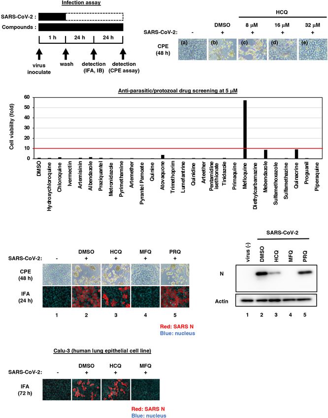
FIGURE 2 | The anti-SARS-CoV-2 activity of MFQ and its derivatives. (A) Chemical structures of MFQ and its derivatives. (B) Extracellular SARS-CoV-2 RNA was
quantified upon treatment with HCQ, MFQ and related compounds PRQ, Quinine and Quinidine at varying concentrations. Calculated inhibitory concentrations of
50, 90, and 99% maximum (IC50 , IC90 , and IC99 ) for each compound are as indicated. (C) Cell viability was measured by MTT assay with the calculated 50%
maximal cytotoxic concentration (CC50 ).
RESULTS Aiming to identify drugs with greater anti-SARS-CoV-2
potential than HCQ, we employed 5 µM for drug screening,
Identification of Mefloquine as a a concentration at which HCQ had no CPE suppression. As
a drug library, we used approved anti-parasitic/anti-protozoal
Potential Inhibitor Against SARS-CoV-2 drugs for following two reasons; (1) In addition to Chloroquine
Infection and HCQ, some drugs such as Ivermectin, Atovaquone, and
In this study, we mainly used VeroE6/TMPRSS2 cells, which quinoline derivatives were reported to demonstrate antiviral
is established by overexpressing transmembrane serine protease activities against other RNA viruses (Mastrangelo et al., 2012;
2 (TMPRSS2) in VeroE6 cells (Nao et al., 2019; Matsuyama Al-Bari, 2015; Cifuentes Kottkamp et al., 2019; DeWald et al.,
et al., 2020), and human lung epithelial-derived Calu-3 cells in 2019). (2) Anti-parasitic/anti-protozoal agents generally reach
a part of experiments, as SARS-CoV-2 infection models. First, high concentrations (i.e., over µM ranges) in the plasma in
we examined the dose dependency of HCQ for antiviral activity clinical settings (Sinha et al., 2014). We thus screened 27
by a cytopathic effect (CPE) assay: VeroE6/TMPRSS2 cells were FDA/EMA/PMDA-approved (or approved in the past) anti-
inoculated with SARS-CoV-2 at an MOI of 0.001 for 1 h, washed parasitic/anti-protozoal drugs at 5 µM by the CPE assay
to remove unbound virus, and incubated for an additional 48 h (Figure 1C and Supplementary Figure 1). By following the
(Figure 1A). SARS-CoV-2 propagation in the cells exhibited scheme shown in Figure 1A, cells at 48 h post-inoculation
an intensive cytopathic effect (Figure 1B, panel b), as reported were fixed, stained with DAPI, and counted to quantify survival
(Matsuyama et al., 2020). HCQ protected cells from SARS- cell numbers. The graph in Figure 1C shows survival cell
CoV-2-induced cytopathology completely at the concentration of numbers relative to that of DMSO-treated cells as a control,
32 µM, remarkably but not completely at 16 µM, and very little and survival cell number relative to that of non-infected cells
at 8 µM (Figure 1B, panels c–e). are shown in Supplementary Figure 1. In this screening,
Frontiers in Microbiology | www.frontiersin.org 5 April 2021 | Volume 12 | Article 651403Shionoya et al. Mefloquine Inhibits SARS-CoV-2 Entry FIGURE 3 | MFQ inhibits the SARS-CoV-2 entry process. (A) SARS-CoV-2 life cycle. SARS-CoV-2 infection is initiated with virus attachment to the host cells that involves the cellular receptor, angiotensin converting enzyme 2 (ACE2), followed by the cleavage of viral Spike (S) proteins by either transmembrane serine protease (TMPRSS2) on the plasma membrane or cathepsins in the endosome/lysosome that induces fusion of viral and host membranes. Viral RNA is translated, processed and replicated to be assembled into progeny virus with viral structural proteins and released extracellularly. (B) Scheme of the time of addition analysis. Compounds were treated at three different times: (a) whole: throughout the assay for 25 h, (b) entry: for the initial 3 h to evaluate the effect on the viral entry process and (c) post-entry: for the last 22 h to evaluate the effect on viral replication/re-infection. Viral RNA levels in the culture supernatant are shown in the graph by setting that upon DMSO treatment as 100%. (C) Virus-cell attachment assay. VeroE6/TMPRSS2 cells were exposed to virus at an MOI of 0.001 at 4◦ C for 5 min with 50 µM MFQ or 100 U/mL Heparin, a SARS-CoV-2 attachment inhibitor used as a positive control. After washing the unbound virus, cell surface-attached virus was extracted and quantified by real-time RT-PCR. (D) Post-attachment assay. For evaluating the activity after virus attachment, from membrane fusion to virus secretion, VeroE6/TMPRSS2 cells preincubated with the virus at an MOI of 1.5 at 4◦ C for 1 h to allow virus attachment were treated with compounds for 6 h at 37◦ C. Extracellular viral RNA was quantified by RT-qPCR. E-64d, a cysteine protease inhibitor, was used as a positive control. (E) Pseudovirus assays carrying the SARS-CoV-2 Spike or hepatitis C virus (HCV) E1E2 envelope. In the SARS-CoV-2 pseudovirus assay, Camostat and E-64d were used as positive controls for inhibiting TMPRSS2 and cysteine protease, respectively (E, left). Bafilomycin A1 (BFA1), which reported to inhibit HCV entry, was used as a positive control for HCV pseudovirus assay (E, right). *p < 0.05 and **p < 0.01. Frontiers in Microbiology | www.frontiersin.org 6 April 2021 | Volume 12 | Article 651403
Shionoya et al.                                                                                                                  Mefloquine Inhibits SARS-CoV-2 Entry

  FIGURE 4 | MFQ shows synergistic anti-SARS-CoV2 activity with replication inhibitor NFV. (A) Viral RNAs in the culture supernatant at 24 h after co-treatment with
  MFQ and NFV were quantified by real-time RT-PCR. Relative values are shown of viral RNA or cell viability to those treated with DMSO control. Cell viability was
  simultaneously measured with a high content image analyzer. [MFQ at 0, 0.83, 1.08, 1.40, 1.82, and 2.37 µM (1.3-fold-dilution); NFV at 0, 2.20, 2.64, and 3.17 µM
  (1.2-fold-dilution)]. (B) The three-dimensional interaction landscapes of NFV and MFQ were evaluated with the Bliss independence model. Orange, white and
  dark-blue colors on the contour plot indicate synergy, additive and antagonism, respectively.

HCQ, Chloroquine and Ivermectin had little effect, while MFQ                          Antiviral Profile of Mefloquine and Other
remarkably protected cells from the virus-induced CPE, with a
                                                                                      Quinoline Derivatives
more than 57-fold increase in surviving cells over those of the
vehicle control (Figure 1C).                                                          To profile the anti-SARS-CoV-2 activity of compounds, we
    We next compared the antiviral activities of MFQ with that                        quantified viral RNA released into the culture supernatant in
of HCQ and an additional Chloroquine derivative, Primaquine                           addition to cell viability at 24 h after virus inoculation upon
(PRQ), as a reference. Cytopathogenicities at 48 h and the viral                      treatment at varying concentrations (0.5, 1, 2, 4, 8, and 16 µM)
N protein expression at 24 h after virus inoculation (a time                          of HCQ, PRQ, MFQ, and other related compounds, Quinine and
before showing CPE) were examined during treatment with                               Quinidine, that possess a quinoline ring (Figures 2A–C). The
each compound at 8 µM (Figures 1D,E): MFQ completely                                  50, 90, and 99% maximal inhibitory concentrations (IC50 , IC90 ,
protected cells from viral propagation-induced CPE and reduced                        and IC99 ) and 50% maximal cytotoxic concentrations (CC50 )
the production of viral protein (lane 4), whereas HCQ weakly                          are shown. HCQ and MFQ demonstrated antiviral activities
exerted an antiviral effect (lane 3), and PRQ had little antiviral                    in a dose-dependent manner, with higher potency for MFQ
effect (lane 5). To examine whether the observed antiviral effects                    than HCQ (Figure 2B). By contrast, PRQ showed marginal
depend on cell types or are generally reproduced beyond cell                          antiviral effects at all concentrations examined (see section
types, we used a human lung epithelial cell line, Calu-3, and                         “Discussion” for structural implication). We also confirmed the
found the robust antiviral activity of MFQ against SARS-CoV-2,                        effect of MFQ on SARS-CoV-2 RNA and the cell viability in
in contrast to much lower HCQ activity (Figure 1F). Therefore,                        Calu-3 cells (Supplementary Figure 2), showing the consistent
we focused on MFQ as a potential anti-SARS-CoV-2 drug in                              anti-SARS-CoV-2 activity at concentration ranges without
subsequent analyses.                                                                  cytotoxic effects.

Frontiers in Microbiology | www.frontiersin.org                                   7                                             April 2021 | Volume 12 | Article 651403
Shionoya et al. Mefloquine Inhibits SARS-CoV-2 Entry
Mefloquine Inhibits the SARS-CoV-2 We further examined the virus entry using a pseudovirus
Entry Process After Virus-Cell carrying the Spike protein derived from SARS-CoV-2 or the
envelope proteins of hepatitis C virus (HCV), another RNA virus
Attachment unrelated to coronavirus (Figure 3E). These pseudoviruses can
SARS-CoV-2 attaches to target cells by the binding of viral evaluate the entry mediated by these Spike or envelope proteins
Spike protein to its receptor, angiotensin-converting enzyme 2 (Bartosch et al., 2003; Hoffmann et al., 2020). The pseudovirus
(ACE2). It is then subjected to Spike cleavage by host proteases, assay showed that SARS-CoV-2 Spike-dependent viral entry was
either TMPRSS2 on the plasma membrane or cathepsins in the significantly inhibited by the TMPRSS2 inhibitor Camostat, and
endosomes, followed by the membrane fusion and the sorting to by MFQ to similar levels to those of E-64d (Figure 3E, left).
the site of replication (entry phase). Viral RNA then replicates However, the assay sensitivity itself was relatively lower than
and assembles with viral structural proteins to produce progeny the SARS-CoV-2 infection assay. Meanwhile, HCV envelope-
virus (replication phase) (Figure 3A; Hoffmann et al., 2020; mediated entry was not affected by MFQ, in contrast to the
Lebeau et al., 2020). reduced entry caused by Bafilomycin A1, a reported HCV entry
We next addressed which step in the viral life cycle MFQ inhibitor (Figure 3E, right). These results cumulatively suggest
inhibits by a series of assays. The time-of-addition analysis, that MFQ inhibited the post-attachment SARS-CoV-2 Spike-
in which compounds are treated at different times, is used dependent entry process.
to evaluate the phase of viral entry and replication separately
(Wang et al., 2020). As previously reported (Wang et al.,
2020), compounds were treated at three different time points
Synergistic Antiviral Activity of
(Figure 3B), either throughout the assay (a; whole life cycle, 1 h Combined Treatment of Mefloquine With
during virus inoculation + 24 h after inoculation), for the initial Nelfinavir
3 h (b; entry phase, 1 h during virus inoculation + 2 h after Combination treatment with multiple agents with different
inoculation), or for the last 22 h (c; post-entry phase, including modes of action is a strategy to improve the outcome of antiviral
replication). In this analysis, RDV, a reported replication inhibitor treatments, including those against human immunodeficiency
(Wang et al., 2020), had no inhibitory effect when applied during virus (HIV) and HCV (Shen et al., 2008; Koizumi et al.,
the initial 3 h (Figure 3B, lane 5), but it decreased viral RNA 2017). We, therefore, examined the combination of MFQ and
in the post-entry phase (Figure 3B, lane 6). By contrast, MFQ a representative anti-SARS-CoV replication inhibitor, Nelfinavir
remarkably reduced viral RNA levels to under 3% when applied (NFV) (Yamamoto et al., 2004). NFV has been suggested to
at the entry phase (Figure 3B, lane 8), but showed much lower inhibit SARS-CoV-2 replication thorough binding with the
antiviral activity (to 24%) when treated after the first round of SARS-CoV-2 main protease by docking simulation (Ohashi et al.,
viral entry (Figure 3B, lane 9). The viral RNA reduction by 2021). Following the experimental scheme in Figure 1A, we
MFQ in lane 9 was likely to the inhibition of second round of treated cells with paired compounds at varying concentrations
infection and thereafter of the produced virus, which occurred for 24 h and quantified viral RNA in the cultured supernatant
during the 22 h. These data suggest that MFQ inhibits the entry by real-time RT-PCR in addition to cell viability by a high
process of SARS-CoV-2. content image analyzer. Viral RNA levels were reduced by a single
We then evaluated the virus-cell attachment in the presence treatment of either MFQ or NFV in a dose-dependent manner,
or absence of MFQ by incubating cells with the virus at 4◦ C to and these was further reduced by combination treatment without
allow viral attachment to the cell surface but not the following any cytotoxicity (Figure 4A). Bliss independence-based synergy
processes. After washing the unattached virus and compounds, plot showed a synergistic antiviral effect in wide concentration
we extracted and quantified the viral RNA on the cell surface. ranges, especially at higher doses (Figure 4B, orange indicates
SARS-CoV-2 RNA from virus attached the surface of the cell synergistic effect).
was drastically reduced in the presence of heparin, an entry
inhibitor for SARS-CoV-2, used as a positive control (Tandon Mathematical Prediction of the
et al., 2020; Tree et al., 2020), while that was not affected by Mefloquine Treatment in Clinical Settings
MFQ treatment (Figure 3C). However, MFQ inhibited the post- Pharmacokinetics data for MFQ and HCQ, including the
attachment phase, ranging from the membrane fusion to virus maximum drug concentration (Cmax ) in the plasma, half-life,
production (Figure 3D): Virus-attached cells were prepared by and the distribution to the lung, are reported (Desjardins et al.,
incubation with a large amount of virus (MOI of 1.5, more than 1979; Jones et al., 1994; Lim et al., 2009; Chhonker et al.,
1,000-fold higher than used in other normal infection assay) at 2018). Mathematical modeling combined with pharmacokinetics,
4◦ C for 1 h followed by washing. The cells were transferred to pharmacodynamics, and the viral dynamics model described in
37◦ C for 6 h in the presence or absence of compounds to induce section “Materials and Methods” (Ohashi et al., 2021) predicted
membrane fusion and subsequent steps up to virus secretion, the dynamics of viral load after MFQ (1,000 mg, once) and HCQ
and viral RNA in the supernatant was quantified. MFQ clearly (400 mg, once per day) administration in patients (Figure 5A, red
reduced the viral RNA levels to almost the same as those when and blue, respectively) and the corresponding time-dependent
treatment with E-64d, a lysosomal/cytosolic cysteine protease antiviral activity of MFQ and HCQ (Figure 5B). The high
inhibitor reported to inhibited SARS-CoV-2 entry (Hoffmann antiviral potential and the long half-life of MFQ (more than
et al., 2020; Hu et al., 2020; Figure 3D). 400 h) (Desjardins et al., 1979) were predicted to exert a
Frontiers in Microbiology | www.frontiersin.org 8 April 2021 | Volume 12 | Article 651403Shionoya et al. Mefloquine Inhibits SARS-CoV-2 Entry
FIGURE 5 | Prediction of the impact of MFQ and HCQ treatment on SARS-CoV-2 dynamics in clinical settings. (A,B) The predicted viral load dynamics without (A,
black), upon MFQ (1,000 mg oral, once) (A, red) or upon HCQ (400 mg oral, once per day) (A, blue) administration and the time-dependent antiviral activity of MFQ
and HCQ (B) predicted by pharmacokinetics/pharmacodynamics/viral-dynamics (PK/PD/VD) models. (C,D) The cumulative viral load calculated as the area under
the curve in (A) and the duration of virus shedding (days) [time from symptom onset to the day achieving a viral load under the detection limit (black horizontal line) in
(A)] were evaluated for non-treatment (black), MFQ treatment (red) or HCQ treatment (blue).
continuous antiviral effect, and a resulting decline of viral load et al., 2020). Pharmacokinetics analyses in healthy volunteers
in a short period of time than HCQ (Figure 5A). Cumulative receiving oral administration of 200 mg HCQ demonstrated a
viral load, which is the area under the curve for the viral load Cmax in the blood of 0.49–0.55 µM (McLachlan et al., 1993),
over the time course, was calculated to be reduced by 6.98% in lower than the concentration ranges having significant anti-
MFQ and 7.87% in HCQ, respectively (Figure 5C). The time SARS-CoV-2 activity. These data led us to identify a drug
until the viral load declines beneath the detectable level is 15.2 possessing a greater anti-SARS-CoV-2 potential. In this study,
days without treatment, but it was calculated to be shortened to HCQ and MFQ demonstrated antiviral activities in a dose-
10.5 days after HCQ treatment, whereas, 9.10 days after MFQ dependent manner, with higher potency for MFQ than HCQ
treatment (Figure 5D). These analyses predict the effectiveness (Figure 2B). By contrast, PRQ showed marginal antiviral effects
of MFQ to reduce the viral load at clinical drug concentrations. at all concentrations examined, suggesting that the hydroxyl and
amino groups in the side chain of MFQ and/or that the position
of the side chain on the quinoline ring are important for the anti-
DISCUSSION SARS-CoV-2 activity. The octanol-water partition coefficient (log
P) values of MFQ, HCQ, Quinine, Quinidine and PRQ were
Given the in vitro anti-SARS-CoV-2 activity and the in vivo calculated to be 4.34, 2.87, 2.48, 2.4, and 1.47, respectively (Ghose
effect on the related coronaviruses (Ko et al., 2020; Liu et al., and Crippen, 1987), which imply that the higher hydrophobicity
2020; Wang et al., 2020; Weston et al., 2020), Chloroquine of MFQ, possibly due to the two trifluoromethyl groups, may
and HCQ have been expected to be effective as anti-COVID-19 contribute to its high antiviral activity. Furthermore, the antiviral
drugs. However, accumulative data have not provided sufficient effects might be associated with the electron density of the
evidence supporting a preferable clinical outcome (Funnell et al., quinoline ring. MFQ, which has two strong electron-withdrawing
2020). The IC50 , IC90 , and IC99 for HCQ calculated in this trifluoromethyl groups in the quinoline ring, shows the strongest
study were 1.94, 7.96, and 37.2 µM, respectively, consistent antiviral activity among the five analogs. HCQ, which has a
with the IC50 values at µM ranges examined in other studies moderate electron-withdrawing chlorine group, has moderate
(Gendrot et al., 2020; Hattori et al., 2020; Liu et al., 2020; Touret antiviral activity. PRQ, Quinine, and Quinidine, having the
Frontiers in Microbiology | www.frontiersin.org 9 April 2021 | Volume 12 | Article 651403Shionoya et al. Mefloquine Inhibits SARS-CoV-2 Entry
electron-donating methoxy group (CH3O) into the quinoline Winkler et al., 2020; Yu J. et al., 2020). However, except for
ring, show decreased antiviral activities. Thus, the higher electron antibodies or vaccine candidates, there are very limited reports
density of the quinoline ring might be related with the stronger at present successfully confirming the reduction of SARS-CoV-
antiviral effects against SARS-CoV-2. 2 viral load in these models by treatment with drug candidates
SARS-CoV-2 entry requires the initial binding of the viral (Park et al., 2020). At this time, however, proposing an additional
Spike protein to its cell surface receptor ACE2, then Spike treatment choice with significant antiviral evidences is urgently
cleavage by either of the two independent host proteases, demanded to combat COVID-19. Interestingly, MFQ showed
endosomal pH-dependent cathepsin or plasma membrane pH- a synergistic effect combined with NFV, a replication inhibitor
independent TMPRSS2 (Hoffmann et al., 2020; Figure 3A). for SARS-associated coronavirus (Yamamoto et al., 2004; Ohashi
Recently, it has been reported that the sensitivity to viral entry et al., 2021; Figure 4). These data would prospect better
inhibitors such as Chloroquine, HCQ and a TMPRSS2 inhibitor clinical outcomes by combined drugs with different modes of
Camostat depends on cell types, so that recommended not action, as used with antiviral therapy against HIV and HCV
to rely only on widely used Vero cell line, but to use rather (Shen et al., 2008; Koizumi et al., 2017). Given the inhibition
TMPRSS2-complemented Vero cells, Calu-3 cells or presumably of viral entry, MFQ is also expected for prophylactic use.
primary respiratory/lung cell culture in an air-liquid interface Its long half-life of approximately 20 days is advantageous
system or organoids as a more physiologically relevant model for achieving a long-lasting antiviral state by a single oral
for airway epithelial cells (Hoffmann et al., 2020; Suzuki et al., administration. Consequently, our analysis highlights the anti-
2020). Due to the poor availability of primary cells, we employed SARS-CoV-2 potency of MFQ, of which efficacy is expected
VeroE6/TMPRSS2 and Calu-3 cells in this study, and discovered to be further evaluated in the future through in vivo or
that MFQ inhibited the viral entry more potently than HCQ clinical testing.
in these TMPRSS2-expressing cells. Importantly, standard MFQ
treatment given to healthy volunteers achieved a plasma Cmax
of 4.58 µM with a long half-life (more than 400 h) (Karbwang DATA AVAILABILITY STATEMENT
and White, 1990), which is within concentration ranges exerting
significant anti-SARS-CoV-2 activity in vitro. Moreover, it has The original contributions presented in the study are included
been reported that the MFQ concentration in the lung was over in the article/Supplementary Material, further inquiries can be
10-fold that of the blood in MFQ-treated human participants directed to the corresponding author/s.
(Jones et al., 1994), expecting an even higher anti-SARS-CoV-
2 effect of MFQ. Our mathematical model analysis (Figure 5)
quantified this prediction, demonstrating a clear reduction AUTHOR CONTRIBUTIONS
in both cumulative viral load in patients and the time for
KW designed the study, critically revised the manuscript, and
viral elimination.
supervised the project. KS screened compounds in the cell-based
The in vitro anti-SARS-CoV-2 activity of MFQ itself has been
screen. KS and MY performed biological experiments. KS and
reported (Fan et al., 2020; Gendrot et al., 2020; Jeon et al., 2020;
KW wrote the manuscript. KS, MY, SI, YI, SF, HO, WS, TT,
Weston et al., 2020), however, they only reported the anti-SARS-
SA, KK, SI, YT, TS, MM, MT, TW, and KW analyzed and
CoV-2 activity in a single cell line (Vero or VeroE6 cells) with
discussed the results.
a single readout (viral RNA or CPE) at only one experimental
condition without mechanistic analysis. In the present study,
in addition to the comparing the activity of MFQ with HCQ FUNDING
and other analogs side-by-side, we characterized the modes of
action and combination treatments. Furthermore, we addressed This work was supported by The Agency for Medical
the clinical antiviral efficacy of MFQ by mathematical prediction, Research and Development (AMED) emerging/re-emerging
a significant scientific novelty. Our time-of-addition, virus-cell infectious diseases project (Grant Nos. JP19fk0108111,
attachment, post attachment and pseudovirus assays suggest that JP19fk0108156, JP20fk0108179, JP20fk0108274, JP20fk0108294,
MFQ inhibits the SARS-CoV-2 entry phase after attachment, JP20fk0108411, and JP20fk0108511), the Japan Society for the
including the viral Spike cleavage/membrane fusion and the Promotion of Science KAKENHI (Grant No. JP20H03499),
following translocation to the replication complex. Detailed the JST MIRAI program; Moonshot R&D (Grant Nos.
analysis of the mode of action is the object of future studies. JPMJMS2021 and JPMJMS2025).
A limitation of our study is the use of antiviral profile data
in cell culture assays but without an in vivo infection model. To
date, SARS-CoV-2 studies have used models including hACE2- ACKNOWLEDGMENTS
transgenic mice, ferrets, cats, hamsters, non-human primates
and mice infected with mouse-adapted SARS-CoV-2 (Bao et al., We thank Dr. Shutoku Matsuyama at Department of Virology
2020; Gao et al., 2020; Golden et al., 2020; Gu et al., 2020; III of National Institute of Infectious Diseases in Tokyo and
Hassan et al., 2020; Imai et al., 2020; Jiang et al., 2020; Kim Dr. Shinichi Saito at Faculty of Sciences Division I of Tokyo
et al., 2020; Richard et al., 2020; Rockx et al., 2020; Rogers University of Science for their technical assistance and discussion.
et al., 2020; Shi et al., 2020; Sia et al., 2020; Sun et al., 2020; We appreciate the TUS (Tokyo University of Science) fund
Frontiers in Microbiology | www.frontiersin.org 10 April 2021 | Volume 12 | Article 651403Shionoya et al. Mefloquine Inhibits SARS-CoV-2 Entry
for strategic research areas for SA. The retrovirus-based SUPPLEMENTARY MATERIAL
pseudoparticle system and human hepatoma cell line, Huh-7
cells were kindly provided by Dr. Francois-Loic Cosset at the The Supplementary Material for this article can be found
University of Lyon and Dr. Francis Chisari at The Scripps online at: https://www.frontiersin.org/articles/10.3389/fmicb.
Research Institute, respectively. 2021.651403/full#supplementary-material
REFERENCES Golden, J. W., Cline, C. R., Zeng, X., Garrison, A. R., Carey, B. D., Mucker,
E. M., et al. (2020). Human angiotensin-converting enzyme 2 transgenic mice
Al-Bari, M. A. (2015). Chloroquine analogues in drug discovery: New directions infected with sars-cov-2 develop severe and fatal respiratory disease. JCI Insight
of uses, mechanisms of actions and toxic manifestations from malaria to 5:e142032. doi: 10.1172/jci.insight.142032
multifarious diseases. J. Antimicrob. Chemother 70, 1608-1621. doi: 10.1093/jac/ Gu, H., Chen, Q., Yang, G., He, L., Fan, H., Deng, Y. Q., et al. (2020). Adaptation
dkv018 of sars-cov-2 in balb/c mice for testing vaccine efficacy. Science 369, 1603-1607.
Bao, L., Deng, W., Huang, B., Gao, H., Liu, J., Ren, L., et al. (2020). The doi: 10.1126/science.abc4730
pathogenicity of sars-cov-2 in hace2 transgenic mice. Nature 583, 830-833. Hassan, A. O., Case, J. B., Winkler, E. S., Thackray, L. B., Kafai, N. M.,
doi: 10.1038/s41586-020-2312-y Bailey, A. L., et al. (2020). A sars-cov-2 infection model in mice
Bartosch, B., Dubuisson, J., and Cosset, F. L. (2003). Infectious hepatitis c demonstrates protection by neutralizing antibodies. Cell 182, 744-753.e4.
virus pseudo-particles containing functional e1-e2 envelope protein complexes. doi: 10.1016/j.cell.2020.06.011
J. Exp. Med. 197, 633-642. doi: 10.1084/jem.20021756 Hattori, S. I., Higshi-Kuwata, N., Raghavaiah, J., Das, D., Bulut, H., Davis, D. A.,
Chhonker, Y S., Sleightholm RL., Li J., Oupický D., and Murry DJ. (2018). et al. (2020). Grl-0920, an indole chloropyridinyl ester, completely blocks
Simultaneous quantitation of hydroxychloroquine and its metabolites in mouse sars-cov-2 infection. mBio 11:e01833-20. doi: 10.1128/mBio.01833-20
blood and tissues using LC–ESI–MS/MS: an application for pharmacokinetic Hoffmann, M., Kleine-Weber, H., Schroeder, S., Krüger, N., Herrler, T., Erichsen,
studies. J. Chromatogr. B 1072: 320-327. S., et al. (2020). Sars-cov-2 cell entry depends on ace2 and tmprss2 and is
Cifuentes Kottkamp, A., De Jesus, E., Grande, R., Brown, J. A., Jacobs, A. R., blocked by a clinically proven protease inhibitor. Cell 181, 271-280.e8. doi:
Lim, J. K., et al. (2019). Atovaquone inhibits arbovirus replication through the 10.1016/j.cell.2020.02.052
depletion of intracellular nucleotides. J. Virol. 93:e00389-19. doi: 10.1128/jvi. Hu, J., Gao, Q., He, C., Huang, A., Tang, N., and Wang, K. (2020). Development
00389-19 of cell-based pseudovirus entry assay to identify potential viral entry inhibitors
Corman, V. M., Landt, O., Kaiser, M., Molenkamp, R., Meijer, A., Chu, D. K. W., and neutralizing antibodies against sars-cov-2. Genes Dis. 7, 551-557. doi: 10.
et al. (2020). Detection of 2019 novel coronavirus (2019-ncov) by real- 1016/j.gendis.2020.07.006
time rt-pcr. Eurosurveillance 25:2000045. doi: 10.2807/1560-7917.Es.2020.25.3. Imai, M., Iwatsuki-Horimoto, K., Hatta, M., Loeber, S., Halfmann, P. J., Nakajima,
2000045 N., et al. (2020). Syrian hamsters as a small animal model for sars-cov-2
Cortegiani, A., Ippolito, M., Ingoglia, G., Iozzo, P., Giarratano, A., and Einav, infection and countermeasure development. Proc. Natl. Acad. Sci. U.S.A. 117,
S. (2020). Update i. A systematic review on the efficacy and safety of 16587-16595. doi: 10.1073/pnas.2009799117
chloroquine/hydroxychloroquine for covid-19. J. Crit. Care 59, 176-190. doi: Jeon, S., Ko, M., Lee, J., Choi, I., Byun, S. Y., Park, S., et al. (2020). Identification
10.1016/j.jcrc.2020.06.019 of antiviral drug candidates against sars-cov-2 from fda-approved drugs.
Desjardins, R. E., Pamplin, C. L., 3rd, von Bredow, J., Barry, K. G., and Canfield, Antimicrob. Agents Chemother 64:e819-e820. doi: 10.1128/aac.00819-20
C. J. (1979). Kinetics of a new antimalarial, mefloquine. Clin. Pharmacol. Ther. Jiang, R. D., Liu, M. Q., Chen, Y., Shan, C., Zhou, Y. W., Shen, X. R., et al.
26, 372-379. doi: 10.1002/cpt1979263372 (2020). Pathogenesis of sars-cov-2 in transgenic mice expressing human
DeWald, L. E., Johnson, J. C., Gerhardt, D. M., Torzewski, L. M., Postnikova, E., angiotensin-converting enzyme 2. Cell 182, 50-58.e8. doi: 10.1016/j.cell.2020.05.
Honko, A. N., et al. (2019). In vivo activity of amodiaquine against ebola virus 027
infection. Sci. Rep. 9:20199. doi: 10.1038/s41598-019-56481-0 Jones, R., Kunsman, G., Levine, B., Smith, M., and Stahl, C. (1994). Mefloquine
Fan, H. H., Wang, L. Q., Liu, W. L., An, X. P., Liu, Z. D., He, X. Q., et al. (2020). distribution in postmortem cases. Forensic. Sci. Int. 68, 29-32. doi: 10.1016/
Repurposing of clinically approved drugs for treatment of coronavirus disease 0379-0738(94)90376-x
2019 in a 2019-novel coronavirus-related coronavirus model. Chin. Med. J. Karbwang, J., and White, N. J. (1990). Clinical pharmacokinetics of mefloquine.
(Engl). 133, 1051-1056. doi: 10.1097/CM9.0000000000000797 Clin. Pharmacokinet 19, 264-279. doi: 10.2165/00003088-199019040-00002
Fukushi, S., Mizutani, T., Saijo, M., Matsuyama, S., Miyajima, N., Taguchi, F., et al. Keyaerts, E., Li, S., Vijgen, L., Rysman, E., Verbeeck, J., Van Ranst, M., et al. (2009).
(2005). Vesicular stomatitis virus pseudotyped with severe acute respiratory Antiviral activity of chloroquine against human coronavirus oc43 infection in
syndrome coronavirus spike protein. J. Gen. Virol. 86, 2269-2274. doi: 10.1099/ newborn mice. Antimicrob. Agents Chemother 53, 3416-3421. doi: 10.1128/aac.
vir.0.80955-0 01509-08
Funnell, S. G. P., Dowling, W. E., Muñoz-Fontela, C., Gsell, P. S., Ingber, D. E., Kim, Y. I., Kim, S. G., Kim, S. M., Kim, E. H., Park, S. J., Yu, K. M., et al. (2020).
Hamilton, G. A., et al. (2020). Emerging preclinical evidence does not support Infection and rapid transmission of sars-cov-2 in ferrets. Cell Host Microbe 27,
broad use of hydroxychloroquine in covid-19 patients. Nat. Commun. 11:4253. 704-709.e2. doi: 10.1016/j.chom.2020.03.023
doi: 10.1038/s41467-020-17907-w Ko, M., Chang, S. Y., Byun, S. Y., Choi, I., d’Alexandry d’Orengiani, A.-L. P. H.,
Gao, Q., Bao, L., Mao, H., Wang, L., Xu, K., Yang, M., et al. (2020). Development Shum, D., et al. (2020). Screening of FDA-approved drugs using a MERS-CoV
of an inactivated vaccine candidate for sars-cov-2. Science 369, 77-81. doi: clinical isolate from South Korea identifies potential therapeutic options for
10.1126/science.abc1932 COVID-19. bioRxiv [Preprint]. doi: 10.1101/2020.02.25.965582
Geleris, J., Sun, Y., Platt, J., Zucker, J., Baldwin, M., Hripcsak, G., et al. (2020). Koizumi, Y., Ohashi, H., Nakajima, S., Tanaka, Y., Wakita, T., Perelson, A. S.,
Observational study of hydroxychloroquine in hospitalized patients with covid- et al. (2017). Quantifying antiviral activity optimizes drug combinations against
19. N. Engl. J. Med. 382, 2411-2418. doi: 10.1056/NEJMoa2012410 hepatitis c virus infection. Proc. Natl. Acad. Sci. U.S.A. 114, 1922-1927. doi:
Gendrot, M., Andreani, J., Boxberger, M., Jardot, P., Fonta, I., Le Bideau, M., 10.1073/pnas.1610197114
et al. (2020). Antimalarial drugs inhibit the replication of sars-cov-2: An Lebeau, G., Vagner, D., Frumence, É., Ah-Pine, F., Guillot, X., Nobécourt, E., et al.
in vitro evaluation. Travel Med. Infect Dis. 37, 101873. doi: 10.1016/j.tmaid. (2020). Deciphering sars-cov-2 virologic and immunologic features. Int. J. Mol.
2020.101873 Sci. 21:5932. doi: 10.3390/ijms21165932
Ghose, A. K., and Crippen, G. M. (1987). Atomic physicochemical parameters Lim HS., Im JS., Cho JY., Bae KS., Klein TA., Yeom JS., et al. (2009).
for three-dimensional-structure-directed quantitative structure-activity Pharmacokinetics of hydroxychloroquine and its clinical implications in
relationships. 2. Modeling dispersive and hydrophobic interactions. J. Chem. chemoprophylaxis against malaria caused by Plasmodium vivax. Antimicrob.
Inf. Comput. Sci. 27, 21-35. doi: 10.1021/ci00053a005 Agents Chemother. 534: 1468-1475.
Frontiers in Microbiology | www.frontiersin.org 11 April 2021 | Volume 12 | Article 651403Shionoya et al. Mefloquine Inhibits SARS-CoV-2 Entry
Liu, J., Cao, R., Xu, M., Wang, X., Zhang, H., Hu, H., et al. (2020). Sun, J., Zhuang, Z., Zheng, J., Li, K., Wong, R. L., Liu, D., et al. (2020). Generation of
Hydroxychloroquine, a less toxic derivative of chloroquine, is effective in a broadly useful model for covid-19 pathogenesis, vaccination, and treatment.
inhibiting sars-cov-2 infection in vitro. Cell Discov. 6:16. doi: 10.1038/s41421- Cell 182, 734-743.e5. doi: 10.1016/j.cell.2020.06.010
020-0156-0 Suzuki, T., Itoh, Y., Sakai, Y., Saito, A., Okuzaki, D., Motooka, D., et al. (2020).
Mastrangelo, E., Pezzullo, M., De Burghgraeve, T., Kaptein, S., Pastorino, B., Generation of human bronchial organoids for sars-cov-2 research. bioRxiv
Dallmeier, K., et al. (2012). Ivermectin is a potent inhibitor of flavivirus [Preprint]. doi: 10.1101/2020.05.25.115600
replication specifically targeting ns3 helicase activity: New prospects for an old Tandon, R., Sharp, J. S., Zhang, F., Pomin, V. H., Ashpole, N. M., Mitra, D.,
drug. J. Antimicrob. Chemother. 67, 1884-1894. doi: 10.1093/jac/dks147 et al. (2020). Effective inhibition of sars-cov-2 entry by heparin and enoxaparin
Matsuyama, S., Nao, N., Shirato, K., Kawase, M., Saito, S., Takayama, I., et al. (2020). derivatives. J Virol. 95:e01987-20. doi: 10.1128/jvi.01987-20
Enhanced isolation of sars-cov-2 by tmprss2-expressing cells. Proc. Natl. Acad. Tang, W., Cao, Z., Han, M., Wang, Z., Chen, J., Sun, W., et al. (2020).
Sci. U.S.A. 117, 7001-7003. doi: 10.1073/pnas.2002589117 Hydroxychloroquine in patients with mainly mild to moderate coronavirus
McLachlan, A. J., Tett, S. E., Cutler, D. J., and Day, R. O. (1993). Absorption disease 2019: Open label, randomised controlled trial. BMJ 369:m1849. doi:
and in vivo dissolution of hydroxycholoroquine in fed subjects assessed using 10.1136/bmj.m1849
deconvolution techniques. Br. J. Clin. Pharmacol. 36, 405-411. doi: 10.1111/j. Tani, H., Shiokawa, M., Kaname, Y., Kambara, H., Mori, Y., Abe, T., et al. (2010).
1365-2125.1993.tb00388.x Involvement of ceramide in the propagation of japanese encephalitis virus.
Mizutani, T., Fukushi, S., Saijo, M., Kurane, I., and Morikawa, S. (2004). J. Virol. 84, 2798-2807. doi: 10.1128/jvi.02499-09
Phosphorylation of p38 mapk and its downstream targets in sars coronavirus- Touret, F., Gilles, M., Barral, K., Nougairède, A., van Helden, J., Decroly, E., et al.
infected cells. Biochem. Biophys. Res. Commun. 319, 1228-1234. doi: 10.1016/j. (2020). In vitro screening of a fda approved chemical library reveals potential
bbrc.2004.05.107 inhibitors of sars-cov-2 replication. Sci. Rep. 10:13093. doi: 10.1038/s41598-
Nao, N., Sato, K., Yamagishi, J., Tahara, M., Nakatsu, Y., Seki, F., et al. 020-70143-6
(2019). Consensus and variations in cell line specificity among human Tree, J. A., Turnbull, J. E., Buttigieg, K. R., Elmore, M. J., Coombes, N., Hogwood,
metapneumovirus strains. PLoS One 14:e0215822. doi: 10.1371/journal.pone. J., et al. (2020). Unfractionated heparin inhibits live wild-type sars-cov-2 cell
0215822 infectivity at therapeutically relevant concentrations. Br. J. Pharmacol. 178,
Ohashi, H., Nishioka, K., Nakajima, S., Kim, S., Suzuki, R., Aizaki, H., et al. (2018). 626-635. doi: 10.1111/bph.15304
The aryl hydrocarbon receptor-cytochrome p450 1a1 pathway controls lipid Wang, M., Cao, R., Zhang, L., Yang, X., Liu, J., Xu, M., et al. (2020). Remdesivir
accumulation and enhances the permissiveness for hepatitis c virus assembly. and chloroquine effectively inhibit the recently emerged novel coronavirus
J. Biol. Chem. 293, 19559-19571. doi: 10.1074/jbc.RA118.005033 (2019-ncov) in vitro. Cell Res. 30, 269-271. doi: 10.1038/s41422-020-
Ohashi, H., Watashi, K., Saso, W., Shionoya, K., Iwanami, S., Hirokawa, T., et al. 0282-0
(2021). Potential anti-COVID-19 agents, cepharanthine and nelfinavir, and Weston, S., Coleman, C. M., Haupt, R., Logue, J., Matthews, K., Li, Y., et al. (2020).
their usage for combination treatment. iScience. 24:102367. doi: 10.1016/j.isci. Broad anti-coronavirus activity of food and drug administration-approved
2021.102367 drugs against sars-cov-2 in vitro and sars-cov in vivo. J. Virol. 94:e01218-e20.
Park, S. J., Yu, K. M., Kim, Y. I., Kim, S. M., Kim, E. H., Kim, S. G., et al. (2020). doi: 10.1128/jvi.01218-20
Antiviral efficacies of fda-approved drugs against sars-cov-2 infection in ferrets. Winkler, E. S., Bailey, A. L., Kafai, N. M., Nair, S., McCune, B. T., Yu, J., et al.
mBio 11:e001114-20. doi: 10.1128/mBio.01114-20 (2020). Sars-cov-2 infection of human ace2-transgenic mice causes severe lung
Richard, M., Kok, A., de Meulder, D., Bestebroer, T. M., Lamers, M. M., Okba, inflammation and impaired function. Nat. Immunol. 21, 1327-1335. doi: 10.
N. M. A., et al. (2020). Sars-cov-2 is transmitted via contact and via the air 1038/s41590-020-0778-2
between ferrets. Nat. Commun. 11:3496. doi: 10.1038/s41467-020-17367-2 Yamamoto, N., Yang, R., Yoshinaka, Y., Amari, S., Nakano, T., Cinatl, J., et al.
Rockx, B., Kuiken, T., Herfst, S., Bestebroer, T., Lamers, M. M., Oude Munnink, (2004). Hiv protease inhibitor nelfinavir inhibits replication of sars-associated
B. B., et al. (2020). Comparative pathogenesis of covid-19, mers, and sars coronavirus. Biochem. Biophys. Res. Commun. 318, 719-725. doi: 10.1016/j.bbrc.
in a nonhuman primate model. Science 368, 1012-1015. doi: 10.1126/science. 2004.04.083
abb7314 Yu, B., Li, C., Chen, P., Zhou, N., Wang, L., Li, J., et al. (2020). Low dose of
Rogers, T. F., Zhao, F., Huang, D., Beutler, N., Burns, A., He, W. T., et al. (2020). hydroxychloroquine reduces fatality of critically ill patients with covid-19. Sci.
Isolation of potent sars-cov-2 neutralizing antibodies and protection from China Life Sci. 63, 1515-1521. doi: 10.1007/s11427-020-1732-2
disease in a small animal model. Science 369, 956-963. doi: 10.1126/science. Yu, J., Tostanoski, L. H., Peter, L., Mercado, N. B., McMahan, K., Mahrokhian,
abc7520 S. H., et al. (2020). DNA vaccine protection against sars-cov-2 in rhesus
Rosenberg, E. S., Dufort, E. M., Udo, T., Wilberschied, L. A., Kumar, J., macaques. Science 369, 806-811. doi: 10.1126/science.abc6284
Tesoriero, J., et al. (2020). Association of treatment with hydroxychloroquine or
azithromycin with in-hospital mortality in patients with covid-19 in New York Conflict of Interest: SI was employed by the Science Groove Inc.
state. Jama 323, 2493-2502. doi: 10.1001/jama.2020.8630
Shen, L., Peterson, S., Sedaghat, A. R., McMahon, M. A., Callender, M., The remaining authors declare that the research was conducted in the absence of
Zhang, H., et al. (2008). Dose-response curve slope sets class-specific any commercial or financial relationships that could be construed as a potential
limits on inhibitory potential of anti-hiv drugs. Nat. Med. 14, 762-766. conflict of interest.
doi: 10.1038/nm1777
Shi, J., Wen, Z., Zhong, G., Yang, H., Wang, C., Huang, B., et al. (2020). Copyright © 2021 Shionoya, Yamasaki, Iwanami, Ito, Fukushi, Ohashi, Saso,
Susceptibility of ferrets, cats, dogs, and other domesticated animals to sars- Tanaka, Aoki, Kuramochi, Iwami, Takahashi, Suzuki, Muramatsu, Takeda, Wakita
coronavirus 2. Science 368, 1016-1020. doi: 10.1126/science.abb7015 and Watashi. This is an open-access article distributed under the terms of the Creative
Sia, S. F., Yan, L. M., Chin, A. W. H., Fung, K., Choy, K. T., Wong, A. Y. L., et al. Commons Attribution License (CC BY). The use, distribution or reproduction in
(2020). Pathogenesis and transmission of sars-cov-2 in golden hamsters. Nature other forums is permitted, provided the original author(s) and the copyright owner(s)
583, 834-838. doi: 10.1038/s41586-020-2342-5 are credited and that the original publication in this journal is cited, in accordance
Sinha, S., Medhi, B., and Sehgal, R. (2014). Challenges of drug-resistant malaria. with accepted academic practice. No use, distribution or reproduction is permitted
Parasite 21:61. doi: 10.1051/parasite/2014059 which does not comply with these terms.
Frontiers in Microbiology | www.frontiersin.org 12 April 2021 | Volume 12 | Article 651403You can also read