Honey reduces the metastatic characteristics of prostate cancer cell lines by promoting a loss of adhesion - PeerJ
←
→
Page content transcription
If your browser does not render page correctly, please read the page content below
Honey reduces the metastatic
                                          characteristics of prostate cancer cell
                                          lines by promoting a loss of adhesion
                                          Sean D.A. Abel1 , Sumit Dadhwal2 , Allan B. Gamble2 and Sarah K. Baird1
                                      1
                                          Department of Pharmacology and Toxicology, University of Otago, Dunedin, New Zealand
                                      2
                                          School of Pharmacy, University of Otago, Dunedin, New Zealand

                                              ABSTRACT
                                              Honey has been shown to have a range of therapeutic effects in humans, with anti-
                                              inflammatory and anti-bacterial effects among those previously characterised. Here,
                                              we examine the possibility of New Zealand thyme, manuka and honeydew honeys, and
                                              their major sugar and phenolic components, reducing the development of metastatic
                                              cancer. Their activity was examined in vitro, in PC3 and DU145 prostate cancer cell
                                              lines, through measuring the compounds’ effects on the metastatic characteristics of
                                              migration, invasion and adhesion. First, the phenolic compounds gallic acid, caffeic
                                              acid, quercetin, kaempferol and chrysin were quantified in the honeys using high
                                              performance liquid chromatography, and found in nanomolar concentrations. In
                                              a Boyden chamber-based migration assay, non-toxic concentrations of thyme and
                                              honeydew honeys reduced cell migration by 20%, and all phenolic compounds except
                                              caffeic acid also lowered migration, although a mixture of only the sugars found in
                                              honey had no effect. All of the honeys, phenolics and the sugar-only mixture reduced
                                              invasive movement of cells through extracellular matrix by up to 75%. Most notably,
                                              each of the three honeys and the sugar-only mixture reduced cell adhesion to collagen I
                                              by 90%. With the exception of quercetin, phenolic compounds did not reduce adhesion.
                                              Therefore, honey and its sugar and phenolic components can lower the metastatic
                                              properties of cancer cells, and may do this by preventing effective cell adhesion to
                                              the extracellular matrix. The sugars and phenol compounds of honey are much more
                                              effective in combination than individually.
Submitted 9 March 2018
Accepted 6 June 2018
Published 3 July 2018
                                          Subjects Cell Biology, Toxicology, Oncology, Pharmacology
Corresponding author
Sarah K. Baird, s.baird@otago.ac.nz       Keywords Honey, Metastasis, Adhesion, Migration, Invasion, Phenolic, Sugar

Academic editor
Maria Deli
                                          INTRODUCTION
Additional Information and
Declarations can be found on              Honey is made from nectar by honeybees (Apis mellifera), and has been shown to
page 16                                   have potential therapeutic benefits in humans. Some of these characteristics, including
DOI 10.7717/peerj.5115                    anti-bacterial, anti-inflammatory, anti-oxidant and anti-hypertensive effects, have been
    Copyright                             demonstrated in vivo and some only in vitro. These properties are usually attributed to the
2018 Abel et al.                          honey’s polyphenol components (Alvarez-Suarez, Giampieri & Battino, 2013).
Distributed under                           The major constituents of honey are sugars, which make up more than 99% of the dry
Creative Commons CC-BY 4.0                weight of honey (White & Doner, 1980). Enzymes, including invertase, diastase and glucose
OPEN ACCESS
                                          oxidase, in the honey stomach of the bees, convert the nectar-derived polysaccharides into

                                          How to cite this article Abel et al. (2018), Honey reduces the metastatic characteristics of prostate cancer cell lines by promoting a loss of
                                          adhesion. PeerJ 6:e5115; DOI 10.7717/peerj.5115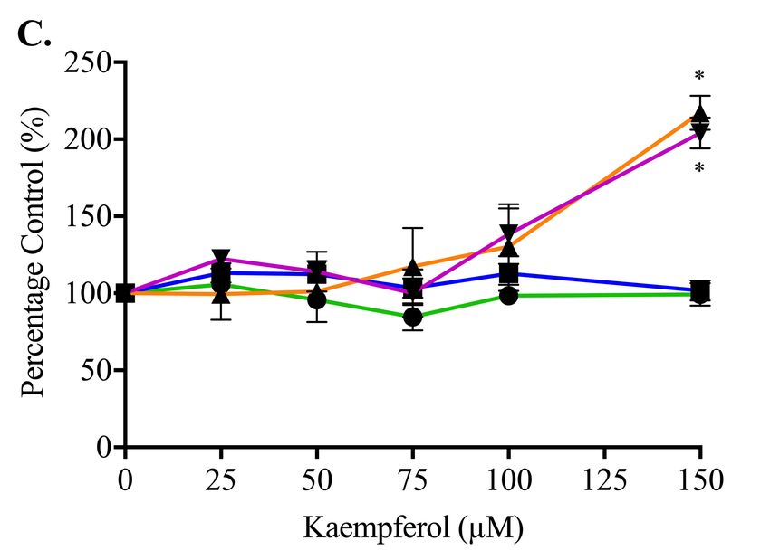
monosaccharides (Winston, 1991). The sugar mixture consists mainly of fructose (40.5%),
                                    glucose (33.5%), maltose (7.5%) and sucrose (1.5%), which is consistent between honeys
                                    regardless of the origin of the nectar (Cooper, Molan & Harding, 2002). However, honey
                                    also has many other components, including amino acids, vitamins, minerals, polyphenols
                                    and enzymes (White, 1978). The polyphenols, including phenolic acids and flavonoids,
                                    make up most of this group. Their composition and proportions vary depending upon the
                                    source of honey, with five of the most prevalent and biologically active being quercetin,
                                    gallic acid, kaempferol, chrysin and caffeic acid (Alvarez-Suarez, Giampieri & Battino, 2013;
                                    Kassim et al., 2010; Erejuwa, Sulaiman & Wahab, 2014).
                                       Honey has also been found to have a cytotoxic effect on cancer cell lines and in tumour-
                                    bearing animal models. It has been shown that honey can inhibit cancer cell proliferation
                                    and induce apoptosis in a range of cancers including breast, colon, liver and prostate
                                    cancers (Fauzi, Norazmi & Yaacob, 2011; Hassan et al., 2012; Jubri et al., 2012; Tomasin &
                                    Gomes-Marcondes, 2011; Tsiapara et al., 2009; Wen et al., 2012).
                                       It is also likely that honeys may have an effect on metastasis, the formation of secondary
                                    tumours at other sites in the body, which is responsible for the majority of cancer deaths
                                    (Mehlen & Puisieux, 2006). In metastasis, cancer cells migrate away from the primary
                                    tumour site, invade through the local tissue and enter the circulation, infiltrate at a
                                    secondary site and finally re-establish tumour growth. The cell processes involved in
                                    metastasis are regulated by receptors such as integrins and intracellular signalling proteins
                                    that control cell adhesion to the surrounding extracellular matrix, degrade physical barriers
                                    such as the basement membrane via proteolysis and increase cell motility (Liotta, Steeg &
                                    Stetler-Stevenson, 1991). The cell moves as a result of cytoskeletal changes mediated by a
                                    cycle of actin polymerisation and depolymerisation controlled by the Rho family GTPases,
                                    which allows formation of membrane protrusions. The protrusions, and the cell body
                                    behind it, attach to and detach from the extracellular matrix in a regulated motion as
                                    the cell moves forward (Pollard & Borisy, 2003). The processes of migration, invasion and
                                    adhesion can each be measured separately in vitro to characterise the mechanism of action
                                    of effects on metastasis.
                                       Two studies have investigated the effect of honey on metastasis using animal models.
                                    Oršolić et al. (2005) showed that 2 g/kg honey given orally daily before intravenous
                                    mammary or fibrosarcoma tumour cell injection into CBA mice reduced tumour formation
                                    in the lungs by around 70%, but had no effect if only given after tumour inoculation. An
                                    earlier study by the same group also examined colon adenocarcinoma in Y59 rats after
                                    intraveneous injection. In this study, it was found that honey, given orally for 10 days
                                    before tumour inoculation at 1 g/kg, reduced tumour nodules in the lungs by around 56%
                                    (Oršolić et al., 2003).
                                       In vitro, any assessment of honey’s potential anti-metastatic activity has been indirect.
                                    Moskwa et al. (2014) measured the activity of matrix metalloproteinase enzymes (MMPs),
                                    important for the invasion process, in U87MG glioblastoma cells, and found that they
                                    could be inhibited up to 95% by honeys, although the dose also caused high cytotoxicity.
                                    Otherwise, any related investigations have used only single polyphenols that are also
                                    found in honey. For example, gallic acid (3.5 µM) has been shown to reduce migration

Abel et al. (2018), PeerJ, DOI 10.7717/peerj.5115                                                                           2/21
in gastric cancer cells AGS by around 75% after 24 h in a scratch wound assay and 60%
                                    in a Boyden chamber assay after 48 h (Ho et al., 2010). Gallic acid also reduced invasion
                                    in the U87 glioma cell line by around 50% at 40 µg/mL (Lu et al., 2010). Caffeic acid
                                    (4 µg/mL) has been shown to reduce invasion in PC3 prostate cancer cells by around 50%
                                    (Lansky et al., 2005).
                                       The aim of this study was to investigate whether whole honeys, or their main components,
                                    sugars or phenolic constituents, might have anti-metastatic properties. We first measured
                                    the concentrations of five major phenolic compounds, and then looked at the three main
                                    characteristics of metastatic cells, using in vitro assays to measure migration, invasion and
                                    adhesion.

                                    MATERIALS AND METHODS
                                    Materials
                                    Dimethyl sulphoxide (DMSO) was from Scharlau (Barcelona, Spain). RPMI-1640 medium,
                                    foetal bovine serum (FBS), penicillin streptomycin (PS), PBS and trypsin were from Gibco
                                    (Carlsbad, CA). Bovine serum albumin (BSA), 3-(4,5-dimethylthiazol-2-yl)-2,5-diphenyl
                                    tetrazolium bromide (MTT), quercetin, caffeic acid, sucrose, maltose, fructose, D-(+)-
                                    glucose, formic acid, methanol, sodium bicarbonate, ethyl acetate, sodium hydroxide
                                    and hydrochloric acid were from Sigma-Aldrich (St. Louis, MO, USA). Thyme, manuka
                                    and honeydew honeys were kindly donated by New Zealand Honey Specialties Limited
                                    (Mosgiel, New Zealand). Kaempferol and chrysin were from Sapphire Biosciences (NSW,
                                    Australia). Gallic acid was from Abcam (Cambridge, UK). Fibronectin, collagen I and
                                             R
                                    Matrigel were from Corning (Corning, NY, USA).

                                    Preparation of honey and compounds
                                    Thyme and manuka honey were from the Central Otago region of New Zealand, produced
                                    from the Thyme bush (Thymus vulgaris) or Manuka tree (Leptospermum scoparium).
                                    Honeydew honey was collected in the Canterbury region of New Zealand, from the Beech
                                    Forest tree (Nothofagus fusca). All honeys were produced by the European honey bee (Apis
                                    mellifera). The sugar-only mixture, an artificial honey, was made by combining 40.5 g
                                    fructose, 33.5 g glucose, 7.5 g maltose and 1.5 g sucrose in 17 ml double distilled water
                                    (ddH2 O) (Cooper, Molan & Harding, 2002). Raw honeys were stored at room temperature,
                                    with minimal light exposure. Stock honey solutions were prepared by dissolving in warmed
                                    serum-free RPMI-1640 medium and sterilized using a 0.22 µm filter. Phenolic compounds
                                    were dissolved in 100% DMSO and filtered. Fresh preparations of honeys and compounds
                                    were made before each experiment.

                                    Preparation of phenolic extracts
                                    The following method was adapted from Wahdan (1998). Thyme, manuka and honeydew
                                    honeys were dissolved in warmed MilliQ water to give a final concentration of 20% (w/v)
                                    (10 mL or 15.48 g honey in 40 mL MilliQ). After extraction, dried samples were redissolved
                                    in 600 µL methanol.

Abel et al. (2018), PeerJ, DOI 10.7717/peerj.5115                                                                           3/21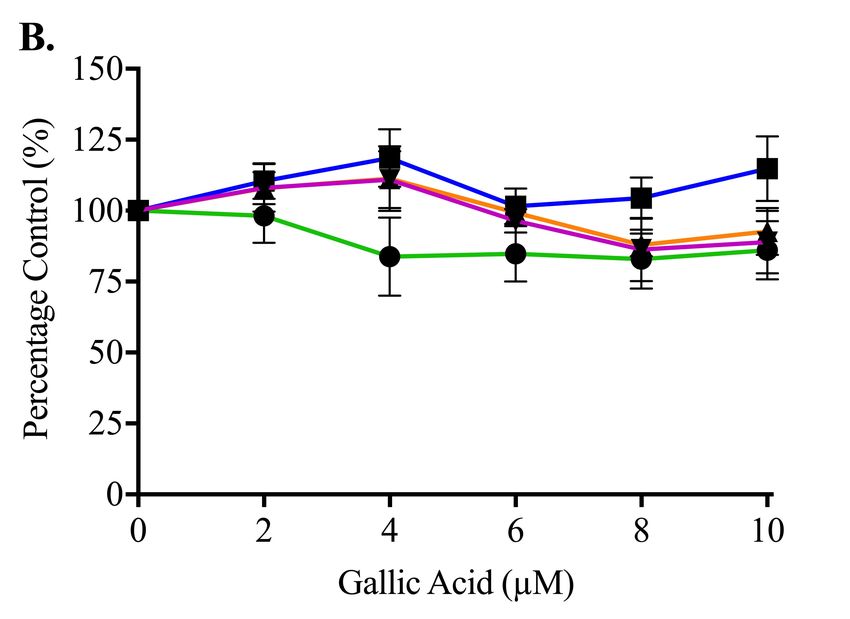
Free phenol extract preparation
                                    Each honey solution (50 mL) was adjusted to pH3.5 using concentrated HCl. Sodium
                                    bisulfite (1 g) and ethyl acetate (50 mL) were mixed with the honey solution in a separating
                                    funnel. The mixture was shaken for 1 min so that phenolic compounds moved into the
                                    organic phase. The honey solution was poured off, and the ethyl acetate layer transferred
                                    into a separate beaker. A further 50 mL ethyl acetate was added to the honey solution.
                                    These extraction steps were repeated six times. The ethyl acetate mixture (300 mL total)
                                    was concentrated using a rotary evaporator under vacuum (∼240 mbar) at 30 ◦ C. Dried
                                    compounds were reconstituted (methanol:ethyl acetate, 1:1), and dried under nitrogen to
                                    be stored at −15 ◦ C.

                                    Total phenol extract preparation
                                    Extra preparation was required in order to hydrolyse phenolic compounds that were bound
                                    to sugars via ester bonds. Each honey solution (25 mL) was combined with NaOH (25 mL,
                                    3N) and hydrolysed at room temperature for 4 h. Following this, pH was adjusted to 3.5
                                    using concentrated HCl, and extraction carried out as for the free phenol extraction above.

                                    Quantification of phenolic compounds by high performance liquid
                                    chromatography (HPLC)
                                    Phenolic separation was carried out using a CBM-20Alite Prominence HPLC (Shimadzu
                                    Corporation, Japan) on a reversed-phase Gemini 5 µm C18 110 Å, LC column (150 x
                                    4.6 mm) (Phenomenex, USA). The method was adapted from Kassim et al. (2010), and
                                    consisted of a gradient system to identify and quantify selected phenols in the extracted
                                    honey samples. The mobile phase was a binary solvent solution consisting of A = 0.25%
                                    formic acid and 2% methanol in water, and B = 100% methanol. The gradient method
                                    was as follows: 0 min 10% B, 20 min 40% B, 30 min 45% B, 50 min 60% B, 52 min
                                    80% B, 60 min 90% B, 62 min 10% B until 65 min. Honey phenolic compounds were
                                    detected using a diode array where the spectra were monitored at 370 nm (kaempferol and
                                    quercetin), 325 nm (caffeic acid), and 270 nm (gallic acid and chrysin).
                                       Calibration curves of standards were used to identify and quantify phenolic compounds
                                    within honey samples. Identification was completed by comparing standard retention
                                    time and absorbance spectrums against samples. Sample spiking was used to increase
                                    the confidence of peak identification. To resolve quantification issues due to split and
                                    shouldered peaks, the valley-to-valley integration method was employed.

                                    Cell culture
                                    Human prostate cancer cell lines PC3 (more metastatic) and DU145 (less metastatic) were
                                    a gift from Prof. Rosengren (University of Otago, New Zealand). Cells were maintained
                                    in RPMI-1640 medium, supplemented with 5% FBS, 100 U/mL penicillin, 100 µg/mL
                                    streptomycin and 2 g/L sodium bicarbonate. Cells were incubated in 5% CO2 /95% O2
                                    humidified air at 37 ◦ C.

                                    Boyden chamber migration assay
                                    The undersides of the Boyden chamber membranes (8 µm pore size, 24 well plates,
                                    Transwells from Corning) were coated with collagen I (15 µL, 150 µg/mL 0.01M HCl in

Abel et al. (2018), PeerJ, DOI 10.7717/peerj.5115                                                                          4/21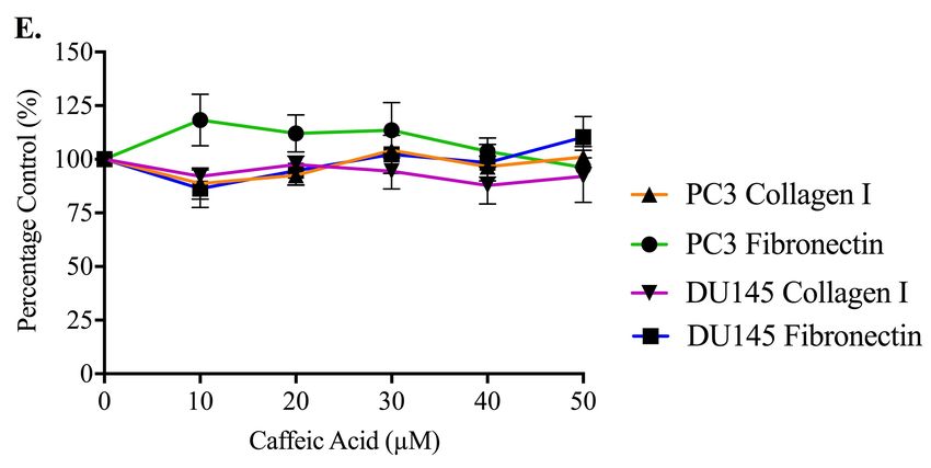
PBS), left to dry under sterile conditions for 1 h, washed twice with PBS and dried for a
                                    further 1 h. 30 × 103 PC3 cells in serum free growth medium were seeded into the upper
                                    chamber. The bottom chamber contained 5% serum-containing growth medium. Both
                                    upper and lower chambers were treated for 48 h with compounds at previously established
                                    maximal non-cytotoxic concentrations as follows: 1% (w/v) honey, 25 µM quercetin,
                                    10 µM gallic acid, 50 µM caffeic acid, 150 µM kaempferol or 100 µM chrysin (Abel &
                                    Baird, 2018). A vehicle control of 0.1% DMSO, the highest concentration added, was used.
                                    Following treatment incubation, MTT solution (0.5 mg/mL) was added to upper and
                                    lower chambers, and plates incubated for a further 3 h (Mosmann, 1983). Following this, a
                                    cotton tip was used to remove formazan crystals from the upper chamber. The undersides
                                    of the inserts were exposed to DMSO to dissolve formazan crystals from migrated cells.
                                    Absorbances were measured at 560 nm. Data were expressed as a percentage of migrated
                                    cells compared to a vehicle control.

                                    Boyden chamber invasion assay
                                    This assay was used to determine the in vitro invasive ability of PC3 prostate cancer cells
                                    through Matrigel R , a solution containing extracellular matrix and basement membrane
                                    proteins resembling the structural parts of the tumour-surrounding stromal environment
                                    (Hughes, Postovit & Lajoie, 2010). The invasion of cells from the inside of the insert towards
                                    the underside was measured after the cells were exposed to compounds at previously
                                    established non-cytotoxic concentrations for 72 h: 0.5% (w/v) honey, 5 µM quercetin,
                                    10 µM gallic acid, 50 µM caffeic acid, 150 µM kaempferol or 100 µM chrysin (Abel &
                                    Baird, 2018). A vehicle control of 0.1% DMSO, the highest concentration added, was used.
                                    Matrigel R was added to the upper chamber of inserts, and after 1 h, was washed with
                                    serum-free growth medium. The remainder of the method proceeded as for the migration
                                    assay, although the treatment duration was 72 h. Data were expressed as a percentage of
                                    migrated cells compared to a vehicle control.

                                    Cell adhesion assay
                                    In order to assess the ability of honey to affect PC3 prostate cancer cell adherence in vitro,
                                    an assay to measure attachment was used (Jin et al., 2000). 96-well plates were coated with
                                    collagen I (20 µL, 5 g/mL, 0.01 M HCl in PBS) or fibronectin (20 µL, 5 µg/mL in PBS) and
                                    dried for 1 h. Plates were washed twice in PBS. BSA (1%, in PBS) was added to each well
                                    for 1 h. Wells were washed twice with PBS and left to dry for a further 2 h. 1 × 10⁴ PC3 or
                                    DU145 cells in growth medium were added to each well. Cells were treated with honey or
                                    compound at concentrations that were non-toxic over 24 h as shown previously24 and were
                                    left to incubate for 30 and 60 min (PC3) or 30, 60 and 90 min (DU145). The concentrations
                                    used were 0.5–5% for honeys, up to 150 µM for quercetin and kaempferol, up to 50 µM for
                                    chrysin and caffeic acid, and up to 10 µM for gallic acid. A vehicle control of 0.1% DMSO,
                                    the highest concentration added, was used. Wells were aspirated and washed twice in PBS.
                                    RPMI 1640 with 5% FBS with MTT (0.5 mg/mL) was added to each well and incubated
                                    for 3 h. The formazan crystals were solubilised in DMSO and absorbances were measured
                                    at 560 nm. Data were expressed as a percentage of vehicle control adhered cells.

Abel et al. (2018), PeerJ, DOI 10.7717/peerj.5115                                                                            5/21
Statistics
                                    Each experiment was set up in triplicate, and repeated three times. Results were expressed
                                    as mean ± SEM using GraphPad Prism 5 (GraphPad, San Diego, CA, USA). Data
                                    were considered significant at P < 0.05 (*). For cytotoxicity experiments, a two-way
                                    ANOVA followed by a Bonferroni post-hoc test was used to determine the effect of
                                    both concentration and time on cell death. Where normality was assumed, outliers were
                                    identified using the Online GraphPad Prism Grubbs’ Test, and were removed from data
                                    sets according to recommendations based on calculated critical Z values.

                                    RESULTS
                                    Quantification of phenolic compounds in honey by HPLC
                                    Since we were interested in quantifying the effect of whole honeys and their main phenolic
                                    constituents on the pro-metastatic properties of prostate cancer cell lines, we first optimised
                                    a method to extract and measure both total and free phenols, including gallic acid, caffeic
                                    acid, quercetin, kaempferol and chrysin, from the thyme, manuka and honeydew honeys.
                                    An HPLC gradient system was developed to ensure sufficient separation of the compounds,
                                    which were identified using calibration curves with standards, verified by sample spiking.
                                    The valley-to-valley integration method was used for analysis, due to frequent split and
                                    shouldered peaks (Fig. 1).
                                       Table 1 shows the concentrations of free and total phenols found in the thyme, manuka
                                    and honeydew honeys, as both µg/100 g of honey and in nM, allowing comparison with the
                                    concentrations used in later experiments. The retention times, in minutes (±SEM, n = 18),
                                    were 4.3 ± 0.02 for gallic acid, 20.3 ± 0.02 for caffeic acid, 35.3 ± 0.12 for quercetin,
                                    41.1 ± 0.1 for kaempferol and 50.0 ± 0.07 for chrysin.
                                       The amounts of each phenol varied widely by up to tenfold between the three honey
                                    types. Gallic acid recorded the highest concentration among the phenolic compounds
                                    throughout all honeys with a total of 1,082.88 ± 16.25 µg/100 g free-phenol extracted
                                    honeydew honey, which becomes a low µM concentration in the 5% (w/v) honey solution
                                    used in later assays. Quercetin recorded the lowest concentration among all phenolic
                                    compounds, being only detectable in free phenol-extracted thyme and manuka honey. A
                                    higher recovery of gallic acid and caffeic acid were observed in most total phenol extracts,
                                    which were in contrast to all other compounds that showed a higher recovery in free phenol
                                    extracts.

                                    Effect of honey and its constituents on prostate cancer cell migration
                                    and invasion
                                    Migration and invasion were tested in Boyden chambers, with the addition of Matrigel for
                                    the invasion assay. For these experiments, only the PC3 cell line was used, since the DU145
                                    cell line is not very migratory, and was found not to move through the Boyden chamber.
                                    The tests were conducted using the previously reported highest non-toxic concentrations
                                    of honey of 1% for the 48 hour-long migration assay, and 0.5% for the 72 hour-long
                                    invasion assay (Abel & Baird, 2018).

Abel et al. (2018), PeerJ, DOI 10.7717/peerj.5115                                                                             6/21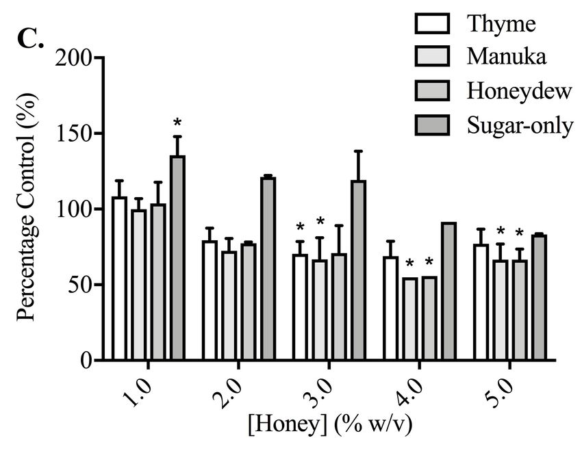
Figure 1 Absorption chromatogram of phenolic compounds in thyme honey using HPLC. Represen-
                                     tative chromatogram of free (A–C) or total (D–F) phenols extracted from thyme honey using ethyl ac-
                                     etate and eluted through a C18 column using HPLC. (A,D) Detectors were set to 370 nm for quercetin
                                     and kaempferol, (B,E) 325 nm for caffeic acid and (C,F) 270 nm for gallic acid and chrysin. Peak retention
                                     times were compared to standards to identify compounds.
                                                                                                    Full-size DOI: 10.7717/peerj.5115/fig-1

Abel et al. (2018), PeerJ, DOI 10.7717/peerj.5115                                                                                          7/21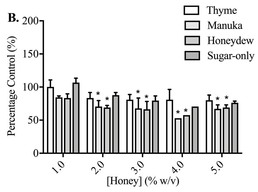
Table 1 HPLC analysis of ethyl acetate extracted phenolic compounds from honey.

  Compound                     Honey                                     [µg/100 g honey]                                          [5% w/v solution] (nM)
                                                           Free                          Total                          Free                            Total
  Gallic Acid                  Thyme                       94.2 ± 2.5                    686.0 ± 15.2                   276.8 ± 7.5                     2016.0 ± 44.6
                               Manuka                      741.5 ± 27.4                  584.5 ± 25.9                   2179.0 ± 80.4                   1718.0 ± 76.0
                               Honeydew                    48.1 ± 6.2                    1082.9 ± 16.3                  141.5 ± 18.2                    3183.0 ± 47.8
  Caffeic Acid                 Thyme                       90.2 ± 2.7                    83.7 ± 2.7                     250.4 ± 7.6                     232.2 ± 7.5
                               Manuka                      44.8 ± 3.5                    131.3 ± 17.1                   124.3 ± 9.8                     364.4 ± 47.3
                               Honeydew                    28.9 ± 2.5                    96.6 ± 9.0                     80.2 ± 7.0                      268.0 ± 25.1
  Quercetin                    Thyme                       38.2 ± 3.2                    –                              63.2 ± 5.3                      –
                               Manuka                      10.6 ± 7.4                    –                              17.5 ± 12.2                     –
                               Honeydew                    –                             –                              –                               –
  Kaempferol                   Thyme                       45.3 ± 13.8                   33.1 ± 12.2                    79.2 ± 24.1                     57.9 ± 0.6
                               Manuka                      9.3 ± 2.6                     12.2 ± 2.0                     16.2 ± 4.6                      21.3 ± 3.5
                               Honeydew                    11.2 ± 1.8                    8.6 ± 0.2                      19.6 ± 3.1                      15.0 ± 0.3
  Chrysin                      Thyme                       53.0 ± 0.3                    38.1 ± 0.4                     104.3 ± 0.5                     75.0 ± 0.7
                               Manuka                      77.6 ± 0.8                    72.1 ± 1.3                     152.6 ± 1.7                     141.9 ± 2.5
                               Honeydew                    46.4 ± 0.5                    26.8 ± 0.6                     91.2 ± 0.9                      52.8 ± 1.1
Notes.
    Compound concentrations (µg/100g honey) were calculated by comparing the area under the peak to standard calibration curves and were expressed as mean ± S.E.M (n = 6).
    Concentrations of each compound were also calculated from within a 5% (w/v) solution of honey used throughout the study (nM).

                                                  The 1% thyme or honeydew honeys caused a small but significant reduction in PC3
                                               cell migration compared to vehicle-treated control of 20.59 ± 4.90% and 18.90 ± 1.62%
                                               respectively (Fig. 2A). The manuka honey and the sugar-only mixture did not cause any
                                               reduction in cell migration, suggesting that non-sugar related compounds in honey may
                                               be responsible for the effect on migration.
                                                  All the non-toxic concentrations of phenolic compounds tested, with the exception of
                                               caffeic acid, caused a decrease in PC3 cell migration (Fig. 2B). Gallic acid (10 µM) caused
                                               the greatest reduction in migration of 49.10 ± 8.33% compared to vehicle (DMSO)-treated
                                               control. Together, these results demonstrated that phenolic compounds found in honey
                                               possess anti-metastatic activity against PC3 cells, however not all compounds have similar
                                               potency.
                                                  When invasion was measured, which looks at PC3 cell movement as well as the capacity
                                               to move through the Matrigel matrix, 0.5% (w/v) thyme, manuka or honeydew honey
                                               caused a significant reduction compared to control of 50.53 ± 4.10%, 60.44 ± 9.71% and
                                               75.32 ± 0.19% respectively (Fig. 2C). The sugar-only mixture caused a 46.77 ± 17.01%
                                               reduction in cell invasion, however it was not statistically significant. These changes were
                                               much greater than the reduction of migration, suggesting that honey particularly reduces
                                               the cells’ ability to move through Matrigel.
                                                  Invasion was also lowered by most of the phenolic compounds, with quercetin (10 µM)
                                               and caffeic acid (50 µM) resulting in reductions of 45.28 ± 0.64% and 14.44 ± 1.17%
                                               respectively (Fig. 2D). Further, chrysin (100 µM) and gallic acid (10 µM) reduced invasion
                                               by 39.57 ± 14.57% and 29.03 ± 15.86% respectively, however these were not statistically

Abel et al. (2018), PeerJ, DOI 10.7717/peerj.5115                                                                                                                     8/21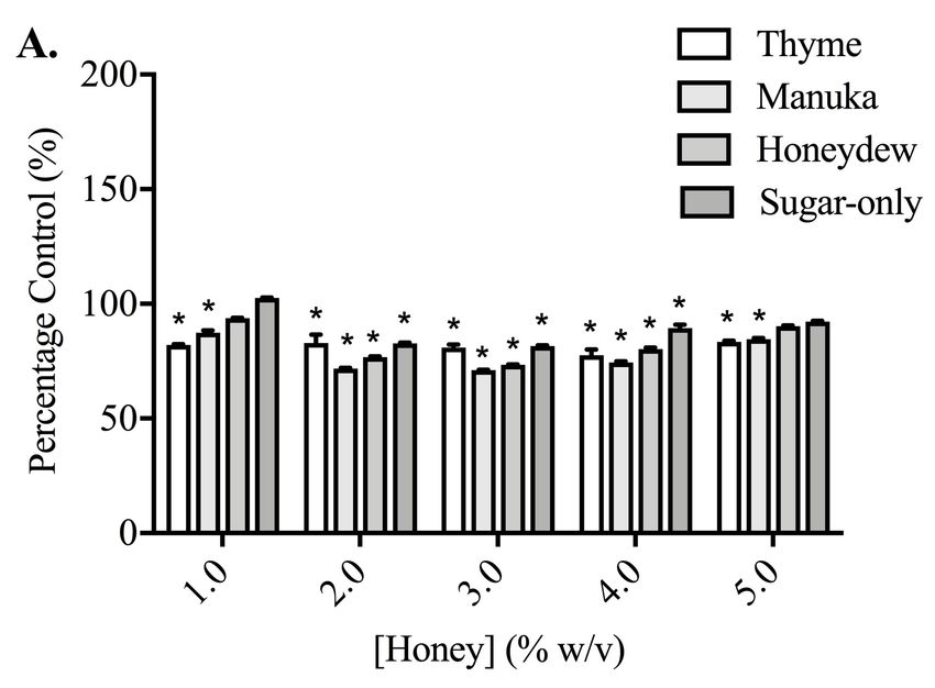
Figure 2

                                     Figure 2 Effect of honey and honey-derived phenolic compounds on PC3 cell migration and inva-
                                     sion. PC3 cells were placed in Boyden chambers and treated with 1% (w/v) thyme, manuka, honeydew or
                                     sugar-only mixture (A) or the phenolics quercetin (10 µM), gallic acid (10 µM), kaempferol (150 µM),
                                     chrysin (100 µM) or caffeic acid (50 µM) (B) for 48 h to measure migration or in Boyden chambers con-
                                     taining Matrigel for 72 h with 0.5 % (w/v) thyme, manuka, honeydew or sugar-only mixture (C) or the
                                     phenolics quercetin (10 µM), gallic acid (10 µM), kaempferol (150 µM), chrysin (100 µM) or caffeic
                                     acid (50 µM) (D) for 72 h to measure invasion. Experiments were completed in triplicate, and values were
                                     expressed as mean cell migration ± S.E.M (n = 2 or 3). Data were analysed using a two-tailed Student’s
                                     t -test. * (p < 0.05) represents a significant difference between individual treatment and the vehicle- un
                                     treated cell control of 0.1% DMSO for phenolics and RPMI-only for honeys.
                                                                                                         Full-size DOI: 10.7717/peerj.5115/fig-2

                                    significant. Finally, kaempferol only recorded a reduction in invasion of 5.04 ± 2.11%.
                                    Overall, the reductions in invasion caused by the phenolic compounds were comparable
                                    to those in migration, with the exception of kaempferol and caffeic acid.

                                    Effects of honey and honey constituents on prostate cancer cell
                                    adhesion
                                    Thyme, manuka and honeydew honeys caused a much greater, concentration-dependent
                                    decrease in cell adhesion, compared to their effects on migration and invasion, in both PC3
                                    and DU145 cells (p < 0.05) (Fig. 3). This was assessed after 30 and 60 min of exposure to
                                    the honeys in PC3 cells, and 60 and 90 min in the DU145 cells, which required longer to
                                    become adherent. DU145 cells were much more sensitive to the effect of the honeys, with
                                    their adhesion to collagen I being nearly completely prevented by 2% (w/v) honey, down
                                    to 8.98 ± 4.65% of vehicle-treated control with honeydew honey after 60 min, whereas

Abel et al. (2018), PeerJ, DOI 10.7717/peerj.5115                                                                                           9/21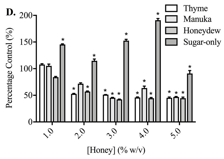
Figure 3

                                     Figure 3 Effect of honey on PC3 and DU145 cell adhesion to collagen I. (A,B) PC3 and (C,D) DU145
                                     cells were left to adhere to plates coated with collagen I for (A) 30, (B,C) 60 or (D) 90 min. Experiments
                                     were completed in triplicate, with results expressed as mean percentage of control ± S.E.M (n = mini-
                                     mum of 3). Individual data were analysed using a two-way ANOVA followed by a Bonferroni post-hoc
                                     test, where p < 0.05 was required for a statistically significant difference. * represents a significant differ-
                                     ence between the vehicle-treated control (0% w/v RPMI only) and individual treatment.
                                                                                                          Full-size DOI: 10.7717/peerj.5115/fig-3

                                    the PC3 cells required 5% (w/v), reaching 8.93 ± 3.69% with honeydew honey at 60 min.
                                    In PC3 cells, the sugar-only mixture was also effective at reducing adhesion, at 3% (w/v)
                                    (33.18 ± 25.05%) and 5% (w/v) (16.51 ± 4.68%) at 60 min, but in the DU145 cells,
                                    lower concentrations of the sugar-only mixture actually increased adhesion (although not
                                    significantly) before strongly lowering it at 5% (w/v), to 21.88 ± 13.01% at 90 min.
                                       When cells were adhering to fibronectin, the results were very different (Fig. 4). In PC3
                                    cells, while the honeys did lower adhesion in a statistically significant way, the effect was
                                    no longer large and nor was it concentration-dependent. The sugar-only mixture and the
                                    honeys gave similar levels of adhesion reduction, with the maximum reduction being found
                                    at 60 min with 4% (w/v) manuka honey, at 67.15 ± 8.33%. In DU145 cells, the effects
                                    were also much smaller than they had been with collagen I, although the reduction was
                                    much greater than with the PC3 cells. Adhesion was lowered to 42.33 ± 1.05% at 90 min
                                    by 3% (w/v) honeydew honey. Again, the effects were not concentration-dependent. With
                                    the DU145 cells, however, the changes in adhesion with the sugar-only mixture were very
                                    different from what was seen with the PC3 cells. With most concentrations, an increase in
                                    adhesion was found, up to 190.97 ± 5.46% with 4% (w/v) at 90 min.

Abel et al. (2018), PeerJ, DOI 10.7717/peerj.5115                                                                                               10/21
Figure 4
Figure 4 Effect of honey on PC3 and DU145 cell adhesion to fibronectin. (A,B) PC3 and (C,D) DU145
cells were left to adhere to plates coated with fibronectin for (A) 30, (B,C) 60 or (D) 90 min. Experiments
were completed in triplicate, with results expressed as mean percentage of control ± S.E.M (n = 3). Indi-
vidual data were analysed using a two-way ANOVA followed by a Bonferroni post-hoc test, where p < 0.05
was required for a statistically significant difference. * represents a significant difference between vehicle-
treated control (0% w/v RPMI only) and individual treatment.
Full-size DOI: 10.7717/peerj.5115/fig-4
Adhesion measurements for both cell lines to collagen I and fibronectin were used to
determine whether the five honey-derived phenolic compounds were responsible for the
loss of cell attachment seen from honey administration (Fig. 5). At 60 min, no compound
had any effect on either PC3 or DU145 cell adhesion to fibronectin at any concentration.
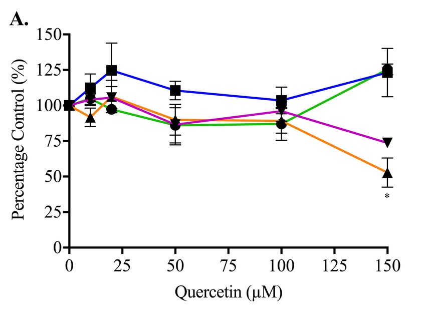
Only quercetin and kaempferol (150 µM) caused any significant change to cell adhesion
to collagen I. Quercetin (150 µM) caused a reduction in PC3 cell adhesion to collagen
I of 47.14 ± 10.27%. Kaempferol (150 µM) caused an increase of PC3 and DU145 cell
adhesion to collagen I of 117.20 ± 11.10% and 104.10 ± 10.09%, respectively.
DISCUSSION
We have shown that honey has anti-metastatic activity in two prostate cancer cell lines,
in an in vitro situation in which their unmetabolised forms are in contact with the cancer
cells. We have also examined some of the constituents of honey, including the major sugars
and five of the main phenolic compounds, and have shown that they can partially inhibit
some metastatic properties as well. Whole honeys, as well as sugars, to a lesser extent,
Abel et al. (2018), PeerJ, DOI 10.7717/peerj.5115 11/21Figure 5
Figure 5 Effect of honey-derived phenolic compounds on PC3 and DU145 cell adhesion. PC3 and
DU145 cells were treated with (A) quercetin (0–150 µM), (B) gallic acid (0–10 µM), (C) kaempferol (0–
150 µM), (D) chrysin (0–50 µM) or (E) caffeic acid (0–50 µM) for 60 min. Experiments were completed
in triplicate, and values were expressed as mean percentage cell viability ± S.E.M (n = 2 or 3). Data were
analysed using a two-way ANOVA followed by a Bonferroni post-hoc test, where p < 0.05 was required
for a statistically significant difference. * represents a significant difference between control and individual
treatment.
Full-size DOI: 10.7717/peerj.5115/fig-5
Abel et al. (2018), PeerJ, DOI 10.7717/peerj.5115 12/21could prevent cell adhesion to collagen I, although phenolic compounds were not effective.
Honeys and quercetin also strongly reduced invasion, but the effect on migration was
much smaller.
We began by measuring the concentrations of five phenolic compounds in whole honeys,
chosen due to their abundance and reported biological activity. An enhanced HPLC gradient
system was developed which improved upon previously reported methodologies used with
honey (Yao et al., 2003) and allowed a more sensitive detection and accurate calculation
of the phenolic concentrations. For example, quercetin was found in thyme and manuka
honeys, whereas using other methods, it has previously been undetectable in thyme honey
(El-Hady & Shaker, 2013).
The phenolic compounds were measured as both free and total phenols. Free phenolic
extract samples contained the unbound phenolic compounds (aglycones) within the honey,
which were readily extracted by ethyl acetate. Total phenolic extract samples contained
both phenolic aglycones and bound phenolic glycosides which required the hydrolysis of
the glycosidic ester bonds to release phenolic aglycones before they could be extracted into
an organic solvent (Wahdan, 1998).
A higher recovery of gallic acid and caffeic acid were observed in total phenol than in free
phenol extracts, which were in contrast to the flavonoid compounds that showed a higher
recovery in free phenol extracts. An explanation for this may be that flavonoids in honey
exist mainly as aglycones and were detected in the free phenol samples fully, however,
the two phenolic acids caffeic acid and gallic acid exist predominantly as glycosides from
pollen, and therefore may require hydrolysis (as used for the ‘total’ samples) (Ferreres et al.,
1991). The only exception was the gallic acid in manuka honey, of which a greater amount
was detected in the free phenol extracts. This could be explained by the fact that manuka
honey has been reported to have a high concentration of glucose oxidase which can produce
hydrogen peroxide (White & Doner, 1980), and radicals including hydrogen peroxide are
able to hydrolyse the glycosidic bonds between phenolics and sugars (Brudzynski et al.,
2011; Hussein et al., 2011). As we found here, gallic acid was previously described as the
predominant phenolic acid in both Australian and New Zealand honeys (Yao et al., 2003;
Yao et al., 2004).
The effect of both whole honey and phenolic compounds on cell migration in Boyden
chambers was limited but significant. The effect was found with both thyme and honeydew
honeys, but the reductions were not as large as those caused by kaempferol and chrysin,
of up to 50%. The sugar-only mixture, manuka honey and other phenolics did not affect
migration. In contrast, Ho et al. (2010) reported that gallic acid (3.5 µM) could inhibit the
migration of gastric cancer AGS cells by 60% in Boyden chambers, but this was measured
after only 6 h of migration following 48 h of exposure to gallic acid in other wells, quite a
different set-up from that used here.
Interestingly, the effect of honeys on invasion, the movement through Matrigel as well
as the Boyden chamber, was much more pronounced, with honeydew honey having the
greatest effect in reducing invasion by around 75%, but with all honeys having a large
significant effect. Of the phenolic compounds, only quercetin and caffeic acid showed
Abel et al. (2018), PeerJ, DOI 10.7717/peerj.5115 13/21statistically significant reductions, but there was a trend towards reduction for chrysin,
gallic acid and the sugar-only mixture as well.
This demonstrates that honeys can inhibit the invasion process more strongly than
the migration process, and suggests that a major point of inhibition is likely to be a part
of a pathway that is more important for invasion than migration. Although we have not
investigated this here, the mechanism of action may be through inhibition of the MMPs,
which are among the proteases expressed at the leading edge of metastasising cells, where
they facilitate the breakdown of the extracellular matrix (Friedl & Wolf, 2003). Ho et al.
(2010) showed that gallic acid could reduce gelatinolytic activity of MMP-2 and MMP-9,
possibly via NF-κB. It has been shown that honey can reduce the expression and nuclear
translocation of NF-κB both in vivo and in vitro (Batumalaie et al., 2013; Hussein et al.,
2013), although honeys were also shown not to inhibit NF-κB activity in glioblastoma cells
U87MG (Moskwa et al., 2014). However, Moskwa et al. (2014) did show that honeys could
reduce enzymatic activity of MMP-2 and MMP-9. Fir honey also inhibited migration of
human keratinocytes through the reduced expression of MMP-9 (Majtan et al., 2013). Lee
et al. (2004) demonstrated that 20 µM quercetin or luteolin could inhibit the secretion
of MMPs in the MIA PaCa-2 cell line. Quercetin (50–100 µM) has also been reported to
downregulate the expression of both MMP-2 and -9 in PC3 cells (Vijayababu et al., 2006).
Phenolic compounds have also been previously shown to affect other parts of the cell
migration process, including both Rho family GTPases and integrin expression, although
this has not been demonstrated in cancer cells. For example, gallic acid (25–100 µM)
inhibited RhoA protein expression and activity in scar-derived fibroblasts after TGFβ
stimulation (Hsieh et al., 2016). In L929 fibroblasts, following treatment with 20 µM
quercetin for 24 h, αV integrin was upregulated by 18%, and β1 integrin was similarly
downregulated (Doersch & Newell-Rogers, 2017). Some of these concentrations were much
higher than those used in our study, however there may have been a cumulative effect of
the combination of lower concentrations of many phenolic compounds.
Another necessary feature of the migration and invasion processes is adhesion to the
extracellular matrix. For the first time, it was shown that over the short 30–90 min timespan
of the adhesion assay using collagen I, honey caused a loss of cell adhesion of more than 90%
in both cell lines, although the DU145 cell line was more sensitive. The sugar constituents
in honey do appear to play a role in this loss of adhesion, but do not account for the full
effect. When fibronectin was used instead of collagen I, the reduction in adhesion was
greatly reduced in the PC3 cells. In the DU145 cells, a decrease in adhesion of up to 50%
was still found, but the sugar-only mixture increased, rather than decreased, adhesion,
suggesting that the mechanism of action of the sugars is very much dependent upon the
protein substrate. The differences in adhesion changes between the two cell types may relate
to their expression of integrins. DU145 cells have been shown to highly express αv and β1
integrins, which preferentially bind to fibronectin, and may reduce migration and result in
a less invasive phenotype compared to PC3 cells (Ruoslahti & Giancotti, 1989; Witkowski
et al., 1993). The increased invasiveness of PC3 cells compared to DU145 cells has instead
been attributed to the expression of α6 and β1 integrins, which bind better to collagen and
laminin (Witkowski et al., 1993; Suyin, Holloway & Dickinson, 2013). Collagens, including
Abel et al. (2018), PeerJ, DOI 10.7717/peerj.5115 14/21collagen I, are deposited at a higher rate in tumours, often in a linearised manner, which
increases stromal stiffness and contributes to cancer cell migration (Zhu et al., 1995;
Egeblad, Rasch & Weaver, 2010). Thus, the possibility of reducing the adhesion of prostate
cells to collagen would, if translatable in vivo, be of potential therapeutic benefit.
By contrast, phenolic compounds do not appear to play a role in reducing prostate cancer
cell adhesion. The highest concentration of quercetin (150 µM) only lowered adhesion
of the PC3 cells attaching to collagen I, and 150 µM kaempferol increased adhesion for
both cell lines on collagen I. Quercetin may affect adhesion through the Epidermal Growth
Factor Receptor, which mediates DU145 cell adhesion to collagen I (Lamb, Zarif & Miranti,
2011) and can be downregulated by quercetin (Kumar et al., 2008; Bhat et al., 2014). We
note that to observe biological activity in our assays with these compounds, much higher
(µM) concentrations of the phenolic compounds were required than are found within the
honey and therefore any effects are very unlikely to be due to a single phenolic compound.
The lack of effect of phenolic compounds on adhesion of prostate cancer cells was
unexpected, given the role of these compounds in the adhesion process in other cell types.
There may be some cell-type or integrin-subtype specificity. For example, chrysin (3 µM
and 10 µM) was shown to inhibit collagen-induced platelet aggregation, by reducing
P-selectin and integrin αIIb β3 signaling (Liu et al., 2016), and caffeic acid at up to 100 µM
could also lower platelet aggregation, through the same two mechanisms (Lu et al., 2015).
Caffeic acid at 1–20 µM also lowered adherence of monocytes to human umbilical vein
endothelial cells, through suppression of six different adhesion molecules and integrins
(Lee et al., 2012).
It is often assumed that phenolic compounds are responsible for the biological activity
of many natural products, including honeys. As honey contains multiple polyphenols, the
effect of combination treatment in vivo and in vitro is of interest. We found that phenolic
compounds were present in honey in the nM range (Table 1), however when individually
used in vitro at µM ranges, they demonstrated minimal anti-metastatic properties. This
suggests that the benefit of honey may be due to a combination of many compounds, and
not individual activities.
Inhibition of cancer cell adhesion by whole honey has not previously been reported.
Maddocks et al. (2013) reported that manuka honey (16–50% w/v) could inhibit the
adhesion of 8 bacteria strains to fibronectin, fibrinogen and collagen. This was thought to
be due to a reduction in fibronectin binding proteins, as well as the inhibition of biofilm
production (Maddocks et al., 2013; Maddocks et al., 2012). In metastasis, the cell must be
able to regulate its attachment to the surroundings in order to move forward. The level of
disruption that treatment with honey causes to this process would leave metastasis unable
to proceed (Bendas & Borsig, 2012).
We have also made the novel finding that the sugar components of honeys, as well as the
phenolics, play a role in its in vitro inhibition of cancer adhesion, migration and invasion.
Sugars may act as antioxidants in the body, in a similar way to that reported for polyphenols.
A single oral administration of honey (1.5 g/kg) in humans was shown to increase the total
phenolic, antioxidant and reducing capacity of the plasma. The sugar-only control, corn
syrup, did not increase the total plasma phenolic levels, however, it did significantly increase
Abel et al. (2018), PeerJ, DOI 10.7717/peerj.5115 15/21plasma antioxidant capacity (Schramm et al., 2003), most likely through the formation of
Maillard products or by acting as reducing sugars (White & Doner, 1980; Maillard, 1912).
This suggests that the activity of honey is due to presence of both phenolics and sugars,
and may enhance its overall biological activity compared to other natural phenol sources
lacking in sugar.
CONCLUSIONS
We have shown that honeys and some of their constituents are able to inhibit pro-metastatic
properties including migration and invasion in prostate cancer cell lines. This is likely to
be related to a blocking of the adhesion process, which has been shown for the first time
to be particularly strongly downregulated by honeys, and is a process that contributes to
both migration and invasion. Further investigation of the mechanisms of action of honey
compound combination effects in metastasis are warranted.
ADDITIONAL INFORMATION AND DECLARATIONS
Funding
This work was funded by the Department of Pharmacology and Toxicology, University of
Otago. The funders had no role in study design, data collection and analysis, decision to
publish, or preparation of the manuscript.
Grant Disclosures
The following grant information was disclosed by the authors:
Department of Pharmacology and Toxicology, University of Otago.
Competing Interests
The authors declare there are no competing interests.
Author Contributions
• Sean D.A. Abel conceived and designed the experiments, performed the experiments,
analyzed the data, prepared figures and/or tables, authored or reviewed drafts of the
paper, approved the final draft.
• Sumit Dadhwal conceived and designed the experiments, performed the experiments,
analyzed the data, contributed reagents/materials/analysis tools, approved the final draft.
• Allan B. Gamble conceived and designed the experiments, analyzed the data, contributed
reagents/materials/analysis tools, approved the final draft.
• Sarah K. Baird conceived and designed the experiments, analyzed the data, contributed
reagents/materials/analysis tools, prepared figures and/or tables, authored or reviewed
drafts of the paper, approved the final draft.
Data Availability
The following information was supplied regarding data availability:
The raw data are provided in a Supplemental File.
Abel et al. (2018), PeerJ, DOI 10.7717/peerj.5115 16/21Supplemental Information
Supplemental information for this article can be found online at http://dx.doi.org/10.7717/
peerj.5115#supplemental-information.
REFERENCES
Abel SDA, Baird SK. 2018. Honey is cytotoxic towards prostate cancer cells but interacts
with the MTT reagent: considerations for the choice of cell viability assay. Food
Chemistry 241:70–78 DOI 10.1016/j.foodchem.2017.08.083.
Alvarez-Suarez J, Giampieri F, Battino M. 2013. Honey as a source of dietary
antioxidants: structures, bioavailability and evidence of protective effects
against human chronic diseases. Current Medicinal Chemistry 20:621–638
DOI 10.2174/092986713804999358.
Batumalaie K, Zaman SS, Mohd YK, Shah II, Devi SS, Qvist R. 2013. Effect of gelam
honey on the oxidative stress-induced signaling pathways in pancreatic hamster cells.
International Journal of Endocrinology 2013:367312 DOI 10.1155/2013/367312.
Bendas G, Borsig L. 2012. Cancer cell adhesion and metastasis: selectins, integrins,
and the inhibitory potential of heparins. International Journal of Cell Biology
2012:676731 DOI 10.1155/2012/676731.
Bhat FA, Sharmila G, Balakrishnan S, Singh PR, Srinivasan N, Arunakaran J. 2014. Epi-
dermal growth factor-induced prostate cancer (PC3) cell survival and proliferation is
inhibited by quercetin, a plant flavonoid through apoptotic machinery. Biomedicine
& Preventive Nutrition 4:459–468 DOI 10.1016/j.bionut.2014.07.003.
Brudzynski K, Abubaker K, St-Martin L, Castle A. 2011. Re-examining the role of
hydrogen peroxide in bacteriostatic and bactericidal activities of honey. Frontiers in
Microbiology 2:213.
Cooper RA, Molan PC, Harding KG. 2002. The sensitivity to honey of Gram-positive
cocci of clinical significance isolated from wounds. Journal of Applied Microbiology
93:857–863 DOI 10.1046/j.1365-2672.2002.01761.x.
Doersch KM, Newell-Rogers MK. 2017. The impact of quercetin on wound healing
relates to changes in aV and b1 integrin expression. Experimental Biology and
Medicine 242:1424–1431 DOI 10.1177/1535370217712961.
Egeblad M, Rasch MG, Weaver VM. 2010. Dynamic interplay between the colla-
gen scaffold and tumor evolution. Current Opinion in Cell Biology 22:697–706
DOI 10.1016/j.ceb.2010.08.015.
El-Hady FKA, Shaker KH. 2013. Honey protects human low density lipoprotein (LDL)
from peroxidation (in vitro study). International Journal of Pharmaceutical Sciences
Review and Research 23:191–197.
Erejuwa OO, Sulaiman SA, Wahab MS. 2014. Effects of honey and its mechanisms of
action on the development and progression of cancer. Molecules 19:2497–2522
DOI 10.3390/molecules19022497.
Abel et al. (2018), PeerJ, DOI 10.7717/peerj.5115 17/21Fauzi AN, Norazmi MN, Yaacob NS. 2011. Tualang honey induces apoptosis and
disrupts the mitochondrial membrane potential of human breast and cervical cancer
cell lines. Food and Chemical Toxicology 49:871–878 DOI 10.1016/j.fct.2010.12.010.
Ferreres F, Tomáas-Barberáan FA, Gil MI, Tomáas-Lorente F. 1991. An HPLC tech-
nique for flavonoid analysis in honey. Journal of the Science of Food and Agriculture
56:49–56 DOI 10.1002/jsfa.2740560106.
Friedl P, Wolf K. 2003. Tumour-cell invasion and migration: diversity and escape
mechanisms. Nature Reviews Cancer 3:362–374
DOI 10.1038/nrc1075.
Hassan MI, Mabrouk GM, Shehata HH, Aboelhussein MM. 2012. Antineoplastic effects
of bee honey and Nigella sativa on hepatocellular carcinoma cells. Integrative Cancer
Therapies 11:354–363 DOI 10.1177/1534735410387422.
Ho HH, Chang CS, Ho WC, Liao SY, Wu CH, Wang CJ. 2010. Anti-metastasis effects
of gallic acid on gastric cancer cells involves inhibition of NF-κB activity and
downregulation of PI3K/AKT/small GTPase signals. Food and Chemical Toxicology
48:2508–2516 DOI 10.1016/j.fct.2010.06.024.
Hsieh SC, Wu CC, Hsu SL, Feng CH, Yen JH. 2016. Gallic acid attenuates TGF- β1-
stimulated collagen gel contraction via suppression of RhoA/Rho-kinase pathway in
hypertrophicscar fibroblasts. Life Sciences 161:19–26 DOI 10.1016/j.lfs.2016.07.011.
Hughes CS, Postovit LM, Lajoie GA. 2010. Matrigel: a complex protein mix-
ture required for optimal growth of cell culture. Proteomics 10:1886–1890
DOI 10.1002/pmic.200900758.
Hussein SZ, Mohd YK, Makpol S, Mohd YAY. 2013. Gelam honey attenuates
carrageenan-induced rat paw inflammation via NF-kappaB pathway. PLOS ONE
8:e72365 DOI 10.1371/journal.pone.0072365.
Hussein SZ, Yusoff KM, Makpol S, Yusof YAM. 2011. Antioxidant capacities and total
phenolic contents increase with gamma irradiation in two types of Malaysian honey.
Molecules 16:6378–6395 DOI 10.3390/molecules16086378.
Jin M, He S, Worpel V, Ryan SJ, Hinton DR. 2000. Promotion of adhesion and migra-
tion of RPE cells to provisional extracellular matrices by TNF-alpha. Investigative
Ophthalmology and Visual Science 41:4324–4332.
Jubri Z, Narayanan NNN, Karim NA, Ngah WZW. 2012. Antiproliferative activity and
apoptosis induction by gelam honey on liver cancer cell line. International Journal of
Applied Science and Technology 2:35–41 DOI 10.9734/BJAST/2012/499.
Kassim M, Achoui M, Mustafa MR, Mohd MA, Yusoff KM. 2010. Ellagic acid, phenolic
acids, and flavonoids in Malaysian honey extracts demonstrate in vitro anti-
inflammatory activity. Nutrition Research 30:650–659
DOI 10.1016/j.nutres.2010.08.008.
Kumar B, Koul S, Khandrika L, Meacham RB, Koul HK. 2008. Oxidative stress is
inherent in prostate cancer cells and is required for aggressive phenotype. Cancer
Research 68:1777–1785 DOI 10.1158/0008-5472.CAN-07-5259.
Abel et al. (2018), PeerJ, DOI 10.7717/peerj.5115 18/21Lamb LE, Zarif JC, Miranti CK. 2011. The androgen receptor induces integrin α6β1 to
promote prostate tumor cell survival via NF-κB and Bcl-xL independently of PI3K
signaling. Cancer Research 71:2739–2749 DOI 10.1158/0008-5472.CAN-10-2745.
Lansky EP, Harrison G, Froom P, Jiang WG. 2005. Pomegranate (Punica granatum)
pure chemicals show possible synergistic inhibition of human PC-3 prostate
cancer cell invasion across Matrigel. Investigational New Drugs 23:121–122
DOI 10.1007/s10637-005-5856-7.
Lee ES, Park SH, Kim MS, Han SY, Kim HS, Kang YH. 2012. Caffeic acid disturbs
monocyte adhesion onto cultured endothelial cells stimulated by adipokine resistin.
Journal of Agricultural and Food Chemistry 60:2730–2739 DOI 10.1021/jf203774y.
Lee LT, Huang YT, Hwang JJ, Lee AYL, Ke FC, Huang CJ, Kandaswami C, Lee PP, Lee
MT. 2004. Transinactivation of the epidermal growth factor receptor tyrosine kinase
and focal adhesion kinase phosphorylation by dietary flavonoids: effect on invasive
potential of human carcinoma cells. Biochemical Pharmacology 67(11):2103–2114
DOI 10.1016/j.bcp.2004.02.023.
Liotta LA, Steeg PS, Stetler-Stevenson WG. 1991. Cancer metastasis and angio-
genesis: an imbalance of positive and negative regulation. Cell 64:327–336
DOI 10.1016/0092-8674(91)90642-C.
Liu G, Xie W, He AD, Da XW, Liang ML, Yao GQ, Xiang JZ, Gao CJ, Ming ZY. 2016.
Antiplatelet activity of chrysin via inhibiting platelet αIIb β3-mediated signaling
pathway. Molecular Nutrition & Food Research 60:1984–1993
DOI 10.1002/mnfr.201500801.
Lu Y, Jiang F, Jiang H, Wu K, Zheng X, Cai Y, Katakowski M, Chopp M, To SST.
2010. Gallic acid suppresses cell viability, proliferation, invasion and angiogenesis
in human glioma cells. European Journal of Pharmacology 641(2–3):102–107
DOI 10.1016/j.ejphar.2010.05.043.
Lu Y, Li Q, Liu YY, Sun K, Fan JY, Wang CS, Han JY. 2015. Inhibitory effect of caffeic
acid on ADP-induced thrombus formation and platelet activation involves mitogen-
activated protein kinases. Scientific Reports 5:13824 DOI 10.1038/srep13824.
Maddocks SE, Jenkins RE, Rowlands RS, Purdy KJ, Cooper RA. 2013. Manuka honey
inhibits adhesion and invasion of medically important wound bacteria in vitro.
Future Microbiology 8:523–1536.
Maddocks SE, Lopez MS, Rowlands RS, Cooper RA. 2012. Manuka honey in-
hibits the development of Streptococcus pyogenes biofilms and causes reduced
expression of two fibronectin binding proteins. Microbiology 158:781–790
DOI 10.1099/mic.0.053959-0.
Maillard LC. 1912. Action of amino acids on sugars. Formation of melanoidins in a
methodical way. Comptes Rendus 154:66–68.
Majtan J, Bohova J, Garcia-Villalba R, Tomas-Barberan FA, Madakova Z, Ma-
jtan T, Majtan V, Klaudiny J. 2013. Fir honeydew honey flavonoids inhibit
TNF- α-induced MMP-9 expression in human keratinocytes: a new action of
honey in wound healing. Archives of Dermatological Research 305(7):619–627
DOI 10.1007/s00403-013-1385-y.
Abel et al. (2018), PeerJ, DOI 10.7717/peerj.5115 19/21Mehlen P, Puisieux A. 2006. Metastasis: a question of life or death. Nature Reviews
Cancer 6:449–459 DOI 10.1038/nrc1886.
Moskwa J, Borawska MH, Markiewicz-Zukowska R, Puscion-Jakubik A, Naliwajko SK,
Socha K, Soroczynska J. 2014. Polish natural bee honeys are anti-proliferative and
anti-metastatic agents in human glioblastoma multiforme U87MG cell line. PLOS
ONE 9:e90533 DOI 10.1371/journal.pone.0090533.
Mosmann T. 1983. Rapid colorimetric assay for cellular growth and survival: application
to proliferation and cytotoxicity assays. Journal of Immunological Methods 65:55–63
DOI 10.1016/0022-1759(83)90303-4.
Oršolić N, Knezević A, Sver L, Terzić S, Hackenberger B, Basić I. 2003. Influence of
honey bee products on transplantable murine tumours. Veterinary and Comparative
Oncology 1:216–226 DOI 10.1111/j.1476-5810.2003.00029.x.
Oršolić N, Terzić S, Šver L, Bašić I. 2005. Honey-bee products in prevention and/or
therapy of murine transplantable tumours. Journal of the Science of Food and
Agriculture 85:363–370 DOI 10.1002/jsfa.2041.
Pollard TD, Borisy GG. 2003. Cellular motility driven by assembly and disassembly of
actin filaments. Cell 112:453–465 DOI 10.1016/S0092-8674(03)00120-X.
Ruoslahti E, Giancotti F. 1989. Integrins and tumor cell dissemination. Cancer Cell
1(4):119–126.
Schramm DD, Karim M, Schrader HR, Holt RR, Cardetti M, Keen CL. 2003. Honey
with high levels of antioxidants can provide protection to healthy human subjects.
Journal of Agricultural and Food Chemistry 51:1732–1735 DOI 10.1021/jf025928k.
Suyin PC, Holloway AF, Dickinson JL. 2013. Integrins in prostate cancer invasion and
metastasis. In: Advances in prostate cancer. London: InTechOpen chapter 25.
Tomasin R, Gomes-Marcondes MC. 2011. Oral administration of aloe vera and honey
reduces Walker tumour growth by decreasing cell proliferation and increasing apop-
tosis in tumour tissue. Phytotherapy Research 25:619–623 DOI 10.1002/ptr.3293.
Tsiapara AV, Jaakkola M, Chinou I, Graikou K, Tolonen T, Virtanen V, Moutsatsou P.
2009. Bioactivity of Greek honey extracts on breast cancer (MCF-7), prostate cancer
(PC-3) and endometrial cancer (Ishikawa) cells: profile analysis of extracts. Food
Chemistry 116:702–708 DOI 10.1016/j.foodchem.2009.03.024.
Vijayababu MR, Arunkumar A, Kanagaraj P, Venkataraman P, Krishnamoorthy
G, Arunakaran J. 2006. Quercetin downregulates matrix metalloproteinases 2
and 9 proteins expression in prostate cancer cells (PC-3). Molecular and Cellular
Biochemistry 287:109–116 DOI 10.1007/s11010-005-9085-3.
Wahdan HA. 1998. Causes of the antimicrobial activity of honey. Infection 26:26–31
DOI 10.1007/BF02768748.
Wen CT, Hussein SZ, Abdullah S, Karim NA, Makpol S, Mohd Yusof YA. 2012. Gelam
and Nenas honeys inhibit proliferation of HT 29 colon cancer cells by inducing
DNA damage and apoptosis while suppressing inflammation. Asian Pacific Journal
of Cancer Prevention 13:1605–1610 DOI 10.7314/APJCP.2012.13.4.1605.
White JW. 1978. Honey. Advances in Food Research 24:287–374
DOI 10.1016/S0065-2628(08)60160-3.
Abel et al. (2018), PeerJ, DOI 10.7717/peerj.5115 20/21White J, Doner LW. 1980. Beekeeping in the United States, agriculture handbook. Honey
composition and properties. Washington, D.C.: US Department of Agriculture,
82–91.
Winston ML. 1991. The biology of the honey bee. Cambridge: Harvard University Press.
Witkowski CM, Rabinovitz I, Nagle RB, Affinito KS, Cress AE. 1993. Characteri-
zation of integrin subunits, cellular adhesion and tumorgenicity of four human
prostate cell lines. Journal of Cancer Research and Clinical Oncology 119:637–644
DOI 10.1007/BF01215981.
Yao L, Datta N, Tomás-Barberán FA, Ferreres F, Martos I, Singanusong R. 2003.
Flavonoids, phenolic acids and abscisic acid in Australian and New Zealand Lep-
tospermum honeys. Food Chemistry 81:59–168.
Yao L, Jiang Y, Singanusong R, D’Arcy B, Datta N, Caffin N, Raymont K. 2004.
Flavonoids in Australian Melaleuca, Guioa, Lophostemon, Banksia and Helianthus
honeys and their potential for floral authentication. Food Research International
37(2):166–174 DOI 10.1016/j.foodres.2003.11.004.
Zhu GG, Risteli L, Mäkinen M, Risteli J, Kauppila A, Stenbäck F. 1995. Immunohisto-
chemical study of type I collagen and type I pN-collagen in benign and malignant
ovarian neoplasms. Cancer 75:1010–1017
DOI 10.1002/1097-0142(19950215)75:43.0.CO;2-O.
Abel et al. (2018), PeerJ, DOI 10.7717/peerj.5115 21/21You can also read