Multiparametric Quantitative MRI in Neurological Diseases - Frontiers
←
→
Page content transcription
If your browser does not render page correctly, please read the page content below
MINI REVIEW
                                                                                                                                               published: 08 March 2021
                                                                                                                                         doi: 10.3389/fneur.2021.640239

                                               Multiparametric Quantitative MRI in
                                               Neurological Diseases
                                               Alexander Seiler 1,2 , Ulrike Nöth 2,3 , Pavel Hok 4 , Annemarie Reiländer 1,2 ,
                                               Michelle Maiworm 1,2,3 , Simon Baudrexel 1,2 , Sven Meuth 5 , Felix Rosenow 1,3,6 ,
                                               Helmuth Steinmetz 1,3 , Marlies Wagner 2,3 , Elke Hattingen 3,7 , Ralf Deichmann 2,3 and
                                               René-Maxime Gracien 1,2,3*
                                               1
                                                 Department of Neurology, Goethe University, Frankfurt, Germany, 2 Brain Imaging Center, Goethe University, Frankfurt,
                                               Germany, 3 Center for Personalized Translational Epilepsy Research (CePTER) Consortium, Goethe University, Frankfurt,
                                               Germany, 4 Department of Neurology, Palacký University Olomouc and University Hospital Olomouc, Olomouc, Czechia,
                                               5
                                                 Department of Neurology, Heinrich Heine University Düsseldorf, Düsseldorf, Germany, 6 Epilepsy Center Frankfurt
                                               Rhine-Main, Center of Neurology and Neurosurgery, University Hospital, Frankfurt, Germany, 7 Department of Neuroradiology,
                                               Goethe University, Frankfurt, Germany

                           Edited by:
                        Amgad Droby,           Magnetic resonance imaging (MRI) is the gold standard imaging technique for diagnosis
              Tel Aviv University, Israel
                                               and monitoring of many neurological diseases. However, the application of conventional
                       Reviewed by:
                 Christine Preibisch,
                                               MRI in clinical routine is mainly limited to the visual detection of macroscopic
              Technische Universität           tissue pathology since mixed tissue contrasts depending on hardware and protocol
                 München, Germany              parameters hamper its application for the assessment of subtle or diffuse impairment
                  Victoria M. Leavitt,
    Columbia University Irving Medical         of the structural tissue integrity. Multiparametric quantitative (q)MRI determines tissue
               Center, United States           parameters quantitatively, enabling the detection of microstructural processes related
                *Correspondence:               to tissue remodeling in aging and neurological diseases. In contrast to measuring
              René-Maxime Gracien
       Rene-Maxime.Gracien@kgu.de
                                               tissue atrophy via structural imaging, multiparametric qMRI allows for investigating
                                               biologically distinct microstructural processes, which precede changes of the tissue
                    Specialty section:         volume. This facilitates a more comprehensive characterization of tissue alterations by
          This article was submitted to
                                               revealing early impairment of the microstructural integrity and specific disease-related
                Applied Neuroimaging,
                a section of the journal       patterns. So far, qMRI techniques have been employed in a wide range of neurological
                 Frontiers in Neurology        diseases, including in particular conditions with inflammatory, cerebrovascular and
       Received: 10 December 2020              neurodegenerative pathology. Numerous studies suggest that qMRI might add valuable
        Accepted: 12 February 2021
         Published: 08 March 2021
                                               information, including the detection of microstructural tissue damage in areas appearing
                            Citation:
                                               normal on conventional MRI and unveiling the microstructural correlates of clinical
 Seiler A, Nöth U, Hok P, Reiländer A,         manifestations. This review will give an overview of current qMRI techniques, the most
  Maiworm M, Baudrexel S, Meuth S,
                                               relevant tissue parameters and potential applications in neurological diseases, such as
 Rosenow F, Steinmetz H, Wagner M,
       Hattingen E, Deichmann R and            early (differential) diagnosis, monitoring of disease progression, and evaluating effects of
  Gracien R-M (2021) Multiparametric           therapeutic interventions.
      Quantitative MRI in Neurological
 Diseases. Front. Neurol. 12:640239.           Keywords: quantitative magnetic resonance imaging, neuroimaging, brain imaging, epilepsy, multiple sclerosis,
     doi: 10.3389/fneur.2021.640239            neurodegeneration

Frontiers in Neurology | www.frontiersin.org                                         1                                          March 2021 | Volume 12 | Article 640239
Seiler et al. Multiparametric qMRI in Neurological Diseases
INTRODUCTION
Conventional magnetic resonance imaging (MRI) techniques are
used in the clinical routine to diagnose and monitor neurological
diseases (1). However, clinical routine MRI of the brain primarily
visualizes macroscopic lesions. In contrast, it is difficult to assess
diffuse or subtle changes with conventional MRI or underlying
pathological principles because it shows mixed contrasts
which depend on protocol settings and hardware parameters
such as the magnetic field strength and inhomogeneities
of the static magnetic field and radiofrequency coil
sensitivities (2).
In contrast, quantitative MRI (qMRI) techniques measure
actual tissue parameters such as the T1-, T2-, T2∗ -relaxation
times or the proton density (PD), largely eliminating hardware
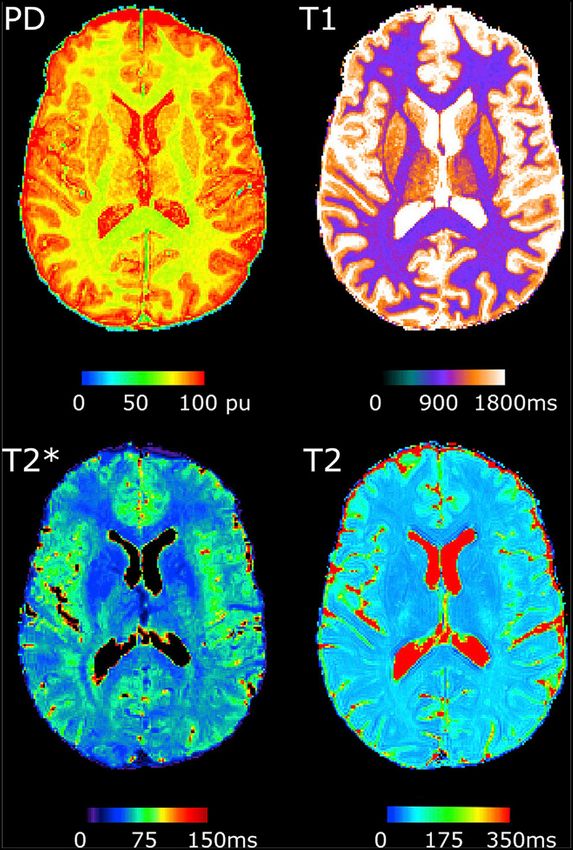
effects (2). Figure 1 demonstrates examples of qMRI maps.
The respective parameter maps provide quantitative parameter
values for each single voxel, which can be compared between
groups, study centers and in longitudinal investigations.
Accordingly, the values can be used to assess diffuse and subtle
changes in tissue composition in neurological diseases. QMRI
measurements are reproducible with only a small scan-rescan
deviation of around 1% for state–of-the-art MR scanners,
whereas deviations between different scanner models are
around 3.5% (3). However, systematic differences between
scanners can be quantified with phantom measurements,
allowing for post-hoc corrections. Furthermore, a small inter-site
bias as well as small inter-site and intra-site coefficient of
variation was observed for multiparametric mapping approaches
with relatively short acquisition times, demonstrating the
potential of these methods for multi-center investigations (4, 5).
While other somewhat more specific MRI techniques such
as perfusion mapping using arterial spin-labeling (ASL) also
FIGURE 1 | Examples of quantitative MRI maps of a single subject. PD, Proton
allow for the quantitative characterization of certain perfusion density. The figure has been published under a Creative Commons license
properties (6–8), this review mainly focuses on quantitative (https://creativecommons.org/licenses/by-nc-nd/4.0/) by Gracien et al. (3).
relaxometry and PD mapping. These increasingly applied
imaging techniques allow for the multiparametric assessment
and characterization of microstructural tissue alterations WHAT CAN BE MEASURED WITH qMRI?
and biochemical processes related to neurological diseases
of different etiologies. The main objective of this review is qMRI techniques measure physical tissue parameter values.
to give a compact but comprehensive overview of current These values depend on the microstructural tissue composition.
and future potential applications for multiparametric qMRI However, most qMRI parameters depend on various
in neurological diseases, including the tissue parameters, microstructural processes and the relative proportions of
which are most relevant for investigating the respective different tissue compartments. In contrast to conventional
diseases, given the underlying pathophysiology and pathological MR imaging used in clinical routine, most qMRI techniques
changes of tissue microstructure, and potential applications in require the acquisition of a series of weighted raw images with
research and clinical practice. Furthermore, we summarize different timing parameters such as different echo or inversion
the major current limitations of qMRI techniques, the times, allowing for exponential fitting of the signal time course
main technical issues to be solved for further improvement and yielding maps of the respective parameter. The resulting
and applications of qMRI techniques for research and acquisition times can be relatively long, depending on the
clinical purposes. number of images in the series. As an example, T2 mapping with
a high number of different echo times allows for the distinction
between different tissue fractions, e.g., the quantification of the
Abbreviations: DTI, diffusion tensor imaging; FCD, focal cortical dysplasia; GM, myelin water content, but requires long experiment durations.
gray matter; MRI, magnetic resonance imaging; MS, multiple sclerosis; MTR,
magnetization transfer ratio; NA, normal-appearing; PD, proton density; qMRI,
Still, the reliable detection of significant differences in qMRI
quantitative MRI; RRMS, relapsing-remitting MS; SVD, small vessel disease; TLE, parameters between a group of patients and a healthy population
temporal lobe epilepsy; WM, white matter. may already be feasible with a low number of echo times and thus
Frontiers in Neurology | www.frontiersin.org 2 March 2021 | Volume 12 | Article 640239Seiler et al. Multiparametric qMRI in Neurological Diseases
a shorter acquisition time. While the parameter PD measures potential of multiparametric qMRI techniques for longitudinal
free tissue water (9, 10), T1 values are positively correlated study designs in MS.
with water content and gliosis and negatively correlated with
iron and myelin content (10, 11). The Magnetization Transfer
Ratio (MTR) is sensitive to the macromolecular content and MULTIPARAMETRIC qMRI APPLICATIONS
the fraction of bound water and commonly used as surrogate IN EPILEPSY
marker of myelin (9). T2 is increased by the free water fraction
but shortened by myelin-bound water (12) and iron deposition Epilepsy is a disease of the brain characterized by an
(13). T2∗ is a marker for the iron content (10, 14). Furthermore, enduring predisposition to generate epileptic seizures (30). The
demyelination is known to prolong the T2∗ relaxation time (15). identification of underlying epileptogenic lesions in patients
Multiparametric qMRI approaches can help to better understand diagnosed with structural epilepsy can be challenging. MRI
and characterize disease related tissue remodeling and damage techniques can help to detect focal cerebral lesions in epilepsy
in neurological diseases. Therefore, multiparametric mapping patients and to quantify diffuse disease-related changes in brain
protocols have been proposed for the simultaneous deduction of tissue. Bernasconi reported that T2 relaxometry can quantify
several parameter maps reflecting multiple tissue properties and hippocampal damage in temporal lobe epilepsy (TLE) even in
microstructural attributes (4, 10). patients with normal conventional MRI and correctly identify
the side of the hippocampal changes in most of the patients
(31). A more recent study employed a multiparametric qMRI
approach including T1/T2 mapping and diffusion tensor imaging
MULTIPARAMETRIC qMRI IN MULTIPLE (DTI) techniques combined with support vector machines to
SCLEROSIS (MS) distinguish TLE patients from healthy control subjects (32).
The accuracy amounted to 88.9%. Furthermore, a prolonged T2
Autoimmune inflammation in MS causes cerebral MS lesions, relaxation time was observed in the amygdala and hippocampus
resulting in clinical symptoms such as the relapses in relapsing- for epilepsy patients with suspected limbic encephalitis, whereas
remitting MS (RRMS). However, macroscopic lesions are known the expected increase of the signal intensity in T2-weighted
to be only the “tip of the iceberg” of tissue damage in MS (16). datasets was not found (33).
Histopathological studies revealed that gliosis, demyelination, Another potential application of qMRI is the detection of
axonal loss, and infiltration of cells of the immune system focal cortical dysplasia (FCD), a highly epileptogenic structural
also occur in tissue which appears normal on conventional cerebral lesion often causing therapy-refractory epilepsy. The
MRI in MS (17, 18). QMRI allows for the quantification of standard methods for improved visualization of FCD described
these microstructural changes in tissue composition induced by by Kassubek et al. (34) and Huppertz et al. (35) are based
MS. Multiparametric qMRI analyses of MS patients revealed on conventional MRI datasets. However, these methods require
increased T1, T2, T2∗ , PD, and decreased magnetization transfer mechanisms for intensity normalization/bias-field correction.
ratio (MTR) values in normal-appearing white matter (WM) and QMRI techniques might be advantageous for FCD detection
cortical gray matter (GM) (19–22). A previous multiparametric since the parameter maps are (intrinsically) corrected for any
qMRI study utilizing surface-based analysis techniques observed hardware bias (2). Nöth et al. (36) developed a method for
spatially inhomogeneous cortical T2-, T2∗ -, and PD-increases improved FCD visualization, utilizing quantitative T1 maps, and
with parameter-specific spatial distribution patterns indicating assessing the cortical extent and the smoothness of the WM-
that in some cortical regions certain microstructural changes GM-junction. Furthermore, a different method for improved
(such as an increased water content) might dominate, while in FCD visualization based on multiparametric qMRI data (T1,
other cortical regions other processes might be more prominent T2, and PD maps) and surface-based analysis techniques has
in MS patients (22). Importantly, qMRI parameter changes in been proposed, measuring the standard deviations of the
normal-appearing tissue were already observed in early stages respective parameters at the WM-cortex junction (37). The
of the disease (23–25). Furthermore, several studies reported a resulting information on junction smoothness and additional
correlation between qMRI parameter values, indicating diffuse measures of the local cortical thickness are then used in
changes in tissue composition in MS, and the clinical/cognitive combination to artificially enhance the signal in conventional
status, thus demonstrating the relevance of diffuse cerebral fluid attenuated inversion recovery (FLAIR) datasets, thus
processes for patients (22, 26). highlighting regions suspicious of FCD. Future studies are
Longitudinal qMRI studies in MS have the potential to required to evaluate the clinical relevance of qMRI in epilepsy
demonstrate the ability of qMRI techniques to assess tissue more closely. Consequently, as demonstrated by the methods
remodeling over time. However, two longitudinal investigations applied for the detection of FCD in epilepsy, complementing
reported no longitudinal changes of mean T1 values in normal- conventional MRI by qMRI allows for an improved visualization
appearing WM or GM in MS (23, 27). The longitudinal study of FCD, thus facilitating a task which is one of the major
by Parry et al. (28) observed decreasing GM T1 values, while a challenges in the diagnosis of epilepsies of unknown origin
different investigation reported increasing cortical PD- and T1- with MRI. Important challenges are the detection of previously
values (29). Future longitudinal MS studies are required which undetected epileptogenic lesions and the discovery of qMRI
utilize advanced data analysis methods minimizing potential biomarkers of epileptogenicity and the seizure onset zone.
sources of bias such as partial volume effects to probe the QMRI may also allow to investigate the contributions of
Frontiers in Neurology | www.frontiersin.org 3 March 2021 | Volume 12 | Article 640239Seiler et al. Multiparametric qMRI in Neurological Diseases
blood-brain barrier dysfunction (38) and neuroinflammation possible consequences with regard to acute treatment and clinical
to initiation and maintenance of epileptogenesis and related management of neurological patients.
cognitive dysfunction (39).
qMRI IN CEREBROVASCULAR DISEASE MULTIPARAMETRIC qMRI IN AGING AND
NEURODEGENERATION
Chronic hemodynamic compromise due to steno-occlusive
disease of the brain-supplying vasculature increases the risk for Given the steadily increasing life expectancy and the increasing
ischemic stroke (40, 41). Furthermore, cerebral hypoperfusion is prevalence of neurodegenerative diseases manifesting at higher
associated with cognitive impairment, even in subjects for whom age in the Western societies, the differentiation between normal
conventional MRI provides no evidence of acute or chronic aging and early stages of neurodegeneration is crucial to identify
ischemic lesions (42). Cerebral changes in tissue composition individuals at risk of cognitive decline. In order to capture
such as microglial activation and demyelination were reported cases in which the process of aging is to a certain degree
in experimental studies on cerebral hypoperfusion (43). Since accelerated or altered by the detrimental impact of various
these pathological tissue alterations are detectable with qMRI, an factors on the microstructural integrity of the brain tissue, it
application of these techniques in patients with cerebrovascular is important to define and characterize microstructural tissue
disease can be expected to be a promising approach. In fact, alterations associated with normal aging. Numerous distinct
significant increases of T2 values were observed in normal- tissue-remodeling processes on the microstructural level are
appearing WM and normal-appearing cortical GM affected by associated with physiological aging. Previous studies employing
chronic hypoperfusion in patients with unilateral high-grade multiparametric qMRI consistently demonstrated decreasing T1
carotid-occlusive disease (44, 45). The relative prolongation values and increasing T2 values with increasing age in the
of the T2 relaxation time correlated with the degree of adult lifespan for both GM and WM (10, 52–57). Besides a
hemodynamic impairment, indicating that T2 mapping detects decreasing overall tissue water fraction and a regression of
gliotic tissue conversion, demyelination and enlargement of dendrites, age-related shortening of T1 has been attributed
the extracellular compartment related to local hypoperfusion to an increasing tissue iron deposition, which seems to be a
(44, 45). In addition, changes of T2 values seem to precede prominent process in brain aging (58). Increasing T2 values have
macroscopic tissue atrophy (44). Therefore, the T2 relaxation been mainly interpreted as the result of demyelination taking
time might be a useful imaging biomarker of early and subtle place with increasing age (49). Concerning quantitative T2∗ ,
hypoperfusion-related microstructural tissue damage. A further the directionality of age-related changes seems to depend on
important application of qMRI in chronic cerebrovascular the tissue type and local microstructural properties (59, 60). In
diseases concerns the characterization of microstructural damage more detail, while in deep GM structures a negative association
in normal-appearing WM in cerebral small vessel disease (SVD). between T2∗ values and age was observed, T2∗ values and age
Previous investigations found significant increases of T2 values were positively correlated in WM (60). This discrepancy may be
within the cerebral WM surrounding WM hyperintensities explained by an age-related iron accumulation in deep GM nuclei
in SVD patients, potentially reflecting microstructural gliosis, leading to a decrease of T2∗ , while prominent demyelination
demyelination, axonal damage and increased tissue water content in WM may lead to a prolongation of T2∗ due to inherent
in these areas (46). Furthermore, due to its sensitivity to the tissue T2 (spin-spin) effects (59, 60). The bidirectional influences of
fluid content, the T1 relaxation time seems to be a promising distinct age-related processes on T2∗ may be the reason for
imaging biomarker with regard to the detection of blood-brain the lack of a correlation between T2∗ values and age in a
barrier disruption as a key mechanism of microstructural WM recent study investigating age-related changes of the cortical GM
damage in SVD (47, 48). It remains to be clarified whether these microstructure with multiparametric qMRI (59). In contrast, the
parameters may be suitable imaging biomarkers of pathological reversible transverse relaxation time T2′ (calculated as 1/T2′ =
tissue remodeling underlying common clinical manifestations 1/T2∗ -1/T2), which is corrected for spin-spin effects, showed
of SVD such as cognitive decline or gait disturbances. In widespread negative associations with age across the cerebral
acute ischemic stroke, an important possible domain of qMRI cortex, most likely due to an age-related cortical iron deposition
is the determination of the time of stroke occurrence in occurring during physiological aging (59). Concerning possible
patients with wake-up stroke or in patients with severe aphasia applications of qMRI in pathological neurodegeneration, several
and unwitnessed symptom onset. Because of the immediate studies suggested that the T2 relaxation time might be
implications for the therapeutical management, this is highly a promising imaging biomarker for differentiating between
relevant. So far, T2 mapping has been employed in clinical pilot cognitively normal elderly subjects and individuals with mild
studies, revealing promising significant associations between cognitive impairment (MCI) or Alzheimer’s disease (AD) (61–
T2 values in the ischemic core and time from symptom 64). Furthermore, the T2 relaxation time seems to be able to
onset (49–51). Future studies are required to confirm the discriminate between cases of MCI and AD within a population
validity of qMRI parameters as indicators of the time since of cognitively impaired elderly individuals, which is an important
symptom onset and to assess their reliability across different finding from a clinical point of view (61–63). Despite revealing
scanners and imaging protocols. In summary, qMRI covers a regionally heterogeneous pattern of qMRI parameter changes
numerous potential applications in cerebrovascular diseases with in subjects with cognitive complaints, a recent multiparametric
a realistic translation into standard patient care, resulting in qMRI study suggested that also T1 and T2∗ mapping may be
Frontiers in Neurology | www.frontiersin.org 4 March 2021 | Volume 12 | Article 640239Seiler et al. Multiparametric qMRI in Neurological Diseases
useful to detect microstructural tissue alterations in MCI patients field strength is identical. Increasing the spatial resolution for
compared to healthy controls (65). In Parkinson’s disease, a T2-, T2∗ -, and T2′ -mapping improves the spatial alignment
widely disseminated application of qMRI techniques is the with other structural datasets during coregistration procedures
evaluation of brain iron accumulation and neuronal loss in the and reduces partial volume effects. However, increased spatial
dopaminergic areas of the brainstem as well as in other basal resolution and a high number of echoes make the qMRI
ganglia structures (66–68). For this purpose, especially T2, T2∗ , maps more prone to artifacts resulting from intra-scan subject
and T1 values were determined in previous studies (69, 70). motion, because of the longer acquisition durations, which
Furthermore, qMRI may provide insights into cortical tissue may be disadvantageous in patients with (acute) neurological
pathology in patients with Parkinson’s disease. So far, especially diseases. It should be noted that qMRI techniques can only
T1 relaxometry was applied for the investigation of cortical assess biological and biochemical processes which have an
involvement (71, 72), in particular for studying the cortical impact on the tissue microstructure. This limits their usefulness
spread-out of the neurodegenerative process and its relation to in neurological diseases which are merely characterized by a
cognitive decline (72). Furthermore, some studies have suggested pathological alteration of the functional connectivity without
the suitability of T2 and T2′ mapping for differentiating between any consequences on the brain microstructure, as may be the
different causes of Parkinsonian syndromes (73, 74), thus case for some movement disorders such as dystonia (75). Future
potentially adding highly relevant information from a clinical studies on neurological diseases might benefit from investigating
point of view. the interaction between functional properties (such as functional
connectivity or brain perfusion) and microstructural tissue
DISCUSSION changes that can be assessed by multiparametric qMRI.
Furthermore, while many of the multiparametric qMRI findings
Multiparametric qMRI provides a set of parameters, which in the different neurological diseases described above are
allow for gaining deeper insights into microstructural tissue plausible and can be interpreted from a pathophysiological point
alterations and impairment of microstructural integrity in a of view, a histological validation—e.g. obtained from appropriate
range of neurological diseases and in physiological aging. and disease-specific animal models—is still lacking in many
Although most of these parameters are not specific for a cases. Therefore, researchers in the field of multiparametric
single distinct microstructural process, predominant sensitivities qMRI should aim at correlating qMRI and histological findings
to certain aspects of microstructural tissue pathology enable in neurological diseases whenever possible. Future research
the comprehensive characterization of disease- or age-related on qMRI methods should focus in particular on reducing
tissue changes. The utility of qMRI in this regard can be acquisition times and improving motion correction algorithms.
further increased if results from preclinical, experimental and This will further enhance the utility of multiparametric qMRI
histological studies are taken into account and are correlated with for the investigation of neurological diseases, also with regard
findings from qMRI investigations. One of the most important to supporting diagnoses and important clinical tasks such as
advantages of multiparametric qMRI is the detection of subtle monitoring disease progression and effects of therapeutical
impairment of the microstructural tissue integrity, which can interventions. Given the relevance of these tasks, more
accrue before measurable tissue volume reduction occurs (44, longitudinal studies using multiparametric qMRI in neurological
59). Therefore, apart from depicting distinct biologically relevant diseases are warranted and needed. Furthermore, it would be
mechanisms of tissue remodeling, qMRI techniques allow for a great step forward if more scanner vendors included qMRI
investigating microstructural processes, which both potentially techniques in the set of release sequences. Recently developed
underlie and precede measurable tissue atrophy (44). A recent open-source software packages for qMRI data processing and
study demonstrated the stability of qMRI results, showing low quantitative parameter mapping, including tools for image
intra-scanner deviations in repeated measurements and a high coregistration and tissue segmentation, will help to further
reproducibility of qMRI findings across different scanner models facilitate the use of qMRI methods, both in research and clinical
(3). Thus, besides the high utility of qMRI for detecting disease- applications (4, 76, 77).
related tissue changes in patients with neurological diseases
compared to healthy control subjects in single center studies,
these findings also suggest a high reliability of qMRI techniques AUTHOR CONTRIBUTIONS
for longitudinal and multicenter studies (3). The combination
AS and R-MG wrote the first draft of the manuscript. All authors
with recent advances in hardware development will help to
reviewed the manuscript, contributed to the manuscript revision
further improve the precision of multiparametric qMRI for the
and approved the submitted version.
characterization of pathological changes in tissue microstructure
in neurological diseases. For instance, the implementation of
qMRI sequences on 7T MRI scanners, which are increasingly FUNDING
used in clinical research, will result in qMRI parameter maps
with a higher spatial resolution. It has to be taken into account This work was supported by the Else Kröner-Fresenius-Stiftung,
that the relaxation times (T1, T2, T2∗ ) depend on the field the State of Hesse with a LOEWE-Grant to the CePTER-
strength. However, this does not affect the comparability of Consortium (http://www.uni-frankfurt.de/67689811) and by the
parameter maps acquired on different scanners, provided the Clinician Scientists program at Goethe University.
Frontiers in Neurology | www.frontiersin.org 5 March 2021 | Volume 12 | Article 640239Seiler et al. Multiparametric qMRI in Neurological Diseases
REFERENCES 20. Gracien RM, Jurcoane A, Wagner M, Reitz SC, Mayer C, Volz S,
et al. Multimodal quantitative MRI assessment of cortical damage in
1. Thompson AJ, Banwell BL, Barkhof F, Carroll WM, Coetzee T, Comi G, et al. relapsing-remitting multiple sclerosis. J Magn Reson Imag. (2016) 44:1600–
Diagnosis of multiple sclerosis: 2017 revisions of the McDonald criteria. The 7. doi: 10.1002/jmri.25297
Lancet Neurology. (2018) 17:162–73. doi: 10.1016/S1474-4422(17)30470-2 21. Lommers E, Simon J, Reuter G, Delrue G, Dive D, Degueldre C, et al.
2. Cercignani M, Dowell NG, Tofts P, editors. Quantitative MRI of the Brain: Multiparameter MRI quantification of microstructural tissue alterations in
Principles of Physical Measurement. Boca Raton FL: CRC Press Taylor & multiple sclerosis. Neuroimage Clin. (2019) 23. doi: 10.1016/j.nicl.2019.101879
Francis Group (2018). 22. van Wijnen A, Petrov F, Maiworm M, Frisch S, Foerch C, Hattingen E,
3. Gracien R-M, Maiworm M, Brüche N, Shrestha M, Nöth U, et al. Cortical quantitative MRI parameters are related to the cognitive status
Hattingen E, et al. How stable is quantitative MRI? – Assessment in patients with relapsing-remitting multiple sclerosis. Eur Radiol. (2020)
of intra- and inter-scanner-model reproducibility using identical 30:1045–53. doi: 10.1007/s00330-019-06437-9
acquisition sequences and data analysis programs. Neuroimage. (2020) 23. Davies GR, Hadjiprocopis A, Altmann DR, Chard DT, Griffin CM, Rashid
207:116364. doi: 10.1016/j.neuroimage.2019.116364 W, et al. Normal-appearing grey and white matter T1 abnormality in early
4. Weiskopf N, Suckling J, Williams G, Correia MM, Inkster B, Tait relapsing-remitting multiple sclerosis: a longitudinal study. Mult. Scler. (2007)
R, et al. Quantitative multi-parameter mapping of R1, PD∗ , MT, 13:169–77. doi: 10.1177/1352458506070726
and R2∗ at 3T: a multi-center validation. Front Neurosci. (2013) 24. Bonnier G, Roche A, Romascano D, Simioni S, Meskaldji D, Rotzinger D,
7:95. doi: 10.3389/fnins.2013.00095 et al. Advanced MRI unravels the nature of tissue alterations in early multiple
5. Weiskopf N, Mohammadi S, Lutti A, Callaghan MF. Advances in MRI-based sclerosis. Ann Clin Transl Neurol. (2014) 1:423–32. doi: 10.1002/acn3.68
computational neuroanatomy: from morphometry to in-vivo histology. Curr 25. Gracien R-M, Reitz SC, Hof SM, Fleischer V, Zimmermann H, Droby A, et al.
Opin Neurol. (2015) 28:313–22. doi: 10.1097/WCO.0000000000000222 Changes and variability of proton density and T1 relaxation times in early
6. Pollock JM, Tan H, Kraft RA, Whitlow CT, Burdette JH, Maldjian JA. Arterial multiple sclerosis: MRI markers of neuronal damage in the cerebral cortex.
spin-labeled MR perfusion imaging: clinical applications. Magn Reson Imaging Eur Radiol. (2016) 26:2578–86. doi: 10.1007/s00330-015-4072-x
Clin N Am. (2009) 17:315–38. doi: 10.1016/j.mric.2009.01.008 26. Parry A, Clare S, Jenkinson M, Smith S, Palace J, Matthews PM.
7. Soldozy S, Galindo J, Snyder H, Ali Y, Norat P, Yagmurlu K, et al. Clinical White matter and lesion T1 relaxation times increase in parallel and
utility of arterial spin labeling imaging in disorders of the nervous system. correlate with disability in multiple sclerosis. J. Neurol. (2002) 249:1279–
Neurosurg Focus. (2019) 47:E5. doi: 10.3171/2019.9.FOCUS19567 86. doi: 10.1007/s00415-002-0837-7
8. Haller S, Zaharchuk G, Thomas DL, Lovblad K-O, Barkhof F, Golay X. Arterial 27. Papadopoulos K, Tozer DJ, Fisniku L, Altmann DR, Davies G, Rashid
spin labeling perfusion of the brain: emerging clinical applications. Radiology. W, et al. TI-relaxation time changes over five years in relapsing-remitting
(2016) 281:337–56. doi: 10.1148/radiol.2016150789 multiple sclerosis. Mult. Scler. (2010) 16:427–33. doi: 10.1177/13524585093
9. Neeb H, Zilles K, Shah NJ. Fully-automated detection of 59924
cerebral water content changes: study of age- and gender- 28. Parry A, Clare S, Jenkinson M, Smith S, Palace J, Matthews PM. MRI
related H2O patterns with quantitative MRI. Neuroimage. (2006) brain T1 relaxation time changes in MS patients increase over time in
29:910–22. doi: 10.1016/j.neuroimage.2005.08.062 both the white matter and the cortex. J Neuroimaging. (2003) 13:234–9.
10. Carey D, Caprini F, Allen M, Lutti A, Weiskopf N, Rees G, et al. doi: 10.1111/j.1552-6569.2003.tb00184.x
Quantitative MRI provides markers of intra-, inter-regional, and age-related 29. Gracien R-M, Reitz SC, Hof S-M, Fleischer V, Droby A, Wahl M,
differences in young adult cortical microstructure. Neuroimage. (2018) et al. Longitudinal quantitative MRI assessment of cortical damage in
182:429–40. doi: 10.1016/j.neuroimage.2017.11.066 multiple sclerosis: A pilot study. J Magn Reson Imaging. (2017) 46:1485–
11. Deichmann R, Gracien R-M. T1: Longitudinal relaxation time. In: Cercignani 90. doi: 10.1002/jmri.25685
M, Dowell NG, Tofts P, editors. Quantitative MRI of the Brain: Principles 30. Fisher RS, Acevedo C, Arzimanoglou A, Bogacz A, Cross JH, Elger CE, et al.
of Physical Measurement. Boca Raton FL: CRC Press Taylor & Francis ILAE official report: a practical clinical definition of epilepsy. Epilepsia. (2014)
Group (2018). p. 73–82. 55:475–82. doi: 10.1111/epi.12550
12. Laule C, Leung E, Lis, D K B, Traboulsee AL, Paty DW, MacKay 31. Bernasconi A, Bernasconi N, Caramanos Z, Reutens DC, Andermann
AL, et al. Myelin water imaging in multiple sclerosis: quantitative F, Dubeau F, et al. T2 relaxometry can lateralize mesial temporal lobe
correlations with histopathology. Mult. Scler. (2006) 12:747–53. epilepsy in patients with normal MRI. Neuroimage. (2000) 12:739–
doi: 10.1177/1352458506070928 46. doi: 10.1006/nimg.2000.0724
13. Burgetova A, Seidl Z, Krasensky J, Horakova D, Vaneckova M. Multiple 32. Cantor-Rivera D, Khan AR, Goubran M, Mirsattari SM, Peters TM. Detection
sclerosis and the accumulation of iron in the Basal Ganglia: quantitative of temporal lobe epilepsy using support vector machines in multi-parametric
assessment of brain iron using MRI t(2) relaxometry. Eur. Neurol. (2010) quantitative MR imaging. Comput Med Imaging Graph. (2015) 41:14–
63:136–43. doi: 10.1159/000279305 28. doi: 10.1016/j.compmedimag.2014.07.002
14. Ghassaban K, Liu S, Jiang C, Haacke EM. Quantifying iron 33. Schievelkamp AH, Jurcoane A, Rüber T, Ernst L, Müller A, Mädler B, et al.
content in magnetic resonance imaging. Neuroimage. (2019) Limbic encephalitis in patients with epilepsy-is quantitative MRI diagnostic?
187:77–92. doi: 10.1016/j.neuroimage.2018.04.047 Clin Neuroradiol. (2019) 29:623–30. doi: 10.1007/s00062-018-0705-1
15. Yao B, Hametner S, van Gelderen P, Merkle H, Chen C, Lassmann H, et al. 34. Kassubek J, Huppertz H-J, Spreer J, Schulze-Bonhage A. Detection and
7 Tesla magnetic resonance imaging to detect cortical pathology in multiple localization of focal cortical dysplasia by voxel-based 3-D MRI analysis.
sclerosis. PLoS ONE. (2014) 9:e108863. doi: 10.1371/journal.pone.0108863 Epilepsia. (2002) 43:596–602. doi: 10.1046/j.1528-1157.2002.41401.x
16. Filippi M, Rocca MA. MRI evidence for multiple sclerosis as a diffuse 35. Huppertz H-J, Grimm C, Fauser S, Kassubek J, Mader I, Hochmuth A,
disease of the central nervous system. J. Neurol. (2005) 252 Suppl 5:v16– et al. Enhanced visualization of blurred gray-white matter junctions in focal
24. doi: 10.1007/s00415-005-5004-5 cortical dysplasia by voxel-based 3D MRI analysis. Epilepsy Res. (2005) 67:35–
17. Filippi M, Rocca MA, Barkhof F, Brück W, Chen JT, Comi G, et al. Association 50. doi: 10.1016/j.eplepsyres.2005.07.009
between pathological and MRI findings in multiple sclerosis. Lancet Neurol. 36. Nöth U, Gracien R-M, Maiworm M, Reif PS, Hattingen E, Knake
(2012) 11:349–60. doi: 10.1016/S1474-4422(12)70003-0 S, et al. Detection of cortical malformations using enhanced synthetic
18. Krämer J, Brück W, Zipp F, Cerina M, Groppa S, Meuth SG. Imaging contrast images derived from quantitative T1 maps. NMR Biomed. (2020)
in mice and men: Pathophysiological insights into multiple sclerosis 33:e4203. doi: 10.1002/nbm.4203
from conventional and advanced MRI techniques. Prog Neurobiol. (2019) 37. Maiworm M, Nöth U, Hattingen E, Steinmetz H, Knake S, Rosenow
182. doi: 10.1016/j.pneurobio.2019.101663 F, et al. Improved visualization of focal cortical dysplasia with surface-
19. Vrenken H, Geurts, Jeroen J G, Knol DL, van Dijk, L Noor, et al. based multiparametric quantitative MRI. Front. Neurosci. (2020)
Whole-brain T1 mapping in multiple sclerosis: global changes 14:622. doi: 10.3389/fnins.2020.00622
of normal-appearing gray and white matter. Radiology. (2006) 38. Prager O, Kamintsky L, La Hasam-Henderson, Schoknecht K, Wuntke V,
240:811–20. doi: 10.1148/radiol.2403050569 Papageorgiou I, et al. Seizure-induced microvascular injury is associated
Frontiers in Neurology | www.frontiersin.org 6 March 2021 | Volume 12 | Article 640239Seiler et al. Multiparametric qMRI in Neurological Diseases
with impaired neurovascular coupling and blood-brain barrier dysfunction. 57. Saito N, Sakai O, Ozonoff A, Jara H. Relaxo-volumetric multispectral
Epilepsia. (2019) 60:14631. doi: 10.1111/epi.14631 quantitative magnetic resonance imaging of the brain over the human
39. Löscher W, Friedman A. Structural, molecular, and functional alterations lifespan: global and regional aging patterns. Magn Reson Imaging. (2009)
of the blood-brain barrier during epileptogenesis and epilepsy: a cause, 27:895–906. doi: 10.1016/j.mri.2009.05.006
consequence, or both? Int J Mol Sci. (2020) 21:591. doi: 10.3390/ijms21020591 58. Gracien R-M, Nürnberger L, Hok P, Hof S-M, Reitz SC, Rüb U, et al.
40. Yamauchi H, Fukuyama H, Nagahama Y, Nabatame H, Nakamura K, Evaluation of brain ageing: a quantitative longitudinal MRI study over 7 years.
Yamamoto Y, et al. Evidence of misery perfusion and risk for recurrent stroke Eur Radiol. (2017) 27:1568–76. doi: 10.1007/s00330-016-4485-1
in major cerebral arterial occlusive diseases from PET. J Neurol Neurosurg 59. Seiler A, Schöngrundner S, Stock B, Nöth U, Hattingen E, Steinmetz H, et al.
Psychiatry. (1996) 61:18–25. doi: 10.1136/jnnp.61.1.18 Cortical aging – new insights with multiparametric quantitative MRI. Aging.
41. Yamauchi Y. Significance of increased oxygen extraction fraction in five- (2020) 12:16195–210. doi: 10.18632/aging.103629
year prognosis of major cerebral arterial occlusive diseases. J Nucl Med. 60. Siemonsen S, Finsterbusch J, Matschke J, Lorenzen A, Ding X-Q, Fiehler J.
(1999) 40:1992. Age-dependent normal values of T2∗ and T2′ in brain parenchyma. AJNR Am
42. Bakker FC, Klijn CJ, Jennekens-Schinkel A, van der Tweel I, Tulleken CA, J Neuroradiol. (2008) 29:950–5. doi: 10.3174/ajnr.A0951
Kappelle LJ. Cognitive impairment in patients with carotid artery occlusion 61. Knight MJ, Wearn A, Coulthard E, Kauppinen RA. T2 relaxometry
and ipsilateral transient ischemic attacks. J. Neurol. (2003) 250:1340– and diffusion tensor indices of the hippocampus and entorhinal cortex
7. doi: 10.1007/s00415-003-0222-1 improve sensitivity and specificity of MRI to detect amnestic mild cognitive
43. Shibata M, Ohtani R, Ihara M, Tomimoto H. White matter lesions and glial impairment and Alzheimer’s disease dementia. J Magn Reson Imaging. (2019)
activation in a novel mouse model of chronic cerebral hypoperfusion. Stroke. 49:445–55. doi: 10.1002/jmri.26195
(2004) 35:2598–603. doi: 10.1161/01.str.0000143725.19053.60 62. Knight MJ, McCann B, Tsivos D, Dillon S, Coulthard E, Kauppinen
44. Seiler A, Deichmann R, Nöth U, Lauer A, Pfeilschifter W, Singer OC, et al. RA. Quantitative T2 mapping of white matter: applications for
Extent of microstructural tissue damage correlates with hemodynamic failure ageing and cognitive decline. Phys Med Biol. (2016) 61:5587–
in high-grade carotid occlusive disease: an mri study using quantitative T2 and 605. doi: 10.1088/0031-9155/61/15/5587
DSC perfusion. Am J Neuroradiol. (2018) 39:1273–9. doi: 10.3174/ajnr.A5666 63. Wearn AR, Nurdal V, Saunders-Jennings E, Knight MJ, Isotalus HK, Dillon S,
45. Seiler A, Brandhofe A, Gracien R-M, Pfeilschifter W, Hattingen E, et al. T2 heterogeneity: a novel marker of microstructural integrity associated
Deichmann R, et al. DSC perfusion-based collateral imaging and with cognitive decline in people with mild cognitive impairment. Alzheimers
quantitative T2 mapping to assess regional recruitment of leptomeningeal Res Ther. (2020) 12:105. doi: 10.1186/s13195-020-00672-9
collaterals and microstructural cortical tissue damage in unilateral steno- 64. House MJ, St Pierre TG, Foster JK, Martins RN, Clarnette R. Quantitative MR
occlusive vasculopathy. J Cereb Blood Flow Metab. (2021) 41:67–81. imaging R2 relaxometry in elderly participants reporting memory loss. Am J
doi: 10.1177/0271678X19898148 Neuroradiol. (2006) 27:430–9.
46. Wagner M, Helfrich M, Volz S, Magerkurth J, Blasel S, Porto L, et al. 65. Granziera C, Daducci A, Donati A, Bonnier G, Romascano D, Roche A,
Quantitative T2, T2∗ , and T2′ MR imaging in patients with ischemic et al. A multi-contrast MRI study of microstructural brain damage in
leukoaraiosis might detect microstructural changes and cortical hypoxia. patients with mild cognitive impairment. Neuroimage Clin. (2015) 8:631–
Neuroradiology. (2015) 57:1023–30. doi: 10.1007/s00234-015-1565-x 9. doi: 10.1016/j.nicl.2015.06.003
47. Muñoz Maniega S, Chappell FM, Valdés Hernández MC, Armitage PA, 66. Pyatigorskaya N, Gallea C, Garcia-Lorenzo D, Vidailhet M, Lehericy S. A
Makin SD, Heye AK, et al. Integrity of normal-appearing white matter: review of the use of magnetic resonance imaging in Parkinson’s disease. Ther
Influence of age, visible lesion burden and hypertension in patients Adv Neurol Disord. (2014) 7:206–20. doi: 10.1177/1756285613511507
with small-vessel disease. J Cereb Blood Flow Metab. (2017) 37:644– 67. Graham JM. Brain iron deposition in Parkinson’s disease imaged
56. doi: 10.1177/0271678X16635657 using the PRIME magnetic resonance sequence. Brain. (2000)
48. Wardlaw JM, Makin SJ, Valdés Hernández MC, Armitage PA, Heye AK, 123:2423–31. doi: 10.1093/brain/123.12.2423
Chappell FM, et al. Blood-brain barrier failure as a core mechanism in cerebral 68. Ordidge RJ, Gorell JM, Deniau JC, Knight RA, Helpern JA. Assessment of
small vessel disease and dementia: evidence from a cohort study. Alzheimer relative brain iron concentrations using T2-weighted and T2∗ -weighted MRI
Dem. (2017) 13:634–43. doi: 10.1016/j.jalz.2016.09.006 at 3 Tesla. Magn Reson Med. (1994) 32:335–41. doi: 10.1002/mrm.1910320309
49. Siemonsen S, Mouridsen K, Holst B, Ries T, Finsterbusch J, Thomalla G, 69. Martin WR, Wieler M, Gee M. Midbrain iron content in early Parkinson
et al. Quantitative t2 values predict time from symptom onset in acute stroke disease: A potential biomarker of disease status. Neurology. (2008) 70:1411–
patients. Stroke. (2009) 40:1612–6. doi: 10.1161/STROKEAHA.108.542548 7. doi: 10.1212/01.wnl.0000286384.31050.b5
50. Duchaussoy T, Budzik J-F, Norberciak L, Colas L, Pasquini M, Verclytte 70. Baudrexel S, Nürnberger L, Rüb U, Seifried C, Klein JC, Deller T,
S. Synthetic T2 mapping is correlated with time from stroke onset: a et al. Quantitative mapping of T1 and T2∗ discloses nigral and
future tool in wake-up stroke management? Eur Radiol. (2019) 29:7019– brainstem pathology in early Parkinson’s disease. Neuroimage. (2010)
26. doi: 10.1007/s00330-019-06270-0 51:512–20. doi: 10.1016/j.neuroimage.2010.03.005
51. McGarry BL, Damion RA, Chew I, Knight MJ, Harston GW, Carone D, 71. Nürnberger L, Gracien R-M, Hok P, Hof S-M, Rüb U, Steinmetz H,
et al. A comparison of T2 relaxation-based MRI stroke timing methods in et al. Longitudinal changes of cortical microstructure in Parkinson’s
hyperacute ischemic stroke patients: a pilot study. J Cent Nerv Syst Dis. (2020) disease assessed with T1 relaxometry. NeuroImage: Clinical. (2017) 13:405–
12:1179573520943314. doi: 10.1177/1179573520943314 14. doi: 10.1016/j.nicl.2016.12.025
52. Cho Z-H, Oh S-H, Kim J-M, Park S-Y, Kwon D-H, Jeong H-J, et al. 72. Klein JC, Rolinski M, Griffanti L, Szewczyk-Krolikowski K, Baig F, Ruffmann
Direct visualization of Parkinson’s disease by in vivo human brain imaging C, et al. Cortical structural involvement and cognitive dysfunction in early
using 7.0T magnetic resonance imaging. Mov Disord. (2011) 26:713– Parkinson’s disease. NMR Biomed. (2018) 31:3900. doi: 10.1002/nbm.3900
8. doi: 10.1002/mds.23465 73. Boelmans K, Holst B, Hackius M, Finsterbusch J, Gerloff C, Fiehler J, et al.
53. Suzuki S, Sakai O, Jara H. Combined volumetric T1, T2 and Brain iron deposition fingerprints in Parkinson’s disease and progressive
secular-T2 quantitative MRI of the brain: age-related global supranuclear palsy. Mov Disord. (2012) 27:421–7. doi: 10.1002/mds.
changes (preliminary results). Magn Reson Imaging. (2006) 24926
24:877–87. doi: 10.1016/j.mri.2006.04.011 74. Forkert ND, Schmidt-Richberg A, Holst B, Münchau A, Handels H,
54. Steen RG, Gronemeyer SA, Taylor JS. Age-related changes in proton T1 values Boelmans K. Image-based classification of parkinsonian syndromes
of normal human brain. J Magn Reson Imaging. (1995) 5:43–8. using T2′ -atlases. Stud Health Technol Inform. (2011) 169:465–9.
55. Breger RK, Yetkin FZ, Fischer ME, Papke RA, Haughton VM, Rimm AA. doi: 10.3233/978-1-60750-806-9-465
T1 and T2 in the cerebrum: correlation with age, gender, and demographic 75. Gracien R-M, Petrov F, Hok P, van Wijnen A, Maiworm M, Seiler
factors. Radiology. (1991) 181:545–7. doi: 10.1148/radiology.181.2.1924802 A, et al. Multimodal quantitative MRI reveals no evidence for
56. Ogg RJ, Steen RG. Age-related changes in brain T1 are correlated with iron tissue pathology in idiopathic cervical dystonia. Front Neurol. (2019)
concentration. Magn Reson Med. (1998) 40:749–53. 10:914. doi: 10.3389/fneur.2019.00914
Frontiers in Neurology | www.frontiersin.org 7 March 2021 | Volume 12 | Article 640239Seiler et al. Multiparametric qMRI in Neurological Diseases
76. Tabelow K, Balteau E, Ashburner J, Callaghan MF, Draganski B, Helms G, Ministries of Science and Arts and of Social Affairs and Integration. HS has
et al. hMRI – A toolbox for quantitative MRI in neuroscience and clinical received speaker’s honoraria from Bayer, Sanofi and Boehringer Ingelheim.
research. Neuroimage. (2019) 194:191–210. doi: 10.1016/j.neuroimage.2019.
01.029 The remaining authors declare that the research was conducted in the absence of
77. Hagiwara A, Warntjes M, Hori M, Andica C, Nakazawa M, any commercial or financial relationships that could be construed as a potential
Kumamaru KK et al. SyMRI of the brain: rapid quantification of conflict of interest.
relaxation rates and proton density, with synthetic MRI, automatic
brain segmentation, and myelin measurement. Invest Radiol. (2017) Copyright © 2021 Seiler, Nöth, Hok, Reiländer, Maiworm, Baudrexel, Meuth,
52:647–57. doi: 10.1097/RLI.0000000000000365 Rosenow, Steinmetz, Wagner, Hattingen, Deichmann and Gracien. This is an open-
access article distributed under the terms of the Creative Commons Attribution
Conflict of Interest: EH has received speaker’s honoraria from BRACCO. License (CC BY). The use, distribution or reproduction in other forums is permitted,
FR has received honoraria for presentations and consultations from Arvelle provided the original author(s) and the copyright owner(s) are credited and that the
Therapeutics, EISAI, UCB Pharma, Novartis Oncology, Medtronic, GW- original publication in this journal is cited, in accordance with accepted academic
Pharmaceuticals, as well as research grants from UCB, European Union, Deutsche practice. No use, distribution or reproduction is permitted which does not comply
Forschungsgemeinschaft, European Science Foundation and the Hessonian with these terms.
Frontiers in Neurology | www.frontiersin.org 8 March 2021 | Volume 12 | Article 640239You can also read