Two RmlC homologs catalyze dTDP 4 keto 6 deoxy d glucose epimerization in Pseudomonas putida KT2440 - Nature
←
→
Page content transcription
If your browser does not render page correctly, please read the page content below
www.nature.com/scientificreports
OPEN Two RmlC homologs catalyze
dTDP‑4‑keto‑6‑deoxy‑d‑glucose
epimerization in Pseudomonas
putida KT2440
Franziska Koller & Jürgen Lassak*
l-Rhamnose is an important monosaccharide both as nutrient source and as building block in
prokaryotic glycoproteins and glycolipids. Generation of those composite molecules requires activated
precursors being provided e. g. in form of nucleotide sugars such as dTDP-β-l-rhamnose (dTDP-l-
Rha). dTDP-l-Rha is synthesized in a conserved 4-step reaction which is canonically catalyzed by the
enzymes RmlABCD. An intact pathway is especially important for the fitness of pseudomonads, as
dTDP-l-Rha is essential for the activation of the polyproline specific translation elongation factor
EF-P in these bacteria. Within the scope of this study, we investigated the dTDP-l-Rha-biosynthesis
route of Pseudomonas putida KT2440 with a focus on the last two steps. Bioinformatic analysis in
combination with a screening approach revealed that epimerization of dTDP-4-keto-6-deoxy-d-
glucose to dTDP-4-keto-6-deoxy-l-mannose is catalyzed by the two paralogous proteins PP_1782
(RmlC1) and PP_0265 (RmlC2), whereas the reduction to the final product is solely mediated by
PP_1784 (RmlD). Thus, we also exclude the distinct RmlD homolog PP_0500 and the genetically linked
nucleoside diphosphate-sugar epimerase PP_0501 to be involved in dTDP-l-Rha formation, other than
suggested by certain databases. Together our analysis contributes to the molecular understanding
how this important nucleotide-sugar is synthesized in pseudomonads.
Rhamnose (Rha) is a naturally occurring sugar being widely distributed among bacteria and p lants1. Rha is
a component of s aponins2, certain bacterial glycans such as r hamnolipids3 or mycolic a cids4, extracellular
polysaccharides5 and even cytosolic proteins6 (Fig. 1A). Incorporation of rhamnose into these compounds
requires an activated precursor which is provided as a nucleotide sugar. To date, two forms of activated Rha
are known to be produced by bacteria: Guanosine diphosphate-α-d-rhamnose (alternative name: 6-deoxy-α-
d-mannose) (GDP-Rha)7 and deoxythymidine-β-l-rhamnose (dTDP-l-Rha)8. While GDP-Rha is synthesized
from mannose-1-phosphate7, the pathway for dTDP-l-Rha starts with glucose-1-phosphate (Glc-1P) (Fig. 1B).
Homologs for the synthesis genes of dTDP-l-Rha, rmlBDAC, can be identified in gram-positive and gram-
negative bacteria1 and according to their number the pathway consists of four steps (Fig. 1B)1. First, a nucleotide
transferase RmlA (also named RfbA12 or R ffH13) transfers a deoxythymidine monophosphate moiety from deoxy-
thymidine triphosphate to Glc-1P accompanied by the release of pyrophosphate. In the second step, a dehydratase
RmlB (also named RfbB14 or RffG13) catalyzes the conversion of dTDP-glucose into dTDP-4-keto-6-deoxy-d-
glucose. The third enzyme—an epimerase RmlC (also named R fbC15)—mediates a double epimerization reaction
leading to the formation of dTDP-4-keto-6-deoxy-l-mannose. Fourth, RmlD (also named RfbD)15 reduces the
C4 keto group of the 4-keto-6-deoxy-l-mannose and with this dTDP-l-Rha synthesis is completed. Notably, the
pathway was shown to be critical or even essential for viability in the human pathogens Streptococcus pyogenes,
S. mutans16 and Mycobacterium tuberculosis17. In the clinically relevant Pseudomonas aeruginosa18, dTDP-l-Rha
is important for the synthesis of r hamnolipids19. These are bacterial surfactants with a rhamnose moiety as head
group and act as a key virulence determinant20. Moreover, in about 10% of all bacteria including pseudomonads,
a protein monorhamnosylation was described in 2015 which is essential for activation of the polyproline specific
translation elongation factor EF-P6. Specifically, the glycosyltransferase EarP transfers a rhamnose moiety onto a
conserved EF-P arginine residue R32 thereby utilizing dTDP-l-Rha as a p recursor6,21–23. In the scope of this study,
we investigated the dTDP-l-Rha biosynthesis pathway of P. putida KT2440 with focus on the epimerization of
TDP-4-keto-6-deoxy-d-glucose. P. putida strains in general are fast-growing and genetically easily a ccessible24.
Department Biology I, Microbiology, Ludwig-Maximilians-Universität München, Planegg/Martinsried, Germany.
*
email: juergen.lassak@lmu.de
Scientific Reports | (2021) 11:11991 | https://doi.org/10.1038/s41598-021-91421-x 1
Vol.:(0123456789)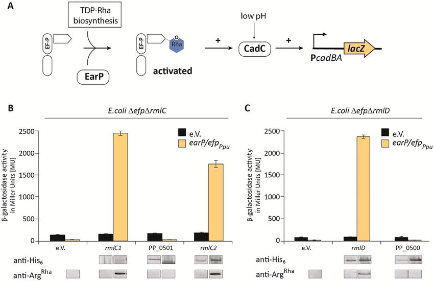
www.nature.com/scientificreports/
Figure 1. Rhamnose as versatile building block in composite biomolecules. (A) l-Rha in bacterial
biomolecules. Top left: Rhamnolipids consisting of a rhamnose moiety and a fatty acid tail in P. aeruginosa9. Top
right: Mycobacterial cell wall containing l-Rha as linking sugar between arabinogalactan and p eptidoglycan9.
acteria6. Bottom middle:
Bottom left: Rhamnosylation of translation elongation factor EF-P in about 10% of all b
biosynthesis of Streptomycin inter alia originating from dTDP-l-Rha10. Bottom right: Glycosylated flagella with
a linking l-Rha moiety in certain Pseudomonads11. (B) dTDP-β-l-rhamnose biosynthesis pathway. Glucose-
1-phosphate thymidylyltransferase, the first enzyme of the pathway, transfers a thymidylmonophosphate
nucleotide to glucose-1-phosphate, which is further oxidated by dTDP-d-glucose 4,6-dehydratase at the C4
hydroxyl group of the saccharide. The double epimerization reaction at positions C3 and C5 is catalyzed by the
dTDP-4-keto-6-deoxy-d-glucose 3,5-epimerase. Finally, the reduction of the C4 keto group by the dTDP-4-
keto-6-deoxy-l-mannose reductase leads to dTDP-l-Rha.
They are a paradigm of metabolically versatile microorganisms being able to recycle organic wastes and are key
players in the maintenance of environmental q uality24.
Following an unbiased approach and utilizing a restriction based genomic library, we identified the two
paralogous proteins PP_1782 (now termed RmlC1) and PP_0265 (now termed RmlC2) as dTDP-4-dehydror-
hamnose 3,5-epimerases while the last step namely the reduction to dTDP-l-Rha seems to be solely catalyzed by
PP_1784 (RmlD). By contrast, two further candidate genes that were identified by database mining and homology
analyses—PP_0500 and PP_0501—are not involved in dTDP-l-Rha biosynthesis. Taken together, our findings
contribute to the molecular understanding how dTDP-l-Rha is synthesized in Pseudomonas putida KT2440.
Results
A screening system that allows for the discovery of dTDP‑l‑Rha synthesis genes. To identify
genes involved in dTDP-l-Rha biosynthesis, we took advantage of cross functionality of pseudomonal EF-P in
Escherichia coli and the fact that activation of the translation factor strictly depends on the nucleotide sugar as
donor substrate. This cannot necessarily be expected, as the E. coli endogenous EF-P significantly differs from
its pseudomonal c ounterpart25: although both proteins alleviate ribosome stalling at polyproline s tretches6,26,
their modes of activation are phylogenetically u nrelated6,27. While E. coli EF-P (EF-PEco) strictly depends on
(R)-β-lysylation22,28–30 and hydroxylation31 of a conserved lysine, Pseudomonas EF-P (EF-PPpu) is rhamnosylated
at an arginine by the glycosyltransferase EarP ( EarPPpu) at the structurally equivalent p osition6,21. Despite these
apparent distinct post-translational modifications, a combination of efpPpu and earPPpu from P. putida KT2440
can compensate for a lack of efp in E. coli (Δefp) as long as the endogenous dTDP-l-Rha pathway remains intact
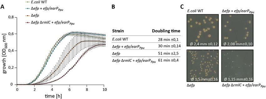
(Fig. 2A,C)6. Interestingly, loss of any synthesis gene—here exemplified with a ΔrmlC strain—does not simply
phenocopy Δefp but even results in more severe growth defects, as can be concluded from the correspond-
ing doubling times (Fig. 2B): E. coli Δefp cross complemented with efp/earPPpu grows twice as fast as the same
strain additionally lacking rmlC (Δefp ΔrmlC). These growth defects are also reflected by the size of the colonies
(Fig. 2C). The differences in growth rates provide us with a selection regime to identify dTDP-4-dehydrorham-
nose-3,5-epimerase genes from a P. putida genomic library.
The library was constructed by partial restriction digestion of the P. putida genome with the dam and CpG
methylation insensitive enzyme StuI (NEB) (Fig. 3). The average fragment size was set to 5 kb to ensure that
at least one gene was completely covered (average gene size: 1.132 kbp). These were cloned into SmaI lin-
earized pBAD33, which allows for high-level expression by induction of the P BAD promoter with l-arabinose32.
Scientific Reports | (2021) 11:11991 | https://doi.org/10.1038/s41598-021-91421-x 2
Vol:.(1234567890)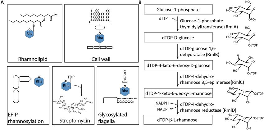
www.nature.com/scientificreports/
Figure 2. Growth analysis of cross complemented E.coli Δefp mutants in dependence of the dTDP-l-Rha
pathway. (A) E. coli MG1655 (E. coli WT), E. coli MG1655 Δefp expressing pBBR MCS2 efp/earPPpu (Δefp
efp/earPPpu), E. coli MG1655 Δefp (Δefp) and E. coli MG1655 Δefp ΔrmlC expressing pBBR MCS2 efp/earPPpu
(Δefp ΔrmlC efp/earPPpu) were grown in LB at 37 °C. Shown is a the mean curve from three independent
biological replicates with standard deviations. (B) Doubling times of E. coli strains listed in A. Doubling times
were calculated from growth analysis from three independent biological replicates. (C) Comparison of colony
size. E. coli MG1655 (E. coli WT), E. coli MG1655 Δefp expressing pBBR MCS2 efp/earPPpu (Δefp efp/earPPpu),
E. coli MG1655 Δefp (Δefp) and E. coli MG1655 Δefp ΔrmlC expressing pBBR MCS2 efp/earPPpu (Δefp ΔrmlC
efp/earPPpu) were plated on LB Agar (1.5%). The mean diameter with respective standard deviations is depicted
at the bottom. Pictures were taken after o/n growth at 37 °C.
Figure 3. Screening strategy for the identification dTDP-l-Rha biosynthesis genes in P. putida. Chromosomal
DNA of P. putida (green) was fragmented by restriction digestion. Fragments with an average size of 5 kb were
then ligated into the arabinose inducible vector (pBAD33). The resulting library was transformed into an E. coli
Δefp PcadBA::lacZ reporter strain that concomitantly lacks either rmlC or rmlD (ΔrmlC/ ΔrmlD) and additionally
encodes earPPpu and efpPpu (orange) of P. putida in trans. Genes cross-complementing ΔrmlC and ΔrmlD recover
the impaired growth phenotype and can be selected by size (white arrows).
Transformation of E. coli DH10B cells with the library revealed ~ 430,000 clones indicating an around 350-fold
coverage of the P. putida KT2440 genome (total length 6.18187 Mbp).
Next, we transformed E. coli Δefp ΔrmlC + efp/earPPpu with the library and cultivated the cells in LB (lysogeny
broth)33,34 containing 0.2% l-arabinose. Considering duplication times (Fig. 2B) and genome coverage, we expect
rmlC copies to accumulate already to a single-digit percentage of the total population within latest two days
(= ~ 16 generations with mutant growth phenotype and ~ 32 for wild-type phenotype), even under unfavorable
circumstances. Indeed, when plating the second overnight culture on LB agar we obtained colonies of two differ-
ent sizes. Consequently, we isolated plasmids from 16 large clones and sequencing identified 12 times PP_1782
and four times PP_0265 as the insert. PP_0265 (from now on rmlC2/RmlC2) resides next to genes encoding a
putative two component signal-transduction system (Fig. 4A). PP_1782 (from now on rmlC1/RmlC1) on the
Scientific Reports | (2021) 11:11991 | https://doi.org/10.1038/s41598-021-91421-x 3
Vol.:(0123456789)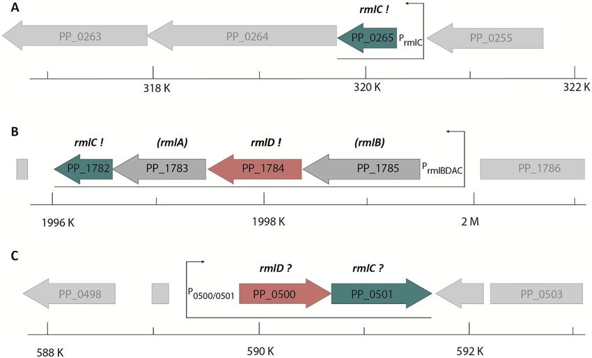
www.nature.com/scientificreports/
Figure 4. Genomic organization of rmlC and rmlD candidate genes in P. putida. (A) PP_0265 gene region, (B)
PP_1782_PP_1784 gene region. (C) PP_0500 and PP_0501 gene region. Putative (?) or validated (!) homologs/
analogs of rmlC and rmlD are shown in green and red respectively. Bottom: position within P. putida genome.
Arrows indicate monocistrons. The scale indicates the position within the P. putida genome.
other hand is the last of four genes in a putative dTDP-l-Rha biosynthesis operon PP_1785-PP_1782 (Fig. 4B). To
substantiate our hypothesis on the dTDP-l-Rha biosynthetic operon, we conducted a second library screen with
E. coli cells now lacking rmlD in addition to efp (Δefp ΔrmlD + efp/earPPpu) instead of rmlC. With this strain we
exclusively enriched clones harboring a copy of PP_1784 (from now on rmlD/RmlD), a homolog of E. coli RmlD.
Thus, we provide experimental evidence that PP_1785-PP_1782 form a rmlBDAC1 operon in P. putida KT2440
and further identified a second gene encoding for an dTDP-4-dehydrorhamnose 3,5-epimerases—RmlC2.
PP_0265/PP_1782 and PP_1784 are dTDP‑4‑dehydrorhamnose 3,5‑epimerases and
dTDP‑4‑dehydrorhamnose reductase, respectively. Our library screen was complemented by data-
base mining and a homology search. In addition to rmlC1 and rmlC2, we found PP_0501 being annotated as
nucleoside diphosphate sugar epimerase of unknown specificity and as such might function as further dTDP-
4-dehydrorhamnose 3,5-epimerase ( String35,36, Pfam37, Uniprot38, Metacyc39 database). However, while RmlC1
& RmlC2 are highly homologous to each other (64% identity), PP_0501 shares no similarities at the sequence
level. Nonetheless and in addition to its annotated function PP_0501 forms an operon with a putative dTDP-
4-dehydrorhamnose reductase gene, PP_050035,36,40 (Fig. 4C). This protein, on the contrary, shares similarities
with RmlD both at the sequence level (29% identity) as well as structurally.
To test the putative role of PP_0500 and PP_0501 in dTDP-l-Rha biosynthesis we made again benefit of EarP
mediated activation of P. putida EF-P and its functionality in E. coli. Hence, we cloned the two genes into pBAD33
simultaneously adding a His6-tag coding sequence for immunodetection in order to ensure proper protein pro-
duction (Fig. 5). rmlC1, rmlC2 and rmlD were also included in the study. The resulting plasmids pBAD33-rmlC1,
pBAD33-rmlC2, pBAD33-PP_0501 as well as pBAD33-rmlD and pBAD33-PP_0500 were introduced into E. coli
Δefp ΔrmlC + efp/earPPpu and Δefp ΔrmlD + efp/earPPpu, respectively. Of note, these are reporter strains in which
EF-P functionality is coupled to LacZ expression (Fig. 5A). Whereas β-galactosidase activity is low in cell with an
incomplete dTDP-l-Rha biosynthesis pathway, introduction of either rmlC1, rmlC2 (Fig. 5B) or rmlD (Fig. 5C)
into the respective mutant strains led to a significant increase. By contrast, neither PP_0500 nor PP_0501 were
able to rescue the ΔefpEco mutant phenotype.
In parallel we analyzed the rhamnosylation status of EF-PPpu utilizing anti-rhamnosylarginine specific anti-
bodies (anti-ArgRha)21,41,42. Immunodetection of EF-PPpu rhamnosylation matched with the reporter expression
levels on the one hand confirming the enzymatic activities of RmlC1, RmlC2 and RmlD as dTDP-4-dehydror-
hamnose 3,5-epimerase and dTDP-4-dehydrorhamnose reductase, respectively. On the other hand, they falsify
speculation and database annotations that attribute PP_0500 and PP_0501 a function in dTDP-l-Rha biosyn-
thesis (String35,36, Pfam37, Uniprot38, Metacyc39 database).
Scientific Reports | (2021) 11:11991 | https://doi.org/10.1038/s41598-021-91421-x 4
Vol:.(1234567890)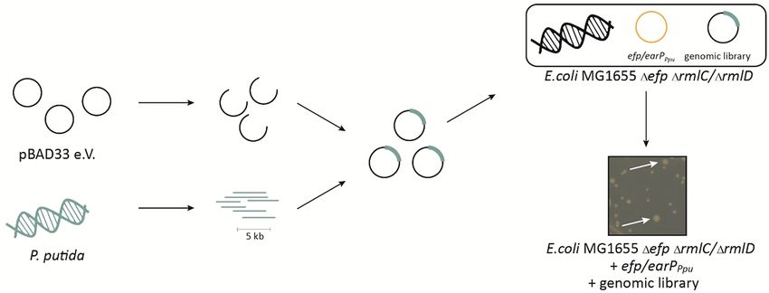
www.nature.com/scientificreports/
Figure 5. Analysis of in vivo activities of activated EF-P in dTDP-l-Rha biosynthesis deletion strains. (A)
β-Galactosidase reporter assay. The assay is based on the lysine decarboxylase acid stress response of E.
coli, the CadABC module43. At low pH, the transcriptional activator CadC activates the promoter of its two
downstream genes ( PcadBA) thereby inducing the expression of lacZ in an E. coli MG1655 PcadBA::lacZ strain.
Proper translation of CadC is dependent on the presence of EF-P which is activated by mono-rhamnosylation,
a reaction catalyzed by the glycosyltransferase EarP using dTDP-l-Rha (blue) as substrate. Thus β-galactosidase
activity can be taken as an indirect readout for functional dTDP-l-Rha biosynthesis. (B,C) Functionalities of
RmlC1, RmlC2, RmlD, PP_0500 and PP_0501 were determined by measuring the β-galactosidase activities of
E. coli MG1655 P cadBA::lacZ Δefp ΔrmlC (B)/ΔrmlD (C) with heterologous expression of a candidate gene from
the pBAD33 vector. The empty vector (e.V.) was included as negative control. Additionally, all strains encoded
the earP/efpPpu operon in trans, being encoded from pBBR MCS2 vectors (grey bars) expressed from the native
promoter. Again, the corresponding empty vector served as control (black bars). All strains were grown o/n in
LB pH 5.8 and activity is given in Miller Units (MU). Means of three independent measurements are shown.
Standard deviations from three independent experiments were determined. Bottom: Western blot analysis of
o/n cultures E. coli depicted in (B) and (C). Rhamnosylated EF-PPpu (EF-PRha) was detected using 0.25 µg/ml
anti-ArgRha. Expression of candidate genes was verified using 0.1 µg/ml anti-His6. Full-length Western Blots and
corresponding SDS-gels are depicted in Fig. S3.
Discussion
In the scope of this study, we have investigated the dTDP-l-Rha pathway of P. putida KT2440 with a focus on the
epimerization of dTDP-4-keto-6-deoxy-d-glucose. Combining an unbiased approach and utilizing a genomic
library, we identified two paralogous proteins RmlC1 and RmlC2. Duplication of rmlC is not restricted to P.
putida KT2440 but certain other pseudomonads such as P. monteilii, P. fulva, P. plecoglossicida or P. asiatica harbor
also two gene copies. In fact, functional redundancy in the dTDP-l-Rha biosynthesis pathway is nothing unu-
sual. As an example, the two enzymes RffH and RffG of E. coli are paralogous to RmlA and RmlB, respectively13.
Such duplications may be useful, e.g., to compensate for bottleneck reactions in the dTDP-l-Rha biosynthesis44.
Such bottlenecks can occur at different stages as the pathway is not only utilized to ultimately generate dTDP-l-
Rha. Specifically, dTDP-4-keto-6-deoxy-d-glucose is also a precursor of dTDP-3-acetamido-α-d-fucose45 and
TDP-d-viosamine46 which are found as part of the glycan pattern in P. syringae47. Similarly, the two paralogs
RmlC1 and RmlC2 in P. putida KT2440 might serve as starting point of similar but so far unknown reactions.
Moreover, gene duplications open the gate for regulated expression in turn allowing the precise adjustment of
the desired ratio of distinct NDP-sugars depending on parts of the dTDP-l-Rha biosynthesis pathway. It would
also allow for the accumulation of educts or products of the preceding reactions such as dTDP-glucose and Glc-
1P. Notably, whereas rmlC1 is part of an operon in which presumably the full dTDP-l-Rha pathway is encoded,
the rmlC2 resides in the vicinity of two genes encoding a two-component system (TCS) of thus far unknown
Scientific Reports | (2021) 11:11991 | https://doi.org/10.1038/s41598-021-91421-x 5
Vol.:(0123456789)www.nature.com/scientificreports/
Feature/genotype References
Plasmid
32
pBAD33 CamR-cassette, p15A origin, araC coding sequence, ara operator
pBBR1MCS2 KanR-cassette, pBBR origin of replication, oriT 50
pBAD33_rmlC1 CamR-cassette, arabinose inducible expression of RmlC1 This study
pBAD33_rmlD CamR-cassette, arabinose inducible expression of RmlD This study
pBAD33_PP_0265 CamR-cassette, arabinose inducible expression of PP_0265 This study
pBAD33_PP_0500 CamR-cassette, arabinose inducible expression of PP_0500 This study
pBAD33_PP_0501 CamR-cassette, arabinose inducible expression of PP_0501 This study
pBBR1MCS2_earP_efp KanR-cassette, earP and efp including the PearP native operon promoter 6,21
Strain
F-λ-endA1 glnV44 thi-1 recA1 relA1 gyrA96 deoR nupG Φ80dlacZΔM15 Δ(lacZYA-
E. coli DH5αλpir 51
argF) U169, hsdR17(rK− mK+)
F– mcrA Δ(mrr-hsdRMS-mcrBC) φ80lacZΔM15 ΔlacX74 recA1 endA1 araD139 Δ (ara-
E. coli DH10B 52
leu)7697 galU galK λ– rpsL(StrR) nupG
E. coli MG1655 K-12 F− λ− ilvG− rfb-50 rph-1 53
E. coli PcadBA::lacZ Δefp MG1655 PcadBA::lacZ Δ(cadBA) Δefp 26
E. coli PcadBA::lacZ Δefp ΔrmlC MG1655 PcadBA::lacZ Δ(cadBA) Δefp ΔrmlC 26
E.coli PcadBA::lacZ Δefp ΔrmlD MG1655 PcadBA::lacZ Δ(cadBA) Δefp ΔrmlD 26
Table 1. Plasmids and strains used in this study.
function. Based on the predicated domain composition, this specific TCS presumably transduces external sig-
nals into gene transcription. One might therefore speculate on regulated expression of rmlC2 according to the
environmental conditions.
While our genomic library revealed two RmlC paralogs in P. putida database mining indicated a further
enzyme with similar activity PP_0501. However, our in vivo rhamnosylation assay disproved the initial hypoth-
esis. Notably, the UDP-N-acetylglucosamine C4-epimerase PelX from P. protegens Pf-5 is structurally the closest
homolog (identity 67%)48. PelX is involved in the biosynthesis of the GalNAc-rich bacterial polysaccharidepoly-
saccharide Pel, that is essential for pellicle biofilm f ormation48,49. One can hence hypothesize, that PP_0501 and
the adjacent putative reductase PP_0500 might be involved in that pathway, instead.
Material and methods
Bacterial strains and growth condition. All strains and plasmids used in this study are listed and
described in Table 1. E. coli cells were grown in Miller modified Lysogeny Broth (LB)33,34 at 37 °C aerobically
under agitation, if not indicated otherwise. LB agar plates contained 1.5% agar. Mean diameters were measured
from 20 colonies from 2 different LB agar plates from of the respective strain after incubation at 37 °C for 16 h.
Growth measurements were conducted in 96 well plates. Therefore, 200 µl LB was inoculated with o/n cultures
at an OD600 0.001. OD600 was monitored in 10-min intervals for 12 h in a Tecan Spark with 240 rpm at 37 °C.
The medium was supplemented with antibiotics at the following concentrations: 50 µg/ml kanamycin sulfate
and 30 µg/ml chloramphenicol. Plasmids carrying the PBAD promoter32 were induced with l-arabinose at a final
concentration of 0.2% (w/v).
Molecular biology methods. Oligonucleotides used in this study are listed and described in the Supple-
mentary Table S1. Plasmid DNA was isolated using the Hi Yield Plasmid Mini Kit from Süd Laborbedarf accord-
ing to manufacturer’s instructions. DNA fragments were purified from agarose gels using the Hi Yield Gel/PCR
DNA fragment extraction kit from Süd Laborbedarf. All restriction enzymes, DNA modifying enzymes and the
Q5 high fidelity DNA polymerase for PCR amplification were purchased from New England BioLabs and used
according to manufacturer’s instructions.
Genomic library. The genomic DNA (gDNA) was isolated from 50 ml o/n culture of P putida KT2440
according to the protocol described in reference54. Further purification was achieved using Phase Lock Gel
(QuantaBio) with Phenol–Chloroform. After the centrifugation, isopropanol precipitation was repeated. The
pellet was resuspended in water, the final amount was 60 µg DNA.
Plasmid DNA was purified as described in “Molecular biology methods” from 12 ml E. coli DH5α cells. The
plasmid DNA was diluted in water, the final amount was 10 µg DNA.
The library was constructed using SmaI (pBAD33 vector) and StuI (gDNA) for digestion resulting in an aver-
age size of 5 kb per insert (Bionexus, Inc.). After ligation, the plasmids were transformed into E. coli DH10 B
(Lucigen). Quality control was done by restriction digest of library clones with BamHI. All restriction enzymes
were produced by New England Biolabs, Frankfurt. The library was reisolated from E. coli DH10B as described
in “Molecular biology methods” and transferred into corresponding reporter strains.
Scientific Reports | (2021) 11:11991 | https://doi.org/10.1038/s41598-021-91421-x 6
Vol:.(1234567890)www.nature.com/scientificreports/
Bioinformatic tools. The multiple sequence alignment was generated using NCBI BLAST55,56 and Clustal
Omega57. Candidate homologues were identified and analysed using S tring35,36, Pfam37, Uniprot38, Metacyc39
databases. Protein structures were predicted using Phyrre258. Illustrations were generated with UCSF Chimera59.
Β‑Galactosidase assay. E. coli MG1655 P
cadBA::lacZ Δefp ΔrmlC/ΔrmlD expressing lacZ under the control
of the cadBA promoter were grown in buffered LB (pH 5.8) overnight (o/n) and harvested by centrifugation.
β-Galactosidase activities were determined as described in reference in biological triplicates and are given in
Miller units (MU)60. Standard deviations from three independent experiments were determined.
SDS‑PAGE and western blotting. Electrophoretic separation of proteins was carried out using
12.5% SDS-PAGE as described by Laemmli61. Separated proteins were visualized in gel using 0.5% (vol/vol)
2-2-2-trichloroethanol62 and detected within a Gel Doc EZ gel documentation system (Bio-Rad). The proteins
were transferred onto a nitrocellulose membrane by vertical Western blotting (4 °C). Antigens were detected
using 0.1 g/ml anti-His6 tag (Abcam, Inc.) or 0.25 g/ml of anti-ArgRha41. Primary antibodies (rabbit) were the
targeted using 0.1 µg/ml anti-rabbit IgG (IRDye 680RD) (donkey) antibodies (Abcam). Target proteins were
visualized via Odyssey CLx Imaging System (LI-COR, Inc).
Received: 11 February 2021; Accepted: 26 May 2021
References
1. Giraud, M. F. & Naismith, J. H. The rhamnose pathway. Curr. Opin. Struct. Biol. 10, 687–696. https://doi.org/10.1016/s0959-
440x(00)00145-7 (2000).
2. Moses, T., Papadopoulou, K. K. & Osbourn, A. Metabolic and functional diversity of saponins, biosynthetic intermediates and
semi-synthetic derivatives. Crit. Rev. Biochem. Mol. Biol. 49, 439–462. https://doi.org/10.3109/10409238.2014.953628 (2014).
3. Soberón-Chávez, G., González-Valdez, A., Soto-Aceves, M. P. & Cocotl-Yañez, M. Rhamnolipids produced by Pseudomonas: From
molecular genetics to the market. Microb. Biotechnol. 14, 136–146. https://doi.org/10.1111/1751-7915.13700 (2021).
4. Ma, Y. et al. Drug targeting Mycobacterium tuberculosis cell wall synthesis: Genetics of dTDP-rhamnose synthetic enzymes and
development of a microtiter plate-based screen for inhibitors of conversion of dTDP-glucose to dTDP-rhamnose. Antimicrob.
Agents Chemother. 45, 1407–1416. https://doi.org/10.1128/aac.45.5.1407-1416.2001 (2001).
5. Gao, M., D’Haeze, W., De Rycke, R., Wolucka, B. & Holsters, M. Knockout of an azorhizobial dTDP-l-rhamnose synthase affects
lipopolysaccharide and extracellular polysaccharide production and disables symbiosis with Sesbania rostrata. Mol. Plant Microbe
Interact. 14, 857–866. https://doi.org/10.1094/mpmi.2001.14.7.857 (2001).
6. Lassak, J. et al. Arginine-rhamnosylation as new strategy to activate translation elongation factor P. Nat. Chem. Biol. 11, 266–270.
https://doi.org/10.1038/nchembio.1751 (2015).
7. Kneidinger, B. et al. Identification of two GDP-6-deoxy-d-lyxo-4-hexulose reductases synthesizing GDP-d-rhamnose in Aneu-
rinibacillus thermoaerophilus L420–91T. J. Biol. Chem. 276, 5577–5583. https://doi.org/10.1074/jbc.M010027200 (2001).
8. Shibaev, V. N. Biosynthesis of bacterial polysaccharide chains composed of repeating units. Adv. Carbohydr. Chem. Biochem. 44,
277–339. https://doi.org/10.1016/s0065-2318(08)60080-3 (1986).
9. Wild, M., Caro, A. D., Hernández, A. L., Miller, R. M. & Soberón-Chávez, G. Selection and partial characterization of a Pseu-
domonas aeruginosa mono-rhamnolipid deficient mutant. FEMS Microbiol. Lett. 153, 279–285. https://d oi.o
rg/1 0.1 111/j.1 574-6 968.
1997.tb12586.x (1997).
10. Wahl, H. P. & Grisebach, H. Biosynthesis of streptomycin. dTDP-dihydrostreptose synthase from Streptomyces griseus and dTDP-
4-keto-l-rhamnose 3,5-epimerase from S. griseus and Escherichia coli Y10. Biochim. Biophys. Acta 568, 243–252. https://doi.org/
10.1016/0005-2744(79)90291-2 (1979).
11. Schirm, M. et al. Structural and genetic characterization of glycosylation of type a flagellin in Pseudomonas aeruginosa. J. Bacteriol.
186, 2523–2531. https://doi.org/10.1128/jb.186.9.2523-2531.2004 (2004).
12. Blankenfeldt, W., Asuncion, M., Lam, J. S. & Naismith, J. H. The structural basis of the catalytic mechanism and regulation of
glucose-1-phosphate thymidylyltransferase (RmlA). EMBO J. 19, 6652–6663. https://doi.org/10.1093/emboj/19.24.6652 (2000).
13. Marolda, C. L. & Valvano, M. A. Genetic analysis of the dTDP-rhamnose biosynthesis region of the Escherichia coli VW187 (O7:K1)
rfb gene cluster: Identification of functional homologs of rfbB and rfbA in the rff cluster and correct location of the rffE gene. J.
Bacteriol. 177, 5539–5546. https://doi.org/10.1128/jb.177.19.5539-5546.1995 (1995).
14. Allard, S. T., Giraud, M. F., Whitfield, C., Messner, P. & Naismith, J. H. The purification, crystallization and structural elucidation
of dTDP-d-glucose 4,6-dehydratase (RmlB), the second enzyme of the dTDP-l-rhamnose synthesis pathway from Salmonella
enterica serovar typhimurium. Acta Crystallogr. D Biol. Crystallogr. 56, 222–225. https://doi.org/10.1107/s0907444999016200
(2000).
15. Graninger, M., Nidetzky, B., Heinrichs, D. E., Whitfield, C. & Messner, P. Characterization of dTDP-4-dehydrorhamnose 3,5-epime-
rase and dTDP-4-dehydrorhamnose reductase, required for dTDP-l-rhamnose biosynthesis in Salmonella enterica serovar Typh-
imurium LT2. J. Biol. Chem. 274, 25069–25077. https://doi.org/10.1074/jbc.274.35.25069 (1999).
16. van der Beek, S. L. et al. Streptococcal dTDP-l-rhamnose biosynthesis enzymes: Functional characterization and lead compound
identification. Mol. Microbiol. 111, 951–964. https://doi.org/10.1111/mmi.14197 (2019).
17. Ma, Y., Pan, F. & McNeil, M. Formation of dTDP-rhamnose is essential for growth of mycobacteria. J. Bacteriol. 184, 3392–3395.
https://doi.org/10.1128/jb.184.12.3392-3395.2002 (2002).
18. Alhede, M., Bjarnsholt, T., Givskov, M. & Alhede, M. Pseudomonas aeruginosa biofilms: Mechanisms of immune evasion. Adv.
Appl. Microbiol. 86, 1–40. https://doi.org/10.1016/b978-0-12-800262-9.00001-9 (2014).
19. Aguirre-Ramírez, M., Medina, G., González-Valdez, A., Grosso-Becerra, V. & Soberón-Chávez, G. The Pseudomonas aeruginosa
rmlBDAC operon, encoding dTDP-l-rhamnose biosynthetic enzymes, is regulated by the quorum-sensing transcriptional regulator
RhlR and the alternative sigma factor σS. Microbiology (Reading, England) 158, 908–916. https://doi.org/10.1099/mic.0.054726-0
(2012).
20. Zulianello, L. et al. Rhamnolipids are virulence factors that promote early infiltration of primary human airway epithelia by Pseu-
domonas aeruginosa. Infect. Immun. 74, 3134–3147. https://doi.org/10.1128/iai.01772-05 (2006).
21. Krafczyk, R. et al. Structural basis for EarP-mediated arginine glycosylation of translation elongation factor EF-P. MBio https://
doi.org/10.1128/mBio.01412-17 (2017).
Scientific Reports | (2021) 11:11991 | https://doi.org/10.1038/s41598-021-91421-x 7
Vol.:(0123456789)www.nature.com/scientificreports/
22. Yanagisawa, T., Sumida, T., Ishii, R., Takemoto, C. & Yokoyama, S. A paralog of lysyl-tRNA synthetase aminoacylates a conserved
lysine residue in translation elongation factor P. Nat. Struct. Mol. Biol. 17, 1136–1143. https://doi.org/10.1038/nsmb.1889 (2010).
23. Rajkovic, A. et al. Cyclic Rhamnosylated Elongation Factor P Establishes Antibiotic Resistance in Pseudomonas aeruginosa. MBio
6, e00823. https://doi.org/10.1128/mBio.00823-15 (2015).
24. Martins dos Santos, V. A. P., Timmis, K. N., Tümmler, B. & Weinel, C. In Pseudomonas: Volume 1 Genomics Life Style and Molecular
Architecture (ed. Ramos, J.-L.) 77–112 (Springer US, 2004).
25. Choi, S. & Choe, J. Crystal structure of elongation factor P from Pseudomonas aeruginosa at 1.75 Å resolution. Proteins 79,
1688–1693. https://doi.org/10.1002/prot.22992 (2011).
26. Ude, S. et al. Translation elongation factor EF-P alleviates ribosome stalling at polyproline stretches. Science 339, 82–85. https://
doi.org/10.1126/science.1228985 (2013).
27. Lassak, J., Wilson, D. N. & Jung, K. Stall no more at polyproline stretches with the translation elongation factors EF-P and IF-5A.
Mol. Microbiol. 99, 219–235. https://doi.org/10.1111/mmi.13233 (2016).
28. Navarre, W. W. et al. PoxA, yjeK, and elongation factor P coordinately modulate virulence and drug resistance in Salmonella
enterica. Mol. Cell. 39, 209–221. https://doi.org/10.1016/j.molcel.2010.06.021 (2010).
29. Gilreath, M. S. et al. β-Lysine discrimination by lysyl-tRNA synthetase. FEBS Lett. 585, 3284–3288. https://doi.org/10.1016/j.febsl
et.2011.09.008 (2011).
30. Pfab, M. et al. Synthetic post-translational modifications of elongation factor P using the ligase EpmA. FEBS J. 288, 663–677.
https://doi.org/10.1111/febs.15346 (2021).
31. Peil, L. et al. Distinct XPPX sequence motifs induce ribosome stalling, which is rescued by the translation elongation factor EF-P.
Proc. Natl. Acad. Sci. U.S.A. 110, 15265–15270. https://doi.org/10.1073/pnas.1310642110 (2013).
32. Guzman, L. M., Belin, D., Carson, M. J. & Beckwith, J. Tight regulation, modulation, and high-level expression by vectors contain-
ing the arabinose P BAD promoter. J. Bacteriol. 177, 4121–4130. https://doi.org/10.1128/jb.177.14.4121-4130.1995 (1995).
33. Bertani, G. Studies on lysogenesis. I. The mode of phage liberation by lysogenic Escherichia coli. J. Bacteriol. 62, 293–300. https://
doi.org/10.1128/jb.62.3.293-300.1951 (1951).
34. Bertani, G. Lysogeny at mid-twentieth century: P1, P2, and other experimental systems. J. Bacteriol. 186, 595–600. https://doi.org/
10.1128/jb.186.3.595-600.2004 (2004).
35. Snel, B., Lehmann, G., Bork, P. & Huynen, M. A. STRING: A web-server to retrieve and display the repeatedly occurring neigh-
bourhood of a gene. Nucleic Acids Res. 28, 3442–3444. https://doi.org/10.1093/nar/28.18.3442 (2000).
36. Szklarczyk, D. et al. STRING v11: Protein–protein association networks with increased coverage, supporting functional discovery
in genome-wide experimental datasets. Nucleic Acids Res. 47, D607–D613. https://doi.org/10.1093/nar/gky1131 (2019).
37. El-Gebali, S. et al. The Pfam protein families database in 2019. Nucleic Acids Res. 47, D427–D432. https://doi.org/10.1093/nar/
gky995 (2019).
38. UniProt: A worldwide hub of protein knowledge. Nucleic Acids Res. 47, D506–D515. https://doi.org/10.1093/nar/gky1049 (2019).
39. Caspi, R. et al. The MetaCyc database of metabolic pathways and enzymes—A 2019 update. Nucleic Acids Res. 48, D445–D453.
https://doi.org/10.1093/nar/gkz862 (2020).
40. Seo, J., Brencic, A. & Darwin, A. J. Analysis of secretin-induced stress in Pseudomonas aeruginosa suggests prevention rather than
response and identifies a novel protein involved in secretin function. J. Bacteriol. 191, 898–908. https://doi.org/10.1128/jb.01443-
08 (2009).
41. Li, X. et al. Resolving the α-glycosidic linkage of arginine-rhamnosylated translation elongation factor P triggers generation of the
first Arg(Rha) specific antibody. Chem. Sci. 7, 6995–7001. https://doi.org/10.1039/c6sc02889f (2016).
42. Gast, D. et al. A set of rhamnosylation-specific antibodies enables detection of novel protein glycosylations in bacteria. Org. Biomol.
Chem. 18, 6823–6828. https://doi.org/10.1039/d0ob01289k (2020).
43. Tetsch, L., Koller, C., Haneburger, I. & Jung, K. The membrane-integrated transcriptional activator CadC of Escherichia coli senses
lysine indirectly via the interaction with the lysine permease LysP. Mol. Microbiol. 67, 570–583. https://d oi.o
rg/1 0.1 111/j.1 365-2 958.
2007.06070.x (2008).
44. Brandis, G. & Hughes, D. The SNAP hypothesis: Chromosomal rearrangements could emerge from positive selection during niche
adaptation. PLoS Genet. 16, e1008615. https://doi.org/10.1371/journal.pgen.1008615 (2020).
45. Pföstl, A. et al. Biosynthesis of dTDP-3-acetamido-3,6-dideoxy-alpha-d-glucose. Biochem. J. 410, 187–194. https://doi.org/10.
1042/bj20071044 (2008).
46. Pandey, R. P., Parajuli, P., Gurung, R. B. & Sohng, J. K. Donor specificity of YjiC glycosyltransferase determines the conjugation of
cytosolic NDP-sugar in in vivo glycosylation reactions. Enzyme Microb. Technol. 91, 26–33. https://doi.org/10.1016/j.enzmictec.
2016.05.006 (2016).
47. Yamamoto, M. et al. Identification of genes involved in the glycosylation of modified viosamine of flagellins in Pseudomonas
syringae by mass spectrometry. Genes 2, 788–803. https://doi.org/10.3390/genes2040788 (2011).
48. Marmont, L. S. et al. PelX is a UDP-N-acetylglucosamine C4-epimerase involved in Pel polysaccharide-dependent biofilm forma-
tion. J. Biol. Chem. 295, 11949–11962. https://doi.org/10.1074/jbc.RA120.014555 (2020).
49. Friedman, L. & Kolter, R. Genes involved in matrix formation in Pseudomonas aeruginosa PA14 biofilms. Mol. Microbiol. 51,
675–690. https://doi.org/10.1046/j.1365-2958.2003.03877.x (2004).
50. Kovach, M. E. et al. Four new derivatives of the broad-host-range cloning vector pBBR1MCS, carrying different antibiotic-resistance
cassettes. Gene 166, 175–176. https://doi.org/10.1016/0378-1119(95)00584-1 (1995).
51. Macinga, D. R., Parojcic, M. M. & Rather, P. N. Identification and analysis of aarP, a transcriptional activator of the 2′-N-acetyl-
transferase in Providencia stuartii. J. Bacteriol. 177, 3407–3413. https://doi.org/10.1128/jb.177.12.3407-3413.1995 (1995).
52. Durfee, T. et al. The complete genome sequence of Escherichia coli DH10B: Insights into the biology of a laboratory workhorse. J.
Bacteriol. 190, 2597–2606. https://doi.org/10.1128/jb.01695-07 (2008).
53. Guyer, M. S., Reed, R. R., Steitz, J. A. & Low, K. B. Identification of a sex-factor-affinity site in E. coli as gamma delta. Cold Spring.
Harb. Symp. Quant. Biol. 45, 135–140. https://doi.org/10.1101/sqb.1981.045.01.022 (1981).
54. Pospiech, A. & Neumann, B. A versatile quick-prep of genomic DNA from gram-positive bacteria. Trends Genet. 11, 217–218.
https://doi.org/10.1016/s0168-9525(00)89052-6 (1995).
55. Database resources of the National Center for Biotechnology Information. Nucleic Acids Res. 44, D7–D19. https://d oi.o
rg/1 0.1 093/
nar/gkv1290 (2016).
56. Altschul, S. F., Gish, W., Miller, W., Myers, E. W. & Lipman, D. J. Basic local alignment search tool. J. Mol. Biol. 215, 403–410.
https://doi.org/10.1016/s0022-2836(05)80360-2 (1990).
57. Sievers, F. & Higgins, D. G. Clustal Omega, accurate alignment of very large numbers of sequences. Methods Mol. Biol. 1079,
105–116. https://doi.org/10.1007/978-1-62703-646-7_6 (2014).
58. Kelley, L. A., Mezulis, S., Yates, C. M., Wass, M. N. & Sternberg, M. J. The Phyre2 web portal for protein modeling, prediction and
analysis. Nat. Protoc. 10, 845–858. https://doi.org/10.1038/nprot.2015.053 (2015).
59. Pettersen, E. F. et al. UCSF Chimera—A visualization system for exploratory research and analysis. J. Comput. Chem. 25, 1605–1612.
https://doi.org/10.1002/jcc.20084 (2004).
60. Miller, J. H. A Short Course in Bacterial Genetics. A Laboratory Manual and Handbook for Escherichia coli and Related Bacteria
(Cold Spring Harbor, 1992).
Scientific Reports | (2021) 11:11991 | https://doi.org/10.1038/s41598-021-91421-x 8
Vol:.(1234567890)www.nature.com/scientificreports/
61. Laemmli, U. K. Cleavage of structural proteins during the assembly of the head of bacteriophage T4. Nature 227, 680–685. https://
doi.org/10.1038/227680a0 (1970).
62. Ladner, C. L., Yang, J., Turner, R. J. & Edwards, R. A. Visible fluorescent detection of proteins in polyacrylamide gels without
staining. Anal. Biochem. 326, 13–20. https://doi.org/10.1016/j.ab.2003.10.047 (2004).
Acknowledgements
This work was funded by the Deutsche Forschungsgemeinschaft research grant LA 3658/1-1 and Research
Training Group GRK2062/1 (Molecular Principles of Synthetic Biology). We thank Ralph Krafczyk and Kirsten
Jung for fruitful discussions. Further we thank Lis Winter, Maximilian Dorok and Martin Erhard for their
contributing results.
Author contributions
All experiments were performed by F.K. F.K. and J.L. designed the study. The manuscript was written by F.K.
and J.L.
Funding
Open Access funding enabled and organized by Projekt DEAL.
Competing interests
The authors declare no competing interests.
Additional information
Supplementary Information The online version contains supplementary material available at https://doi.org/
10.1038/s41598-021-91421-x.
Correspondence and requests for materials should be addressed to J.L.
Reprints and permissions information is available at www.nature.com/reprints.
Publisher’s note Springer Nature remains neutral with regard to jurisdictional claims in published maps and
institutional affiliations.
Open Access This article is licensed under a Creative Commons Attribution 4.0 International
License, which permits use, sharing, adaptation, distribution and reproduction in any medium or
format, as long as you give appropriate credit to the original author(s) and the source, provide a link to the
Creative Commons licence, and indicate if changes were made. The images or other third party material in this
article are included in the article’s Creative Commons licence, unless indicated otherwise in a credit line to the
material. If material is not included in the article’s Creative Commons licence and your intended use is not
permitted by statutory regulation or exceeds the permitted use, you will need to obtain permission directly from
the copyright holder. To view a copy of this licence, visit http://creativecommons.org/licenses/by/4.0/.
© The Author(s) 2021
Scientific Reports | (2021) 11:11991 | https://doi.org/10.1038/s41598-021-91421-x 9
Vol.:(0123456789)You can also read