Josa Prado, F., Luo, J., Rubin, P., Henley, J., & Wilkinson, K. (2019). Developmental profiles of SUMOylation pathway proteins in rat cerebrum and ...
←
→
Page content transcription
If your browser does not render page correctly, please read the page content below
Josa Prado, F., Luo, J., Rubin, P., Henley, J., & Wilkinson, K. (2019).
Developmental profiles of SUMOylation pathway proteins in rat cerebrum
and cerebellum. PLoS ONE, 14(2), [e0212857].
https://doi.org/10.1371/journal.pone.0212857

Publisher's PDF, also known as Version of record
License (if available):
CC BY
Link to published version (if available):
10.1371/journal.pone.0212857

Link to publication record in Explore Bristol Research
PDF-document

This is the final published version of the article (version of record). It first appeared online via PLOS at
https://doi.org/10.1371/journal.pone.0212857 . Please refer to any applicable terms of use of the publisher.

University of Bristol - Explore Bristol Research
General rights

This document is made available in accordance with publisher policies. Please cite only the published
version using the reference above. Full terms of use are available:
http://www.bristol.ac.uk/pure/about/ebr-terms
RESEARCH ARTICLE

                                                     Developmental profiles of SUMOylation
                                                     pathway proteins in rat cerebrum and
                                                     cerebellum
                                                     Fernando Josa-Prado ID1,2*, Jia Luo2, Philip Rubin2, Jeremy M. Henley2, Kevin
                                                     A. Wilkinson2*
                                                     1 Universidad Alfonso X el Sabio, Avda, de la Universidad, Madrid, España, 2 School of Biochemistry,
                                                     Medical Sciences Building, University of Bristol, University Walk, Bristol, United Kingdom

a1111111111                                          * f.josaprado@gmail.com(FJP); kevin.wilkinson@bristol.ac.uk(KAW)
a1111111111
a1111111111
a1111111111
a1111111111                                          Abstract
                                                     Protein SUMOylation regulates multiple processes involved in the differentiation and matu-
                                                     ration of cells and tissues during development. Despite this, relatively little is known about
                                                     the spatial and temporal regulation of proteins that mediate SUMOylation and deSUMOyla-
    OPEN ACCESS                                      tion in the CNS. Here we monitor the expression of key SUMO pathway proteins and levels
Citation: Josa-Prado F, Luo J, Rubin P, Henley JM,   of substrate protein SUMOylation in the forebrain and cerebellum of Wistar rats during
Wilkinson KA (2019) Developmental profiles of
                                                     development. Overall, the SUMOylation machinery is more highly-expressed at E18 and
SUMOylation pathway proteins in rat cerebrum and
cerebellum. PLoS ONE 14(2): e0212857. https://       decreases thereafter, as previously described. All of the proteins investigated are less abun-
doi.org/10.1371/journal.pone.0212857                 dant in adult than in embryonic brain. Furthermore, we show for first time that the profiles dif-
Editor: Fernando de Castro, Instituto Cajal-CSIC,    fer between cerebellum and cerebrum, indicating differential regional regulation of some of
SPAIN                                                the proteins analysed. These data provide further basic observation that may open a new
Received: November 30, 2018                          perspective of research about the role of SUMOylation in the development of different brain
                                                     regions.
Accepted: February 11, 2019

Published: February 22, 2019

Copyright: © 2019 Josa-Prado et al. This is an
open access article distributed under the terms of
the Creative Commons Attribution License, which      Introduction
permits unrestricted use, distribution, and
reproduction in any medium, provided the original    SUMOylation is the covalent attachment of a 97-residue protein, SUMO (Small Ubiquitin-
author and source are credited.                      related MOdifier), to lysine residues on target proteins. SUMOylation is best characterised for
Data Availability Statement: All relevant data are   modifying nuclear proteins involved in genome integrity, nuclear structure and transcription
within the manuscript and its Supporting             [1, 2] but it is now clear that SUMOylation is also important for extranuclear signal transduc-
Information files.                                   tion, trafficking and modification of cytosolic and integral membrane proteins. Several hun-
Funding: This work was supported by: to JMH:         dred SUMOylation substrates have been validated and many more candidate substrates have
Project “SyMBaD” Synapses: from molecules to         been identified by proteomic studies [3–5].
higher brain function and diseases / Project ID          There are three SUMO paralogues (SUMO-1-3) in vertebrates. SUMO-2 and SUMO-3 are
238608 / FP7-PEOPLE-ITN-2008 - Marie Curie           identical except for three residues, but share only ~50% sequence identity with SUMO-1.
Action: "Networks for Initial Training" / European
                                                     While some substrates can be modified by both SUMO-1 and SUMO-2/3, SUMO proteins are
Commission / https://cordis.europa.eu/project/rcn/
92985_en.html / Joint award between 14 academic
                                                     functionally heterogeneous and show distinct patterns of conjugation under both resting con-
and commercial partners co-ordinated by              ditions and in response to cell stress. For example, under resting conditions there is very little
University of Bordeaux; SUMOBRAIN -                  unconjugated SUMO-1 whereas there is a large free pool of SUMO-2/3 [6]. However, in

PLOS ONE | https://doi.org/10.1371/journal.pone.0212857 February 22, 2019                                                                                   1 / 15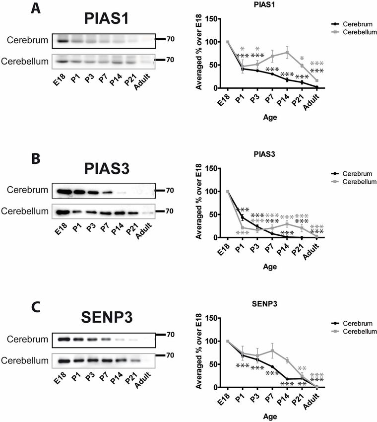
SUMOylation during rat brain development

‘Mechanisms and consequences of synaptic               response to a variety of stressors, SUMO-2/3 conjugation is dramatically increased while
SUMOylation in health and disease’ / Proposal n˚       SUMO-1 conjugation is relatively unchanged [6–13]. The functional consequences of SUMO
232881 / European Research Council / https://
                                                       attachment are in many cases poorly understood and can vary greatly depending on the
cordis.europa.eu/project/rcn/90355_es.html / (Sole
applicant). FJP and KAW received no specific           substrate.
funding for this work. The funders had no role in         The SUMOylation state of substrate proteins is a dynamic balance between conjugation
study design, data collection and analysis, decision   and deconjugation. Briefly, inactive precursor SUMO is matured by SUMO-specific proteases
to publish, or preparation of the manuscript.          (SENPs) to expose a C-terminal diglycine motif, which is activated by an ATP-dependent E1
Competing interests: The authors have declared         enzyme, formed by a heterodimer of SAE1 and SAE2 [14]. E1 passes the activated SUMO onto
that no competing interests exist.                     the specific and unique SUMO conjugating E2 enzyme Ubc9 via a transesterification reaction
                                                       [15, 16]. Ubc9, often in conjunction with a growing number of identified E3 ligase enzymes,
                                                       then catalyses SUMOylation of the substrate.
                                                          SUMO is removed from substrates by the isopeptidase activity of the SENPs, the same
                                                       enzymes required for pro-SUMO maturation. There are six mammalian SENPs (SENP1-3 and
                                                       SENP5-7; for reviews, see [17–19]). SENP1 and SENP2 have a broad specificity for SUMO-1
                                                       and SUMO-2/3 and are involved in both maturation and deconjugation while SENP3 and
                                                       SENP5 favour SUMO-2/3 over SUMO-1. They selectively remove SUMO-2/3 from substrate
                                                       proteins and do not play a role in SUMO maturation. SENP6 and 7 are primarily involved in
                                                       the editing of poly-SUMO chains. Furthermore, three proteins distinct from the SENP family
                                                       have also been reported to function as deSUMOylating enzymes [19].
                                                          SUMOylation is involved in multiple signalling cascades in neurons and is strongly impli-
                                                       cated in many neurological and neurodegenerative diseases including Huntington’s disease,
                                                       Alzheimer’s disease, Parkinson’s disease and stroke (for reviews see [20–23]). Importantly,
                                                       protein modification by SUMO2/3 is neuroprotective against metabolic cell stress [24, 25, 26].
                                                       Furthermore, we have shown that SUMOylation is a key modulator of synaptic transmission
                                                       and plasticity via direct modulation of kainate receptors [27–29] and presynaptic proteins
                                                       such as Syntaxin 1A [30] and Synapsin Ia [31] and indirect regulation of AMPAR trafficking
                                                       in synaptic scaling [32] and LTP [29]. Recently, it has been shown that SUMOylation might be
                                                       important in neuronal development through modification of the transcription factors FOXP1
                                                       and MEF2A [33, 34].
                                                          Here we set out to define the developmental profiles of SUMO machinery proteins and the
                                                       extent of total protein SUMOylation in fractions of rat cerebrum and cerebellum. The aim of
                                                       this work was to provide a framework for defining how protein conjugation by SUMO-1 and
                                                       SUMO-2/3 changes during brain development.

                                                       Materials and methods
                                                       Brain lysates
                                                       Lysates were prepared from crude cerebrum and cerebellum from Wistar rats at the indicated
                                                       ages. Animals were bred by an in-house animal unit and sacrificed by cervical dislocation in
                                                       accordance with UK Home Office Schedule 1 guidelines. All procedures were approved by the
                                                       Animal Welfare and Ethics Review Body (AWERB) at the University of Bristol (approval refer-
                                                       ence number UIN UB/18/004). In all cases brains from at least 3 separate rats were used. Seven
                                                       developmental stages were investigated: embryonic day 18 (E18) and postnatal ages (in days)
                                                       P1, P3, P7, P14, P21 and adult (pregnant rat from 12 weeks onwards). Female rats were used
                                                       for all samples except E18, which were not sexed. For E18 more embryos were sacrificed to get
                                                       sufficient tissue and samples were taken from different litters. Following sacrifice, brains were
                                                       immediately taken and the cerebellum and cerebrum separated. These were then homogenised
                                                       in 25mM HEPES, 150 mM NaCl, 1x protease inhibitor cocktail (Complete tablets, Roche), pH
                                                       7.5 supplemented with 20 mM NEM (to avoid deSUMOylation after cell lysis).

PLOS ONE | https://doi.org/10.1371/journal.pone.0212857 February 22, 2019                                                                           2 / 15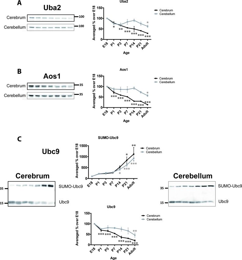
SUMOylation during rat brain development

                                              The volume of buffer used was proportional to the wet weight of the tissue (10 ml of buffer
                                           per gram of tissue) and samples were homogenized with a Teflon and glass homogeniser
                                           (around 10 passes). The homogenate was then collected and solubilised by the addition of 1%
                                           Triton X-100 and 0.1% SDS (final concentration) and incubation on a rotating wheel at 4˚C
                                           for 40 minutes. Cellular debris was separated by centrifugation at 16,000g for 10 minutes and
                                           the insoluble pellet discarded. The total protein concentration in the soluble fraction was mea-
                                           sured by Bradford assay and samples mixed with 6x Laemmli buffer, boiled and diluted to a
                                           concentration of 1 mg/ml.

                                           Subcellular fractionation
                                           Fractions were obtained by differential centrifugation [35]. Briefly, tissue from either cerebrum
                                           or cerebellum of adult rat was homogenised in 10 ml/g of tissue in Tris-sucrose buffer (10mM
                                           Tris-HCl, pH7.4, 0.32M sucrose, 20mM NEM and protease inhibitors). The crude nuclear
                                           fraction (N) was obtained by centrifugation at 412g for 10 minutes and this P1 fraction was
                                           washed twice in the same buffer. The P1 supernatant (S1) was centrifuged at 10,000g for 20
                                           minutes to obtain a crude synaptosomal fraction (P2). This was resuspended and loaded onto
                                           a discontinuous 0.32, 0.8, 1.2M sucrose gradient and centrifuged at 53,000g for 2 hours. The
                                           final synaptosomal (Sy) fraction was collected from the interphase between the 0.8M and 1.2M
                                           sucrose layers. The cytosolic fraction (S3) was obtained by centrifuging S2 at 20,000g for 1
                                           hour. All fractions were solubilised in 0.1% SDS and 1% Triton X-100. Protein concentration
                                           was measured by Bradford assay and samples equalized to the same protein concentration in
                                           Laemmli buffer for SDS-PAGE.

                                           Western blotting
                                           Samples from each age point were subjected to SDS-PAGE on 8–15% polyacrylamide gels,
                                           depending on the protein being detected. For SUMO blots, gradient gels (4–20%) were used.
                                           Separated proteins were transferred to nitrocellulose membrane and immunoblotted. The pri-
                                           mary antibodies used were: β-III-tubulin (1:5000, T8660), β-actin (1:2500, A5441), Syntaxin1A
                                           (1:5000, S0664) all from Sigma-Aldrich. PSD95 (1:1000, AB1596), GluA1 (1:1000, AB2263),
                                           NMADR1 (1:1000, AB9864) from Millipore. Aos1 (1:200, sc-46766), Uba2 (1:200, sc-376305),
                                           SUMO-1 (1:500, sc-5308) from Santa Cruz Biotechnology. Ubc9 (1:250, 610749) from BD Bio-
                                           sciences; PIAS1 (1:2000, 77231) from ABCAM; PIAS3 (1:1000, AP1245a) from Abgent; SENP3
                                           (1:4000, D20A10) Cell Signaling Technology; SUMO-2/3 (1:100, Clone 8A2, produced in
                                           house) from DRHB. Luminescent or fluorescent signal was captured with a Li-COR Odyssey
                                           Fc system and quantified using Image Studio 2.1 provided by Li-COR. For SUMO quantifica-
                                           tion the whole lane was selected and quantified as well as individual bands with strong signal.

                                           Statistical analysis
                                           1-way ANOVA with Newman-Keuls post-hoc test was used for analysis of the differences
                                           between time points in cerebrum or cerebellum compared to E18, which was designated as
                                           100%. For determining differences between cerebrum and cerebellum in each time point for
                                           each protein two-way ANOVA with Bonferroni post-hoc test was used (� = p�0.05, �� =
                                           p�0.01, ��� = p�0.001).

                                           Results
                                           In order to address how profiles of protein SUMOylation and SUMO pathway proteins are
                                           altered in cerebellum and cerebrum during development, we homogenised rat brains at

PLOS ONE | https://doi.org/10.1371/journal.pone.0212857 February 22, 2019                                                               3 / 15
SUMOylation during rat brain development
various stages of development (embryonic day 18, and post-natal days 1, 3, 7, 14, 21 and
adult). Importantly, due to their small size, the embryonic day 18 (E18) brains were not dis-
sected into cerebrum and cerebellum, and instead we prepared whole homogenate from these
brains. However, this approach had the added advantage of meaning that all cerebellum and
cerebrum samples from the other time points were run against the same reference E18 sam-
ples, allowing direct comparison of changes in protein levels between the brain regions.
To validate our homogenate samples, we initially investigated the developmental profiles of
several marker proteins (S1 and S2 Figs). Cytoskeletal proteins commonly used as loading con-
trols, such as β-III-tubulin and β-actin, showed some developmental variation thus not being
suitable as loading control (S1 Fig). Nonetheless, we loaded equal amounts of protein for each
sample using Bradford assay to measure total protein concentration. We also monitored the
profiles of several synaptic proteins: PSD95, syntaxin1A, GluA1 and NR1. Importantly, the
profiles of PSD95 [36, 37], GluA1 [38] and NR1 [39–41] were consistent with previous litera-
ture, validating our samples (S2 Fig).
The samples show the expected changes and developmental trends in synaptic proteins,
with respect to the literature, and are thus a reliable set of samples upon which to study the
profile of proteins of the SUMOylation machinery, as well as global SUMOylation.
Profiles of SUMOylation machinery proteins
We next determined the developmental profiles of various SUMO pathway proteins. The E1
ligase in the SUMOylation pathway comprises a heteromer of Uba2 and Aos1. In the cere-
brum, Uba2 steadily and continuously decreases with age (Fig 1A). At P7 levels are signifi-
cantly less than at from E18 (55.2% ± 1.8%) and in adult levels are 30.0% ± 0.8% of the E18
values. In cerebellum, Uba2 displays a slightly different profile with an initial decrease at P1
(74.7% ± 4.7%) followed by a stabilisation of levels until ~P21 followed by a further decrease in
the adult (62.3% ± 5.7%; Fig 1A). As expected, since they act as a dimer, the profiles for Aos1
match those for Uba2 in both cerebrum and cerebellum. In cerebrum, Aos1 levels continu-
ously decrease with age to 13.5% ± 1.2% of the E18 value in adult. In cerebellum, Aos1 is
expressed at levels similar from E18 until P14 (92.4% ± 6.1%) and thereafter decreases to
57.0% ± 4.3% in adult (Fig 1B).
The sole SUMO E2 conjugating enzyme, Ubc9, has been previously reported to be SUMOy-
lated [42, 43]. We detected a robust SUMOylated band for Ubc9 so we profiled and compared
both SUMOylated and non-SUMOylated Ubc9 bands (Fig 1C). Unmodified Ubc9 decreases
over development with 20.3% ± 3.5% of the E18 levels present in adult cerebrum. Similarly, in
the cerebellum, unmodified Ubc9 levels decrease, albeit slightly less rapidly, to 46.1% ± 10.2%
of the E18 level in adult.
In stark contrast, we observed age-dependent increases in SUMOylated Ubc9. In both cere-
brum and cerebellum we observed an ~10-fold increase in SUMOylated Ubc9 in adult com-
pared to E18. These observations are consistent with findings from another group that
reported a similar increase in SUMOylated Ubc9 [43].
SUMO E3 ligases can determine the specificity of substrate SUMOylation [3]. We moni-
tored two proteins of the PIAS family of RING type E3 ligases, PIAS1 and PIAS3 [44]. Levels
of PIAS1 decrease from E18 to P1 in cerebrum (41.6% ± 3.8%) and cerebellum (46.8% ±
14.5%). In cerebrum the decrease continues with very low levels of PIAS1 in adults compared
to E18 (2.4% ± 1.3%; Fig 2A). However, in cerebellum PIAS1 levels showed a distinct profile.
After the initial decrease between E18 and P1, PIAS1 levels stabilize/recover slightly before
decreasing to 16.4% ± 1.5% in adult. The developmental profile of PIAS3 showed a similar pat-
tern to PIAS1 but displayed an even greater initial decrease around birth. In cerebellum and
PLOS ONE | https://doi.org/10.1371/journal.pone.0212857 February 22, 2019 4 / 15SUMOylation during rat brain development
Fig 1. Developmental profiles of SUMO E1 and E2 enzymes. Upper panels, Representative immunoblots and
quantification of the age-dependent profiles of E1 enzymes Uba2 and Aos1. Lower panels, SUMOylated and non-
SUMOylated Ubc9 show inverse developmental profiles. Graphs show the levels of immunoreactivity at different ages
expressed as a percentage of the levels present in E18 brain. (n = 3, � = p�0.05, �� = p�0.01, ��� = p�0.001).
https://doi.org/10.1371/journal.pone.0212857.g001
cerebrum PIAS3 levels at P1 were 21.4% ± 2.9% and 43.4% ± 6.5%, respectively, of those at
E18. PIAS3 levels in the cerebrum continue to decrease with age and are almost undetectable
in adult (0.03% ± 0.016%). Similarly, in adult cerebellum PIAS3 levels decrease to 2.9% ± 1.1%
of those at E18 (Fig 2B).
We also monitored levels of SENP3 as a representative of the SENP family of deSUMOylat-
ing proteins. The age-dependent profile was similar to those of the E3 ligases with a steady
reduction in SENP3 levels from E18 to adult. At P21 SENP3 levels were 18.9% ± 2.6% and
24.3% ± 7.8% and in adult 1.4% ± 0.4% and 1.0% ± 0.2% of the E18 level in cerebrum and cere-
bellum, respectively. Interestingly, the decrease in cerebellum starts after P7 while in cerebrum
is continuous from E18 (Fig 2C).
Overall, we can see that, in general, SUMO machinery proteins hold their levels higher in
cerebellum than cerebrum, at least between P7 and P21, to decrease and be closer to each
other by adult
Age-dependent changes SUMO-1 and SUMO-2/3 conjugation
We assessed levels of SUMO conjugation using anti-SUMO-1 and anti-SUMO-2/3 antibodies.
In both cases, we subjected the entire lane of the Western blot to densitometry to define the
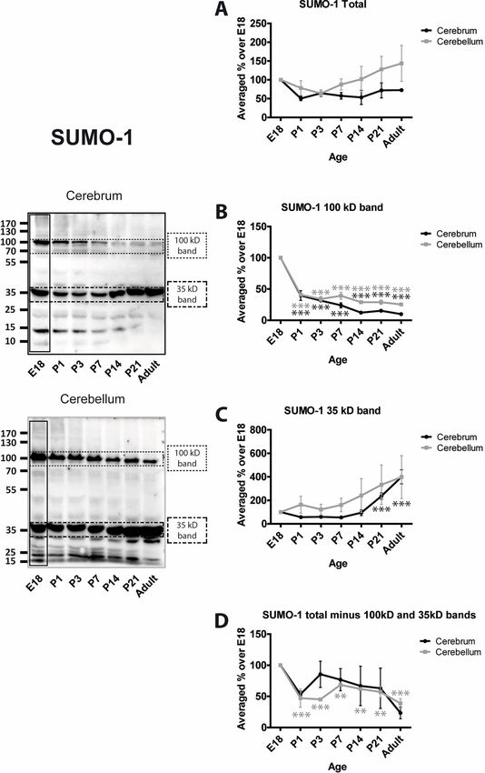
total amount of protein SUMOylation. In addition, two very prominent bands at 100 and 35
PLOS ONE | https://doi.org/10.1371/journal.pone.0212857 February 22, 2019 5 / 15SUMOylation during rat brain development
Fig 2. Developmental profiles of SUMO E3 ligases and the deSUMOylating enzyme SENP3. Representative blots
for PIAS1, PIAS3 and SENP3 and graphs showing the quantified levels of immunoreactivity from cerebrum and
cerebellum homogenates at the designated age points. Graphs show the levels of immunoreactivity at different ages
expressed as a percentage of the levels present in E18 brain. (n = 3, � = p�0.05, �� = p�0.01, ��� = p�0.001).
https://doi.org/10.1371/journal.pone.0212857.g002
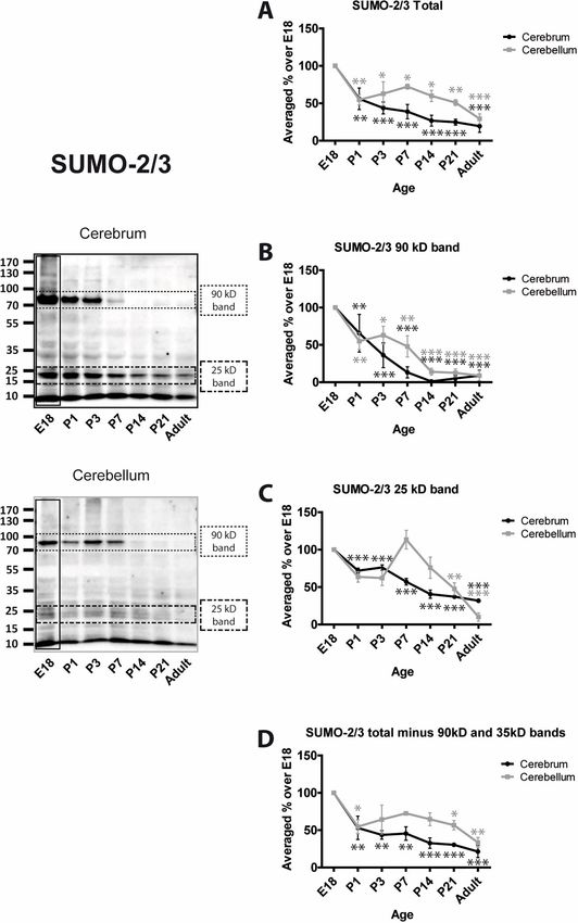
kDa in SUMO-1 blots, and 90 and 25 kDa in SUMO-2/3 blots were analysed separately and,
because these bands make up a large proportion of the entire SUMO signal, they were sub-
tracted from the ‘total’ SUMO values.
Levels of total SUMO-1 conjugation in cerebrum remain relatively stable during develop-
ment Levels of total SUMO-1 conjugation in cerebellum appear to increase slightly with age
but the differences were not significantly different from E18 (Fig 3A). As noted, however,
these values are very strongly influenced by the two major immunoreactive bands. SUMO-1
conjugation to the 100 kDa band in both cerebrum and cerebellum decreases rapidly around
birth and then remains relatively constant at a much lower level (Fig 3B). In contrast, SUMOy-
lation of the 35 kDa band remains stable and appears to increase with time (Fig 3C). Levels of
SUMO-1 conjugation to all other proteins (i.e the entire lane minus the 100 and 35 kDa
bands) appear variable. There is an initial decrease with age in the cerebrum (P1; 53.3% ±
5.3%) and remain relatively stable thereafter. Levels of total SUMO-1 conjugation in cerebel-
lum decrease and remain significantly lower at all subsequent age points tested (Fig 3D).
Using the 8A2 antibody the profile for total SUMO-2/3 conjugation decreases with age in
both cerebrum and cerebellum. There is an initial decrease around birth to 53.1% ± 17.7% and
54.7% ± 9.2%, respectively, at P1 and 21.4% ± 8.0% and 33.5% ± 6.7% in adult (Fig 4A).
SUMO-2/3 conjugation to the 90 kDa band displays a strong age-dependent decrease with
PLOS ONE | https://doi.org/10.1371/journal.pone.0212857 February 22, 2019 6 / 15SUMOylation during rat brain development
Fig 3. Levels of SUMO-1 conjugation to substrate proteins. Representative blots and quantification of the intensity
of the whole SUMO-1 lane, the indicated bands and the whole lane minus the indicated bands. Total SUMO-1
immunoreactivity was quantified by taking the whole lanes (vertical black rectangle). Since the 100 kDa and 35 kDa
bands are very intense and exhibit different profiles, they were subtracted from the total and the remaining signal
plotted (Total (-) bands). This represents the level of SUMOylation of the other proteins in the sample. Graphs show
the levels of immunoreactivity at different ages expressed as a percentage of the levels present in E18 brain. (n = 3, � =
p�0.05, �� = p�0.01, ��� = p�0.001).
https://doi.org/10.1371/journal.pone.0212857.g003
little conjugation evident in cerebrum at P14 or thereafter and similar, but less pronounced,
reductions in cerebellum (Fig 4B). SUMO-2/3-ylation of the 25 kDa band decreases steadily in
cerebrum with age but in cerebellum the levels are not reduced by P14. However, in both
brain regions SUMO-2/3 conjugation to this substrate is significantly decreased in P21 and
PLOS ONE | https://doi.org/10.1371/journal.pone.0212857 February 22, 2019 7 / 15SUMOylation during rat brain development
Fig 4. Levels of SUMO-2/3 conjugation to substrate proteins. Representative blots and quantification of the intensity
of the whole SUMO-2/3 lane, the indicated bands and the whole lane minus the indicated bands. Total SUMO-2/3
immunoreactivity was quantified by taking the whole lanes (vertical black rectangle). Since the 90 kDa and 25 kDa
bands are very intense and exhibit different profiles, they were subtracted from the total and the remaining signal
plotted (Total (-) bands). This represents the level of SUMOylation of the other proteins in the sample. Graphs show
the levels of immunoreactivity at different ages expressed as a percentage of the levels present in E18 brain. (n = 3, � =
p�0.05, �� = p�0.01, ��� = p�0.001).
https://doi.org/10.1371/journal.pone.0212857.g004
adult. (Fig 4C). The profile of SUMO-2/3 conjugation to all other proteins (i.e. the entire lane
minus the 90 and 25 kDa bands) almost exactly mirrors the total SUMO-2/3 (Fig 4D).
Interestingly, the differential trend between cerebrum and cerebellum of SUMO-2/3 conju-
gation resembles that of the SUMOylation machinery proteins, which was not the case for
SUMO-1.
PLOS ONE | https://doi.org/10.1371/journal.pone.0212857 February 22, 2019 8 / 15SUMOylation during rat brain development
Fig 5. SUMO machinery and substrate proteins profiles in subcellular fractions. Representative blots of nuclear
(N), synaptosomal (Sy) and cytosolic (Cy) fractions from adult rat cerebrum and cerebellum. 15μg of protein was
loaded in each lane. A) fraction markers, B) SUMO machinery proteins, C) Proteins SUMOylated by SUMO-1, D)
Proteins SUMOylated by SUMO-2/3.
https://doi.org/10.1371/journal.pone.0212857.g005
Intracellular localisation of the SUMOylation machinery and SUMO
substrates
To investigate the compartmentalisation of SUMO pathway proteins we prepared nuclear (N),
synaptosomal (Sy) and cytosolic (Cy) fractions from adult cerebrum and cerebellum (Fig 5).
The integrity of the fractions was determined by Western blotting for marker proteins. Lamin
B and PSD95 were selected as markers for nucleus and synaptosomes, respectively. Each of
these markers was enriched in their appropriate fraction (Fig 5A).
The E1 components Uba2 and Aos1 were present in all fractions but more abundant in
cytosol in both cerebrum and cerebellum (Fig 5B). Ubc9 showed marked differences in the rel-
ative abundance of its unmodified and higher molecular weight SUMOylated form (Fig 5B).
Interestingly, in cerebellum there was a reciprocal relationship between the bands with Ubc9
predominantly present in its modified form in the nucleus and synaptosomes but the unmodi-
fied form most prevalent in the cytosol. In cerebrum, the most abundant band was the modi-
fied form in synaptosmes. However, the differences in both forms of Ubc9 between nucleus
and cytosol, was not as marked as in cerebellum. PIAS1 and PIAS3 were abundant in the
nucleus but were not detected in the synaptosomal fraction. Both PIAS proteins showed higher
expression levels in cerebellum than in cerebrum (Fig 5B). Finally, SENP3 was present in the
nuclear and cytosolic fractions but not in the synaptosomes and its levels of expression was
highest in the cerebrum nuclear fraction (Fig 5B). Strikingly, in cerebellum, it was almost
PLOS ONE | https://doi.org/10.1371/journal.pone.0212857 February 22, 2019 9 / 15SUMOylation during rat brain development
equally portioned between nucleus and cytosol (Fig 5B). However, it is consistent with a role
for SENP3 in the cytosol and at mitochondria [26, 45].
Proteins conjugated to SUMO-1 were present in all fractions, albeit at lower levels in the
synaptosomes than in the other two fractions. The pattern of SUMO-1 substrates was similar
between cerebrum and cerebellum although less signal was detected in synaptosomes prepared
from cerebellum. Some differences were apparent between the cerebral and cerebellar nuclear
and cytosolic fractions between those two regions (Fig 5C). Interestingly, SUMO-2/3 was pres-
ent in all of the fractions and, in both regions, the synaptosomal fraction was labelled with
approximately the same intensity as the nuclear and cytosolic fractions indicating a particular
enrichment of SUMO-2/3 conjugation at synapses compared to SUMO-1. The most notable
feature for SUMO-2/3 is the very intense immunoreactive bands between 10 and 35 kDa in the
nuclear fraction. The lowest band likely corresponds to free SUMO2/3, which is much more
abundant in the nucleus than in any other fraction (Fig 5D).
Discussion
Taken together our results provide further evidence that SUMOylation machinery and SUMO
conjugation in general is most active prior to and around birth in brain. Thereafter levels
decrease with age. Our data broadly supports previous research by Loriol et al., 2012 [43] and
later by Hasegawa et al. 2014 [46], which also observed a decrease in SUMOylation and SUMO
pathway proteins throughout development. Moreover, we have extended these findings by
analysing additional proteins from the SUMOylation machinery, such as the E1 enzyme Uba2,
the deSUMOylating enzyme SENP3 and the E3 ligases PIAS 1 and PIAS3.
To validate our homogenates prior to investigating the SUMOylation machinery, we mea-
sured the developmental profiles of several key brain proteins. Importantly, our developmental
profiles match previous studies for GluA1 [38, 47] NR1 [39–41] and PSD95 [36, 37], thus vali-
dating our experimental methodology and analysis (S2 Fig).
In contrast to PSD95 and syntaxin1A, which increase with age, all of the SUMO machinery
proteins we investigated show a similar general pattern of decline during development in both
cerebrum and cerebellum. Furthermore, the developmental pattern for SUMO conjugation is
similar between cerebrum and cerebellum. However, it is important to take into account the
fact that we are measuring the net signal from a complex mix of substrates. For individual pro-
teins SUMOylation may increase, remain unchanged or decrease during development. This is
exemplified by analysis of two prominent SUMO1 substrate bands at 100 kDa and 35 kDa,
which display different conjugation profiles. It should also be noted that we are measuring lev-
els under basal conditions, and the animals were not subjected to, for example, metabolic
stress, which can radically alter protein SUMOylation.
While we were primarily interested in how the levels of SUMOylation and SUMO machin-
ery proteins change within either cerebrum or cerebellum during development, the design of
our experimental approach also allows us to make direct comparisons of the levels of proteins
of interest between these two regions. Since, for each time point tested, each set of cerebrum
and cerebellum samples were run against the same whole brain E18 homogenate, these E18
samples essentially act as an internal control allowing direct comparison of protein levels
between cerebrum and cerebellum. Therefore, we have plotted the expression levels of the pro-
teins of interest examined in cerebrum versus cerebellum in S3 and S4 Figs.
This analysis demonstrates that total SUMO1 conjugation levels in cerebrum and cerebel-
lum do not differ significantly over development; however, the 100kDa band is slightly but sig-
nificantly higher in cerebellum from P7 onwards. In contrast, total SUMO2/3 conjugation
PLOS ONE | https://doi.org/10.1371/journal.pone.0212857 February 22, 2019 10 / 15SUMOylation during rat brain development
exhibits a peak at P7 in cerebellum that does not occur in cerebrum, largely due to a dramatic
increase in abundance of the 25 kDa SUMO2/3-reactive band in cerebellum.
In terms of the SUMOylation machinery, we found that Uba2 and Aos1 expression in cere-
bellum rise above the levels in cerebrum from P3 and remain that way through adulthood.
Furthermore, Ubc9, PIAS1, PIAS3 and SENP3 all experience a variable peak, occurring from
P7 to P21, of significantly higher levels of expression in cerebellum than in cerebrum. Broadly
speaking, these results indicate that the expression of the SUMOylation machinery is signifi-
cantly higher in cerebellum than cerebrum throughout development. While these changes are
not reflected in gross changes in total SUMO1 or SUMO2/3 conjugation in cerebellum versus
cerebrum, this may be expected due to the higher levels of enzymes that both promote and
counteract protein SUMOylation in cerebellum. Nonetheless, these findings suggest that
SUMOylation may be more dynamically regulated in cerebellum than cerebrum during
development.
Interestingly, we also observed that while the profile of Ubc9 + SUMO-Ubc9 is not signifi-
cantly different through development in cerebrum or cerebellum, the level of Ubc9 increases
in cerebellum by P14 and P21, compared to E18. Strikingly, the ratio of SUMO-Ubc9 to Ubc9
increases more during development in cerebrum than in cerebellum, being approximately
3-fold higher in cerebrum by the adult stage (S4 Fig). Thus, during adulthood cerebrum con-
tains around 3 times more SUMOylated Ubc9 to Ubc9 than cerebellum. It has been previously
shown in the literature that SUMOylation of Ubc9 modifies its target discrimination [42, 48,
49]. Moreover, the classical function of Ubc9 as an E2 may convert to an E3 ligase type of func-
tion when SUMOylated [50]. However, the implications of this finding in brain development
and function are unknown, which could make this line of research an attractive target for
investigation in the future.
Previous studies have investigated age- and activity-dependent changes in SUMOylation
pathways in the brain [51–53]. Overall, their findings are consistent with our results, showing
relatively high levels of SUMOylated substrates and SUMO machinery proteins early in devel-
opment followed by an age-dependent decline. This reduction is consistent with the fact that
during neuronal outgrowth and synapse development there is intense transcriptional and
translational activity, processes in which SUMOylation is well-established to participate [54].
Similarly, SUMOylation is a key regulator of multiple protein interactions involved in synapse
formation, stabilisation and function [3, 22]. There is intense activity in these processes during
development, which decreases as neuronal architecture and connectivity develop and mature
networks become established. The differences between the cerebrum and cerebellum may
reflect the different influence of the SUMO pathway in cell type specificity, anatomical and
functional characteristics, rates of maturation and in protein targets.
Conclusion
In summary, in this work, our results describe a relatively consistent general developmental
profile for the regulation of SUMO machinery proteins and SUMOylated substrates, with
regional variations between cerebrum and cerebellum. This overall picture provides a clear
context for future studies focused on how specific SUMO machinery proteins or defined
SUMOylation substrate proteins in different CNS regions and cellular compartments are indi-
vidually regulated during development.
Supporting information
S1 Fig. Developmental profiles of common marker proteins. The cytoskeletal proteins β-III-
tubulin and β-actin, were monitored as general markers, commonly used as protein loading
PLOS ONE | https://doi.org/10.1371/journal.pone.0212857 February 22, 2019 11 / 15SUMOylation during rat brain development
control. Representative immunoblots of cerebrum and cerebellum. Graphs show the levels of
immunoreactivity at different ages expressed as a percentage of the levels present in E18 brain.
(n = 3, � = p�0.05, �� = p�0.01, ��� = p�0.001).
(TIFF)
S2 Fig. Developmental profiles of synaptic marker proteins. The postsynaptic protein
PSD95, the presynaptic protein syntaxin1A and the AMPA and NMDA neurotransmitter
receptor subunits GluA1 and NR1 (NMDAR1) were monitored as general markers of synapse
formation. Representative immunoblots of cerebrum and cerebellum. Graphs show the levels
of immunoreactivity at different ages expressed as a percentage of the levels present in E18
brain. (n = 3, � = p�0.05, �� = p�0.01, ��� = p�0.001).
(TIFF)
S3 Fig. Differences between expression profiles in cerebellum and cerebrum of common
and synaptic marker proteins. The cytoskeletal proteins β-III-tubulin and β-actin, and the
postsynaptic protein PSD95, the presynaptic protein syntaxin1A and the AMPA and NMDA
neurotransmitter receptor subunits GluA1 and NR1 (NMDAR1) were monitored in two brain
regions over time. Their immunoreactivity profiles expressed as a percentage of the levels pres-
ent in E18 brain in cerebellum vs. cerebrum were compared for each time point. (n = 3, � =
p�0.05, �� = p�0.01, ��� = p�0.001).
(TIFF)
S4 Fig. Differences between expression profiles in cerebellum and cerebrum of SUMOyla-
tion machinery proteins and SUMO1 and SUMO2/3 conjugated proteins. The SUMOyla-
tion machinery proteins Aos1, Uba2, Ubc9, PIAS1, PIAS3, SENP3 and SUMO1 and SUMO2/
3 conjugated proteins were monitored in two brain regions over time. Their immunoreactivity
profiles expressed as a percentage of the levels present in E18 brain in cerebellum vs. cerebrum
were compared for each time point. (n = 3, � = p�0.05, �� = p�0.01, ��� = p�0.001).
(TIFF)
S1 Table. Mean and standard error of the mean (SEM). This table includes the numerical
data of the time courses performed for different proteins in cerebrum and cerebellum.
(XLSX)
Acknowledgments
Fernando Josa Prado was an ESR fellow funded by the MSCA ITN “SyMBaD”. We are grateful
to the European Commission (MSCAs and European Research Council) for funding this
work.
Author Contributions
Conceptualization: Fernando Josa-Prado, Kevin A. Wilkinson.
Data curation: Fernando Josa-Prado.
Formal analysis: Fernando Josa-Prado.
Funding acquisition: Jeremy M. Henley.
Investigation: Fernando Josa-Prado.
Methodology: Fernando Josa-Prado, Philip Rubin, Kevin A. Wilkinson.
Project administration: Jeremy M. Henley.
PLOS ONE | https://doi.org/10.1371/journal.pone.0212857 February 22, 2019 12 / 15SUMOylation during rat brain development
Resources: Jia Luo.
Supervision: Jeremy M. Henley, Kevin A. Wilkinson.
Validation: Kevin A. Wilkinson.
Visualization: Fernando Josa-Prado.
Writing – original draft: Fernando Josa-Prado.
Writing – review & editing: Fernando Josa-Prado, Jeremy M. Henley, Kevin A. Wilkinson.
References
1. Hannoun Z, Greenhough S, Jaffray E, Hay RT, Hay DC. Post-translational modification by SUMO. Toxi-
cology. 2010; 278(3):288–93. Epub 2010/08/03. https://doi.org/10.1016/j.tox.2010.07.013 PMID:
20674646.
2. Praefcke GJ, Hofmann K, Dohmen RJ. SUMO playing tag with ubiquitin. Trends Biochem Sci. 2012; 37
(1):23–31. Epub 2011/10/25. S0968-0004(11)00152-6 [pii] https://doi.org/10.1016/j.tibs.2011.09.002
PMID: 22018829.
3. Wilkinson KA, Henley JM. Mechanisms, regulation and consequences of protein SUMOylation. Bio-
chem J. 2010; 428(2):133–45. Epub 2010/05/14. BJ20100158 [pii] https://doi.org/10.1042/BJ20100158
PMID: 20462400.
4. Gareau JR, Lima CD. The SUMO pathway: emerging mechanisms that shape specificity, conjugation
and recognition. Nat Rev Mol Cell Biol. 2011; 11(12):861–71. nrm3011 [pii] https://doi.org/10.1038/
nrm3011 PMID: 21102611.
5. Hendriks IA, Vertegaal AC. A comprehensive compilation of SUMO proteomics. Nat Rev Mol Cell Biol.
2016; 17(9):581–95. https://doi.org/10.1038/nrm.2016.81 PMID: 27435506.
6. Saitoh H, Hinchey J. Functional heterogeneity of small ubiquitin-related protein modifiers SUMO-1 ver-
sus SUMO-2/3. J Biol Chem. 2000; 275(9):6252–8. PMID: 10692421.
7. Cimarosti H, Ashikaga E, Jaafari N, Dearden L, Rubin P, Wilkinson KA, et al. Enhanced SUMOylation
and SENP-1 protein levels following oxygen and glucose deprivation in neurones. J Cereb Blood Flow
Metab. 2012; 32(1):17–22. Epub 2011/10/13. https://doi.org/10.1038/jcbfm.2011.146 PMID: 21989481;
PubMed Central PMCID: PMC3308141.
8. Cimarosti H, Lindberg C, Bomholt SF, Rønn LCB, Henley JM. Increased protein SUMOylation following
focal cerebral ischemia. Neuropharmacology. 2008;Available online 6 October 2007: https://doi.org/10.
1016/j.neuropharm.2007.09.010 PMID: 17991493
9. Lee YJ, Miyake S, Wakita H, McMullen DC, Azuma Y, Auh S, et al. Protein SUMOylation is massively
increased in hibernation torpor and is critical for the cytoprotection provided by ischemic preconditioning
and hypothermia in SHSY5Y cells. J Cereb Blood Flow Metab. 2007; 27(5):950–62. https://doi.org/10.
1038/sj.jcbfm.9600395 PMID: 16955077.
10. Lee YJ, Mou Y, Maric D, Klimanis D, Auh S, Hallenbeck JM. Elevated Global SUMOylation in Ubc9
Transgenic Mice Protects Their Brains against Focal Cerebral Ischemic Damage. PLoS One. 2011; 6
(10):e25852. Epub 2011/10/22. https://doi.org/10.1371/journal.pone.0025852 PMID: 22016779.
11. Yang W, Ma Q, Mackensen GB, Paschen W. Deep hypothermia markedly activates the small ubiquitin-
like modifier conjugation pathway; implications for the fate of cells exposed to transient deep hypother-
mic cardiopulmonary bypass. J Cereb Blood Flow Metab. 2009; 29(5):886–90. Epub 2009/02/26.
jcbfm200916 [pii] https://doi.org/10.1038/jcbfm.2009.16 PMID: 19240742.
12. Yang W, Sheng H, Homi HM, Warner DS, Paschen W. Cerebral ischemia/stroke and small ubiquitin-
like modifier (SUMO) conjugation—a new target for therapeutic intervention? J Neurochem. 2008; 106
(3):989–99. Epub 2008/04/16. JNC5404 [pii] https://doi.org/10.1111/j.1471-4159.2008.05404.x PMID:
18410505.
13. Yang W, Sheng H, Warner DS, Paschen W. Transient global cerebral ischemia induces a massive
increase in protein sumoylation. J Cereb Blood Flow Metab. 2007. https://doi.org/10.1038/sj.jcbfm.
9600523 PMID: 17565359.
14. Gong L, Li B, Millas S, Yeh ET. Molecular cloning and characterization of human AOS1 and UBA2, com-
ponents of the sentrin-activating enzyme complex. FEBS Lett. 1999; 448(1):185–9. PMID: 10217437.
15. Johnson ES. Protein modification by SUMO. Annu Rev Biochem. 2004; 73:355–82. https://doi.org/10.
1146/annurev.biochem.73.011303.074118 PMID: 15189146.
16. Hay RT. SUMO: a history of modification. Mol Cell. 2005; 18(1):1–12. https://doi.org/10.1016/j.molcel.
2005.03.012 PMID: 15808504.
PLOS ONE | https://doi.org/10.1371/journal.pone.0212857 February 22, 2019 13 / 15SUMOylation during rat brain development
17. Yeh ET. SUMOylation and De-SUMOylation: wrestling with life’s processes. J Biol Chem. 2009; 284
(13):8223–7. R800050200 [pii] https://doi.org/10.1074/jbc.R800050200 PMID: 19008217.
18. Mukhopadhyay D, Dasso M. Modification in reverse: the SUMO proteases. Trends Biochem Sci. 2007;
32(6):286–95. https://doi.org/10.1016/j.tibs.2007.05.002 PMID: 17499995.
19. Hickey CM, Wilson NR, Hochstrasser M. Function and regulation of SUMO proteases. Nat Rev Mol Cell
Biol. 2012; 13(12):755–66. Epub 2012/11/24. https://doi.org/10.1038/nrm3478 PMID: 23175280;
PubMed Central PMCID: PMC3668692.
20. Anderson DB, Wilkinson KA, Henley JM. Protein SUMOylation in neuropathological conditions. Drug
News Perspect. 2009; 22(5):255–65. Epub 2009/07/18. 1378636 [pii] https://doi.org/10.1358/dnp.2009.
22.5.1378636 PMID: 19609463.
21. Sarge KD, Park-Sarge OK. Sumoylation and human disease pathogenesis. Trends Biochem Sci. 2009;
34(4):200–5. Epub 2009/03/14. S0968-0004(09)00027-9 [pii] https://doi.org/10.1016/j.tibs.2009.01.004
PMID: 19282183.
22. Wilkinson KA, Nakamura Y, Henley JM. Targets and consequences of protein SUMOylation in neurons.
Brain Res Rev. 2010; 64(1):195–212. Epub 2010/04/13. S0165-0173(10)00043-3 [pii] https://doi.org/
10.1016/j.brainresrev.2010.04.002 PMID: 20382182.
23. Henley JM, Carmichael RE, Wilkinson KA. Extranuclear SUMOylation in Neurons. Trends Neurosci.
2018; 41(4):198–210. https://doi.org/10.1016/j.tins.2018.02.004 PMID: 29530319.
24. Datwyler AL, Lattig-Tunnemann G, Yang W, Paschen W, Lee SL, Dirnagl U, et al. SUMO2/3 conjuga-
tion is an endogenous neuroprotective mechanism. J Cereb Blood Flow Metab. 2011. Epub 2011/08/
25. jcbfm2011112 [pii] https://doi.org/10.1038/jcbfm.2011.112 PMID: 21863037.
25. Guo C, Hildick KL, Luo J, Dearden L, Wilkinson KA, Henley JM. SENP3-mediated DeSUMOylation of
dynamin-related protein 1 Promotes Cell Death Following Ischemia. EMBO Journal. 2013;in press.
26. Guo C, Wilkinson KA, Evans AJ, Rubin PP, Henley JM. SENP3-mediated deSUMOylation of Drp1 facil-
itates interaction with Mff to promote cell death. Sci Rep. 2017; 7:43811. https://doi.org/10.1038/
srep43811 PMID: 28262828; PubMed Central PMCID: PMCPMC5338345.
27. Martin S, Nishimune A, Mellor JR, Henley JM. SUMOylation regulates kainate-receptor-mediated syn-
aptic transmission. Nature. 2007; 447(7142):321–5. Epub 2007/05/09. nature05736 [pii] https://doi.org/
10.1038/nature05736 PMID: 17486098.
28. Chamberlain SE, Gonzalez-Gonzalez IM, Wilkinson KA, Konopacki FA, Kantamneni S, Henley JM,
et al. SUMOylation and phosphorylation of GluK2 regulate kainate receptor trafficking and synaptic
plasticity. Nat Neurosci. 2012; 15(6):845–52. nn.3089 [pii] https://doi.org/10.1038/nn.3089 PMID:
22522402.
29. Jaafari N, Konopacki FA, Owen TF, Kantamneni S, Rubin P, Craig TJ, et al. SUMOylation is required
for glycine-induced increases in AMPA receptor surface expression (ChemLTP) in hippocampal neu-
rons. PLoS One. 2013; 8(1):e52345. https://doi.org/10.1371/journal.pone.0052345 PMID: 23326329;
PubMed Central PMCID: PMCPMC3543417.
30. Craig TJ, Anderson D, Evans AJ, Girach F, Henley JM. SUMOylation of Syntaxin1A regulates presyn-
aptic endocytosis. Sci Rep. 2015; 5:17669. https://doi.org/10.1038/srep17669 PMID: 26635000;
PubMed Central PMCID: PMCPMC4669484.
31. Tang LT, Craig TJ, Henley JM. SUMOylation of synapsin Ia maintains synaptic vesicle availability and
is reduced in an autism mutation. Nat Commun. 2015; 6:7728. https://doi.org/10.1038/ncomms8728
PMID: 26173895; PubMed Central PMCID: PMCPMC4504226.
32. Craig TJ, Jaafari N, Petrovic MM, Jacobs SC, Rubin PP, Mellor JR, et al. Homeostatic Synaptic Scaling
Is Regulated by Protein SUMOylation. J Biol Chem. 2012; 287(27):22781–8. Epub 2012/05/15.
M112.356337 [pii] https://doi.org/10.1074/jbc.M112.356337 PMID: 22582390; PubMed Central
PMCID: PMC3391081.
33. Rocca DL, Wilkinson KA, Henley JM. SUMOylation of FOXP1 regulates transcriptional repression via
CtBP1 to drive dendritic morphogenesis. Sci Rep. 2017; 7(1):877. https://doi.org/10.1038/s41598-017-
00707-6 PMID: 28408745; PubMed Central PMCID: PMCPMC5429823.
34. Zhu B, Carmichael RE, Solabre Valois L, Wilkinson KA, Henley JM. The transcription factor MEF2A
plays a key role in the differentiation/maturation of rat neural stem cells into neurons. Biochem Biophys
Res Commun. 2018; 500(3):645–9. https://doi.org/10.1016/j.bbrc.2018.04.125 PMID: 29678571;
PubMed Central PMCID: PMCPMC5956278.
35. Gray EG, Whittaker VP. The isolation of nerve endings from brain: An electron-microscopic study of cell
fragments derived by homogenisation and centrifugation. J Anat. 1962; 96:79–88. PMID: 13901297
36. Buckby LE, Mummery R, Crompton MR, Beesley PW, Empson RM. Comparison of neuroplastin and
synaptic marker protein expression in acute and cultured organotypic hippocampal slices from rat.
PLOS ONE | https://doi.org/10.1371/journal.pone.0212857 February 22, 2019 14 / 15SUMOylation during rat brain development
Brain Res Dev Brain Res. 2004; 150(1):1–7. Epub 2004/05/06. https://doi.org/10.1016/j.devbrainres.
2004.01.006 PMID: 15126032.
37. Miyazaki H, Yamazaki M, Watanabe H, Maehama T, Yokozeki T, Kanaho Y. The small GTPase ADP-
ribosylation factor 6 negatively regulates dendritic spine formation. FEBS Lett. 2005; 579(30):6834–8.
Epub 2005/12/06. S0014-5793(05)01382-7 [pii] https://doi.org/10.1016/j.febslet.2005.11.022 PMID:
16325184.
38. Jourdi H, Iwakura Y, Narisawa-Saito M, Ibaraki K, Xiong H, Watanabe M, et al. Brain-derived neuro-
trophic factor signal enhances and maintains the expression of AMPA receptor-associated PDZ pro-
teins in developing cortical neurons. Dev Biol. 2003; 263(2):216–30. Epub 2003/11/05.
S0012160603004445 [pii]. PMID: 14597197.
39. Chen L, Cooper NG, Mower GD. Developmental changes in the expression of NMDA receptor subunits
(NR1, NR2A, NR2B) in the cat visual cortex and the effects of dark rearing. Brain Res Mol Brain Res.
2000; 78(1–2):196–200. Epub 2000/07/13. S0169328X00000760 [pii]. PMID: 10891601.
40. Curras MC, Dao J. Developmental plasticity of NR1 and NR2B subunit expression in the supraoptic
nucleus of the rat hypothalamus. Brain Res Dev Brain Res. 1998; 109(1):1–12. Epub 1998/08/26.
S0165380698000601 [pii]. PMID: 9706387.
41. Ling W, Chang L, Song Y, Lu T, Jiang Y, Li Y, et al. Immunolocalization of NR1, NR2A, and PSD-95 in
rat hippocampal subregions during postnatal development. Acta Histochem. 2012; 114(3):285–95.
Epub 2011/07/02. S0065-1281(11)00095-X [pii] https://doi.org/10.1016/j.acthis.2011.06.005 PMID:
21719075.
42. Knipscheer P, Flotho A, Klug H, Olsen JV, van Dijk WJ, Fish A, et al. Ubc9 sumoylation regulates
SUMO target discrimination. Mol Cell. 2008; 31(3):371–82. Epub 2008/08/12. S1097-2765(08)00424-3
[pii] https://doi.org/10.1016/j.molcel.2008.05.022 PMID: 18691969.
43. Loriol C, Parisot J, Poupon G, Gwizdek C, Martin S. Developmental regulation and spatiotemporal
redistribution of the sumoylation machinery in the rat central nervous system. PLoS One. 2012; 7(3):
e33757. Epub 2012/03/23. https://doi.org/10.1371/journal.pone.0033757 PMID: 22438991.
44. Schmidt D, Muller S. Members of the PIAS family act as SUMO ligases for c-Jun and p53 and repress
p53 activity. Proc Natl Acad Sci U S A. 2002; 99(5):2872–7. https://doi.org/10.1073/pnas.052559499
PMID: 11867732.
45. Guo C, Hildick KL, Luo J, Dearden L, Wilkinson KA, Henley JM. SENP3-mediated deSUMOylation of
dynamin-related protein 1 promotes cell death following ischaemia. EMBO J. 2013; 32(11):1514–28.
https://doi.org/10.1038/emboj.2013.65 PMID: 23524851; PubMed Central PMCID: PMCPMC3671254.
46. Hasegawa Y, Yoshida D, Nakamura Y, Sakakibara S. Spatiotemporal distribution of SUMOylation com-
ponents during mouse brain development. J Comp Neurol. 2014; 522(13):3020–36. https://doi.org/10.
1002/cne.23563 PMID: 24639124.
47. Martin LJ, Furuta A, Blackstone CD. AMPA receptor protein in developing rat brain: glutamate receptor-
1 expression and localization change at regional, cellular, and subcellular levels with maturation. Neuro-
science. 1998; 83(3):917–28. Epub 1998/03/04. S0306-4522(97)00411-9 [pii]. PMID: 9483574.
48. Ho CW, Chen HT, Hwang J. UBC9 autosumoylation negatively regulates sumoylation of septins in Sac-
charomyces cerevisiae. J Biol Chem. 2011; 286(24):21826–34. https://doi.org/10.1074/jbc.M111.
234914 PMID: 21518767; PubMed Central PMCID: PMCPMC3122237.
49. McManus FP, Bourdeau V, Acevedo M, Lopes-Paciencia S, Mignacca L, Lamoliatte F, et al. Quantita-
tive SUMO proteomics reveals the modulation of several PML nuclear body associated proteins and an
anti-senescence function of UBC9. Sci Rep. 2018; 8(1):7754. https://doi.org/10.1038/s41598-018-
25150-z PMID: 29773808; PubMed Central PMCID: PMCPMC5958138.
50. Klug H, Xaver M, Chaugule VK, Koidl S, Mittler G, Klein F, et al. Ubc9 sumoylation controls SUMO
chain formation and meiotic synapsis in Saccharomyces cerevisiae. Mol Cell. 2013; 50(5):625–36.
https://doi.org/10.1016/j.molcel.2013.03.027 PMID: 23644018.
51. Hashiyama K, Shigenobu S, Kobayashi S. Expression of genes involved in sumoylation in the Drosoph-
ila germline. Gene Expr Patterns. 2009; 9(1):50–3. Epub 2008/08/30. S1567-133X(08)00089-6 [pii]
https://doi.org/10.1016/j.gep.2008.08.001 PMID: 18755298.
52. La Salle S, Sun F, Zhang XD, Matunis MJ, Handel MA. Developmental control of sumoylation pathway
proteins in mouse male germ cells. Dev Biol. 2008; 321(1):227–37. Epub 2008/07/08. S0012-1606(08)
01010-5 [pii] https://doi.org/10.1016/j.ydbio.2008.06.020 PMID: 18602382.
53. Lu H, Liu B, You S, Xue Q, Zhang F, Cheng J, et al. The activity-dependent stimuli increase SUMO
modification in SHSY5Y cells. Biochem Biophys Res Commun. 2009; 390(3):872–6. Epub 2009/10/21.
S0006-291X(09)02043-9 [pii] https://doi.org/10.1016/j.bbrc.2009.10.065 PMID: 19840774.
54. Cubenas-Potts C, Matunis MJ. SUMO: A Multifaceted Modifier of Chromatin Structure and Function.
Dev Cell. 2012; 24(1):1–12. Epub 2013/01/19. S1534-5807(12)00540-0 [pii] https://doi.org/10.1016/j.
devcel.2012.11.020 PMID: 23328396.
PLOS ONE | https://doi.org/10.1371/journal.pone.0212857 February 22, 2019 15 / 15You can also read