Exploring the relationship of Homalosilpha and Mimosilpha (Blattodea, Blattidae, Blattinae) from a morphological and molecular perspective ...
←
→
Page content transcription
If your browser does not render page correctly, please read the page content below
Exploring the relationship of Homalosilpha
                                    and Mimosilpha (Blattodea, Blattidae,
                                    Blattinae) from a morphological and
                                    molecular perspective, including a
                                    description of four new species
                                    Shuran Liao, Yishu Wang, Duting Jin, Rong Chen, Zongqing Wang and
                                    Yanli Che
                                    College of Plant Protection, Southwest University, Chongqing, Beibei, China

                                        ABSTRACT
                                        This study utilized six genes (12S, 16S, 18S, 28S, COII and H3) from a total of 40 samples
                                        to construct maximum likelihood (ML) and Bayesian inference (BI) phylogenetic
                                        trees in order to infer the relationships between the morphologically similar genera
                                        Homalosilpha Stål, 1874 and Mimosilpha Bey-Bienko, 1957. The phylogenetic analysis
                                        showed the two genera have a close relationship and were recovered as sister groups
                                        based on ML and BI analyses. Four new species are described among these samples, i.e.,
                                        Homalosilpha obtusangula sp. nov., Homalosilpha recta sp. nov., Homalosilpha alba sp.
                                        nov. and Homalosilpha clavellata sp. nov. based on morphological and COI data. A key
                                        to the worldwide Homalosilpha is provided.

                                    Subjects Biodiversity, Entomology, Molecular Biology, Taxonomy, Zoology
                                    Keywords Bayesian inference, Habitat, Key, Maximum likelihood, Sister group

                                    INTRODUCTION
Submitted 26 June 2020              Genus Homalosilpha Stål, 1874 is remarkable in the family of Blattidae for its flat pronotum
Accepted 30 November 2020
Published 13 January 2021
                                    decorated with black or white markings (Princis, 1966a; Roth, 1999). Stål (1874) established
                                    Homalosilpha with Periplaneta ustulata Burmeister, 1838 as the type species. Then Kirby
Corresponding author
Yanli Che, shirleyche2000@126.com   (1904) transferred three Periplaneta Burmeister, 1838 species to Homalosilpha. Homalosilpha
Academic editor                     can be easily distinguished from Periplaneta by the discoidal pronotum whose surface
Tony Robillard                      is scattered with various spots, sides are not deflexed and with greatest width at the
Additional Information and          middle (Shelford, 1910). Subsequently, two species were reported from Uganda and
Declarations can be found on        China by Shelford (1908), Shelford (1910). Later, Princis (1966a) described two species
page 22
                                    from Congo and Indonesia and provided a key for eight Homalosilpha species. In 1969,
DOI 10.7717/peerj.10618
                                    Bey-Bienko described four species, of which three species were from China. Kumar
    Copyright                       (1975) synonymized H. vicina Brunner von Wattenwyl, 1865 with H. cruralis Shelford,
2021 Liao et al.
                                    1908 because the diagnostic characters used, the color of the tibiae and the anterior
Distributed under                   and posterior margin of pronotum, were variable and should be treated as intraspecific
Creative Commons CC-BY 4.0
                                    variation. After the examination of specimens from Malaysia and Indonesia by Roth (1999),
OPEN ACCESS                         Homalosilpha quadrimaculata was described on the basis of the distinct orange spots on

                                    How to cite this article Liao S, Wang Y, Jin D, Chen R, Wang Z, Che Y. 2021. Exploring the relationship of Homalosilpha and Mimosilpha
                                    (Blattodea, Blattidae, Blattinae) from a morphological and molecular perspective, including a description of four new species. PeerJ 9:e10618
                                    http://doi.org/10.7717/peerj.10618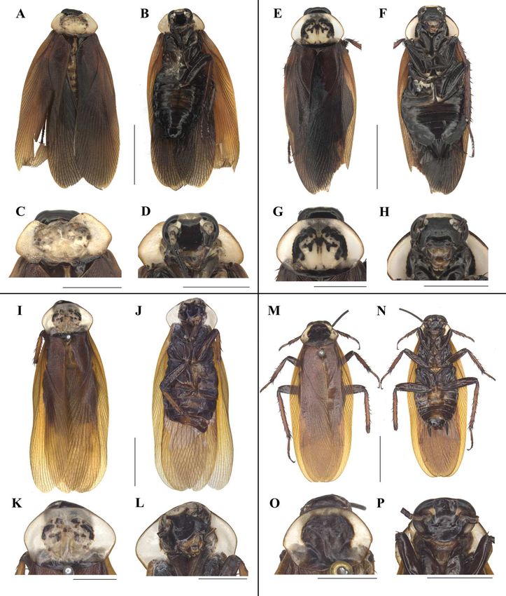
the pronotum. Up to now, a total of twelve species of Homalosilpha had been reported
                                    worldwide (Beccaloni, 2014), of which, H. arcifera Bey-Bienko, 1969, H. gaudens Shelford,
                                    1910, H. kryzhanovskii Bey-Bienko, 1969, H. ustulata (Burmeister, 1838), and H. valida
                                    Bey-Bienko, 1969 are distributed in China. Without professional taxonomic knowledge
                                    about Blattodea, it is challenging to identify the Homalosilpha species owing to their high
                                    similarity in appearance (Liao SR & Che YL, pers. obs., 2018–2019). Therefore, simple and
                                    accurate methods are needed to help the identification of Homalosilpha species.
                                        Genus Mimosilpha was established with M. disticha as type species from Yunnan, China.
                                    It is strongly similar to Homalosilpha in appearance (Bey-Bienko, 1957). The main difference
                                    between Mimosilpha and Homalosilpha is that there are three rows of spines along the edge
                                    of the hind tibiae of Homalosilpha, while Mimosilpha only has two rows of spines. Only
                                    one Mimosilpha species has been recorded in the world (Beccaloni, 2014).
                                        DNA barcodes proved to be an effective tool to aid the identification of the similar
                                    species and even resolve the problem of sexual dimorphism in cockroaches (Evangelista,
                                    Buss & Ware, 2013; Che et al., 2017; Liao, Wang & Che, 2019). Multi-gene combination
                                    phylogenetic trees are increasingly applied in cockroach systematics to explore the problem
                                    of paraphyly (Inward, Beccaloni & Eggleton, 2007; Ware et al., 2008; Legendre et al., 2015),
                                    establish new taxa (Evangelista et al., 2019), infer possible sister group (Djernæs, Klass
                                    & Eggleton, 2015; Wang et al., 2017), and then revise the taxonomy (Djernæs, 2018).
                                    These studies have all been demonstrated to be informative and successful in revealing
                                    relationships among different groups via multi-gene analysis. Mimosilpha and Homalosilpha
                                    are very close in morphology with a confused taxonomy, so it is urgent to sequence six
                                    genes to infer the relationship of these two genera.
                                        In this study, 13 COI sequences of Homalosilpha and Mimosilpha species were obtained
                                    in order to help distinguish species when combined with morphological data. We also
                                    sequenced three mitochondrial genes (12S, 16S and COII ) and three nuclear genes (18S,
                                    28S and H3) to explore the phylogenetic relationship of Homalosilpha and Mimosilpha.
                                    Moreover, we illustrate four new Homalosilpha species from China based on morphological
                                    characters and DNA barcoding.

                                    MATERIALS AND METHODS
                                    Morphological study
                                    Morphological terminology used in this paper mainly follows McKittrick (1964), Roth
                                    (2003) and Li et al. (2018).
                                       The maculae on the pronotum provided effective information for the identification of
                                    Homalosilpha species, which could be grouped into three types (Shelford, 1910; Princis,
                                    1966a; Bey-Bienko, 1969; Roth, 1999): (1) white spots scattered on the black pronotum,
                                    represented by H. decorata and H. quadrimaculata, (2) one large central dark spot on the
                                    pale brown pronotum, represented by H. hanni, H. nigricans and H. gaudens, and (3) the
                                    symmetrical multiple spots and stripes on the disk of the pale brown pronotum, represented
                                    by the remaining Homalosilpha species. The third type is more complex than the former
                                    two, so we herein address the specific part of the maculae for description as shown in Fig. 1.

Liao et al. (2021), PeerJ, DOI 10.7717/peerj.10618                                                                           2/27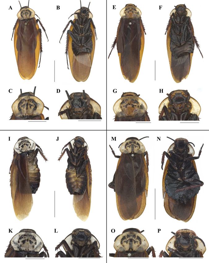
Figure 1 The pronotum of H. obtusangula sp. nov. (A) Anterior pattern. (B) Small spot. (C) Lateral
                                     pattern. (D) Central pattern. (E) Vertical pattern. (F) Posterior band. Scale bars = 5 mm.
                                                                                                       Full-size DOI: 10.7717/peerj.10618/fig-1

                                       All specimens were measured by vernier caliper for the body length from the anterior
                                    to the posterior, for the body length including tegmina from the anterior to the tip
                                    of tegmina, and pronotum length × width at the longest and the widest part. Genital
                                    segments of the examined specimens were macerated and photographs (Figs. 1–9) of
                                    the specimens were taken as previously described in Liao, Wang & Che (2019). The type
                                    materials are deposited in the Institute of Entomology, College of Plant Protection,
                                    Southwest University, Chongqing, China (SWU).

                                    DNA extraction, PCR, and sequencing
                                    We sampled seven genes of 13 species (Tables S1 and S2) from Homalosilpha in this study:
                                    mitochondrial 12S, 16S, cytochrome c oxidase subunit I (COI ) and subunit II (COII ),
                                    and nuclear 18S, 28S, and histone H3. Total DNA was extracted from the leg tissue of
                                    samples according to the Hipure Tissue DNA Mini Kit (Tsingke Biological Technology,
                                    Beijing, China). All fragments were amplified using PCR; primers for amplifications are
                                    given in Table 1. Reactions were carried out in volumes of 25 µL, containing 22 µL of 1×1
                                    T3 supper mix (Tsingke Biological Technology, Beijing, China), 1 µL of each primer and
                                    1 µL of DNA template, except for COII containing 12.5 µL T2 mix (Tsingke Biological
                                    Technology, Beijing, China), 8.5 µL of ultrapure water, 1 µL of each primer and 2 µL of
                                    DNA template. The amplification conditions were: initial denaturation at 98 ◦ C for 2 min,
                                    followed by 35 cycles for 10 s at 98 ◦ C, 10 s at 43−55 ◦ C, and 15 s at 72 ◦ C, with a final
                                    extension of 2 min at 72 ◦ C; however for COII: initial denaturation at 94 ◦ C for 5 min,

Liao et al. (2021), PeerJ, DOI 10.7717/peerj.10618                                                                                         3/27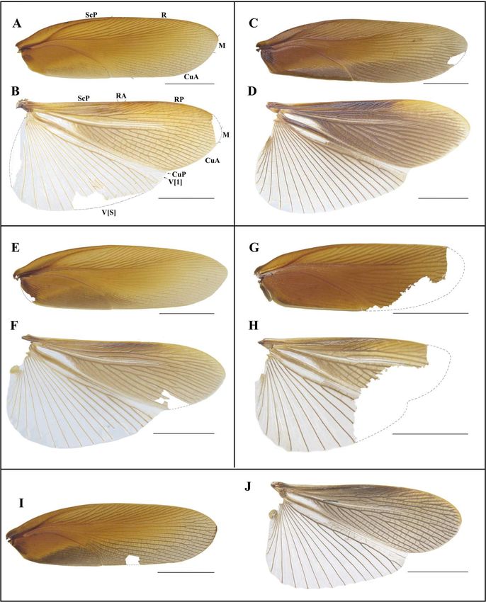
Figure 2 Maximum likelihood (ML) tree of the cockroaches based on COI. ML tree derived from COI
                                     gene analysis following GTRGAMMA model with 1,000 bootstrap replicates.
                                                                                              Full-size DOI: 10.7717/peerj.10618/fig-2

                                    followed by 35 cycles for 45 s at 94 ◦ C, 45 s at 50 ◦ C, and 45 s at 72 ◦ C, with a final extension
                                    of 10 min at 72 ◦ C. All sequences were deposited in GenBank (accession numbers in Tables
                                    S1 and S2).

                                    Sequence processing and phylogenetic analyses
                                    In this study, a total of 14 COI sequences, whose lengths were 658 bp, were combined
                                    with one Protagonista, two Periplaneta, one Rhabdoblatta, one Brephallus and one mantid
                                    sequence to infer species delimitation analysis for Homalosilpha and Mimosilpha (Table
                                    S1). Intraspecific and interspecific genetic divergence values are quantified based on the
                                    Kimura 2-parameter (K2P) distance model (Kimura, 1980), using MEGA 7 (Kumar, Stecher
                                    & Tamura, 2016).
                                       To infer the relationship of Homalosilpha and Mimosilpha, we included sequence
                                    data from 22 Blattidae taxa (ingroup including seven Homalosilpha and one Mimosilpha
                                    species) and 12 outgroup taxa (Table S2). These analyses were performed based on six
                                    genes (12S, 16S, 18S, 28S, COII and H3). These data was aligned by online Mafft 7
                                    (https://mafft.cbrc.jp/alignment/server/), and the methods were the same as Wang et al.
                                    (2017). Specifically, the Q-INS-i algorithm was selected for non-coding protein genes (12S,

Liao et al. (2021), PeerJ, DOI 10.7717/peerj.10618                                                                                 4/27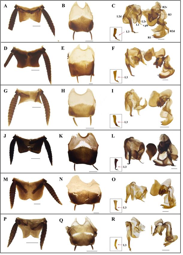
16S, 18S, 28S), the G-INS-i algorithm was selected for coding protein genes (COII, H3) and
                                    used with other parameters at their default values. Alignments of sequences were inspected
                                    visually and manually adjusted in MEGA 7.0. Poorly aligned characters were removed but
                                    these were limited. The remaining bases are as following: 424nt for 12S, 447nt for 16S,
                                    1814nt for 18S, 596nt for 28S, 665nt for COII, 328nt for H3.
                                       The molecular data set was divided into eight partitions (partitioned by gene: 12S,
                                    16S, 18S, 28S, COII _pos1, COII _pos2, H3_pos1, and H3 _pos2). We excluded the
                                    third codon positions of the protein-coding genes (COII and H3) because of the high
                                    level of mutational saturation. The third codon position (pos3) (ISS = 0.977) was much
                                    more saturated than the first and second codon position (pos12) (ISS = 0.493) after
                                    using Xia’s method implemented in DAMBE 5.0 (Xia, 2013). Phylogenetic analyses were
                                    constructed using maximum likelihood (ML) and Bayesian inference methods. ML
                                    analyses were performed using RaxML 7.7.1 (Stamatakis, Hoover & Rougemont, 2008), and
                                    BI analyses were performed using MrBayes 3.2 (Ronquist et al., 2012). ML analyses was
                                    performed using RAxML v.7.7.1 (Stamatakis, Hoover & Rougemont, 2008). We used the
                                    GTRGAMMA model for the combined datasets with 1,000 bootstrap replicates. For BI
                                    analyses, PartitionFinder v.1.1.1 (Lanfear et al., 2012) was utilized to choose models and
                                    model selection was based on BIC. For eight partitions, the best-fitting substitution models
                                    are as follows: GTR+I+G: 12S, 16S; TIMef+I+G: 18S, H3_pos1, H3_pos 2; TrN+I+G: 28S,
                                    COII _pos1, COII _pos2. Two independent sets of Markov chains were run, each with one
                                    cold and three heated chains for 5,000,000 generations, and every 1,000th generation was
                                    sampled. Convergence was inferred when a standard deviation of split frequencies < 0.01
                                    was completed. The ESS values are all more than 200.

                                    RESULTS
                                    Species delimitation based on COI and morphological data
                                    In this study, we acquired 14 COI sequences representing seven Homalosilpha and 1
                                    Mimosilpha species. All of the new sequences have been deposited in GenBank with
                                    accession numbers MW201581–MW201594 (Table S1). The COI sequences we acquired
                                    had a relatively high AT content (67.2%), with an average nucleotide composition of
                                    A = 31%, T = 36.2%, C = 16.5%, and G = 16.3%. Sequence analysis revealed that 109
                                    (16.57%) sites were variable, of which 79 (12%) sites were parsimoniously informative.
                                    ML analysis revealed that clades from the same species, constituted monophyletic groups
                                    with high support values (Fig. 2). In particular, the male and female of H. clavellata sp.
                                    nov., which have different markings on the pronotum, could be matched by COI data.
                                       We observed the lowest and largest K2P interspecies genetic distance (Table 2) among
                                    these species. The lowest distance 0.031 was between Homalosilpha sp. and H. clavellata
                                    sp. nov., the largest distance 0.084 between H. nigricans and H. arcifera, H. arcifera and H.
                                    kryzhanovskii, as well as M. disticha and H. kryzhanovskii. Morphologically, although having
                                    a low genetic distance, Homalosilpha sp. and H. clavellata sp. nov. show distinct differences
                                    in the maculae on the pronotum, the band in the interocular space (H. clavellata sp. nov.:
                                    yellow and straight transverse band, Homalosilpha sp.: yellow and tortuous transverse

Liao et al. (2021), PeerJ, DOI 10.7717/peerj.10618                                                                          5/27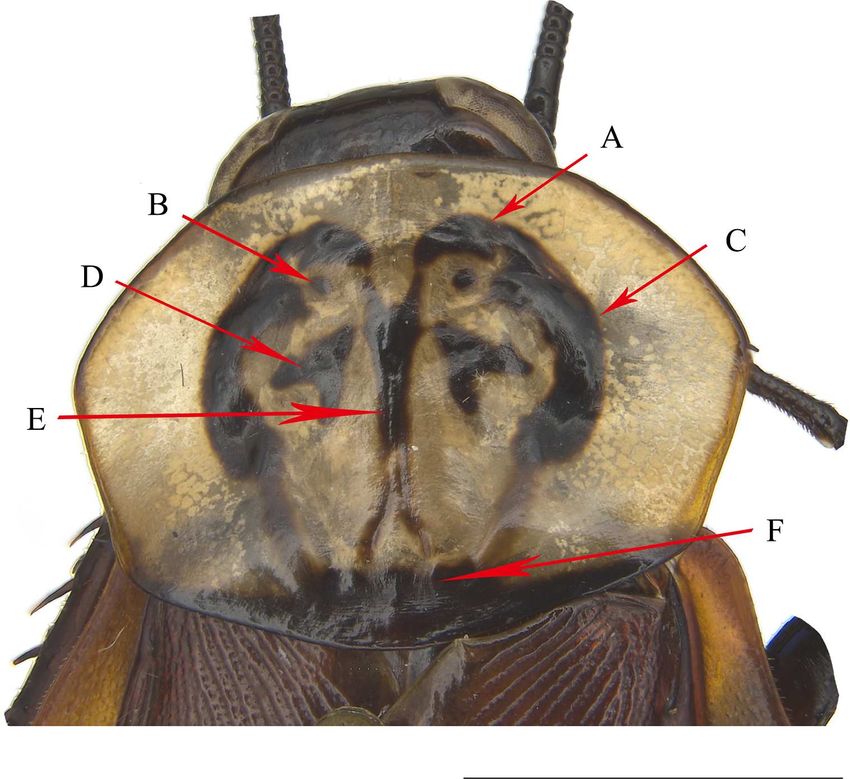
Figure 3 Maximum likelihood (ML) tree of the cockroaches based on six genes. ML tree derived from
                                     analysis of combined data 12S, 16S, 18S, 28S, COII, and H3 genes. Branch labels are support for our anal-
                                     yses in the following order: bootstrap supports of the maximum-likelihood tree, Bayesian posterior prob-
                                     abilities of the Bayesian tree; asterisks (*) indicate 100% support for a given analysis. The topology shown
                                     was totally similar to that derived from BI analysis.
                                                                                                       Full-size DOI: 10.7717/peerj.10618/fig-3

                                    band) and even in the female genitalia (H. clavellata: anterior of basivalvula with spots, the
                                    shape of first valvule uniform, Homalosilpha sp.: surface of basivalvula with fold, the end
                                    of the first valvula swollen). Finally, a total of 8 Homalosilpha and Mimosilpha species were
                                    recovered after combining the results of molecular data with morphological data, of which
                                    4 species are new to science: H. obtusangula sp. nov., H. recta sp. nov., H. alba sp. nov.,
                                    and H. clavellata sp. nov. More details about these new Homalosilpha species are provided
                                    below.

                                    Relationship of Homalosilpha and Mimosilpha inferred from two
                                    phylogenetic analyses
                                    We acquired 12 12S, 12 16S, 11 18S, 12 28S, 10 COII, 12 H3 sequences with accession
                                    numbers MW218556–MW218578, MW219581–MW218604 and MW201809–MW201830
                                    (Table S2). For the concatenate dataset (12S, 16S, 18S, 28S, COII, H3), our likelihood
                                    and Bayesian phylogenetic analyses yielded totally identical topologies with generally

Liao et al. (2021), PeerJ, DOI 10.7717/peerj.10618                                                                                           6/27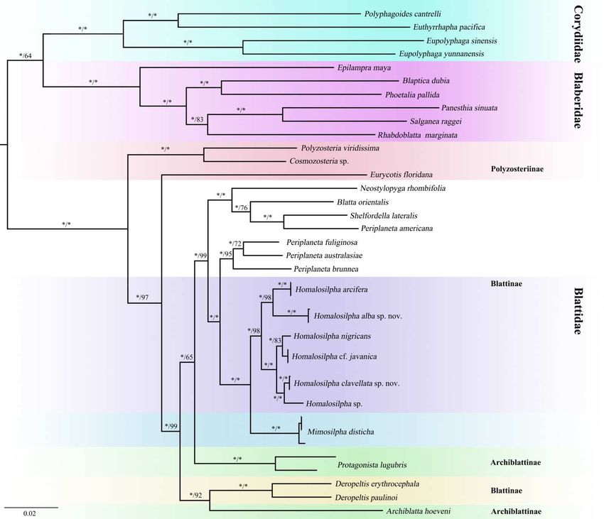
Figure 4 Photographs of middle and hind tibiae of Homalosilpha and Mimosilpha species. (A–B)
                                     H. nigricans Princis, 1966a. (C–D) H. alba sp. nov. (E–F) M. disticha. Middle and hind tibiae are seen on
                                     dorsal view. Scale bars = 5 mm.
                                                                                                      Full-size DOI: 10.7717/peerj.10618/fig-4

                                    high node support (Fig. 3 and Fig. S1). All genera of Blattidae clustered together and
                                    formed a highly supported monophyletic group (MLB = 100, BPP = 100). Five recognized
                                    major lineages of Blattidae from ML and BI inferences were recovered, represented by
                                    members of Polyzosteriinae, Archiblattinae and Blattinae. According to our inferred trees,
                                    Polyzosteriinae, Archiblattinae and Blattinae were paraphyletic with high support values.
                                       The topology derived from ML and BI analyses shows that all Homalosilpha members
                                    cluster together and are recovered as the sister group of Mimosilpha (MLB = 100, BPP
                                    = 100). H. clavellata sp. nov. and Homalosilpha sp. were recovered as sister groups with
                                    strong support values, which is consistent with the inference based on COI data. And the
                                    result is same as the trees of seven genes (12S, 16S, 18S, 28S, COI, COII, H3) (Figs. S2 and
                                    S3).
                                       The electronic version of this article in Portable Document Format (PDF) will represent
                                    a published work according to the International Commission on Zoological Nomenclature
                                    (ICZN), and hence the new names contained in the electronic version are effectively
                                    published under that Code from the electronic edition alone. This published work and the
                                    nomenclatural acts it contains have been registered in ZooBank, the online registration
                                    system for the ICZN. The ZooBank LSIDs (Life Science Identifiers) can be resolved and
                                    the associated information viewed through any standard web browser by appending the
                                    LSID to the prefix http://zoobank.org/. The LSID for this publication is: [74F6BF88-9FFD-
                                    40DB-9C8B-D7650491F0EE]. The online version of this work is archived and available
                                    from the following digital repositories: PeerJ, PubMed Central and CLOCKSS.

                                    Taxonomy

Liao et al. (2021), PeerJ, DOI 10.7717/peerj.10618                                                                                        7/27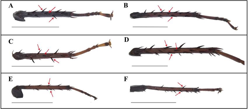
Homalosilpha Stål, 1874
                                    Homalosilpha Stål, 1874: 13. Type species: Homalosilpha ustulata Burmeister, 1838. Kirby,
                                    1904: 143; Shelford, 1910: 19; Hanitsch, 1915: 112; Princis, 1966a: 49; Princis, 1966b: 457;
                                    Roth, 1999: 172.
                                    Diagnosis. Body medium to large, flat, smooth, generally brownish to blackish. Antennae
                                    long, slender. Pronotum flat, decorated with maculae, sides not deflexed, greatest width
                                    near the middle. Tegmina and wings extending considerably beyond the apex of the
                                    abdominal tip. Legs long, front femora Type A2 , the outer edge of the middle and hind
                                    tibiae each with three rows of spines (Fig. 4); tarsal claws simple, symmetrical. Supra-anal
                                    plate broad. Cerci long. Subgenital asymmetrical. Male genitalia: left and right phallomere
                                    all consisting of three parts. L1 fold sclerite; margin of L2d with serration; posterior of
                                    L2v produced with a spiny projection to the end; L3 unciform, the terminal bifurcate. R1
                                    foot-shaped, left margin with two spines; terminal of R2d projected, R2v folded sclerite;
                                    R3 broad and folded sclerite. In the nymph, the body shows zebra-like stripes.

                                    Checklist of Homalosilpha worldwide

                                      Homalosilpha alba sp. nov. China
                                      Homalosilpha arcifera Bey-Bienko, 1969 China, Vietnam
                                      Homalosilpha contraria (Walker, 1868) Philippines
                                      Homalosilpha decorata (Serville, 1838) Indonesia (Sumatra), Indonesia (Java Island),
                                      Borneo Island
                                      Homalosilpha gaudens Shelford, 1910 China, Vietnam
                                      Homalosilpha haani Princis, 1966a Indonesia
                                      Homalosilpha clavellata sp. nov. China
                                      Homalosilpha javanica Bey-Bienko, 1969 Indonesia
                                      Homalosilpha kryzhanovskii Bey-Bienko, 1969 China
                                      Homalosilpha nigricans Princis, 1966 Congo Democratic Republic
                                      Homalosilpha obtusangula sp. nov. China
                                      Homalosilpha recta sp. nov. China
                                      Homalosilpha quadrimaculata Roth, 1999 Malaysia
                                      Homalosilpha ustulata (Burmeister, 1838) China, Myanmar, Malaysia,
                                      Indonesia, Philippines
                                      Homalosilpha valida Bey-Bienko, 1969 China
                                      Homalosilpha vicina (Brunner von Wattenwyl, 1865) Guinea, Cameroon, Uganda
                                      Homalosilpha sp. China

                                    Key to Homalosilpha species worldwide
                                      1. Pronotum mostly black . . . 2
                                      Pronotum mostly white. . . 3
                                      2. Pronotum with two vertical white stripes near the lateral margin . . . H. decorata
                                      Pronotum with four white spots respectively at the anterior margin and middle. . . H.
                                      quadrimaculata
                                      3. Pronotum with a large black spot at center . . . 4

Liao et al. (2021), PeerJ, DOI 10.7717/peerj.10618                                                                         8/27
Pronotum with multiple black spots or stripes . . . 6
4. The large spot connecting both the front and hind margins . . . H. haani
The large spot only connected with the hind margin . . . 5
5. Pronotum broadest near the hind margin, trapeziform . . . H. nigricans
Pronotum broadest near the middle . . . H. gaudens
6. Face pale yellow . . . H. arcifera
Face black . . . 7
7. Vertex yellow to yellowish brown . . . 8
Vertex black . . . 10
8. Abdomen with yellow spots . . . H. valida
Abdomen brownish black to dark . . . 9
9. The hind margin of supra-anal plate slightly concave, lateral angles blunt . . . H. recta
sp. nov.
The hind margin of supra-anal plate round, lateral angles sharp . . . H. alba sp. nov.
10. Interocular space with a yellow straight band . . . 11
Interocular space with a yellow band . . . H. ustulata
11. The lateral pattern and vertical pattern of pronotum connected with posterior band,
spots on both sides of the lateral pattern . . . H. vicina
The middle of pronotum with spots or spots and stripes, the both sides of lateral pattern
without spots. . . 12
12. Pronotum without vertical pattern . . . H. contraria
Pronotum with vertical pattern . . . 13
13. The vertical pattern of pronotum with one stripe . . . 14
The vertical pattern of pronotum with spots . . . 16
14. The vertical pattern with one short and bold stripe . . . H. clavellata sp. nov.
The vertical pattern with one long stripe and posterior vertical pattern bifurcated . . . 15
15. The vertical pattern with pinstripe and the anterior pattern connected with lateral
pattern . . . H. obtusangula sp. nov.
The vertical pattern with black stripe and the central pattern connected with vertical
pattern . . . Homalosilpha sp.
16. The hind margin of supra-anal plate with two sharp valves . . . H. javanica
The hind margin of supra-anal plate with two round valves . . . H. kryzhanovskii
Homalosilpha obtusangula Liao et Che sp. nov.
(Figs. 5A–5D, 7A and 7B, 8A–8C)
urn:lsid:zoobank.org:act:8713E5A8-D25B-4DFB-9E44-3B1E719AE2FC
Holotype. CHINA: Tibet: male (SWUB125-H) Beibeng Township, Mêdog County,
2013.VII.31, Xinglong Bai & Junsheng Shan leg.
Paratype. 1 female (SWUB125-P1), CHINA: Yunnan: Mt. Fenshuiling, Jinping County,
2016.V, Tianlong He leg.
Liao et al. (2021), PeerJ, DOI 10.7717/peerj.10618 9/27Etymology. The species epithet comes from the Latin word obtusangulus in reference to
the lateral angles of supra-anal plate being obtuse.
Description. Male. Body length 24.4 mm; overall length including tegmen 33.8 mm;
pronotum length × width 6.4 × 8.4 mm. Female. Body length 26.5 mm; overall length
including tegmen 36.3 mm; pronotum length × width 6.8 × 8.9 mm.
Coloration. Similar in both sexes. Body generally dark brown (Figs. 5A and 5B). Interocular
space with a whitish transverse band. Pronotum white, margins black outlined, hind
margin black; center of the pronotum with symmetrical blackish markings (Figs. 5C and
5D). Tegmen reddish brown, wing hyaline, brownish except the anal area (Figs. 7A and
7B). Tibiae and tarsi dark reddish brown (Fig. 5B).
Head with vertex unsheltered by pronotum. Interocular distance slightly narrower than
the distance between antennae sockets. Pronotum nearly heptagonal, broadest near the
middle; the middle of pronotum with black and symmetrical patterns, the anterior pattern
connected with lateral pattern, the lateral pattern arc-shaped, central pattern dart-like, the
posterior of vertical pattern bifurcated, the posterior band bold (Figs. 5C and 5D). Male
supra-anal plate broad and symmetrical, nearly trapezoidal, but with a deep, obtuse-angled
invagination at the middle of hind margin; male subgenital plate asymmetrical, left hind
corner more protruded than on the right, styli similar, long (Figs. 8A and 8B).
Male genitalia. Left phallomere: L1 fold sclerite; margin of L2d with small serration,
posterior of L2v produced as a spiny projection to the end; L3 unciform, the base broad,
tapering down, the terminal bifurcate, one branch short, the other long (Fig. 8C). Right
phallomere: R1 foot-shaped, left margin with two spines; terminal of R2d sharp, R2v fold
sclerite; R3 broad and fold sclerite (Fig. 8C).
Remarks. H . obtusangula sp. nov. resemble H. kryzhanovskii, but can be distinguished
from H. kryzhanovskii by the following characters: (1) the pronotum with stripes in the
former, while the latter with spots; (2) the hind margin of supra-anal plate deeply concave,
while the latter, hind margin slightly concave; (3) the lateral corner sharp in the former,
while the latter, the lateral corner rounded
Homalosilpha recta Liao et Che sp. nov.
(Figs. 5E–5H, 7C and 7D, 8D–8F)
urn:lsid:zoobank.org:act:88B30874-14A8-4035-B200-262CF60E09D6
Holotype. CHINA: Yunnan: male (SWUB126-H) Banhong Township, Cangyuan County,
1130 m, 2008.VII.16-18, Jishan Xu & Zhenhua Gao leg.
Etymology. The species epithet ‘rectus’ refers to the hind margin of supra-anal plate and
subgenital plate being straight.
Description. Male. Body length 28.3 mm; overall length including tegmen 39.9 mm;
pronotum length × width 7.3 × 10.6 mm.
Coloration. Body generally dark brown (Figs. 5E and 5F). Vertex and the area between
eyes and ocelli white. Pronotum white, subhyaline, with black outline weak, hind margin
blackish weakly, middle of the pronotum with sparse, symmetrical blackish markings (Figs.
Liao et al. (2021), PeerJ, DOI 10.7717/peerj.10618 10/275G and 5H). Tegmina yellowish brown, wings hyaline, dark brownish except the anal area
(Figs. 7C and 7D). Tibiae and tarsi dark reddish brown.
Head with vertex unsheltered by pronotum. Interocular distance narrower than distance
between antennae sockets. Pronotum nearly hexagonal, anterior and hind margins slightly
outward, broadest near the middle; the anterior pattern triangular, the lateral pattern
and central pattern with two parts, the vertical pattern with only one spot, the posterior
band indistinct (Figs. 5E and 5F). Male supra-anal plate broad and symmetrical, nearly
trapezoidal, with a very shallow, obtuse-angled invagination at the middle of hind margin,
hind lateral angles slightly protruded laterally; subgenital plate of male asymmetrical,
quadrate, right lateral hind corner narrowed, styli similar, long (Figs. 8D and 8E).
Male genitalia. Left phallomere: L1 fold sclerite; posterior margin of L2d with small spines,
posterior of L2v produced as a spiny projection to the end; L3 unciform, the base broad,
tapering down, the terminus bifurcate (Fig. 8F). Right phallomere: R1 foot-shaped, left
margin with two unequal spines, the spine on the top longer than the bottom; terminal of
R2d unciform, R2v fold sclerite; R3 broad (Fig. 8F).
Remarks. H. recta sp. nov. is similar to H. kryzhanovskii (Figs. 6I–6L, 8M–8O) and H.
alba sp. nov. (Figs. 5I–5L, 8G–8I), but can be distinguished from H. kryzhanovskii by the
following characters: (1) the composition of black spots in pronotum; (2) vertex white,
while in H. kryzhanovskii black; and (3) the hind margin of supra-anal plate slightly concave
and with lateral sides curving laterally, however in H. kryzhanovskii the two hind angles
nearly round. Furthermore, it can be distinguished from H. alba sp. nov. by the following
characters: (1) the composition of black spots with slight difference; and (2) the hind
margin of the supra-anal plate slightly concave and blunt, however in H. alba, the lateral
angles of the supra-anal plate are sharp.
Homalosilpha alba Liao et Che sp. nov.
(Figs. 5I–5L, 7E and 7F, 8G–8I)
urn:lsid:zoobank.org:act:ED3EEF8E-FD76-47F7-AA5A-ABE9F3CA6724
Holotype. CHINA: Hainan: male (SWUB127-H) Yajiang Scenery Spot, Mt Bawangling,
Cangjiang County, 1130 m, 2015.IV.30, Lu Qiu & Qikun Bai leg.
Paratypes. China: Hainan: 1 female (SWUB127-P1). Maogan Township, Baoting County,
2015.IV.11-12, Lu Qiu & Qi-kun Bai leg; 1 female (SWUB127-P2). Mt Diaoluoshan,
Lingshui County, 1964.III.28, Si-Kong Liu; 1 female (SWU) Ledong County, 1954.V.4,
Keren Huang leg.
Etymology. The species epithet is derived from the Latin word ‘albus’, referring to the
vertex being white.
Description. Male. Body length 27.2 mm; overall length including tegmen 33.1 mm;
pronotum length × width 6.1 × 8.5 mm. Female. Body length 28.3 mm; overall length
including tegmen 36.4 mm; pronotum length × width 7 × 9.9 mm.
Coloration: Similar in both sexes. Body dark brown (Figs. 5I and 5J). Vertex and the area
between eyes and ocelli white. Pronotum white, margins black outlined, hind margin black
Liao et al. (2021), PeerJ, DOI 10.7717/peerj.10618 11/27Figure 5 Photographs of bodies, pronotums and faces of four species of Homalosilpha. (A–D) H. ob-
tusangula sp. nov., male holotype. (E–H) H. recta sp. nov., male holotype. (I–L) H. alba sp. nov., male
holotype. (M–P) Homalosilpha sp., female. Scale bars = 10 mm (A, B, E, F, I, J, M, N); 5 mm (C, D, G, H,
K, L, O, P).
Full-size DOI: 10.7717/peerj.10618/fig-5
(Figs. 5K and 5L). Tegmina reddish brown, wings hyaline, brown except the anal area
(Figs. 7E and 7F). Middle of the abdomen yellowish brown. Tibiae and tarsi reddish brown
(Fig. 5L).
Liao et al. (2021), PeerJ, DOI 10.7717/peerj.10618 12/27Figure 6 Photographs of bodies, pronotums and faces of two species of Homalosilpha and one species
of Mimosilpha. (A–D) H. clavellata sp. nov., male holotype. (E–H) H. clavellata sp. nov., female paratype.
(I–L) H. kryzhanovskii. (M–P) M. disticha. Scale bars = 10 mm (A, B, E, F, I, J, M, N); 5 mm (C, D, G, H,
K, L, O, P).
Full-size DOI: 10.7717/peerj.10618/fig-6
Head with vertex unsheltered by pronotum. Interocular distance narrower than the
distance between antennae sockets. Pronotum nearly a hexagon, anterior and hind
margins slightly outward, broadest near the middle; the anterior pattern and central
pattern triangular, the lateral pattern with three parts, the vertical pattern with only one
spot, the posterior band bold and with two spots on the band (Fig. 5K). Male supra-anal
plate symmetrical, nearly trapezoidal, hind margin widely concave, lateral hind angles
Liao et al. (2021), PeerJ, DOI 10.7717/peerj.10618 13/27Figure 7 Photographs of tegmen and hind wings of Homalosilpha. (A–B) H. obtusangula sp. nov. (C–
D) H. recta sp. nov.; (E–F) H. alba sp. nov. (G–H) Homalosilpha sp. (I–J) H. clavellata sp. nov. Scale bars
= 10 mm.
Full-size DOI: 10.7717/peerj.10618/fig-7
sharp and protruded outward; subgenital plate of male asymmetrical, left hind corner
angled while right hind corner round, styli similar (Figs. 8G and 8H).
Male genitalia: Left phallomere: L1 lamellar; margin of L2d with small spines, posterior
of L2v produced with a spiny projection to the end; L3 with a spiny projection, the basal
broad, tapering down, the terminus bifurcate, the lengths equal (Fig. 8I). Right phallomere:
Liao et al. (2021), PeerJ, DOI 10.7717/peerj.10618 14/27Figure 8 Photographs of male genitalia of Homalosilpha and Mimosilpha species. (A–C) H. obtusan-
gula sp. nov., holotype. (D–F) H. recta sp. nov., holotype. (G–I) H. alba sp. nov., holotype. (J–L) H. clavel-
lata sp. nov., holotype. (M–O) H. kryzhanovskii. (P–R) M. disticha. A, D, G, J, M, P, supra-anal plate, dor-
sal view; B, E, H, K, N, Q, subgenital plate, ventral view; C, F, I, L, O, R, phallomere, dorsal view. Scale
bars = 1 mm.
Full-size DOI: 10.7717/peerj.10618/fig-8
Liao et al. (2021), PeerJ, DOI 10.7717/peerj.10618 15/27Figure 9 Habitats of Homalosilpha. and Mimosilpha species from China. (A) M. disticha found
on lichenous and mossy tree trunk (Dadugang, Jinghong, Yunnan). (B) H. kryzhanovskii found under
the bark (Dadugang, Jinghong, Yunnan). (C) Nymphs of M. distincta, one was eating a dipteran insect
(Dadugang, Jinghong, Yunnan). (D) A female Homalosilpha sp. (Mt. Ailaoshan, Yunnan). (E) A nymph of
Homalosilpha species from Mengla, Xishuangbanna, Yunan (collected by Jianyue Qiu and Hao Xu). (A–C)
photographed by Xinran Li. (D–F) photographed by Lu Qiu.
Full-size DOI: 10.7717/peerj.10618/fig-9
R1 foot-shaped, left margin with two equal spines; terminal of R2d unciform, R2v fold
sclerite; R3 with complicated and broad sclerite (Fig. 8I).
Remarks. H. alba is similar to H. kryzhanovskii (Figs. 6I–6L, 8M–8O) but can be
distinguished from H. kryzhanovskii by the following characters: (1) the black spots smaller
Liao et al. (2021), PeerJ, DOI 10.7717/peerj.10618 16/27Table 1 Primers used in this study.
Genes Forward/ Sequences (5′ -3′ ) References
Reverse
12S F ATCTATGTTACGACTTAT Inward, Beccaloni & Eggleton (2007)
R AAACTAGGATTAGATACCC Kambhampati (1995)
16S F CGCCTGTTTAACAAAAACAT Simon et al. (1994)
R CGCCTGTTTAACAAAAACAT Cognato & Vogler (2001)
18S F CTGGTTGATCCTGCCAGT Hillis & Dixon (1991)
R TAATGATCCTTCCGCAGGTTCACCT Halanych, Lutz & Vrijenhoek (1998)
28S F ACACGGACCAAGGAGTCTAAC
Inward, Beccaloni & Eggleton (2007)
R GTCCTGCTGTCTTAAGCAACC
COII F AGAGCWTCACCTATTATAGAAC
Park et al. (2004)
R GTARWACRTCTGCTGCTGTTAC
H3 F ATGGCTCGTACCAAGCAGACVGC
Inward, Beccaloni & Eggleton (2007)
R ATATCCTTRGGCATRATRGTGAC
COI F GGTCAACAAATCATAAGATATTGG
Folmer et al. (1994)
R TAAACTTCAGGGTGACCAAAAAATCA
Homalosilpha clavellata Liao et Che sp. nov.
(Figs. 6A–6H, 7G and 7H, 8J–8L)
urn:lsid:zoobank.org:act:944DAC55-EAA7-4FB5-955B-5D5EBE3E7654
and thinner than H. kryzhanovskii in pronotum; and (2) the lateral angles of supra-anal
plate sharp, however in H. kryzhanovskii nearly round.
Holotype. CHINA: Yunnan: male (SWUB128-H), Tongbiguan Township, Yingjiang
County, 1130 m, 2018.IV.30, Lu Qiu & Wenbo Deng leg.
Paratype. 1 female (SWUB128-P1), CHINA: Yunnan: Tongbiguan Township, Yingjiang
County, 1130 m, 2018.IV.30, Lu Qiu & Wenbo Deng leg.
Etymology. This species epithet is derived from the Latin word ‘clavellatus’ referring to the
terminal of R2d being produced in a stick shape.
Description. Male. Body length 22.8 mm; overall length including tegmina 29.0 mm;
pronotum length × width 4.4 × 7.1 mm. Female. Body length 23.2 mm; overall length
including tegmina 29.3 mm; pronotum length × width 5.5 × 8.1 mm.
Coloration: body generally black. Interocular space with a white transverse band, distal
half of clypeus white. Pronotum white, margins black outlined; center of the pronotum
with symmetrical blackish markings (Figs. 6A and 6B). Tegmen reddish brown, wings
hyaline, blackish brown except the anal area (Figs. 7G and 7H). Tibiae and tarsi reddish
brown (Fig. 6B).
Head with vertex unsheltered by pronotum. Interocular distance slightly narrower than
the distance between antennae sockets. Pronotum nearly hexagonal, broadest on the basal
half but near the middle, the anterior pattern connected with lateral pattern, the lateral
pattern bold, the vertical pattern connected with posterior pattern, the posterior band bold
and with two spots on the band (since the pronotum of the holotype male is deformed, thus
Liao et al. (2021), PeerJ, DOI 10.7717/peerj.10618 17/27Liao et al. (2021), PeerJ, DOI 10.7717/peerj.10618
Table 2 Pairwise genetic divergence and the variance of the underlying distribution of distances calculated by using K2P model and bootstrap method respectively
using cytochrome oxidase subunit I (COI ) gene sequences in MEGA.
Species Accession 1 2 3 4 5 6 7 8 9 10 11 12
number
Homalosilpha cf. javanica MW201581
Homalosilpha cf. javanica MW201582 0.000
Homalosilpha nigricans MW201583 0.036 0.036
Homalosilpha clavellata sp. nov. MW201584 0.044 0.044 0.039
Homalosilpha clavellata sp. nov. MW201585 0.044 0.044 0.039 0.000
Homalosilpha sp. MW201586 0.054 0.054 0.047 0.031 0.031
Homalosilpha kryzhanovskii MW201587 0.051 0.051 0.046 0.035 0.035 0.056
Homalosilpha arcifera MW201588 0.079 0.079 0.084 0.072 0.072 0.083 0.084
Homalosilpha alba sp. nov. MW201590 0.070 0.070 0.081 0.064 0.064 0.079 0.077 0.047
Homalosilpha alba sp. nov. MW201591 0.072 0.072 0.082 0.065 0.065 0.081 0.079 0.049 0.002
Mimosilpha disticha MW201592 0.081 0.081 0.077 0.074 0.074 0.079 0.084 0.075 0.075 0.077
Mimosilpha disticha MW201593 0.081 0.081 0.077 0.074 0.074 0.079 0.084 0.075 0.075 0.077 0.000
Mimosilpha disticha MW201594 0.077 0.077 0.077 0.069 0.069 0.077 0.081 0.070 0.074 0.075 0.006 0.006
18/27this description is only based on the female paratype) (Figs. 6A–6H). Male supra-anal plate
broad and symmetrical, nearly trapezoidal, but with a shallow, obtuse-angled invagination
at the center of hind margin; male subgenital plate asymmetrical, left hind corner more
protruded than that of the right, styli similar, thin (Figs. 8J and 8K).
Male genitalia: Left phallomere: L1 broad and lamellar; margin of L2d with small and
dense spines, posterior of L2v produced as an acerose spiny projection to the end; L3 with
a spiny projection, the basal broad, tapering down, the terminal bifurcate (Fig. 8L). Right
phallomere: R1 foot-shaped, posterior with two spines; terminal of R2d clubbed, R2v fold
sclerite and the end with a spine; R3 with broad sclerite (Fig. 8L).
Remarks. H . clavellata sp. nov. is similar to H. arcifera, but can be distinguished by the
following characters: (1) different irregular maculae on the pronotum, large in H. clavellata
sp. nov., while H. arcifera, small; (2) face and body black in H. clavellata sp. nov., while in
H. arcifera, yellowish white. Besides, the pronotum of the male holotype is deformed with
posterior and anterior margins deeply concave. We utilized DNA barcoding to pair this
species, and the genetic distance between them was 0.00%.
Homalosilpha sp.
(Figs. 5M–5P, 7I–7J)
Material examined. 2 females (SWUB129-1, SWUB129-2), CHINA: Yunnan: Yaonan
Village, Mt Ailao, Xinping County, 2018.V.24, Lu Qiu & Zhiwei Dong leg.
Description. Female. Body length 22.8–23.5 mm; overall length including tegmen 29.0–30.2
mm; pronotum length × width 4.4–5.0 × 6.9–7.1 mm.
Coloration: Body generally black. Interocular space with a white transverse band. Pronotum
white, margins with black outline, hind margin black; center of the pronotum with
symmetrical blackish markings. Tegmina brown, wings hyaline. Abdomen brown. Tibiae
and tarsi yellow brown.
Head with vertex unsheltered by pronotum. Interocular distance slightly narrower
than the distance between antennae sockets. Pronotum nearly hexagonal, broadest near
the middle; the anterior pattern polygonal, the lateral pattern with two patterns, the
central pattern connected with vertical pattern, the vertical pattern bold and the posterior
bifurcated, the posterior band bold. Supra-anal plate broad, symmetrical and with ridges,
cerci with obvious segmentation. Subgenital plate symmetrical, divided in two valves.
Natural History. Individuals were captured during night on the village roads (Lu Qiu,
personal observation).
Mimosilpha Bey-Bienko, 1957
Mimosilpha Bey-Bienko, 1957: 905. Type species: Mimosilpha disticha Bey-Bienko, 1957
Princis, 1966b: 459.
Diagnosis. Body small. The diagnoses are similar to Homalosilpha except the outer edge of
middle and hind tibiae with two rows of spines (Fig. 4) and the margin of L2d with bigger
serration (Fig. 8).
Liao et al. (2021), PeerJ, DOI 10.7717/peerj.10618 19/27Mimosilpha disticha Bey-Bienko, 1957
(Figs. 6M–6P, 8P–8R)
Mimosilpha disticha Bey-Bienko, 1957: 905; Princis, 1966b: 459
Material examined. 11 males, 7 females (SWUB130-1, SWUB130-2. . . SWUB130-17,
SWU130-18), CHINA: Yunnan: Dadugang, Jinghong City, Xishuangbanna, 2014.IV.28-
29, Xinran Li & Hongguang Liu leg; 1 male (SWU130B-19), CHINA: Yunnan: Jinghong
City, Xishuangbanna, 545 m, 1974.V.14-15, Io Chou & Feng Yuan leg.
Redescription. Male. Body length 18.2–20.5 mm; overall length including tegmen 22.6–24.9
mm; pronotum length × width 3.7–4.6× 5.4–6.3 mm. Female. Body length 19.2–22.4 mm;
overall length including tegmen 21.3–25.7 mm; pronotum length × width 4.3–4.7 ×
5.7–6.5 mm.
Coloration. Similar in both sexes. Body dark brown (Figs. 6M and 6N). Pronotum white,
with a large central black spot on middle disk, margins with black outline, hind margin
black (Figs. 6O and 6P). Tegmen dark brown, wing hyaline, brown except the anal area.
Abdomen brown to dark brown. Tibiae and tarsi brown (Fig. 6N).
Head with vertex unsheltered by pronotum. Interocular distance narrower than the
distance between antennae sockets. Pronotum nearly hexagonal, broadest near the middle,
large black spot connected to the hind margin. Legs with sparse spines. Supra-anal plate
of male broad and symmetrical, nearly trapezoidal, posterior margin widely concave, the
lateral margin concave, both lateral hind angles sharp; male subgenital plate asymmetrical,
left hind corner round, hind margin wavy (Figs. 8P and 8Q).
Male genitalia: Left phallomere: L1, L2 and L3. L1 lamellar; margin of L2d with small
spines, posterior of L2v produced a spiny projection to the end; L3 with a spiny projection,
the basal broad, tapering down, the terminal bifurcated (Fig. 8R). Right phallomere: R1,
R2 and R3. R1 foot-shaped, posterior with two spines; terminal of R2d unciform, R2v fold
sclerite; R3 simple and broad (Fig. 8R).
DISCUSSION
Due to the similar pattern bearing in the pronotum by some Homalosilpha species (Bey-
Bienko, 1969), i.e., H. arcifera, H. javanica, H. kryzhanovskii, H. ustulata, H. valida, H.
alba sp. nov., H. recta sp. nov., it is very challenging to distinguish them based only on
morphological data. Fortunately, our results show that DNA-based species delimitation
methods perform well for Homalosilpha species, and also did well in matching male and
female. Therefore, COI can be recommended as a single DNA barcode for Homalosilpha.
Homalosilpha is closely related to Mimosilpha not only in appearance but also in
phylogenetic analysis. Apart from the slight difference in body size (Homalosilpha: medium
to large, Mimosilpha: small), the diagnostic character given by Bey-Bienko (1957) to separate
Mimosilpha and Homalosilpha was the number of rows of spines existing in the edge of hind
tibiae (Homalosilpha: 3, Mimosilpha: 2). ML and BI analyses showed that Homalosilpha and
Mimosilpha were monophyletic groups. Homalosilpha was recovered as the sister group of
Mimosilpha.
Liao et al. (2021), PeerJ, DOI 10.7717/peerj.10618 20/27Periplaneta americana, whose tibiae are also ornamented with strong spines, usually
depend on their spines to brace themselves against the sides of a burrow and provide a
stable platform for the transmission of force (Bell, Nalepa & Roth, 2007). Sometimes spines
at the tip of tibiae will assist the tarsal claws to climb rough surfaces (Bell, Nalepa & Roth,
2007). It can be inferred that the spines on the tibiae of Mimosilpha and Homalosilpha also
play a certain role in the process of movement. According to information observed so far (25
specimens of Homalosilpha and 25 specimens of Mimosilpha, including 12 Homalosilpha
species from original description), we inferred the number of rows of spines to be stable
in different individuals and even in nymphs. In field collection, one H. kryzhanovskii was
found on wooden poles and two Homalosilpha sp. were found on the road, but more
Mimosilpha disticha were found on tree trunks covered with lichen and moss (Li XR, pers.
obs., 2014) (Fig. 9). We speculate that different rows of spines exhibited by Mimosilpha and
Homalosilpha on the edge of hind tibiae might reflect adaptation to different habitats. So
we do not treat Mimosilpha as the junior synonym of Homalosilpha although only minor
morphological differences exist between them. We will explore more evidence concerning
this relationship later.
Although our data are insufficient, the ML and BI phylogenetic analyses also reflected
that Polyzosteriinae, Archiblattinae and Blattinae are paraphyletic with high support
values. At the beginning, some scholars proposed that Archiblatta should be placed in
Blattinae because of the valvular structure of the female subgenital plate (Shelford, 1910;
Bruijning, 1948); but Roth (2003) still recognized Archiblattinae as a subfamily because
of the absence or greatly reduced femoral armament. Murienne (2009) concatenated 12S
and H3 to construct ML and BI, and the results revealed Archiblattinae (Archiblatta) and
Blattinae form a monophyletic group. However, Djernæs, Klass & Eggleton (2015) proposed
that Polyzosteriinae (Drymaplaneta, Eurycotis), Archiblattinae (Archiblatta) and Blattinae
(Periplaneta, Deropeltis) might be artificial and not natural groups based on a phylogenetic
tree combining 8 genes (12S, 16S, 18S, tRNA-Leu, COII, tRNA-Lys, H3). Bourguignon et
al. (2018) also discovered Archiblattinae (Protagonisa) and Blattinae are paraphyletic via
mitochondrial phylogeny. As these issues are reflected by our results, the monophyly of
Polyzosteriinae, Archiblattinae and Blattinae should be questioned and more attention paid
to solving this dilemma. Therefore, additional sampling of Archiblattinae and Blattinae
will help improve our understanding of Blattidae in the future.
CONCLUSION
Our study shows that molecular methods based on COI data delimit MOTUs for
Homalosilpha that are highly consistent with those based on morphological characters.
Therefore, COI is recommended as an effective DNA Barcode for Homalosilpha. Our
phylogeny based on mitochondrial and nuclear genes reveals the close relationship
of Homalosilpha and Mimosilpha, but the introduction of more samples is therefore
recommended to better improve our understanding of those highly similar genera.
Although limited samples of Blattidae are included in our study, the paraphyly of
Polyzosteriinae, Archiblattinae and Blattinae is found with high support values and
Liao et al. (2021), PeerJ, DOI 10.7717/peerj.10618 21/27our study will pave the way for a better understanding of the relationship among those
subfamilies.
Terminology abbreviations
L1, L2, L3 sclerites of the left phallomere
L2d/R2d L2/R2 dorsal
R1, R2, R3 sclerites of the right phallomere
L2v/R2v L2/R2 ventral
v.ph ventral phallomere
CuA cubitus anterior
CuP cubitus posterior
RP radius posterior
ScP subcosta posterior
Pcu postcubitus
M media
R radius
RA radius anterior
V vannal veins
ACKNOWLEDGEMENTS
We thank all collectors in this paper for their efforts in collecting specimens. We also would
like to thank John Richard Schrock for proofreading the English.
ADDITIONAL INFORMATION AND DECLARATIONS
Funding
This work was supported by the National Natural Science Foundation of China (Nos.
31772506, 31872271) and a Program of the Ministry of Science and Technology of the
People’s Republic of China (2015FY210300). The funders had no role in study design, data
collection and analysis, decision to publish, or preparation of the manuscript.
Grant Disclosures
The following grant information was disclosed by the authors:
National Natural Science Foundation of China: 31772506, 31872271.
Program of the Ministry of Science and Technology of the People’s Republic of China:
2015FY210300.
Competing Interests
The authors declare there are no competing interests.
Liao et al. (2021), PeerJ, DOI 10.7717/peerj.10618 22/27Author Contributions
• Shuran Liao conceived and designed the experiments, performed the experiments,
analyzed the data, prepared figures and/or tables, authored or reviewed drafts of the
paper, and approved the final draft.
• Yishu Wang, Duting Jin and Rong Chen performed the experiments, prepared figures
and/or tables, and approved the final draft.
• Zongqing Wang conceived and designed the experiments, authored or reviewed drafts
of the paper, and approved the final draft.
• Yanli Che conceived and designed the experiments, analyzed the data, authored or
reviewed drafts of the paper, and approved the final draft.
Data Availability
The following information was supplied regarding data availability:
12S data are available at GenBank: MW218556 to MW218567.
16S data are available at GenBank: MW218581 to MW218592
18S data are available GenBank: MW218568 to MW218578
28S data are available GenBank: MW218593 to MW218604
COII data are available GenBank: MW201821 to MW201830
H3 data are available GenBank: MW201809 to MW201820
COI data are available GenBank: MW201581 to MW201594
The other GenBank accession numbers were recorded in Tables S1 and S2.
All specimens were kept in College of Plant Protection, Southwest University, and their
accession numbers as follows:
Homalosilpha obtusangula Liao et Che sp. nov.: SWUB125-H, SWUB125-P1
Homalosilpha recta Liao et Che sp. nov.: SWUB126-H
Homalosilpha alba Liao et Che sp. nov.: SWUB127-H, SWUB127-P1, SWUB127-P2,
SWU B127-P3
Homalosilpha clavellata Liao et Che sp. nov.: SWUB128-H, SWUB128-P1
Homalosilpha sp.: SWUB129-1, SWUB129-2
Mimosilpha disticha BeyBienko1957: SWUB130-1, SWUB130-2 - SWUB130-17,
SWU130-18, SWU130B-19
New Species Registration
The following information was supplied regarding the registration of a newly described
species:
Publication LSID: urn:lsid:zoobank.org:pub:74F6BF88-9FFD-40DB-9C8B-D7650491F0EE
Homalosilpha clavellata sp. nov. LSID: urn:lsid:zoobank.org:act:944DAC55-EAA7-
4FB5-955B-5D5EBE3E7654
Homalosilpha obtusangula sp. nov. LSID: urn:lsid:zoobank.org:act:8713E5A8-D25B-
4DFB-9E44-3B1E719AE2FC
Homalosilpha recta sp. nov. LSID: urn:lsid:zoobank.org:act:88B30874-14A8-4035-
B200-262CF60E09D6
Homalosilpha alba sp.nov. LSID: urn:lsid:zoobank.org:act:ED3EEF8E-FD76-47F7-
AA5A-ABE9F3CA6724.
Liao et al. (2021), PeerJ, DOI 10.7717/peerj.10618 23/27Supplemental Information
Supplemental information for this article can be found online at http://dx.doi.org/10.7717/
peerj.10618#supplemental-information.
REFERENCES
Beccaloni GW. 2014. Cockroach species file online. Version 5.0/5.0. World Wide Web
electronic publication. Available at http:// Cockroach.SpeciesFile.org (accessed on 18
December 2019).
Bell WJ, Nalepa CA, Roth LM. 2007. Cockroaches: ecology, behavior, and natural history.
The Johns Hopkins University Press.
Bey-Bienko GY. 1957. Blattoidea of Szechuan and Yunnan. Communication I. Entomo-
logicheskoe Obozrenie 36:895–915.
Bey-Bienko GY. 1969. New genera and species of cockroaches (Blattoptera) from tropical
and subtropical Asia. Entomologica Review 48:528–548
DOI 10.1016/j.sbspro.2015.07.500.
Bourguignon T, Tang Q, Ho SYW, Juna F, Wang ZQ, Arab DA, Cameron SL, Walker J,
Rentz D, Evans TA, Lo N. 2018. Transoceanic dispersal and plate tectonics shaped
global cockroach distributions: evidence from mitochondrial phylogenomics.
Molecular Biology and Evolution 35:970–983 DOI 10.1093/molbev/msy013.
Bruijning CFA. 1948. Studies on Malayan Blattidae. Zoologische Mededelingen 29:1–174.
Brunner von Wattenwyl C. 1865. Nouveau Système des Blattaires. Vienna: G-Braumüller,
1–426.
Burmeister H. 1838. Handbouch der Entomologie. Berlin: Reimer, 397–756.
Che YL, Gui SH, Lo N, Ritchie A, Wang ZQ. 2017. Species delimitation and phylogenetic
relationships in Ectobiid cockroaches (Dictyoptera, Blattodea) from China. PLOS
ONE 12(1):e0169006 DOI 10.1371/journal.pone.0169006.
Cognato AI, Vogler AP. 2001. Exploring data interaction and nucleotide alignment in a
multiple gene analysis of Ips (Coleoptera: Scolytinae). Systematic Biology 50:758–780
DOI 10.1080/106351501753462803.
Djernæs M. 2018. Biodiversity of Blattodea—the cockroaches and termites: science and
society. Insect Biodiversity 2:359–387 DOI 10.1002/9781118945582.ch14.
Djernæs M, Klass KD, Eggleton P. 2015. Identifying possible sister groups of Cryptocer-
cidae+Isoptera: a combined molecular and morphological phylogeny of Dictyoptera.
Molecular Phylogenetics and Evolution 84:284–303 DOI 10.1016/j.ympev.2014.08.019.
Evangelista DA, Buss L, Ware JL. 2013. Using DNA barcodes to confirm the presence
of a new invasive cockroach pest in New York City. Journal of Economic Entomology
106:2275–2279 DOI 10.1603/EC13402.
Evangelista DA, Wipfler B, Béthoux O, Donath A, Fujita M, Kohli KM, Legendre F, Liu
SL, Machida R, Misof B, Peters RS, Podsiadlowski L, Rust J, Schuette K, Tollenaar
W, Ware JL, Wappler T, Zhou X, Meusemann K, Simon S. 2019. An integrative
phylogenomic approach illuminates the evolutionary history of cockroaches and
Liao et al. (2021), PeerJ, DOI 10.7717/peerj.10618 24/27termites (Blattodea). Proceedings of the Royal Society B: Biological Sciences 286:1–9
DOI 10.1098/rspb.2018.2076.
Folmer O, Black M, Hoeh W, Lutz R, Vrijenhoek RC. 1994. DNA primers for ampli-
fication of mitochondrial cytochrome c oxidase subunit I from diverse metazoan
invertebrates. Molecular Marine Biology and Biotechnology 3:294–299.
Halanych KM, Lutz RA, Vrijenhoek RC. 1998. Evolutionary origins and age
of vestimentiferan tube–worms. Cahiers De Biologie Marine 39:355–358
DOI 10.1515/botm.1998.41.1-6.113.
Hanitsch R. 1915. Malayan Blattidae. Part I. Journal Straits Branch Royal Asiatic Society
69:17–178.
Hillis DM, Dixon MT. 1991. Ribosomal DNA: molecular evolution and phylogenetic
inference. The Quarterly Review of Biology 66:411–453 DOI 10.1086/417338.
Inward D, Beccaloni G, Eggleton P. 2007. Death of an order: a comprehensive molecular
phylogenetic study confirms that termites are eusocial cockroaches. Biology Letters
3(3):331–335 DOI 10.1098/rsbl.2007.0102.
Kambhampati S. 1995. A phylogeny of cockroaches and related insects based on DNA se-
quence of mitochondrial ribosomal RNA genes. Proceedings of the National Academy
of Sciences of the United States of America 92:2017–2020 DOI 10.1073/pnas.92.6.2017.
Kimura M. 1980. A simple method for estimating evolutionary rates of base substitutions
through comparative studies of nucleotide sequences. Journal of Molecular Evolution
16:111–120 DOI 10.1007/bf01731581.
Kirby WF. 1904. A synonymic catalogue of orthoptera Vol. 1. Orthoptera Euplexoptera
Cursoria, et Gressoria 18:61–209.
Kumar R. 1975. A review of the cockroaches of West Africa and the Congo basin
(Dictyoptera: Blattaria). Bulletin de l’Institut fondamental d’Afrique noire (Sciences
naturelles) 37:27–121.
Kumar S, Stecher G, Tamura K. 2016. MEGA7: molecular evolutionary genetics analysis
version 7.0 for bigger datasets. Molecular Biology and Evolution 33:1870–1874
DOI 10.1093/molbev/msw054.
Lanfear R, Calcott B, Ho SYW, Guindon S. 2012. PartitionFinder: combined selection of
partitioning schemes and substitution models for phylogenetic analyses. Molecular
Biology and Evolution 29:1695–1701 DOI 10.1093/molbev/mss020.
Legendre F, Nel A, Svenson GJ, Robillard T, Pellens R, Grandcolas P. 2015. Phylogeny
of Dictyoptera: dating the origin of cockroaches, praying mantises and termites
with molecular data and controlled fossil evidence. PLOS ONE 10(7):e0130127
DOI 10.1371/journal.pone.0130127.
Li XR, Zheng YH, Wang CC, Wang ZQ. 2018. Old method not old-fashioned: paral-
lelism between wing venation and wing-pad tracheation of cockroaches and a revi-
sion of terminology. Zoomorphology 137:519–533 DOI 10.1007/s00435-018-0419-6.
Liao SR, Wang ZQ, Che YL. 2019. A new genus and a new species in the subfam-
ily Polyzosteriinae (Blattodea, Blattidae) from China. ZooKeys 852:85–100
DOI 10.3897/zookeys.852.33325.
Liao et al. (2021), PeerJ, DOI 10.7717/peerj.10618 25/27McKittrick FA. 1964. Evolutionary studies of cockroaches. Cornell University Agricultural
Experiment Station Memoir 389:1–197.
Murienne J. 2009. Molecular data confirm family status for the tryonicus–lauraesilpha
group (Insecta: Blattodea: Tryonicidae). Organisms Diversity and Evolution 9:44–51
DOI 10.1016/j.ode.2008.10.005.
Park YC, Maekawa K, Matsumoto T, Santoni R, Choe JC. 2004. Molecular phylogeny
and biogeography of the Korean woodroaches Cryptocercus spp. Molecular Phyloge-
netics and Evolution 30:450–464 DOI 10.1016/S1055-7903(03)00220-3.
Princis K. 1966a. Kleine Beitrage zur Kenntnis der Blattarien und ihrer Verbreitung. IX.
Opuscula Entomologica 31:43–60.
Princis K. 1966b. Blattariae: Suborbo [sic] Blattoidea. Fam.: Blattidae, Nocticolidae.
In: Beier M, ed. Orthopterorum Catalogus. Pars 8. ’s-Gravenhage (The Hague,
Netherlands): Uitgeverij. Dr. W. Junk, 402–614.
Ronquist F, Teslenko M, Van der Mark P, Ayres DL, Darling A, Höhna S, Larget B, Liu
L, Huelsenbeck JP. 2012. MrBayes 3.2: efficient Bayesian phylogenetic inference
and model choice across a large model space. Systematic Biology 61:539–542
DOI 10.1093/sysbio/sys029.
Roth LM. 1999. Descriptions of new taxa, redescriptions, and records of cockroaches,
mostly from Malaysia and Indonesia (Dictyoptera: Blattaria). Oriental Insects
33:109–185 DOI 10.1080/00305316.1999.10433789.
Roth LM. 2003. Systematics and phylogeny of cockroaches (Dictyoptera: Blattaria).
Oriental Insects 37:1–186 DOI 10.1080/00305316.2003.10417344.
Shelford R. 1908. New species of Blattidae in the collection of the Deutsche Ento-
mologische National-Museum, (Orthoptera). Deutsche Entomologische Zeitschrift
1908:115–131.
Shelford R. 1910. Orthoptera: Blattidae: Blattinae in P. Wytsman. Genera Insectorum: fasc
109:1–27.
Simon C, Frati F, Beckenbach A, Crespi B, Liu H, Flook P. 1994. Evolution, weighting,
and phylogenetic utility of mitochondrial gene sequences and a compilation of
conserved polymerase chain reaction primers. Annals of the Entomological Society of
America 87(6):651–701 DOI 10.1093/aesa/87.6.651.
Stål C. 1874. Recherches sur le système des Blattaires. Bihang Till K. Svensk. Vet-Akad
Handlingar 2:3–18.
Stamatakis A, Hoover P, Rougemont J. 2008. A rapid bootstrap algorithm for the
RAxML web servers. Systematic Biology 57:758–771 DOI 10.1080/10635150802429642.
Wang ZQ, Shi Y, Qiu ZW, Che YL, Lo N. 2017. Reconstructing the phylogeny of
Blattodea: robust support for interfamilial relationships and major clades. Scientific
Reports 7:3903 DOI 10.1038/s41598-017-04243-1.
Ware JL, Litman J, Klass KD, Spearman LA. 2008. Relationships among the major
lineages of Dictyoptera: the effect of outgroup selection on dictyopteran tree
topology. Systematic Entomology 33:429–450 DOI 10.1111/j.1365-3113.2008.00424.x.
Liao et al. (2021), PeerJ, DOI 10.7717/peerj.10618 26/27You can also read