Bacterial processing of glucose modulates C. elegans lifespan and healthspan - Nature
←
→
Page content transcription
If your browser does not render page correctly, please read the page content below
www.nature.com/scientificreports
OPEN Bacterial processing of glucose
modulates C. elegans lifespan
and healthspan
Samuel F. Kingsley1,5, Yonghak Seo1,5, Calista Allen2, Krishna S. Ghanta3, Steven Finkel2 &
Heidi A. Tissenbaum1,4*
Intestinal microbiota play an essential role in the health of a host organism. Here, we define how
commensal Escherichia coli (E. coli) alters its host after long term exposure to glucose using a
Caenorhabditis elegans-E. coli system where only the bacteria have direct contact with glucose. Our
data reveal that bacterial processing of glucose results in reduced lifespan and healthspan including
reduced locomotion, oxidative stress resistance, and heat stress resistance in C. elegans. With chronic
exposure to glucose, E. coli exhibits growth defects and increased advanced glycation end products.
These negative effects are abrogated when the E. coli is not able to process the additional glucose
and by the addition of the anti-glycation compound carnosine. Physiological changes of the host C.
elegans are accompanied by dysregulation of detoxifying genes including glyoxalase, glutathione-
S-transferase, and superoxide dismutase. Loss of the glutathione-S-transferase, gst-4 shortens C.
elegans lifespan and blunts the animal’s response to a glucose fed bacterial diet. Taken together,
we reveal that added dietary sugar may alter intestinal microbial E. coli to decrease lifespan and
healthspan of the host and define a critical role of detoxification genes in maintaining health during a
chronic high-sugar diet.
Approximately 0.1–5% of the human microbiome consists of the bacterium E. coli, which is normally thought
to present little to no harm to its hosts (reviewed in1,2). Despite this small fraction, E. coli is responsible for
thousands of illnesses each year due to its rapid colonization of the human gut2,3. Overall, the microbiota of the
intestine plays a major role in human health and disease and undergoes large changes as we age4,5. Additionally,
recent studies suggest that a high sugar diet can alter the gut microbiota leading to age-associated illness6–9. Direct
evidence of the overall importance of the human intestinal microbiota particularly with respect to carbohydrate
metabolism, and mechanistic analysis has not been possible due to its species complexity, containing hundreds
of types of organisms. In addition, there is limited opportunity to perform experiments on humans and as such,
human microbiome studies are defined primarily by fecal samples which mainly represent only a small portion
of the large intestine microbial community.
Caenorhabditis elegans present a highly malleable proxy to determine how diet affects microbes and the reper-
cussions imparted onto their host. In the laboratory, the C. elegans diet usually consists of a single strain of E.
coli, which then inhabits its intestine in a commensal relationship. As bacterivores, C. elegans have an obligatory
symbiotic relationship with microbes as their food source that provide dietary nutritional supplementation, for
example providing vitamins and essential amino acids10–12. Recent studies have shown that altering the bacterial
diet can cause changes in C. elegans lifespan and h ealthspan13–15. Here, we have developed a C. elegans-E. coli
system that allows direct modification of both the diet (E. coli) and the host (C. elegans) in response to changes
in the environment. This model system takes advantage of C. elegans as a premiere system for studies on the
aging process. Moreover, our studies highlight the effects of altering the health of E. coli, which is a significant
component of the human intestinal microbiota.
Previously, studies on the effects of a high-glucose diet in C. elegans involved adding glucose either to the
agar media directly or to the top of the agar medium growth p late16–21. This high glucose diet led to a decreased
lifespan, reduced healthspan (locomotion), and changes in fat storage. In these previous studies, the additional
1
Department of Molecular, Cell and Cancer Biology, University of Massachusetts Medical School, Worcester,
MA 01605, USA. 2Molecular and Computational Biology Section, Department of Biological Sciences, University
of Southern California, Los Angeles, CA 90089, USA. 3RNA Therapeutics Institute, University of Massachusetts
Medical School, Worcester, MA 01605, USA. 4Program in Molecular Medicine, University of Massachusetts
Medical School, Worcester, MA 01605, USA. 5These authors contributed equally: Samuel F. Kingsley and Yonghak
Seo. *email: Heidi.Tissenbaum@umassmed.edu
Scientific Reports | (2021) 11:5931 | https://doi.org/10.1038/s41598-021-85046-3 1
Vol.:(0123456789)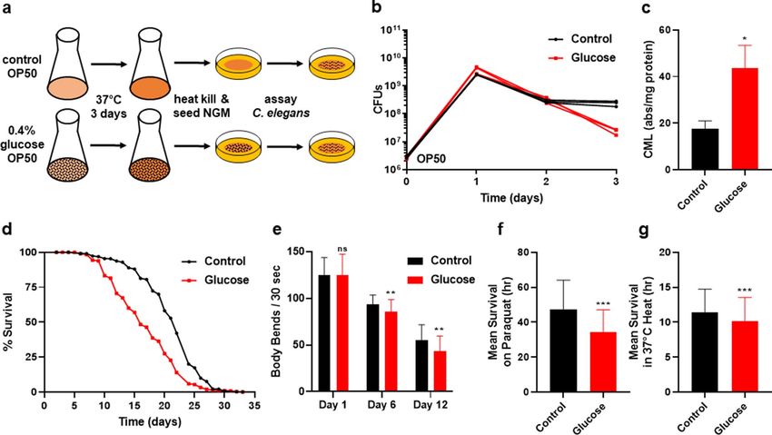
www.nature.com/scientificreports/
glucose was in contact with both bacteria as well as C. elegans. Therefore, we questioned the mechanism of the
high glucose effect: was the effect attributable to direct contact with sugar by the worm, the bacteria, or both?
These previous published protocols have variations with regard to how the glucose was applied to the agar,
whether the bacteria were alive or dead, how the bacteria were killed, whether the bacteria had any direct contact
with the g lucose16–21, and the age of animal exposed to the g lucose22,23. Moreover, previous studies that tried to
separate the bacterial contribution to effects on lifespan used a bacterial mutant in the major bacterial glucose
transporter20. However, these pts mutants can still transport sugar at substantial, although reduced, rates24,25. To
separate the effects of C. elegans consuming a glucose fed bacteria diet, here, we developed a new experimental
procedure based on previous studies 26. In this new protocol, prior to seeding the bacteria on the plate, the bac-
teria are incubated with or without glucose for three days. Therefore, this protocol is unique since it allows us to
control contact between the glucose and C. elegans.
Added dietary sugar has been associated with an alarming rise in a multitude of debilitating medical condi-
tions including obesity, diabetes, cardiovascular and neurodegenerative diseases, such as Alzheimer’s disease,
due to its impact on the generation of advanced glycation end products (AGEs). A group of heterogeneous
compounds, AGEs, include proteins, lipids, and nucleic acids that become glycated as a result of exposure to
sugars. Glycation results from a non-enzymatic reaction where the carbonyl group of reducing sugars is cova-
lently coupled to proteins, lipids, and/or nucleic acids. Both exogenous and endogenous sources contribute to
the levels of AGEs. Endogenous formation of AGEs occurs continuously at low levels, but exogenous sources,
including consumption of a high-sugar diet, can drastically increase this pool. As humans age and in certain
diseased states, AGE levels rise and are associated with increased cardiovascular risk, diabetes, chronic kidney
disease, and Alzheimer’s disease.
Many studies in animal model systems show that dietary consumption of exogenous AGEs contribute to
oxidative stress and inflammation which could contribute to a number of chronic disease states (reviewed
in27,28). Additionally, in cell culture dietary AGEs have been shown to affect inflammatory r esponse29, but there
are mixed results for the effects of dietary AGEs in human t rials27. Emerging studies also suggest that dietary
AGEs can contribute to the onset of organ damage, affect metabolic control, and therefore impact global h ealth28.
We find that our C. elegans-E. coli system, where we modify the health of the microbiota through modulat-
ing E. coli’s dietary sugar, results in changes in lifespan and healthspan of C. elegans. Increased added dietary
sugar for solely the bacteria results in decreased lifespan, decreased healthspan including decreased movement
in liquid, and decreased oxidative stress resistance. Addition of the anti-glycation compound carnosine to the
bacteria, ameliorates the negative effects of glucose in the diet on C. elegans. Added dietary sugar suppresses
C. elegans oxidative stress resistance, most notably through suppression of the glutathione-S-transferase, gst-4,
expression. Our data reveal a central role for C. elegans gst-4 in the regulation of a high-sugar diet. Both a glucose-
fed bacterial diet as well as loss of gst-4 shorten lifespan. In addition, loss of gst-4 blunts the animals’ response
to a glucose fed bacterial diet. Taken together, our model system allows dissection of the intestinal microbiota
on a level not possible in humans, with univariable analysis of both the microbiota and the host which reveals
the intimate connection between oxidative stress, host responses, and a high-sugar/high AGE-producing diet.
Results
Previously, studies on the effects of glucose toxicity in C. elegans altered several variables including the age of
animal exposed to the g lucose22,23, whether the bacterial diet was alive or dead, and the duration of and exposure
time of the E. coli and C. elegans to g lucose16–21. Additionally, the added glucose was in contact with both the E.
coli and C. elegans. To determine the mechanisms leading to the effects of C. elegans consuming a bacterial diet
high in glucose, we developed a new experimental procedure based on our previous bacterial s tudies26. In the
new system, OP50 E. coli was inoculated in LB media supplemented with various glucose concentrations (rang-
ing from 0 to 0.8%), and incubated for an extended period of 3 days. This was followed by heat-killing of the E.
coli which was then seeded onto nematode growth medium (NGM) plates for physiological and biochemical
assays (Fig. 1a). Heat-killing the bacterial diet prevented any possible effects of bacterial proliferation. The 3-day
glucose exposure resulted in the OP50 E. coli exhibiting a significant decrease in colony forming units (Fig. 1b;
P < 0.01). The optical density of the glucose-fed E. coli was not significantly changed, indicating that C. elegans
were consuming similar biomass (Supplementary Figure S1a, P < 0.05), even though over time bacterial cells
showed a significant loss of viability. We also confirmed that glucose-fed E. coli had elevated levels of intracellular
glucose (Supplementary Figure S1b). Similar to our previous studies with a different strain of E. coli supplemented
with glucose26, the 3-day incubation of OP50 E. coli with glucose led to an increased concentration of the AGE
carboxymethyl-lysine (CML) as detected by ELISA (Fig. 1c). CML, is a major AGE and CML regulation and the
extent of its accumulation is used extensively as a proxy for total AGE c oncentration26,30,31. Therefore, the OP50
E. coli shown in Fig. 1b supplemented with 0.4% glucose shows an increase in CML along with the loss of CFU.
Together, based on our previous research26 and our initial studies, we selected the 3-day incubation period with
supplementation of 0.4% glucose for our experimental paradigm. The experimental system presented here is
unique since the glucose only directly interacts with the bacteria, never directly contacting the animals.
C. elegans consuming the glucose-fed E. coli diet show a significant reduction (~ 30%) in lifespan compared
to animals fed a control diet with no added glucose (Fig. 1d, P < 0.01, Supplementary Table S1). Importantly, the
reduced lifespan was also accompanied by a reduction in healthspan, as shown in Fig. 1e, where animals consum-
ing the glucose fed bacterial diet, show decreased healthspan as quantified by movement in liquid/swimming/
thrashing. To further evaluate health parameters, oxidative and heat stress resistance were tested on C. elegans
after consuming the glucose-fed E. coli diet for 6 days (Fig. 1f–g). We selected day 6 as the time point for further
analysis as this time reflected differences that could be observed prior to the onset of normal age-related effects
on C. elegans viability. An additional phenotype of consuming a glucose-fed bacterial diet is that animals show
Scientific Reports | (2021) 11:5931 | https://doi.org/10.1038/s41598-021-85046-3 2
Vol:.(1234567890)
www.nature.com/scientificreports/
Figure 1. C. elegans consumption of glucose fed bacteria. (a) Experimental protocol used for OP50 E. coli
processing of glucose and C. elegans assays. (b) Effect of 0.4% glucose supplementation on E. coli colony
forming units over time (P = 0.002 at day 3). (c) Detection of Carboxymethyl-lysine (CML), an AGE product,
by ELISA Assay performed on control and 0.4% glucose fed E. coli. (d) Lifespan of wild type C. elegans treated
with 0% control and 0.4% glucose fed OP50 E. coli; Statistics in Supplementary Table S1, S2. (e) Healthspan—
Movement in liquid/swimming/thrashing of C. elegans consuming control or 0.4% glucose fed E. coli, N = 232.
(f) Oxidative stress resistance of wild type C. elegans consuming 0% control and or 0.4% glucose fed E. coli for
6 days, measured by mean survival on paraquat, N = 328. (g) Heat stress resistance (mean survival at 37 °C) of
wild type C. elegans consuming 0% control and 0.4% glucose fed E. coli for 6 days, N = 366. Statistical analysis
of histograms compared C. elegans consuming 0% control with C. elegans consuming 0.4% glucose fed E. coli at
the same time point using an unpaired two-tailed t test with GraphPad Prism 8.0 (https://www.graphpad.com).
Symbols as follows: (ns = not significant, *P ≤ 0.05, **P ≤ 0.005, ***P ≤ 0.001). Data shown is a compilation from
at least 3 biological replicates.
a consistent reduction in their overall body size over time (Supplementary Figure S1c). Together, our results
illustrate that consumption of the glucose-fed bacterial diet significantly reduces lifespan and healthspan which
mirrors results seen in more traditional glucose supplementation experiments16–21. Further, these physiological
effects are observed in an environment where the worms never directly encounter the additional glucose.
Next, we examined how bacterial processing of glucose affects the health of C. elegans using three different
approaches. First, we supplemented E. coli with glucose prior to the start of the culture (Pre glucose; method
as in Fig. 1a) and compared this with glucose supplementation after the 3 day incubation (Post glucose). In the
latter method (Post glucose), glucose was readily available only to C. elegans, in comparison to the Pre glucose
diet, in which glucose was incubated only with the E. coli. As shown in Fig. 2a, lifespan was shortened only when
E. coli was able to process the glucose in the Pre-glucose treatment. Remarkably, although the post glucose sup-
plementation had a higher amount of glucose available to the animal (80×; Fig. 2g), consumption of the Post
glucose diet showed no change in lifespan (Fig. 2a). In addition to the effect on lifespan, oxidative stress resist-
ance was reduced only in the Pre glucose diet (Fig. 2d). We also assayed healthspan as measured by movement
of the animals in liquid as animals age. Movement of the animals in liquid required a greater time to observe
any effect and was not substantially reduced by the Post glucose supplementation until day 12 (Supplementary
Figure S2a). Therefore, it is the processing of glucose by the bacteria that decreases lifespan, oxidative stress
resistance, and locomotion-healthspan.
A second method to examine the effects of bacterial metabolism of glucose on C. elegans was the use of the
synthetic glucose analog 2-deoxy-D-glucose (2-DG). When consumed, 2-DG is phosphorylated by hexokinase
which then cannot be further processed and therefore is used as a glycolytic inhibitor. We treated the OP50 E.
coli with 2-DG for 3 days, similar to previous experiments. C. elegans consuming OP50 E. coli supplemented
with 2-DG exhibit a normal lifespan without changes in either resistance to oxidative stress or healthspan (move-
ment in liquid; Fig. 2b,e, and Supplementary Figure S2b). Thus, consuming the 2-DG fed bacteria diet results in
phenotypes opposite to those seen with the glucose fed bacterial diet.
Scientific Reports | (2021) 11:5931 | https://doi.org/10.1038/s41598-021-85046-3 3
Vol.:(0123456789)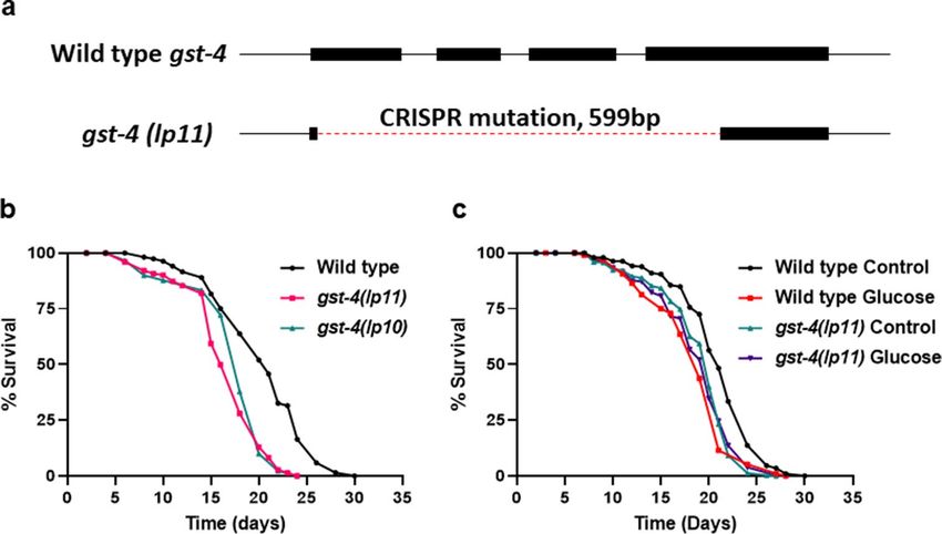
www.nature.com/scientificreports/
Figure 2. Interfering with bacterial metabolism of glucose alters its effects on C. elegans. (a) Lifespan of wild
type C. elegans consuming either 0% control, 0.4% glucose pre culture, or 0.4% glucose post culture OP50 E.
coli, statistics in Supplementary Table S1, S2. (b) Lifespan of wild type C. elegans consuming either 0% control,
0.4% glucose fed, or 0.4% 2-deoxy-glucose (2-DG) fed OP50 E. coli, statistics in Supplementary Tables S1, S2.
(c) Lifespan of wild type C. elegans consuming either 0% control, 0.4% glucose, 50 mM carnosine, or 0.4%
glucose + 50 mM carnosine fed OP50 E. coli, statistics in Supplementary Tables S1, S2. (d) Oxidative stress
resistance of wild type C. elegans consuming either 0% control, 0.4% glucose pre culture or 0.4% glucose post
culture OP50 E. coli, measured by mean survival on paraquat, N = 570. (e) Oxidative stress resistance of wild
type C. elegans consuming either 0% control, 0.4% glucose, or 0.4% 2-deoxy-glucose (2-DG) fed OP50 E. coli,
measured by mean survival on paraquat media, N = 215. (f) Oxidative stress resistance of wild type C. elegans
consuming either 0% control, 0.4% glucose, 50 mM carnosine, or 0.4% glucose + 50 mM carnosine fed OP50
E. coli, measured by mean survival on paraquat, N = 155. (g) Glucose assay of the OP50 E. coli bacterial diet
in (a)–(f), results normalized to protein concentration. Statistical analysis of histograms compared C. elegans
consuming 0% control with C. elegans consuming 0.4% glucose fed E. coli at the same time point using an
unpaired two-tailed t test with GraphPad Prism 8.0 (https://www.graphpad.com). Symbols as follows: (ns = not
significant, *P ≤ 0.05, **P ≤ 0.005, ***P ≤ 0.001). Data shown is a compilation from at least 3 biological replicates.
Third, to further elucidate the connection between bacterial metabolism of glucose and host health, we supple-
mented the bacterial diet with the dipeptide carnosine in combination with glucose. We have previously observed
that the anti-glycation properties of carnosine reduces the toxic effects of glucose in E. coli26. As shown in Fig. 2c,
glucose plus carnosine-fed E. coli significantly extends C. elegans lifespan and ameliorates the effect of added
glucose alone (P < 0.01). Interestingly, there is little to no effect of carnosine on C. elegans when the compound
is mixed within the agar of the plate then seeded with heat-killed E. coli (Supplementary Figure S2c). Addition-
ally, glucose concentrations within the E. coli are decreased by 3× when the glucose-fed E. coli are supplemented
with carnosine (Fig. 2g). C. elegans consuming this glucose and carnosine diet are protected from the decrease
in oxidative stress resistance that occurs in glucose supplementation alone (Fig. 2f). Therefore, across three dif-
ferent methods, we reveal that the bacterial processing of glucose dictates C. elegans lifespan and healthspan.
Animals consuming the glucose-fed bacteria diet exhibit a shortened lifespan and decreased stress resistance.
Therefore, we next examined whether the expression levels of key genes involving environmental defenses could
account for these observed phenotypes using both transgenic GFP animals as well as RT-qPCR. We used known
stress-related transgenic GFP strains to observe any potential changes in the dynamics of expression associated
with consumption of glucose-fed bacteria within the host over time. To determine the downstream signaling
pathway in the host that responds to the glucose-fed bacteria diet, we screened distinct pathways induced by
oxidative stress (gst-4, sod-3)32, heat stress (hsp-16.2), the mitochondrial unfolded protein response (UPR) (hsp-
6)33,34, the glyoxalase system (glod-4)35, the transcription factor skn-136, and the insulin/IGF-1 signaling pathway
transcription factor C. elegans FOXO/daf-1637–40 (Fig. 3a and Supplementary Figures S3a–b, S4a–d, S5a–b).
Notably, as shown in Fig. 3a, we observed blunted expression of both gst-4::GFP and glod-4::GFP when
animals consumed the glucose-fed bacteria, as well as an increase in sod-3::GFP. To remove any bias associated
with the visual assay, we quantified the fluorescence of those animals (Fig. 3b). Then, to confirm the validity of
our findings, we performed RT-qPCR on wild-type animals under the same conditions. We find consistent gene
expression changes when assayed by RT-qPCR (Fig. 3c).
Scientific Reports | (2021) 11:5931 | https://doi.org/10.1038/s41598-021-85046-3 4
Vol:.(1234567890)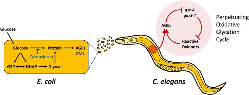
www.nature.com/scientificreports/
Figure 3. Effects of bacterial metabolism of glucose on host C. elegans gene expression. (a) Fluorescent imaging
of transgenic C. elegans gst-4::gfp, sod-3::gfp, or glod-4::gfp, after 6 days consuming either 0% control, 0.4%
glucose, 0.4% 2-deoxy-glucose (2-DG), 50 mM carnosine, or 0.4% glucose + 50 mM carnosine fed E. coli for
6 days. See methods for complete genotype oftransgenic C. elegans. (b) Fluorescence quantification of animals
in (a): transgenic C. elegans gst-4::gfp, sod-3::gfp, or glod-4::gfp, consuming either 0% control, 0.4% glucose, 0.4%
2-deoxy-glucose (2-DG), 50 mM carnosine, or 0.4% glucose + 50 mM carnosine, fed E. coli for 6 days, N = 336.
(c) RTqPCR of wild type C. elegans consuming either 0% control or 0.4% glucose fed E. coli for 6 days. Statistical
analysis of histograms compared C. elegans consuming 0% control with C. elegans consuming 0.4% glucose
fed E. coli at the same time point using an unpaired two-tailed t test with GraphPad Prism 8.0 (https://www.
graphpad.com). Symbols as follows: (ns = not significant, *P ≤ 0.05, **P ≤ 0.005, ***P ≤ 0.001). Data shown is a
compilation from at least 3 biological replicates.
Next, we tested whether gene expression changes paralleled our findings in Fig. 2 with the use of the glucose
analog 2-DG, which did not alter C. elegans lifespan or healthspan. As shown in Fig. 3a, we observed gene expres-
sion levels opposite to glucose when animals were fed a bacterial diet supplemented with 2-DG. This finding was
confirmed with fluorescence quantification (Fig. 3b). Therefore, in response to a glucose-fed diet, we observed a
decrease in lifespan and healthspan associated with a downregulation of several key detoxification genes.
Further, we examined GFP expression changes in C. elegans when fed a diet consisting of E. coli fed carnosine
alone or in combination with glucose, as carnosine ameliorated the shorter lifespan induced by glucose (Fig. 2).
C. elegans consuming carnosine-fed E. coli exhibit a mild increase in lifespan and also show a slightly higher
level of gst-4::GFP, sod-3::GFP, and glod-4::GFP expression (Fig. 3a,b). When both carnosine and glucose are
fed to the bacteria, lifespan is greater than glucose alone and gst-4::GFP fluoresces significantly brighter, while
sod-3::GFP and glod-4::GFP show similar expression to carnosine alone (Fig. 3a,b; P < 0.01). Furthermore, C.
elegans consuming carnosine-fed E. coli exhibit longer lifespan, protection from the decline in oxidative stress
resistance and changes in gene expression when compared to consuming the glucose-fed bacterial diet insulin/
IGF-1signaling (IIS) (Figs. 2f, 3a,b).
We also screened two signaling pathways: Detoxification- skn-1 (C. elegans NRF2) and IIS- daf-16 (C. elegans
FOXO) for gene expression changes in response to consumption of the glucose fed bacterial diet using both
transgenic GFP animals as well as RT-qPCR. As shown in Supplementary Figure S3a, the glucose-fed bacte-
rial diet did not change either expression levels of skn-1::GFP or nuclear translocation. The skn-1::GFP results
were consistent with the RTqPCR results shown in Supplementary Figure S3b where mRNA of skn-1 was not
Scientific Reports | (2021) 11:5931 | https://doi.org/10.1038/s41598-021-85046-3 5
Vol.:(0123456789)www.nature.com/scientificreports/
Figure 4. Generation and characterization of C. elegans gst-4 mutant. (a) Diagram of DNA sequences of wild
type C. elegans genomic gst-4 and the gst-4(lp11) mutant. Lines represent introns, rectangles represent exons,
and dashed line represents the lp11 allele CRISPR deletion. (b) Lifespan of wild type, gst-4(lp10), and gst-4(lp11),
statistics in Supplementary Table S1, S2. (c) Lifespan of wild type and gst-4(lp11) mutants consuming either 0%
control or 0.4% glucose fed OP50 E. coli, statistics in Supplementary Tables S1, S2.
significantly changed with the glucose-fed bacterial diet. Since skn-1 showed no changes in expression in response
to the glucose-fed bacterial diet, we further tested one of its target genes, gcs-1, often used as a surrogate for skn-
141. There was neither a change in gcs-1::GFP expression nor gcs-1 mRNA levels (Supplementary Figures S3a and
S3b). Additionally, the heat stress marker hsp-16.2 was consistently downregulated with the glucose-fed bacte-
rial diet as determined by both GFP fluorescence and mRNA expression (Supplementary Figures S3a and S3b).
We also examined the IIS pathway in response to the glucose-fed bacterial diet initially with nuclear trans-
location of daf-16a::GFP. We did not observe any changes in nuclear translocation for daf-16a::GFP but did
observe a significant reduction in total fluorescence per animal (Supplementary Figures S4a and S4b; P < 0.01)
and RT-qPCR showed a 20% increase in daf-16 mRNA (Supplementary Figure S4c). Further analysis of DAF-16
activity was performed by quantifying expression of 14 well-known transcriptional targets of DAF-1637,38,42. Of
these DAF-16 targets, 10/14 were significantly changed with the glucose fed bacteria; sod-3, cpr-2, ctl-1, and dod-6
upregulated; hsp-12.6, ZK742.4, fat-7, scl-1, ctl-2, and dod-3 downregulated (Supplementary Figure S4d, P < 0.01).
Therefore overall, our data suggest that DAF-16 is partly involved in the response to a glucose-fed bacterial diet.
We observed a consistent upregulation of sod-3 and sod-5 with the glucose-fed bacterial diet, which presum-
ably should increase resistance to oxidative damage43. However, a recent report by Dues et al44 examined the
total superoxide dismutase capacity of C. elegans. They found that sod-3 and sod-5 together only account for 1.7%
of all sod expression while sod-1, sod-2, and sod-4 together amass 98.2% of the total sod mRNA. In accordance
with these findings, we mathematically extrapolated our RTqPCR results onto the published expression ratios
to estimate total sod expression (Supplementary Figures S5a–b). Animals consuming glucose fed bacteria show
a ~ 50% decrease in sod-1 and sod-2 expression which overshadows any increase in sod-3 expression. Therefore
overall, animals consuming the glucose-fed bacterial diet exhibit a reduction of sod expression to only 60% of
total wild type sod mRNA capacity (Supplementary Figure S5b). Therefore, across multiple experimental para-
digms, our data confirm that bacterial processing of glucose promotes a decrease in lifespan as well as a reduc-
tion in health, and stress resistance. These phenotypical changes are accompanied by differential regulation of
glyoxalase, glutathione-S-transferases, and superoxide dismutase—all of which should enzymatically detoxify
the glycolytic effects of glucose.
Our data reveal that glucose-fed bacteria which are high in CML (AGEs) promote an environment that causes
animals to exhibit signs of oxidative stress including strong and consistent suppression of gst-4 (glutathione
transferase-4) involved in the regulation of the Phase II oxidative stress r esponse45. Analysis of transcriptional
activation of gst-4 is often used as a proxy for oxidative stress tolerance46. Therefore, we next examined a loss of
function allele of gst-4 generated by CRISPR/Cas9.
We generated two independent isolates of the gst-4 mutation, lp10 and lp11 by CRISPR/Cas9. As shown in
Fig. 4a, sequencing lp11 revealed a deletion of 599 bp and spans all 4 exons of wild type gst-4. Next, we tested the
gst-4 mutants for lifespan and show that the gst-4 mutation results in a decrease in lifespan (Fig. 4b). We further
examined whether loss of gst-4 interfered with the effects of the glucose fed bacterial diet. As shown in Fig. 4c,
mutation in gst-4 abrogates the response to the glucose fed bacterial diet. Therefore, our results indicate that
gst-4 is necessary to respond to a glucose-fed bacterial diet. Together, our data reveal that dietary AGEs promote
oxidative stress in the host dependent on the Phase II oxidative stress response.
Scientific Reports | (2021) 11:5931 | https://doi.org/10.1038/s41598-021-85046-3 6
Vol:.(1234567890)www.nature.com/scientificreports/
Discussion
As bacterivores, the diet of C. elegans in the laboratory typically consists of a single E. coli strain, which then
inhabits its gut in a commensal relationship. Here, we test the C. elegans-E. coli interaction in response to changes
in the dietary environment, specifically with the addition of glucose. Previous studies examining a high glucose
diet involved several different methods. A given percentage of glucose, typically 2%, is either added to the NGM
agar before cooling or added to the top of the NGM agar filled plate after it has cooled. Bacteria, the diet for C.
elegans, would then be spread onto the NGM agar plate. In such an experimental paradigm, there is a three-way
interaction: E. coli is exposed to the glucose, C. elegans is exposed to the glucose, and C. elegans consumes the E.
coli exposed to the g lucose16–21,42,47,48. As shown in our model in Supplementary Figure S6, with these methods,
glucose has both direct and indirect contact with C. elegans.
In contrast, our C. elegans-E. coli system specifically examines bacterial processing of glucose and its effect
on C. elegans. Addition of glucose to the bacterial culture precedes any exposure to C. elegans. These glucose-fed
bacteria are then seeded onto the NGM agar plate thereby illustrating the indirect effect of glucose on C. elegans.
With this method, we find that C. elegans consuming the glucose-fed E. coli diet have a 24% reduction in lifespan
and a reduction in healthspan (locomotion) and oxidative stress resistance (Fig. 1, Supplementary Table S1).
Therefore, both the indirect and direct effects of glucose result in poor health as animals age16–21.
Our data also illustrate that the duration of direct contact between the glucose and the bacteria is critical. As
shown in Figure S7, similar to findings by Lee et al20, pre-incubating the bacteria overnight (16 h) does not confer
any changes in lifespan (Supplementary Figure S7). Based on our previous studies26, we considered that perhaps
the E. coli pre-exposure to glucose required a longer incubation. Subsequently, we chose a 3-day incubation with
glucose prior to seeding the E. coli onto the NGM plate. In effect, our C. elegans-E. coli system reduces the vari-
ability of bacterial metabolism by imposing a consistent window in which bacteria process glucose. Addition of
glucose to the bacterial culture allows processing by the bacteria to occur preceding any exposure to C. elegans.
Our data reveal that the specific bacterial processing of glucose negatively affected the health of C. elegans
within three different methods to separate, inhibit, and suppress bacterial processing (Fig. 2). First, we supple-
mented the E. coli culture with the same 0.4% concentration of glucose after the bacterial diet was heat-inacti-
vated. This direct application of glucose had no effect on C. elegans lifespan perhaps since this is a relatively low
amount of glucose when compared to previous reports utilizing 2% per plate. However, when processed by the
bacteria, 0.4% glucose drastically changes the health of the host. Secondly, we incubated the E. coli with glucose
analog 2-DG to inhibit glycolysis within the bacteria. In contrast to glucose fed bacteria, 2-DG fed bacteria
result in healthier animals with a normal lifespan. In a third set of experiments, we incubated the E. coli culture
with carnosine, an anti-glycation compound, that has the ability to lower glucose toxicity through reducing the
accumulation of A GEs26. We observed that carnosine alone, as well as carnosine in combination with glucose,
resulted in healthier animals compared with consumption of a glucose-fed bacterial diet. Therefore, across
three methods, our data reveal that the bacterial processing of glucose dictates the phenotypes of the animals
consuming this diet.
Consistently, the high AGE E. coli diet significantly shortened mean lifespan of C. elegans. However, when
fed the glucose and carnosine treated E. coli, lifespan was reduced only by ~ 7% (Supplementary Table S1). Addi-
tionally, when applied directly to the NGM media with heat-killed bacteria, carnosine had little to no effect on
C. elegans lifespan. Thus, benefits were only observed when carnosine was processed by the bacteria. Carnosine
treated E. coli also increased C. elegans healthspan as measured by resistance to oxidative stress, both alone and in
the presence of glucose. This additional oxidative stress resistance may be attributed to the ability of carnosine to
reduce the glucose burden of the bacteria by lowering total AGEs consumed by C. elegans. Together, these results
suggest that carnosine used as a prebiotic intervention may have the potential to effect resident microbiota and
alleviate dietary glucose related oxidative stress.
Previous studies in mammals have linked oxidative stress to consumption of a high AGE diet28,49–51. We show
that when animals consume the glucose-fed bacterial diet, they are subject to high levels of oxidative stress. We
surveyed a broad panel of stress reporter genes when animals are subject to the glucose-fed bacterial diet (Fig. 3,
Supplementary Figures 3–5). Animals consuming the glucose-fed bacterial diet exhibit a down regulation of
gst-4, both in gst-4::GFP expression and mRNA levels. Interestingly, our data show that the microbial processing
of glucose inhibits the expression of gst-4, as 2-DG treated bacteria did not confer this same effect. Further, our
analyses reveal that the glucose-fed bacterial diet promotes an environment of constant oxidative stress such that
when C. elegans age on this diet, they become more susceptible to oxidative stress. Furthermore, our data reveal
that loss of gst-4 prevents the animal from responding to the glucose-fed bacterial diet (Fig. 4). It should be noted
that we examined multiple other glutathione-S-transferases including gst-10, gst-29 and found that they were all
downregulated. This indicates the possibility that the bacterial processing of glucose suppresses the animal’s abil-
ity to respond to this oxidant rich environment, which may perpetually cause chronic vulnerability to oxidants.
Our data suggest a model shown in Fig. 5. Uptake of glucose by the bacteria leads to increased processing of
glucose and results in more bacterial-generated AGEs. Consumption of this glucose-fed bacterial diet high in
AGEs promotes a transcriptional response including suppression of gst-4 and glod-4. Enzymes such as gst-4 and
glod-4 function to prevent reactive oxidants such as glyoxal and methylglyoxal from nonenzymatically binding
to proteins forming more AGEs32,35. Inhibition of gst-4 and glod-4 result in a perpetuating oxidative glycation
cycle that results in even more AGEs produced and further increase of oxidative stress.
We suggest that using our C. elegans-E. coli system, it is possible that we mimic dietary effects on bacteria
within the human digestive tract. We use a 3-day bacterial culture supplemented with glucose to analyze the
effect onto its host. We suggest this is similar to the entrenched bacteria within the human digestive tract that
are dependent on nutrient intake by their host. Interestingly, the physiological manifestation of Crohn’s disease
is characterized by bacterially derived persistent inflammation of the intestine and colon. In fact, recent clinical
Scientific Reports | (2021) 11:5931 | https://doi.org/10.1038/s41598-021-85046-3 7
Vol.:(0123456789)www.nature.com/scientificreports/
Figure 5. Model of E. coli glucose processing effects on C. elegans. Glucose is processed/metabolized within the
E. coli to produce AGEs. Carnosine supplementation abrogates this formation. Consumption of the glucose-fed
bacteria with successive digestion and absorption leads to C. elegans transcriptional changes. High AGEs from
the diet suppress gst-4 and glod-4; two genes which function to prevent reactive oxidants such as glyoxal and
methylglyoxal from nonenzymatically binding with proteins and forming more AGEs. Thus, the inhibition of
gst-4 and glod-4 cause a perpetuating oxidative glycation cycle, further limiting C. elegans ability to respond to
AGEs/oxidants.
studies find that patients suffering from Crohn’s disease and Irritable Bowel Syndrome exhibit oxidative stress52.
Therefore, the blunted transcriptional response observed when animals consume the prebiotic carnosine with
the glucose-fed bacteria could aid future models of the dynamics involved in persistent bacterial inflammation
in cases of Crohn’s disease and Irritable Bowel Syndrome.
Taken together, our data show that bacterial processing of glucose resulted in reduced lifespan and reduced
stress resistance in their host even though the host never directly encountered the additional glucose. High
levels of AGEs in the C. elegans diet are accompanied with downregulation of stress response genes. This overall
transcriptional response by C. elegans in response to consumption of glucose-fed bacteria is protected by the
preventative mechanisms of action of carnosine. Consumption of the high glucose/highly glycated/increased
AGEs diet shortens lifespan, reduces healthspan and promotes oxidative stress.
Methods
Strains. All C. elegans strains were maintained at 16 °C using standard C. elegans techniques except where
indicated53. Strains used in this study: N2, LGIV—HT2335 gst-4(lp10), HT2336 gst-4(lp11).
The following transgenes were used: TJ356 [daf-16a::gfp], LD1008[ldEx9 [skn-1::GFP + rol-6(su1006)]],
BC15643 [dpy-5(e907) I; sEx15643[rCes C16C10.10::GFP + pCeh361]], CL2166 [dvIs19 [(pAF15)gst-4p::GFP::NLS]
III.], CF1553 [muIs84 [Psod-3::GFP]], CL2070 [dvIs70[Phsp-16.2::GFP + rol-6(su1006)]], LD1171 [ldIs3 [Pgcs-
1::GFP + rol-6(su1006)]], Some strains were provided by the CGC, which is funded by NIH Office of Research
Infrastructure Programs (P40 OD01044).
Lifespan assay. All lifespan assays were performed at 20 °C. Strains were semi-synchronized by allowing
gravid adults to lay eggs on standard NGM plates overnight, and animals developed for several days until they
reached L4. Then, ∼100 L4s were transferred to NGM or treatment plates and kept at 20°(∼33 animals per plate).
Animals were scored by gently tapping with a platinum wire every 2–3 days. Animals that did not respond were
scored as dead. Animals that crawled off the plate, bagged, or died from vulva bursting were censored from the
analysis. Each figure shows data collected from all trials. The mean lifespan, standard deviation, and P values
were calculated via two-tailed unpaired t tests (Supplementary Table S1). Survival graphs and statistical analyses
were produced using GraphPad Prism 8.0 (https://www.graphpad.com).
Glucose/carnosine fed E. coli diet. Standard Nematode Growth Media (NGM)53 was autoclaved at
121 °C for 25 min, then cooled at room temperature (RT) and 100 µg/mL ampicillin (Fisher Scientific) was
added prior to pouring plates. Plates dried for 3 days at RT before seeding with E. coli. The OP50 E. coli was
grown for 3 days in LB only (control), or LB supplemented either pre-culture or post-culture with 0.4% D-(+)-
glucose (Sigma), 0.4% 2-Deoxy-D-glucose (Sigma), and/or 50 mM L-carnosine (Sigma). After 3 days, the E. coli
cultures were subjected to a 65 °C heat bath for 30 min to inhibit further growth and then seeded onto the NGM
plate. The E. coli OP50 diet was streaked onto an LB agar plate and cultured at 37 °C overnight to confirm lack of
growth. Plates were stored at 4 °C until use.
Scientific Reports | (2021) 11:5931 | https://doi.org/10.1038/s41598-021-85046-3 8
Vol:.(1234567890)www.nature.com/scientificreports/
E. coli fitness (survival) assay. Overnight cultures were inoculated from frozen stocks. 100 μl of each
strain was spread on each plate in triplicate and allowed to dry by evaporation. At each time point, the num-
ber of viable cells was determined by “coring” a section of the lawn with a sterile Pasteur pipet (inner diame-
ter ~ 1.5 mm), resuspending each core in 50 μl of LB, and titering by serial dilution and plating onto LB medium;
the limit of detection is < 100 cfu/ml54,55.
E. coli AGE assay. ELISA was used to detect carboxymethyl-lysine levels as in Pepper et al26 with 100 μl of
supersensitive, 3,3′,5,5′-tetramethylbenzidine (TMB) (Sigma) used as the color reagent and read at 630 nm on a
BioTek ELx808 absorbance reader.
Glucose assay. OP50 E. coli was grown for 3 days in LB only (control), or LB supplemented either pre-
culture or post-culture with 0.4% D-(+)-glucose (Sigma), 0.4% 2-Deoxy-D-glucose (Sigma), and 50 mM L-Car-
nosine (Sigma). After 3 days, the E. coli cultures were subjected to a 65 °C heat bath for 30 min to inhibit further
growth. Samples were diluted in reaction buffer to accurately detect glucose levels and frozen at − 20 °C until
use. Glucose amount was determined using the Amplex Red Glucose/Glucose Oxidase Assay Kit (Invitrogen), in
triplicate. Protein concentration was then measured with Pierce Coomassie Plus (Bradford) Assay Kit (Thermo
Scientific). Glucose levels were standardized to protein levels, and then used to graph over three replicate experi-
ments using GraphPad Prism 8.0 (https://www.graphpad.com).
RNA extraction and RT‑qPCR. Approximately 200 synchronized L4 stage animals were transferred to
treatment plates and grown at 20 °C for 6 days, then washed off the plates with M9 buffer and rinsed twice
with DEPC-treated water. Total RNA was isolated using TRIzol Reagent with the Direct-zol RNA MiniPrep
(Zymo Research). After RNA extraction, first-strand cDNA was synthesized from 1.0 μg of total RNA using
dNTPs, Oligo(dT)12–18 and SuperScript II Reverse Transcriptase (Invitrogen). Quantitative PCR was done with
an Applied Biosystems StepOne Plus Real-Time PCR system with Power SYBR Green PCR Master Mix (Applied
Biosystems) per the manufacturer’s instructions, with triplicates done for each of three biological replicates,
and act-1 used as the endogenous control for relative expression normalization. Sequences of primers can be
found in Supplementary Table S3. Specificity of PCR amplification was determined by the melting curve for
each reaction. The threshold cycle (CT) for each primer set was automatically determined by StepOne Soft-
ware v2.3 (https://www.thermofisher.com/us/en/home/technical-resources/software-downloads/StepOne-and-
StepOnePlus-Real-Time-PCR-System.html). Relative fold changes of gene expression were calculated using the
2−△△Ct method. The RQ values were then used to graph relative gene expression over at least three replicate
experiments using GraphPad Prism 8.0 (https://www.graphpad.com).
Healthspan: body bends/movement in liquid media/thrashing/swimming. Synchronized L4
stage animals were transferred to treatment plates and incubated at 20 °C for 1, 3, 6, or 12 days. On the experi-
mental day, individual animals were picked onto an unseeded NGM plates, then 30 μL of M9 buffer was pipetted
onto each animal. After 5 s, the number of body bends was recorded over 30 s using a mounted DMK 21AF04
camera (The Imaging Source) outfitted onto a dissecting microscope. At least 20 animals were recorded per
strain or treatment and the average body bends per minute for each of the samples was calculated and graphed
using GraphPad Prism 8.0 (https://www.graphpad.com).
Resistance to oxidative stress. Paraquat plates were made by adding 1 mL of 250 mM paraquat solution
to corresponding treatment plates. Then plates were put on a shaker for 1 h, followed by 1.5 h in a laminar flow
hood to ensure plates were dry with the paraquat evenly distributed. Synchronized L4 stage animals were trans-
ferred to treatment/paraquat plates and incubated at 20 °C for 6 days. Animals were then transferred to paraquat
plates at 20 °C and scored twice daily for survival. Animals were touched with a platinum wire, and those that did
not respond were scored as dead. Two independent replicates were performed, and the mean survival calculated
using GraphPad Prism 8.0 (https://www.graphpad.com).
Resistance to heat stress. Synchronized L4 stage animals were transferred to treatment plates and incu-
bated at 20 °C for 6 days. Animals were transferred to 37 °C and then scored every 2 h for survival. Animals were
touched with a platinum wire, and those that did not respond were scored as dead. Two independent replicates
were performed, and the mean survival was calculated using GraphPad Prism 8.0 (https://www.graphpad.com).
Fluorescence imaging and quantification. Synchronized L4 stage animals were transferred to treat-
ment plates and incubated at 20 °C for 1, 3, 6, or 12 days. On the experimental day, 10–12 animals were then
picked onto 2% agarose pad on a glass slide and immobilized with 100 mM N aN3. Fluorescence imaging of GFP
was done using a Zeiss Axioskop 2 plus fitted with a Hamamatsu ORCA-ER camera and a FITC filter. Images
taken were in grayscale. Quantification of GFP expression was performed using ImageJ Software 1.52a (https
://imagej.nih.gov/ij/index.html). In the photos, each worm at least 95% was in frame was outlined by hand and
then measured for minimum, maximum, and average pixel intensity within the defined area. The minimum
pixel intensity recorded was then subtracted to remove background fluorescence interference. The average pixel
intensity per worm compiled across 2 or more batch experiments was then plotted using GraphPad Prism 8.0
(https://www.graphpad.com).
Scientific Reports | (2021) 11:5931 | https://doi.org/10.1038/s41598-021-85046-3 9
Vol.:(0123456789)www.nature.com/scientificreports/
CRISPR/Cas9 generation of a C. elegans mutant. Guide RNA sequences were designed using http://
crispor.tefor.net56 and genome editing was performed as described previously57,58. Cas9, tracrRNA and two crR-
NAs targeting the coding region of gst-4 were injected as ribonucleoprotein (RNP) complexes into gonads of N2
hermaphrodites. Using Prf4:rol-6(su1006) as injection marker, F1 Roller progeny were cloned and genotyped for
deletions. Genotyping was performed with oligos flanking the guide binding sites to identify mutants that lack
the entire region between the crRNA target sites. Genotyping PCR primer sequences were designed utilizing
the aid of IDT DNA PrimerQuest tool and were manufactured by Invitrogen. Two such deletion mutants were
isolated as heterozygotes and later homozygosed to obtain gst-4(lp10) and gst-4(lp11). After PCR, the sequence
was confirmed with DNA sequencing performed by GENEWIZ, Inc.
Received: 25 September 2020; Accepted: 17 February 2021
References
1. Blount, Z. D. The unexhausted potential of E. coli. Elife https://doi.org/10.7554/eLife.05826 (2015).
2. Delmas, J., Dalmasso, G. & Bonnet, R. Escherichia coli: The good, the bad and the ugly. Clin. Microbiol. OpenAccess https://doi.
org/10.4172/2327-5073.1000195ClinMicrobiol2015,4:2,2-8 (2015).
3. Tenaillon, O., Skurnik, D., Picard, B. & Denamur, E. The population genetics of commensal Escherichia coli. Nat. Rev. Microbiol.
8, 207–217. https://doi.org/10.1038/nrmicro2298 (2010).
4. Finlay, B. B., Pettersson, S., Melby, M. K. & Bosch, T. C. G. The microbiome mediates environmental effects on aging. BioEssays
41, e1800257. https://doi.org/10.1002/bies.201800257 (2019).
5. Claesson, M. J. et al. Gut microbiota composition correlates with diet and health in the elderly. Nature 488, 178–184. https://doi.
org/10.1038/nature11319 (2012).
6. Brahe, L. K., Astrup, A. & Larsen, L. H. Can we prevent obesity-related metabolic diseases by dietary modulation of the gut
microbiota?. Adv Nutr 7, 90–101. https://doi.org/10.3945/an.115.010587 (2016).
7. Nagpal, R. et al. Gut microbiome and aging: Physiological and mechanistic insights. Nutr. Healthy Aging 4, 267–285. https://doi.
org/10.3233/NHA-170030 (2018).
8. Baye, E., Kiriakova, V., Uribarri, J., Moran, L. J. & de Courten, B. Consumption of diets with low advanced glycation end products
improves cardiometabolic parameters: Meta-analysis of randomised controlled trials. Sci. Rep. 7, 2266. https://doi.org/10.1038/
s41598-017-02268-0 (2017).
9. Yacoub, R. et al. Advanced glycation end products dietary restriction effects on bacterial gut microbiota in peritoneal dialysis
patients; a randomized open label controlled trial. PLoS ONE 12, e0184789. https://doi.org/10.1371/journal.pone.0184789 (2017).
10. Bass, T. M., Weinkove, D., Houthoofd, K., Gems, D. & Partridge, L. Effects of resveratrol on lifespan in Drosophila melanogaster
and Caenorhabditis elegans. Mech. Ageing Dev 128, 546–552. https://doi.org/10.1016/j.mad.2007.07.007 (2007).
11. Maynard, C. & Weinkove, D. Bacteria increase host micronutrient availability: Mechanisms revealed by studies in C. elegans. Genes
Nutr. 15, 4. https://doi.org/10.1186/s12263-020-00662-4 (2020).
12. Zecic, A., Dhondt, I. & Braeckman, B. P. The nutritional requirements of Caenorhabditis elegans. Genes Nutr. 14, 15. https://doi.
org/10.1186/s12263-019-0637-7 (2019).
13. Cabreiro, F. & Gems, D. Worms need microbes too: Microbiota, health and aging in Caenorhabditis elegans. EMBO Mol. Med. 5,
1300–1310. https://doi.org/10.1002/emmm.201100972 (2013).
14. Schifano, E. et al. Virulence behavior of uropathogenic Escherichia coli strains in the host model Caenorhabditis elegans. Microbi-
ologyopen 8, e00756. https://doi.org/10.1002/mbo3.756 (2019).
15. Revtovich, A. V., Lee, R. & Kirienko, N. V. Interplay between mitochondria and diet mediates pathogen and stress resistance in
Caenorhabditis elegans. PLoS Genet. 15, e1008011. https://doi.org/10.1371/journal.pgen.1008011 (2019).
16. Schulz, T. J. et al. Glucose restriction extends Caenorhabditis elegans life span by inducing mitochondrial respiration and increasing
oxidative stress. Cell Metab. 6, 280–293 (2007).
17. Schlotterer, A. et al. C. elegans as model for the study of high glucose- mediated life span reduction. Diabetes 58, 2450–2456. https
://doi.org/10.2337/db09-0567 (2009).
18. Choi, S. S. High glucose diets shorten lifespan of Caenorhabditis elegans via ectopic apoptosis induction. Nutr. Res. Pract. 5, 214–218.
https://doi.org/10.4162/nrp.2011.5.3.214 (2011).
19. Garcia, A. M. et al. Glucose induces sensitivity to oxygen deprivation and modulates insulin/IGF-1 signaling and lipid biosynthesis
in Caenorhabditis elegans. Genetics 200, 167–184. https://doi.org/10.1534/genetics.115.174631 (2015).
20. Lee, S. J., Murphy, C. T. & Kenyon, C. Glucose shortens the life span of C. elegans by downregulating DAF-16/FOXO activity and
aquaporin gene expression. Cell Metab. 10, 379–391. https://doi.org/10.1016/j.cmet.2009.10.003 (2009).
21. Liggett, M. R., Hoy, M. J., Mastroianni, M. & Mondoux, M. A. High-glucose diets have sex-specific effects on aging in C. elegans:
Toxic to hermaphrodites but beneficial to males. Aging 7, 383–388. https://doi.org/10.18632/aging.100759 (2015).
22. Alcantar-Fernandez, J. et al. High-glucose diets induce mitochondrial dysfunction in Caenorhabditis elegans. PLoS ONE 14,
e0226652. https://doi.org/10.1371/journal.pone.0226652 (2019).
23. Alcantar-Fernandez, J., Navarro, R. E., Salazar-Martinez, A. M., Perez-Andrade, M. E. & Miranda-Rios, J. Caenorhabditis elegans
respond to high-glucose diets through a network of stress-responsive transcription factors. PLoS ONE 13, e0199888. https://doi.
org/10.1371/journal.pone.0199888 (2018).
24. Jahreis, K., Pimentel-Schmitt, E. F., Bruckner, R. & Titgemeyer, F. Ins and outs of glucose transport systems in eubacteria. FEMS
Microbiol. Rev. 32, 891–907. https://doi.org/10.1111/j.1574-6976.2008.00125.x (2008).
25. Steinsiek, S. & Bettenbrock, K. Glucose transport in Escherichia coli mutant strains with defects in sugar transport systems. J.
Bacteriol. 194, 5897–5908. https://doi.org/10.1128/JB.01502-12 (2012).
26. Pepper, E. D., Farrell, M. J., Nord, G. & Finkel, S. E. Antiglycation effects of carnosine and other compounds on the long-term
survival of Escherichia coli. Appl. Environ. Microbiol. 76, 7925–7930. https://doi.org/10.1128/AEM.01369-10 (2010).
27. Snelson, M. & Coughlan, M. T. Dietary advanced glycation end products: Digestion, metabolism and modulation of gut microbial
ecology. Nutrients https://doi.org/10.3390/nu11020215 (2019).
28. Aragno, M. & Mastrocola, R. Dietary sugars and endogenous formation of advanced glycation endproducts: Emerging mechanisms
of disease. Nutrients https://doi.org/10.3390/nu9040385 (2017).
29. van der Lugt, T. et al. Dietary advanced glycation endproducts induce an inflammatory response in human macrophages in vitro.
Nutrients https://doi.org/10.3390/nu10121868 (2018).
30. Ikeda, K. et al. N (epsilon)-(carboxymethyl)lysine protein adduct is a major immunological epitope in proteins modified with
advanced glycation end products of the Maillard reaction. Biochemistry 35, 8075–8083. https://doi.org/10.1021/bi9530550 (1996).
Scientific Reports | (2021) 11:5931 | https://doi.org/10.1038/s41598-021-85046-3 10
Vol:.(1234567890)www.nature.com/scientificreports/
31. Shibayama, R., Araki, N., Nagai, R. & Horiuchi, S. Autoantibody against N(epsilon)-(carboxymethyl)lysine: an advanced glycation
end product of the Maillard reaction. Diabetes 48, 1842–1849. https://doi.org/10.2337/diabetes.48.9.1842 (1999).
32. Sheehan, D., Meade, G., Foley, V. M. & Dowd, C. A. Structure, function and evolution of glutathione transferases: Implications for
classification of non-mammalian members of an ancient enzyme superfamily. Biochem. J. 360, 1–16. https: //doi.org/10.1042/0264-
6021:3600001 (2001).
33. Benedetti, C., Haynes, C. M., Yang, Y., Harding, H. P. & Ron, D. Ubiquitin-like protein 5 positively regulates chaperone gene
expression in the mitochondrial unfolded protein response. Genetics 174, 229–239. https://doi.org/10.1534/genetics.106.061580
(2006).
34. Shore, D. E., Carr, C. E. & Ruvkun, G. Induction of cytoprotective pathways is central to the extension of lifespan conferred by
multiple longevity pathways. PLoS Genet. 8, e1002792. https://doi.org/10.1371/journal.pgen.1002792 (2012).
35. Allaman, I., Belanger, M. & Magistretti, P. J. Methylglyoxal, the dark side of glycolysis. Front. Neurosci. 9, 23. https: //doi.org/10.3389/
fnins.2015.00023 (2015).
36. Papp, D., Csermely, P. & Soti, C. A role for SKN-1/Nrf in pathogen resistance and immunosenescence in Caenorhabditis elegans.
PLoS Pathog. 8, e1002673. https://doi.org/10.1371/journal.ppat.1002673 (2012).
37. Kwon, E. S., Narasimhan, S. D., Yen, K. & Tissenbaum, H. A. A new DAF-16 isoform regulates longevity. Nature 466, 498–502.
https://doi.org/10.1038/nature09184 (2010).
38. Murphy, C. T. et al. Genes that act downstream of DAF-16 to influence the lifespan of Caenorhabditis elegans. Nature 424, 277–283.
https://doi.org/10.1038/nature01789 (2003).
39. Ogg, S. et al. The Fork head transcription factor DAF-16 transduces insulin-like metabolic and longevity signals in C. elegans.
Nature 389, 994–999. https://doi.org/10.1038/40194 (1997).
40. Lin, K., Dorman, J. B., Rodan, A. & Kenyon, C. daf-16: An HNF-3/forkhead family member that can function to double the life-
span of Caenorhabditis elegans. Science 278, 1319–1322 (1997).
41. An, J. H. & Blackwell, T. K. SKN-1 links C. elegans mesendodermal specification to a conserved oxidative stress response. Genes
Dev. 17, 1882–1893. https://doi.org/10.1101/gad.1107803 (2003).
42. Seo, Y., Kingsley, S., Walker, G., Mondoux, M. A. & Tissenbaum, H. A. Metabolic shift from glycogen to trehalose promotes lifespan
and healthspan in Caenorhabditis elegans. Proc. Natl. Acad. Sci. U S A 115, E2791–E2800. https: //doi.org/10.1073/pnas.171417 8115
(2018).
43. Doonan, R. et al. Against the oxidative damage theory of aging: Superoxide dismutases protect against oxidative stress but have
little or no effect on life span in Caenorhabditis elegans. Genes Dev. 22, 3236–3241. https://doi.org/10.1101/gad.504808 (2008).
44. Dues, D. J. et al. Uncoupling of oxidative stress resistance and lifespan in long-lived isp-1 mitochondrial mutants in Caenorhabditis
elegans. Free Radic. Biol. Med. 108, 362–373. https://doi.org/10.1016/j.freeradbiomed.2017.04.004 (2017).
45. Kahn, N. W., Rea, S. L., Moyle, S., Kell, A. & Johnson, T. E. Proteasomal dysfunction activates the transcription factor SKN-1 and
produces a selective oxidative-stress response in Caenorhabditis elegans. Biochem. J. 409, 205–213. https://doi.org/10.1042/BJ200
70521(2008).
46. Detienne, G., Van de Walle, P., De Haes, W., Schoofs, L. & Temmerman, L. SKN-1-independent transcriptional activation of glu-
tathione S-transferase 4 (GST-4) by EGF signaling. Worm 5, e1230585–e1230585. https://doi.org/10.1080/21624054.2016.12305
85 (2016).
47. Teshiba, E., Miyahara, K. & Takeya, H. Glucose-induced abnormal egg-laying rate in Caenorhabditis elegans. Biosci. Biotechnol.
Biochem. 80, 1436–1439. https://doi.org/10.1080/09168451.2016.1158634 (2016).
48. Mendler, M. et al. daf-16/FOXO and glod-4/glyoxalase-1 are required for the life-prolonging effect of human insulin under high
glucose conditions in Caenorhabditis elegans. Diabetologia 58, 393–401. https://doi.org/10.1007/s00125-014-3415-5 (2015).
49. Clarke, R. E., Dordevic, A. L., Tan, S. M., Ryan, L. & Coughlan, M. T. Dietary advanced glycation end products and risk factors for
chronic disease: A systematic review of randomised controlled trials. Nutrients 8, 125. https://doi.org/10.3390/nu8030125 (2016).
50. Gupta, A. & Uribarri, J. Dietary advanced glycation end products and their potential role in cardiometabolic disease in children.
Horm. Res. Paediatr. 85, 291–300. https://doi.org/10.1159/000444053 (2016).
51. Lubitz, I. et al. High dietary advanced glycation end products are associated with poorer spatial learning and accelerated Abeta
deposition in an Alzheimer mouse model. Aging Cell 15, 309–316. https://doi.org/10.1111/acel.12436 (2016).
52. Alzoghaibi, M. A. Concepts of oxidative stress and antioxidant defense in Crohn’s disease. World J. Gastroenterol. 19, 6540–6547.
https://doi.org/10.3748/wjg.v19.i39.6540 (2013).
53. Stiernagle, T. Maintenance of C. elegans 1–11 (WormBook, 2006).
54. Kraigsley, A. M. & Finkel, S. E. Adaptive evolution in single species bacterial biofilms. FEMS Microbiol. Lett. 293, 135–140. https
://doi.org/10.1111/j.1574-6968.2009.01526.x (2009).
55. Kram, K. E. & Finkel, S. E. Culture volume and vessel affect long-term survival, mutation frequency, and oxidative stress of
Escherichia coli. Appl. Environ. Microbiol. 80, 1732–1738. https://doi.org/10.1128/AEM.03150-13 (2014).
56. Haeussler, M. et al. Evaluation of off-target and on-target scoring algorithms and integration into the guide RNA selection tool
CRISPOR. Genome Biol. 17, 148. https://doi.org/10.1186/s13059-016-1012-2 (2016).
57. Dokshin, G. A., Ghanta, K. S., Piscopo, K. M. & Mello, C. C. Robust genome editing with short single-stranded and long, partially
single-stranded DNA donors in Caenorhabditis elegans. Genetics 210, 781–787. https: //doi.org/10.1534/geneti cs.118.301532 (2018).
58. Paix, A., Folkmann, A., Rasoloson, D. & Seydoux, G. High efficiency, homology-directed genome editing in Caenorhabditis elegans
using CRISPR-Cas9 ribonucleoprotein complexes. Genetics 201, 47–54. https://doi.org/10.1534/genetics.115.179382 (2015).
Acknowledgements
We are grateful to members of the Tissenbaum and Finkel lab for advice and suggestions, Susan Lee and Evelyn
Caez for technical support, Dr. Craig Mello for advice and support with generation of the gst-4 mutant, and Dr.
Jeremy Van Raamsdonk for advice and primer sequences. Some of the C. elegans strains were kindly provided by
the Caenorhabditis Genetics Center, which is funded by NIH Office of Research Infrastructure Programs (P40
OD010440). H.A.T. is a William Randolph Hearst Investigator. This project was funded in part by a Grant from
the American Diabetes Association (1-17-IBS-176) to H.A.T. and S.F., a Grant from the U.S. Army Research
Office (W911NF1210321) to S.F., and an endowment from the William Randolph Hearst Foundation to H.A.T.
Author contributions
S.K., Y.S., C.A., S.F. and H.A.T. designed research; S.K., Y.S., C.A., and K.G. performed experiments; S.K., Y.S.,
and H.A.T. analyzed data; S.K. produced figure models; and S.K. and H.A.T. wrote the manuscript.
Competing interests
The authors declare no competing interests.
Scientific Reports | (2021) 11:5931 | https://doi.org/10.1038/s41598-021-85046-3 11
Vol.:(0123456789)www.nature.com/scientificreports/
Additional information
Supplementary Information The online version contains supplementary material available at https://doi.
org/10.1038/s41598-021-85046-3.
Correspondence and requests for materials should be addressed to H.A.T.
Reprints and permissions information is available at www.nature.com/reprints.
Publisher’s note Springer Nature remains neutral with regard to jurisdictional claims in published maps and
institutional affiliations.
Open Access This article is licensed under a Creative Commons Attribution 4.0 International
License, which permits use, sharing, adaptation, distribution and reproduction in any medium or
format, as long as you give appropriate credit to the original author(s) and the source, provide a link to the
Creative Commons licence, and indicate if changes were made. The images or other third party material in this
article are included in the article’s Creative Commons licence, unless indicated otherwise in a credit line to the
material. If material is not included in the article’s Creative Commons licence and your intended use is not
permitted by statutory regulation or exceeds the permitted use, you will need to obtain permission directly from
the copyright holder. To view a copy of this licence, visit http://creativecommons.org/licenses/by/4.0/.
© The Author(s) 2021
Scientific Reports | (2021) 11:5931 | https://doi.org/10.1038/s41598-021-85046-3 12
Vol:.(1234567890)You can also read