HIGHLY SENSITIVE NON ENZYMATIC ELECTROCHEMICAL GLUCOSE SENSOR BASED ON DUMBBELL SHAPED DOUBLE SHELLED HOLLOW NANOPOROUS CUO/ZNO MICROSTRUCTURES ...
←
→
Page content transcription
If your browser does not render page correctly, please read the page content below
www.nature.com/scientificreports
OPEN Highly sensitive non‑enzymatic
electrochemical glucose sensor
based on dumbbell‑shaped
double‑shelled hollow nanoporous
CuO/ZnO microstructures
Zahra Haghparas1, Zoheir Kordrostami1*, Mohsen Sorouri2*, Maryam Rajabzadeh2 &
Reza Khalifeh2
A high-performance non-enzymatic glucose sensor based on hybrid metal-oxides is proposed.
Dumbbell-shaped double-shelled hollow nanoporous CuO/ZnO microstructures (CuO/ZnO-DSDSHNM)
were prepared via the hydrothermal method using pluronic F-127 as a surfactant. This structure is
studied by various physicochemical characterizations such as scanning electron microscopy, X-ray
diffraction spectroscopy, inductively coupled plasma atomic emission spectroscopy, elemental
mapping techniques, X-ray photoelectron spectroscopy, and transmission electron microscopy. This
unique CuO/ZnO-DSDSHNM provides both a large surface area and an easy penetrable structure
facilitating improved electrochemical reactivity toward glucose oxidation. The prepared CuO/
ZnO-DSDSHNM was used over the glassy carbon electrode (GCE) as the active material for glucose
detection and then coated by Nafion to provide the proposed Nafion/CuO/ZnO-DSDSHNM/GCE.
The fabricated glucose sensor exhibits an extremely wide dynamic range from 500 nM to 100 mM,
a sensitivity of 1536.80 µA mM−1 cm−2, a low limit of detection of 357.5 nM, and a short response
time of 1.60 s. The proposed sensor also showed long-term stability, good reproducibility, favorable
repeatability, excellent selectivity, and satisfactory applicability for glucose detection in human serum
samples. The achieved high-performance glucose sensing based on Nafion/CuO/ZnO-DSDSHNM/GCE
shows that both the material synthesis and the sensor fabrication methods have been promising and
they can be used in future researches.
Electronics and Chemistry researchers have to work jointly to make fast and sensitive electrochemical glucose
sensors realizable. This collaboration is vital to detect a silent killer called diabetes which might develop danger-
ous complications like damages in the kidney, nerves, eyes, and cardiovascular problems1. Extremely elevated
blood glucose level is found to be one of the leading causes of death in the world. A new study in 2017 estimates
that diabetes is the 3rd (not 7th as reported by CDC) leading cause of death2. Since most diabetics do not rec-
ognize early signs of diabetes and since ignoring it has cruel costs, regular monitoring and sensitive detection of
the blood glucose level is vital to diagnose and control this life-threatening illness.
Today, most of the commercially available electrochemical glucose biosensors have been fabricated with
glucose oxidase enzyme, which is the most preferred enzyme for blood glucose d etection3,4. They have some
advantages such as sensitive amperometric response, selectivity, and direct point care a ssays4,5. However, research-
ers are never satisfied with the current technology and thus trying to replace the enzymatic glucose sensors with
enzyme-free sensors. The reason is that the enzymatic sensors suffer from several enzyme drawbacks such as
enzyme denaturation, low stability, weak enzyme immobilization on the electrode, and complex enzyme purifi-
cation process3,5−7. In fact, the unavoidable drawbacks of enzymes, are important obstacles, that have restricted
the progress in enzymatic glucose s ensors8.
1
Department of Electrical and Electronic Engineering, Shiraz University of Technology, Shiraz, Iran. 2Department of
Chemistry, Shiraz University of Technology, Shiraz, Iran. *email: kordrostami@sutech.ac.ir; sorouri@sutech.ac.ir
Scientific Reports | (2021) 11:344 | https://doi.org/10.1038/s41598-020-79460-2 1
Vol.:(0123456789)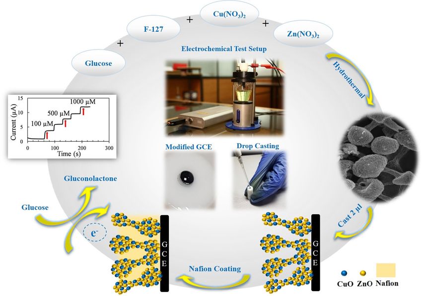
www.nature.com/scientificreports/
Enzyme-free glucose sensors seem to be promising alternatives that eliminate the problems associated with
the enzymes, however, current non-enzymatic glucose sensors still require noticeable improvements in the
sensitivity and selectivity to be used for commercial purposes4.
Since the electrode surface plays an important role in designing non-enzymatic glucose sensors, synthesizing
more sensitive materials with new morphologies that improve all the sensor performance parameters without
sacrificing any of them is currently an attractive topic of research and d evelopment9. Despite the exciting pro-
gresses achieved in fabricating micro and nano structures to date, it is still desirable yet challenging to synthesis
high-quality complex multi-shelled hollow spheres including more than one metal o xides10−13.
Qi et al. have reviewed the properties and applications of multi-shelled hollow micro/nano structures
which shows that the additional shells can improve the performance because they provide additional sites for
interactions14. The micro/nano hollow structures provide advantages such as large contact area between electrode
and electrolyte, high permeability, more active sites, low density, and shorter path of electron transfer which are
very interesting for sensor a pplications9,15−17. Hollow structures prevent the agglomeration of nanoparticles owing
to their interior voids and this helps in better accommodation of volume changes in successive electrochemical
measurements9,17−19.
The outstanding catalytic activity of transition metal oxides such as ZnO and CuO in alkaline solutions toward
glucose detection have been reported numerously in previous researches20−23. Unlike expensive noble metals
these transition metal oxides are cost-effective, widely used for sensor fabrication, and less affected by interfering
species8,9,24. ZnO and CuO are inexpensive semiconductors, non-toxic and both have excellent electrochemical
and catalytic properties. It is possible to synthesize different morphologies of ZnO and CuO by using simple
methods and low temperatures. By tuning the process parameters, the morphologies of these metal oxides can
be controlled. These parameters include the carbon template source, the concentration of metal salts, and the
time of the hydrothermal process.
Because of the variety of morphologies and high surface area to volume ratio of ZnO and CuO nanostructures,
they are known as promising materials and a universal choice of researchers for building high-performance
glucose sensors6.
Also, ZnO nanostructures with favorable characteristics such as large surface area, high crystallinity, and
good electrical properties can be modified by other nanomaterials to provide enhanced sensing properties.
On the other hand, the CuO nanostructures have unique advantages such as the high surface area, low density,
presence of active sites even in the interior space, and favorable permeation25. ZnO is a low-cost, broad bandgap
energy (3.37 eV), high-mobility, non-toxic mineral compound considered as an n-type semiconductor. CuO is
a p-type semiconductor with a narrow bandgap (1.2–1.9 eV) that can enhance the electrochemical properties in
combination with ZnO26,27. Because of their excellent electrochemical behavior, most of the works have used one
of the CuO or ZnO nanostructures individually to develop their biosensors. More recently, hybrid nanostructured
materials based on ZnO have been p roposed6,22,27−31. They exhibit larger surface-to-volume ratios which in turn
activates the electrocatalytic properties of the materials further. Besides, because of the high electron mobilities in
ZnO based hybrid materials, the transfer of the electrons to the supporting electrodes becomes easier and faster6.
Some researchers have studied the hybrid and hollow structures toward glucose detection by designing new
metal oxides m orphologies9. Recently, non-enzymatic glucose sensors have been fabricated using Cu/Cu2O/
CuO32, Cu2O33, core-sheath nanocarbon20, double-shelled C uCo2O434, CuO/Cu2O composite35, CuxS36, NiS37,
NiO , CO3O4 hollow spheres, hollow CuO/Pani , carbon nanofibers42, cubic CuOx/NiOy nanocomposite9,
38,39 40 41
cubic NiO porous architecture17, and N iCo2O4 Nanocages43. Ahmad et al. directly grew ZnO nanorods (NRs) on
the FTO electrode surface (ZnO NRs/FTO) using a low-temperature hydrothermal method and functionalized
them with CuO (CuO-ZnO NRs/FTO electrode) to enhance the electrochemical activity for glucose detection.
The electrochemical sensing of CuO-ZnO NRs/FTO electrode exhibited a high sensitivity of 2961.7 μA.mM−1.
cm−2, linear range up to 8.45 mM, and a low limit of detection (LOD) of 0.40 μM6. Zhou et al. obtained a three-
dimensional (3D) porous ZnO–CuO hierarchical nanocomposites (HNCs) through one-step electrospinning
together with ordinary hot press and calcination process. The optimum ZnO–CuO HNCs electrode presented
a high sensitivity of 3066.4 µA mM−1 cm−2, a linear range up to 1.6 mM, and a practical LOD of 0.21 m M28.
Ridhuan et al. fabricated ZnO nanorods on a seeded substrate which were prepared by the sol–gel method,
wherein a seed layer was coated onto ITO substrates. The Nafion/GOx/ZnO NRs/ITO electrode which was used
as a modified electrode for glucose detection displayed a linear response to glucose ranging from 0.05 to 1 mM,
with a sensitivity of 48.75 μA/mM5.
In this paper, a hybrid material has been achieved via the hydrothermal method based on the combination
of two transition metal oxides (CuO/ZnO) in a form of dumbbell-shaped double-shelled hollow nanoporous
microstructures and also used as a non-enzymatic high sensitivity glucose biosensor. Our sensor benefits from
enhanced sensing characteristics due to the appropriate combination of two kinds of semiconductor oxides.
The excellent electrochemical performance of CuO/ZnO-DSDSHNM as a non-enzymatic glucose sensor was
verified when outstanding sensor parameters were obtained. As another step in the enrichment of our scientific
report, we also fabricated pure ZnO and CuO hollow spheres separately and showed that the sensing ability of
the ZnO-CuO hybrid material is much higher than them.
Results and discussion
Structural and morphological properties. The proposed non-enzymatic glucose sensor was designed
based on Nafion/CuO/ZnO-DSDSHNM/GCE. As brought into the methods section, the unique structure of
CuO/ZnO-DSDSHNM was achieved via the hydrothermal method. The CuO/ZnO-DSDSHNM has been used
as the sensing material which has been deposited on a glassy carbon electrode (GCE) by using a casting pro-
cedure. The schematic representation of the sensor construction steps is summarized in Fig. 1. The SEM of as-
Scientific Reports | (2021) 11:344 | https://doi.org/10.1038/s41598-020-79460-2 2
Vol:.(1234567890)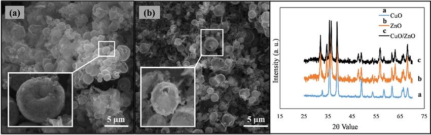
www.nature.com/scientificreports/
Figure 1. Schematic illustration of the construction and application of CuO/ZnO-DSDSHNM non-enzymatic
biosensor for the determination of glucose in human blood samples.
synthesized CuO/ZnO-DSDSHNM is shown in Fig. 2a,b. As the main morphology of the material particles, the
dumbbell shape structures are clearly observed. In many cases, the size of the heads at each side of the dumbbell
is not equal (asymmetrical dumbbell). The hollow nature of the synthesized structure is signified at the inset in
Fig. 2a. The hollow nature of these particles could be directly related to the presence of carbon microspheres as a
hard template. During the hydrothermal process, the carbon microspheres (Formed in situ from glucose precur-
sor) served as the template for the fabrication of the final hollow multi-shelled nanostructure. The removal of
the template during the calcination step under the air atmosphere is responsible for the formation of a hollow
multi-shelled nanostructure by the strategy of “hard template synthesis”. Using the oxygen-containing air as the
calcination atmosphere causes the conversion of the template to CO and facilitates the removal of the carbon
template. According to the SEM micrograph at the inset of Fig. 2b, the nanostructured details of each particle
could be observed. In addition, the integration of nanoparticles to form the final micron-sized particles has
rendered the nanoporous nature of the synthesized particles which is also clear in the SEM micrograph. The
formation of the nanoporous surfaces could be attributed to the presence of pluronic F-127 as a surfactant in the
synthesis procedure. The holes over some particles are remarked by arrows in Fig. 2a which implies the hollow
nature of these particles. Further observation utilizing transmission electron microscopy (TEM) confirmed the
results obtained from the analysis of the SEM images. The TEM images indicate the double-shelled structures of
CuO/ZnO. As can be seen in Fig. 2c,d, the dumbbell-shaped CuO/ZnO structures are composed of two layers.
The inner shell diameter is 170.74 nm and the outer shelled diameter is 393.63 nm (Fig. 2d). Figure 3a,b repre-
sent the SEM images of separate CuO and ZnO microspheres (CuO-MS and ZnO-MS), respectively. Accord-
ing to the SEM micrograph of the CuO microstructures, the formation of bean-like particles with an average
diameter of 1.50 µm from small nanoparticulate blocks could be observed (Fig. 3a)44. In the case of ZnO micro-
structures (Fig. 3b) it has been shown that they have an average size of 1.50 µm. Also, the nanoparticulate, as
well as the hollow nature of the synthesized structures, has been observed. The X-ray diffraction spectroscopy
(XRD) of CuO-MS, ZnO-MS, and CuO/ZnO-DSDSHNM are represented in Fig. 3C. The XRD pattern of CuO
corresponds to the JCPDS # 00–045-0937. The diffraction peaks at 2θ values of 32.60, 35.60, 38.80, 48.92, 53.68,
58.40, 61.68, 66.48, 68.20, 72.72 and 75.28 could be indexed as (− 110), (002), (111), (− 202), (020), (202), (− 113),
(− 311), (− 220), (311) and (− 222) planes, respectively44. According to diffractogram b, the ZnO XRD planes
at 2θ values of 31.80, 34.40, 36.25, 47.50, 56.60, 62.95, 66.50 and 68.05 could be indexed as (100), (002), (101),
(102), (110), (103), (200) and (201) planes in correspondence with JCPDS # 00–036-1451. Finally, the XRD pat-
tern of CuO/ZnO-DSDSHNM was illustrated as diffractogram c. It is also obvious that the characteristic peaks
of both CuO and ZnO at 2θ values of 34.40, 47.50, 62.95, 66.50 and 68.05 are remarkable in the diffractogram
of CuO/ZnO-DSDSHNM. According to obtained XRD peaks, the phase purity of CuO and ZnO are conserved
and the corresponding peaks are obvious.
Scientific Reports | (2021) 11:344 | https://doi.org/10.1038/s41598-020-79460-2 3
Vol.:(0123456789)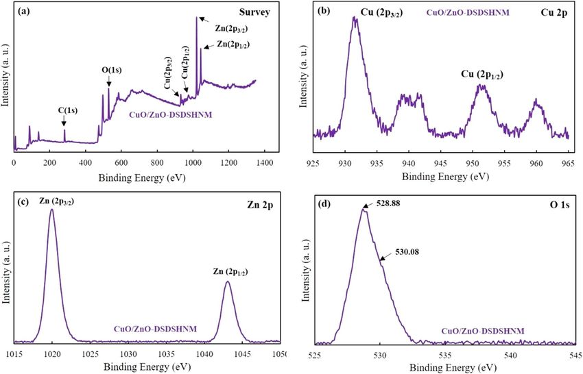
www.nature.com/scientificreports/
Figure 2. (a) And (b) typical SEM micrographs of CuO/ZnO-DSDSHNM, (C), and (d) TEM images of CuO/
ZnO-DSDSHNM.
Figure 3. SEM micrographs of (a) CuO-MS and (b) ZnO-MS, (c) XRD patterns of as-synthesized CuO/ZnO-
DSDSHNM.
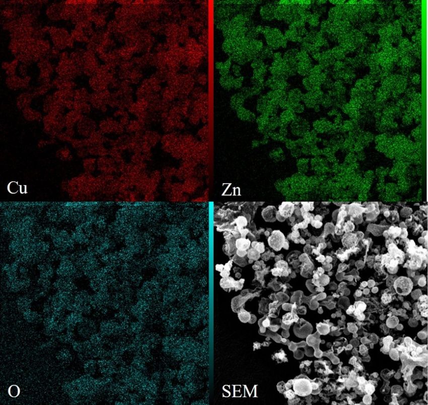
The inductively coupled plasma atomic emission spectroscopy (ICP-AES) showed that the atomic ratio of
Cu over Zn is 1.4 which shows a very close distribution. In addition, the elemental mapping of the CuO/ZnO-
DSDSHNM is depicted in Fig. 4. The uniform distribution of Cu (red), Zn (green), and O (blue) over the entire
sample could be deduced. It can be seen that the ICP results are in almost good agreement with the elemental
mapping results. Conclusively, the homogeneous structure of particles as a composite of CuO and ZnO materi-
als has been verified.
X-ray photoelectron spectroscopy (XPS) of the samples was studied to further investigate the chemical state
of the composing elements and also the surface composition of the CuO/ZnO-DSDSHNM samples. The XPS
Scientific Reports | (2021) 11:344 | https://doi.org/10.1038/s41598-020-79460-2 4
Vol:.(1234567890)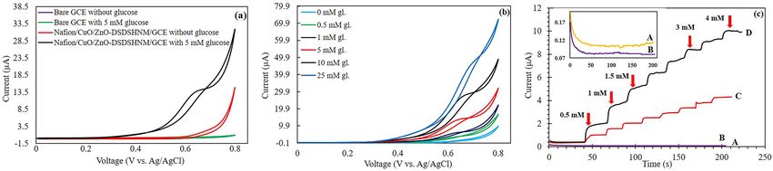
www.nature.com/scientificreports/
Figure 4. The elemental mapping technique of CuO/ZnO-DSDSHNM.
results have been shown in Fig. 5. The survey spectrum illustrates that Cu, Zn, O, and C are present in the sam-
ples which verifies the successful synthesis of CuO/ZnO-DSDSHNM (Fig. 5a). There are two dominant peaks
in the spectrum of Cu at 931.68 eV (Cu 2 p3/2) and 951.58 eV (Cu 2 p1/2). These peaks can respectively be fitted
to Cu2+ (930.98 and 950.98 eV) and C u+ (932.18 and 953.38 eV). The presence of the component CuO has been
validated by the existence of two satellite peaks at ∼940 and ∼960 eV that correspond to the Cu2+ oxidation state
(Fig. 5b)4,45−47. As can be seen in Fig. 5c, high-resolution Zn 2p spectra of CuO/ZnO-DSDSHNM showed two
symmetric peaks. The presence of the Zn in the matrix (in + 2 oxidation state) can be concluded from the peaks
at ∼1020.08 eV (Zn 2 p3/2) and ∼1043.28 eV (Zn 2 p1/2)48. Since the O 1 s peak is asymmetric (Fig. 5d), it can be
concluded that the surface is comprised of two components of the oxygen. One of the components located at
∼528.88 eV is attributed to the O2− ions bonded with Z n2+ or Cu+/Cu2+ ions, whereas the second one which is
2−
located at ∼530.08 eV belongs to the O ions in oxygen-deficient regions. The obtained XPS results verify that
the oxygen vacancy defects are present on the surface of the synthesized samples49−51.
Electrochemical properties. The GCE has been modified with the synthesized hollow structures and the
Nafion/CuO/ZnO-DSDSHNM/GCE was used as a modified working electrode for measurements. The electro-
chemical CV tests for different working electrodes were performed in 0.50 M NaOH solution at the scan rate
of 50 mVs−1 within the potential range of 0 to + 0.80 V. The CVs shown in Fig. 6a illustrate the responses of the
bare and modified GCE in the absence and presence of 5 mM glucose. The CVs shown in Fig. 6a illustrates the
responses of the bare and modified GCE in the absence and presence of 5 mM glucose. As shown in Fig. 6a, in the
absence of glucose, no response can be observed for bare GCE and Nafion/CuO/ZnO-DSDSHNM/GCE. How-
ever, when glucose is added, a distinguishable response was revealed, which demonstrated the reasonable perfor-
mance of CuO/ZnO-DSDSHNM toward glucose detection. The CVs shown in Fig. 6a illustrates the responses
of the bare and modified GCE in the absence and presence of 5 mM glucose. As shown in Fig. 6a, in the absence
of glucose, no response can be observed for bare GCE and Nafion/CuO/ZnO-DSDSHNM/GCE. However, when
glucose is added, a distinguishable response was revealed, which demonstrated the reasonable performance of
CuO/ZnO-DSDSHNM toward glucose detection. The CV responses for the CuO/ZnO-DSDSHNM modified
GCE to different glucose concentrations have been presented in Fig. 6b. As can be seen, by increasing the glucose
concentration from 0 to 25 mM the current has also been increased. As Fig. 6b demonstrates, there are remark-
able differences between the CVs which shows that the fabricated sensor is very sensitive to the changes in the
glucose concentration and the sensor is reacting very sensitively to the increase in the glucose concentration. In
Scientific Reports | (2021) 11:344 | https://doi.org/10.1038/s41598-020-79460-2 5
Vol.:(0123456789)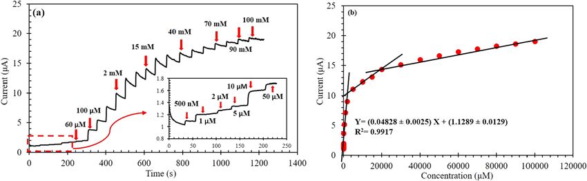
www.nature.com/scientificreports/
Figure 5. XPS spectra of CuO/ZnO-DSDSHNM showing full scan survey (a) and corresponding peaks in the
high-resolution spectra for Cu 2p (b), Zn 2p (c), and O 1 s (d).
Figure 6. (a) The CV responses of the bare and the modified GCE with CuO/ZnO-DSDSHNM at the scan
rate of 50 mVs−1 in the absence and presence of 5 mM glucose. (b)The CVs of Nafion/CuO/ZnO-DSDSHNM/
GCE at the scan rate of 50 m Vfs−1 in 0.5 M NaOH solution at different glucose concentrations. (c) i–t curves of
(A) bare GCE, (B) Nafion/ZnO-MS/GCE, (C) Nafion/CuO-MS/GCE, and (D) Nafion/CuO/ZnO-DSDSHNM/
GCE by successive addition of 0.50 mM of glucose to the final concentration of 4 mM. The inset plot of (c) is the
magnified view of the (A) bare GCE and (B) Nafion/ZnO-MS/GCE by successive addition glucose in the range
of 0.50–4 mM.
order to examine the behavior of the sensor quantitatively, we performed chronoamperometric tests to deter-
mine the calibration curve and other performance parameters. The results of the amperometry tests are shown
in Fig. 6c. The electrochemical properties of ZnO-MS, CuO-MS, CuO/ZnO-DSDSHNM, and bare GCE have
been studied in Fig. 6c and provided trustworthy chemical information. The amperometry tests were performed
in the potential of + 0.60 V by the successive addition of glucose from 0.50 to 4 mM. As can be seen in the inset
plot of Fig. 6c, bare GCE and ZnO-MS have no electrochemical activity for glucose detection. CuO-MS modified
GCE has a weaker electrochemical activity and a lower sensitivity versus glucose concentration44 in comparison
to the CuO/ZnO-DSDSHNM. Comparing the responses of CuO and ZnO with our proposed hybrid material
CuO/ZnO in Fig. 6c reveals that the CuO response is much larger than ZnO. Although ZnO has no significant
response to the glucose in the non-enzymatic detection process, however, it has a strong contribution to the
enhancement of electrochemical properties of the sensor when it combines with CuO and the proposed hybrid
material (CuO/ZnO-DSDSHNM) is formed6,28. The integration of metal oxide semiconductors of p-type and
n-type enhances the concentration of charge carriers due to better charge separation. In addition, due to the
heterogeneous function, an increase in the stability and catalytic activity is a chievable26,27. As a result of Fig. 6c,
the integration of these two semiconductors provides a more favorable electrochemical activity and outstanding
responses in comparison with the Nafion/CuO-MS/GCE electrode.
Scientific Reports | (2021) 11:344 | https://doi.org/10.1038/s41598-020-79460-2 6
Vol:.(1234567890)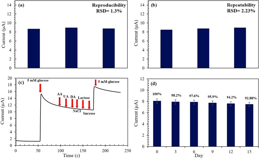
www.nature.com/scientificreports/
Figure 7. CV responses of CuO/ZnO-DSDSHNM in 0.5 M NaOH at various scan rates ranging from 10 to 250
mVs−1 (10, 20, 30, 40, 50, 60, 70, 80, 90, 100, 125, 150, 175, 200, 225 and 250 m
Vs−1) in 10 mM glucose. Inset:
corresponding two plots of anodic peaks versus the (a) square root of scan rate and (b) scan rate.
The mechanism of the nonenzymatic electrochemical oxidation of glucose over CuO/ZnO-DSDSHNM elec-
trode could be explained by the semiconductive nature of CuO/ZnO52. Herein, despite several types of research
that have introduced Cu(3 +) as the active intermediate for the oxidation of glucose53, the formation of holes
over the semi-conductive surface of CuO/ZnO is considered responsible.
According to Fig. 6, the voltammetry peak related to the conversion of Cu(2 +) to Cu(3 +) was not observ-
able at CuO/ZnO-DSDSHNM in alkaline electrolyte. Instead, also in the absence of glucose, a plateau around
0.5–0.7 V vs. Ag/AgCl was observable. The capacitive nature of this plateau has been approved p reviously52
which could be attributed to the adsorption of hydroxyl ions in the electrolyte to the surface of CuO/ZnO elec-
trode as a p-type semiconductor. The sharp increase of the current after this plateau also could be attributed to
the oxidation of these adsorbed hydroxyl species to O2 in the high anodic potentials. Following the presence
of glucose in the electrolyte, it seems that this electrochemical O2 evolution is amplified and the enhancement
of plateau is remarkable. Although the well-shaped peak of glucose oxidation over CuO/ZnO-DSDSHNM was
not apparent, but the gradual increase of plateau current by successive addition of glucose concentration was
encountered (Fig. 6b). Herein, for better distinguishing of the response to various concentrations of glucose, the
chronoamperometric technique was chosen instead of cyclic voltammetry. In addition to the above-mentioned
electrochemical evidences, the presence of O vacancies over the electrode surface was previously approved by
using XPS analysis (Fig. 5).
Due to the application of the unique semiconductive hybrid nanostructures of CuO/ZnO in the construc-
tion of CuO/ZnO-DSDSHNM electrode, the adsorption of O H− species from alkaline electrolyte occurred
remarkably. In the absence of glucose, the further oxidation of these adsorbed species caused the sharp current
increase at the extreme of the anodic sweep. However, in the presence of glucose, some interactions of the ana-
lyte with the electrode surface was e xpectable52. According to interactions of glucose with the electrode surface,
the amplification of the oxidation process of O H− species occurred and in turn, the current has been increased.
The increase of current was linearly related to the glucose concentration and this has been successfully used for
sensing application.
According to Fig. 6c, the ZnO-MS modified electrode has no response to the glucose in the non-enzymatic
detection process which has no specific property of redox reaction like CuO, however, the role of ZnO in the
proposed sensor is to accelerate the electron transfer, which demonstrates the high performance of CuO/ZnO-
DSDSHNM as evidenced in Fig. 6c.
To survey the kinetics of glucose oxidation on the surface of the Nafion/CuO/ZnO-DSDSHNM/GCE, the
effect of the scan rates on the CV currents has been studied for 10 mM of glucose in 0.50 M NaOH solution. Fig-
ure 7 shows the CV responses of glucose oxidation at Nafion/CuO/ZnO-DSDSHNM/GCE at different potential
scan rates ranging from 10 to 250 mVs−1. As illustrated at the inset (a) of Fig. 7, the peak currents exhibited a
linear relation versus the square root of the scan rate, demonstrating that the oxidation of glucose at the modified
electrode is under the diffusion-controlled behavior. In contrast, as depicted in the inset (b) of Fig. 7, the plot of
peak currents versus the scan rate is not favorably linear which indicates the absence of adsorptive b ehavior30,31,44.
Sensor optimization. The working electrode surface should be modified with CuO/ZnO-DSDSHNM. To
enhance the performance of the sensor, it is necessary to cast an optimum amount of CuO/ZnO-DSDSHNM on
the GCE surface. Optimizing this parameter will increase the accuracy and sensitivity of the sensor. In order to
determine the best concentration of the CuO/ZnO-DSDSHNM, the CV tests were performed at different con-
centrations of the sample. According to figure S1, the optimum value of this parameter has been selected 5 mg/
ml. After that, a coating of Nafion is casted on the surface of CuO/ZnO-DSDSHNM. This coating prevents the
falling of nanoparticles from the GCE surface and prevents interfering species from penetrating. It also increases
the reproducibility and repeatability of the sensor. Therefore, as another step for optimization, it is necessary to
optimize the concentration of the Nafion. Different concentrations of Nafion were prepared and CV tests were
Scientific Reports | (2021) 11:344 | https://doi.org/10.1038/s41598-020-79460-2 7
Vol.:(0123456789)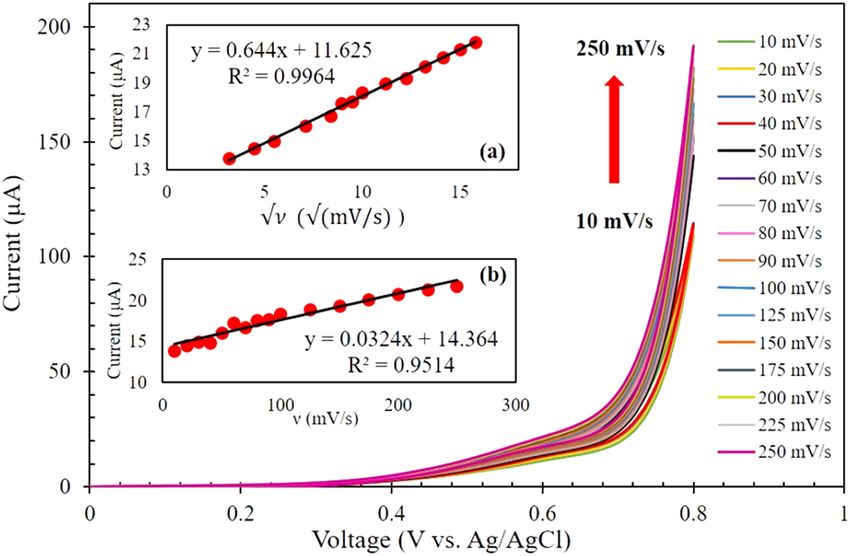
www.nature.com/scientificreports/
Figure 8. (a) Amperometric response of Nafion/CuO/ZnO-DSDSHNM/GCE at 0.60 V in 0.5 M NaOH
solution by successive addition of glucose concentration from 500 nM to 100 mM. Inset: the magnified view of
the low concentration range of glucose (500 nM–50 μM). (b) Calibration plot of the sensor responses versus the
glucose concentrations.
carried out in the presence of 5 mM glucose. Figure S2 shows the results of the different Nafion concentrations
and accordingly, it is obvious that the optimum concentration is 0.50 wt%. Because the GCE and the CuO/
ZnO-DSDSHNM will be oxidized in the absence of the analyte in NaOH solution, several consecutive cycles of
the CV measurements have been performed. These cycles were repeated in the absence of glucose until they are
overlapped. This way we are sure that by adding glucose, the response of the electrode is only due to the glucose
oxidation. The measurements showed that after 10 cycles the proposed sensor behaves in exactly the same way
as the last cycle. Thus, to stabilize the results, 10 cycles of CVs must be run before the test starts. After the elec-
trode activation, the best voltage range for improved sensing abilities should be selected. This is one of the most
important parameters that is very effective in the performance of the sensor. Based on figure S3, the best results
for modified GCE were observed in the range of 0–0.80 V. Optimizing the NaOH concentration results in the
maximum production of OH−, in which case the oxidation of glucose will be as efficient as possible. According
to figure S4, the linear increasing trend of current versus glucose concentration was observed in 0.50 M NaOH
solution which also resulted in a higher current. As another design parameter, the best voltage of the glucose
oxidation for the amperometry test has to be determined which enables the reliable performance of the sensor.
In order to do that, the current–time (i-t) curves of Nafion/CuO/ZnO-DSDSHNM/GCE with the addition of
5 mM glucose in 0.5 M NaOH solution were acquired at different oxidation voltages (Figure S5). According to
figure S5, the highest amount of glucose oxidation occurs at a voltage of 0.60 V.
Non‑enzymatic glucose sensing based on CuO/ZnO‑DSDSHNM. For further highlighting and
confirming the superior electro-catalytic activity of CuO/ZnO-DSDSHNM, amperometry test was carried out.
Figure 8a demonstrates the typical i-t curve of CuO/ZnO-DSDSHNM with successive addition of different glu-
cose concentrations at an optimum applied potential of + 0.60 V under the stirring condition in 0.5 M NaOH
solution. By successive additions of various glucose concentrations from 500 nM to 100 mM the consecutive
increase in current has been observed. In addition, the inset of Fig. 8a illustrates the magnified view of the low
concentration ranges of glucose. Obviously, the first current steps appeared for 500 nM glucose, suggesting
the high detection sensitivity of the sensor. The corresponding calibration curve can be described by a power
plot (Figure S6) which could be broken into three linear ranges with the following regression equation for the
first linear range: I(μA) = (0.04828 ± 0.0025) C(μM) + (1.1289 ± 0.0129) (R2 = 0.9917) for 0.5 µM–2 mM glucose
(Fig. 8b). The sensitivity can be calculated as the ratio of the slope to the electrode area. The novel electrochemi-
cal glucose biosensor was designed, constructed, and optimized so that it has a wide dynamic range from 500 nM
to 100 mM which included three linear ranges 500 nM–2 mM, 2 mM–20 mM, and 20 mM–100 mM, and an
outstanding sensitivity of 1536.80 ± 79.60 µA mM−1 cm−2. Upon the addition of 500 nM of glucose, the Nafion/
CuO/ZnO-DSDSHNM/GCE reached a steady-state with a very sharp response. It raised to 90% of the response
within 1.60 s which is highly favorable comparing with similar sensors which are brought in Table 1. Also, the
limit of detection is calculated as 357.5 nM which shows that the sensor has provided a very low detection limit.
This indicates that the proposed sensor has a high capability for fast electrocatalytic detection of glucose.
The analytical performance of the as-prepared CuO/ZnO-DSDSHNM sensing system for glucose detection is
superior to or comparable with many previously enzymatic and non-enzymatic glucose s ensor4,30,31,54 (Table 1).
Reproducibility, repeatability, selectivity, and stability tests. Reproducibility, repeatability, selec-
tivity, and stability are critical parameters for the investigation of the performance of sensing devices. Repeat-
ability means the absence of incompatibility between consecutive measurements by the same electrode, whilst
reproducibility is the vicinity of the results acquired by means of a number of the same modified electrodes with
the same measurement procedure. The CV responses of three different electrodes with successive addition of
5 mM glucose were recorded at the optimal condition. The relative standard deviation (RSD) of the response
currents for different CuO/ZnO-DSDSHNM electrodes was only 1.3%, suggesting good reproducibility and
precision (Fig. 9a). The repeatability was examined by three times of measurements by a single modified GCE
Scientific Reports | (2021) 11:344 | https://doi.org/10.1038/s41598-020-79460-2 8
Vol:.(1234567890)www.nature.com/scientificreports/
Sensitivity (μA. Detection limit Dynamic range
Electrode Voltage (V) mM−1.cm−2) (μM) (mM) Response time (s) References
Nafion/CuO/ZnO-
0.60 1536.80 0.357 0.0005–100 1.60 This work
DSDSHNM/GCE
Nafion/GOx/ZnO/ 48.75 0.05–1.00 5
− 0.70 60 10
ITO 3.87 1.00–20.00
6
CuO-ZnO NRs/FTO 0.62 2961.70 0.40 0.001–8.45www.nature.com/scientificreports/
Sample (mM) Founded (mM) RSD (%) Recovery (%)
2.57 2.63 0.12 102.33
4.09 3.92 0.97 95.84
5.80 6.08 0.1 104.83
8.47 8.63 0.4 101.89
Table 2. Recovery and relative standard deviation (RSD) of Nafion/CuO/ZnO-DSDSHNM/GCE for detecting
glucose in a real serum sample.
of the sample into the 30 ml of 0.50 M NaOH at the potential of + 0.60 V (versus Ag/AgCl). The analysis was
performed via the standard addition method. The same aliquots of serum samples were spiked by the known
concentrations of glucose and 30 µl of each sample was injected into the 30 ml of NaOH solution. The electro-
chemical analysis of each sample was done by using Nafion/CuO/ZnO-DSDSHNM/GCE three times. Anywhere
which was necessary, the volume correction was performed. The Nafion/CuO/ZnO-DSDSHNM/GCE glucose
sensor electrode provided an acceptable recovery ranging from 95.84 to 104.83% for the human blood serum
samples as shown in Table 2.
Conclusion
A new double-shelled hollow nanoporous CuO/ZnO microstructure was successfully fabricated by a hydro-
thermal method, designed for non-enzymatic electrochemical detection of glucose. As a result, an efficient
and rapid electrochemical glucose biosensor was fabricated and optimized. The results revealed that the sensor
optimizations performed have been successful and the proposed sensor showed a wide dynamic detection range
of 500 nM–100 mM, remarkable sensitivity of 1536.8 ± 79.6 µA mM−1 cm−2, and very low LOD of 357.5 nM. The
Nafion/CuO/ZnO-DSDSHNM modified GCE exhibited excellent reproducibility and repeatability, favorable
stability, and satisfactory selectivity toward glucose sensing in a 0.5 M NaOH solution. The cost-effective synthesis
procedure and the outstanding catalytic performance suggest that the CuO/ZnO-DSDSHNM material may be
promising for non-enzymatic glucose sensing in practical analysis applications.
Methods
Materials. Zinc Nitrate (Zn(NO3)2), Copper Nitrate (Cu(NO3)2), Sodium Hydroxide (NaOH), Aluminum
Oxide (Al2O3), AA, DA, lactose, sucrose, NaCl, Nafion, glucose, and pluronic F-127 were purchased from Sigma-
Aldrich, Merck and DuPont and human blood serum was taken from the hospital. In this study, all the materials
were of analytical grade and used as received without any further purification. All solutions were prepared by DI
water (3 μS/cm) generated from the Deltino water purifying system.
Apparatus. XRD was taken by BRUKER D8 Advanced. SEM and elemental mapping data were obtained
by using TESCAN Mira3. The TEM images were taken with digital charge-coupled device JEOL-JEM-2010
equipped with a CCD camera. XPS experiments were measured using an ESCAlab 250 Analytical XPL Spec-
trometer with a monochromatic Al Ka source. ICP-AES test was performed by ICP-MS ELANDRC-E Perkin-
Elmer.The Electrochemical measurements were performed by using IVIUM Vertex potentiostat/galvanostat
electrochemical analyzer. All experiments were conducted using a three-electrode system with a glassy carbon-
based working electrode (2 mm diameter), an Ag/AgCl reference electrode (3 M KCl), and a platinum wire for
the counter electrode (1 cm length and 1 mm diameter). The CVs and choronoamperometric measurements
were performed with an electrochemical analyzer in a 0.50 M NaOH solution.
Synthesis of materials. Three types of hollow structured materials have been synthesized. As the main
material, a unique dumbbell-shaped double-shelled hollow nanoporous CuO/ZnO microstructures was synthe-
sized. For comparison of electrocatalytic activity, the CuO-MS and ZnO-MS also were synthesized via a similar
method. The procedure for synthesizing the above-mentioned materials are as follows:
First, 3 g of glucose, 1 g of F-127 as a surfactant were dissolved in 150 ml deionized water and the solution
was sonicated for 10 min. Then, 2 ml of 1 M of Zn(NO3)2 solution and 2 ml of 1 M of Cu(NO3)2 solution were
added to the mixture. The mixture of glucose, surfactant and both metal salts was placed into the autoclave and
the hydrothermal process was directed for 24 h at 180 ֯C. The resulted product was washed three times with
distilled water and dried in an oven at 80 ֯C overnight. The final structure was achieved via the calcination of the
product from the hydrothermal step at 550 ֯C for 270 min.
The synthesis of CuO-MS and ZnO-MS was achieved via the same procedure with the same amount of rea-
gents but by using the single salt of each metal at processes.
Electrode preparation (Nafion/CuO/ZnO‑DSDSHNM/GCE). At first step, the GCE was polished by
means of alumina slurry over a Pad, and washed three times with DI water and ethanol, respectively. To fabricate
the Nafion/CuO/ZnO-DSDSHNM/GCE, the solution of 5 mg/ml of synthesized material in ethanol was pre-
pared and ultrasonically homogenized for 20 min. After that, 2 μl of CuO/ZnO-DSDSHNM solution was casted
on the surface of GCE until it dried at room temperature. Next, 2 μl of 0.5 wt% Nafion solution was casted on
the catalyst layer at GCE and then dried at ambient conditions. Henceforth, the Nafion/CuO/ZnO-DSDSHNM/
GCE electrode was prepared for performing electrochemical measurements.
Scientific Reports | (2021) 11:344 | https://doi.org/10.1038/s41598-020-79460-2 10
Vol:.(1234567890)www.nature.com/scientificreports/
Statement. All experiments and methods were performed in accordance with relevant guidelines and regu-
lations. All experimental protocols were approved by the ethics committee of the laboratory of Shahid Beheshti
Hospital. Informed consent was obtained from all subjects.
Received: 24 August 2020; Accepted: 8 December 2020
References
1. Clinic, M. The essential diabetes book. In Oxmoor houses (2012).
2. Stokes, A. & Preston, S. H. Deaths attributable to diabetes in the United States: comparison of data sources and estimation
approaches. PLoS ONE 12, e0170219 (2017).
3. Onay, A. et al. Amperometric glucose sensor based on the glucose oxidase enzyme immobilized on graphite rod electrode modified
with Fe3O4-CS-Au magnetic nanoparticles. Ionics 24, 4015–4022 (2018).
4. Foroughi, F., Rahsepar, M., Hadianfard, M. J. & Kim, H. Microwave-assisted synthesis of graphene modified CuO nanoparticles
for voltammetric enzyme-free sensing of glucose at biological pH values. Microchim. Acta 185, 57 (2018).
5. Ridhuan, N. S., Abdul Razak, K. & Lockman, Z. Fabrication and characterization of glucose biosensors by using hydrothermally
grown ZnO nanorods. Sci. Rep. 8, 1–12 (2018).
6. Ahmad, R. et al. Highly efficient non-enzymatic glucose sensor based on CuO modified vertically-grown ZnO nanorods on
electrode. Sci. Rep. 7, 1–10 (2017).
7. Anusha, J. R. et al. Amperometric glucose biosensor based on glucose oxidase immobilized over chitosan nanoparticles from
gladius of Uroteuthis duvauceli. Sens. Actuators, B 215, 536–543 (2015).
8. Cheng, C. et al. 3D Network and 2D paper of reduced graphene oxide/Cu2O composite for electrochemical sensing of hydrogen
peroxide. Anal. Chem. 90, 1983–1991 (2017).
9. Long, L., Liu, X., Chen, L., Li, D. & Jia, J. A hollow CuOx/NiOy nanocomposite for amperometric and non-enzymatic sensing of
glucose and hydrogen peroxide. Microchim. Acta 186, 74 (2019).
10. Khalifeh, R., Karimi, M., Rajabzadeh, M., Hafizi, A. & Nogorani, F. S. (2020) Synthesis and morphology control of nano CuAl2O4
hollow spheres and their application as an efficient and sustainable catalyst for CO2 fixation. J. CO2 Util. 41, 101233 (2020).
11. Rajabzadeh, M., Khalifeh, R., Eshghi, H. & Bakavoli, M. A facile hydrothermal synthesis of novel hollow triple-shell C uNiFe2O4
nanospheres with robust catalytic performance in the Suzuki-Miyaura coupling reaction. J. Catal. 360, 261–269 (2018).
12. Wang, H. et al. (2020) Dual-defects adjusted crystal-field splitting of LaCo 1–x NixO3−δ hollow multishelled structures for efficient
oxygen evolution. Angew. Chem. Int. Ed. 59(44), 19691–19695 (2020).
13. Wang, H. et al. Hollow multishelled structure of heterogeneous C o3O4–CeO2−x nanocomposite for CO catalytic oxidation. Adv.
Funct. Mater. 29, 1806588 (2019).
14. Qi, J. et al. Multi-shelled hollow micro-/nanostructures. Chem. Soc. Rev. 44, 6749–6773 (2015).
15. Sohn, J. H., Cha, H. G., Kim, C. W., Kim, D. K. & Kang, Y. S. Fabrication of hollow metal oxide nanocrystals by etching cuprous
oxide with metal(ii) ions: approach to the essential driving force. Nanoscale 5, 11227 (2013).
16. Nai, J., Tian, Y., Guan, X. & Guo, L. Pearson’s principle inspired generalized strategy for the fabrication of metal hydroxide and
oxide nanocages. J. Am. Chem. Soc. 135(43), 16082–16091 (2013).
17. He, G. et al. Sensitive nonenzymatic electrochemical glucose detection based on hollow porous NiO. Nanoscale Res. Lett. 13, 3
(2018).
18. Li, H., Liu, A. & Che, H. Synthesis of hierarchical Cu2O octahedra@Ni(OH)2 nanosheets core-shell heterostructures for high-
performance supercapacitor. Mater. Sci. Semicond. Process. 91, 115–123 (2019).
19. Cao, C.-Y., Guo, W., Cui, Z.-M., Song, W.-G. & Cai, W. Microwave-assisted gas/liquid interfacial synthesis of flowerlike NiO hollow
nanosphere precursors and their application as supercapacitor electrodes. J. Mater. Chem. 21, 3204 (2011).
20. Abunahla, H. et al. MOMSense: metal-oxide-metal elementary glucose sensor. Sci. Rep. 9, 1–10 (2019).
21. Zhang, J., Sun, Y., Li, X. & Xu, J. Fabrication of porous NiMn2O4 nanosheet arrays on nickel foam as an advanced sensor material
for non-enzymatic glucose detection. Sci. Rep. 9, 1–13 (2019).
22. Qian, J. et al. Non-enzymatic glucose sensor based on ZnO–CeO2 whiskers. Mater. Chem. Phys. 239, 122051 (2020).
23. Gong, X. et al. High-performance non-enzymatic glucose sensors based on CoNiCu alloy nanotubes arrays prepared by electro-
deposition. Front. Mater. 6, 3 (2019).
24. Li, S.-J. et al. Enzyme-free glucose sensor using a glassy carbon electrode modified with reduced graphene oxide decorated with
mixed copper and cobalt oxides. Microchim. Acta 183, 1813–1821 (2016).
25. Wang, X., Feng, J., Bai, Y., Zhang, Q. & Yin, Y. Synthesis, properties, and applications of hollow micro-/nanostructures. Chem. Rev.
116, 10983–11060 (2016).
26. Chen, L. et al. High-performance photoelectronic sensor using mesostructured ZnO nanowires. ACS Sens. 2, 1567–1572 (2017).
27. Daemi, S., Ghasemi, S. & Ashkarran, A. A. Electrospun CuO–ZnO nanohybrid: tuning the nanostructure for improved ampero-
metric detection of hydrogen peroxide as a non-enzymatic sensor. J. Colloid Interface Sci. 550, 180–189 (2019).
28. Zhou, C. et al. Ultrasensitive non-enzymatic glucose sensor based on three-dimensional network of ZnO–CuO hierarchical
nanocomposites by electrospinning. Sci. Rep. 4, 7382 (2014).
29. SoYoon, S., Ramadoss, A., Saravanakumar, B. & Kim, S. J. Novel Cu/CuO/ZnO hybrid hierarchical nanostructures for non-
enzymatic glucose sensor application. J. Electroanal. Chem. 717–718, 90–95 (2014).
30. Muthuchamy, N., Atchudan, R., Edison, T. N. J. I., Perumal, S. & Lee, Y. R. High-performance glucose biosensor based on green
synthesized zinc oxide nanoparticle embedded nitrogen-doped carbon sheet. J. Electroanal. Chem. 816, 195–204 (2018).
31. Zhang, Y. et al. Three-dimensional macroporous carbon supported hierarchical ZnO-NiO nanosheets for electrochemical glucose
sensing. J. Alloys Compd. 698, 800–806 (2017).
32. Lin, L.-Y., Karakocak, B. B., Kavadiya, S., Soundappan, T. & Biswas, P. A highly sensitive non-enzymatic glucose sensor based on
Cu/Cu2O/CuO ternary composite hollow spheres prepared in a furnace aerosol reactor. Sens. Actuators, B 259, 745–752 (2018).
33. Chen, K. et al. Synthesis of hollow nanospherical cuprous oxide supported by nitrogen-doped reduced graphene oxide and its
application to enzyme-free glucose sensing. ChemistrySelect 4, 7027–7034 (2019).
34. Cheng, D., Wang, T., Zhang, G., Wu, H. & Mei, H. A novel nonenzymatic electrochemical sensor based on double-shelled C uCo2O4
hollow microspheres for glucose and H2O2. J. Alloys Compd. 819, 153014 (2020).
35. Zhang, F. et al. Facile synthesis of uniform CuO/Cu2O composite hollow microspheres for a non-enzymatic glucose sensor. Mater.
Res. Express 6, 115049 (2019).
36. Cao, M. et al. Highly efficient non-enzymatic glucose sensor based on CuxS hollow nanospheres. Appl. Surf. Sci. 492, 407–416
(2019).
Scientific Reports | (2021) 11:344 | https://doi.org/10.1038/s41598-020-79460-2 11
Vol.:(0123456789)www.nature.com/scientificreports/
37. Wei, C. et al. NiS hollow spheres for high-performance supercapacitors and non-enzymatic glucose sensors. Chem. Asian J. 10,
679–686 (2015).
38. Huang, W. et al. 3D NiO hollow sphere/reduced graphene oxide composite for high-performance glucose biosensor. Sci. Rep. 7,
1–11 (2017).
39. Liu, S., Yu, B. & Zhang, T. A novel non-enzymatic glucose sensor based on NiO hollow spheres. Electrochim. Acta 102, 104–107
(2013).
40. Yin, H., Nie, Q., Cui, Z. & He, X. Hollow C o3O4 microspheres assembled with nanocrystals for non-enzymatic glucose sensor.
Micro Nano Lett. 11, 151–155 (2016).
41. Liu, T. et al. Fabrication of hollow CuO/PANI hybrid nanofibers for non-enzymatic electrochemical detection of H2O2 and glucose.
Sens. Actuators, B 286, 370–376 (2019).
42. Li, Y., Cai, R., Lü, R., Gao, L. & Qin, S. Template synthesis of the C u2O nanoparticle-doped hollow carbon nanofibres and their
application as non-enzymatic glucose biosensors. R. Soc. Open Sci. 5, 181474 (2018).
43. Feng, Y. et al. MOF-derived spinel N iCo2O4 hollow nanocages for the construction of non-enzymatic electrochemical glucose
sensor. Electroanalysis 32, 571–580 (2019).
44. Haghparas, Z., Kordrostami, Z., Sorouri, M., Rajabzadeh, M. & Khalifeh, R. Fabrication of non-enzymatic electrochemical glucose
sensor based on nano-copper oxide micro hollow-spheres. Biotechnol. Bioprocess Eng. 25(4), 528–535 (2020).
45. Ghijsen, J. et al. Electronic structure of C u2O and CuO. Phys. Rev. B 38, 11322 (1988).
46. Wang, X., Ge, C.-Y., Chen, K. & Zhang, Y. X. An ultrasensitive non-enzymatic glucose sensors based on controlled petal-like CuO
nanostructure. Electrochim. Acta 259, 225–232 (2018).
47. Chakraborty, P., Dhar, S., Debnath, K. & Mondal, S. P. Glucose and hydrogen peroxide dual-mode electrochemical sensing using
hydrothermally grown CuO nanorods. J. Electroanal. Chem. 833, 213–220 (2019).
48. Xia, W. et al. Nanoplate-built ZnO hollow microspheres decorated with gold nanoparticles and their enhanced photocatalytic and
gas-sensing properties. ACS Appl. Mater. Inter. 7, 11824–11832 (2015).
49. Kim, J., Kim, W. & Yong, K. CuO/ZnO heterostructured nanorods: photochemical synthesis and the mechanism of H2S gas sens-
ing. J. Phys. Chem. C 116, 15682–15691 (2012).
50. Ahmad, R. et al. Ammonium ion detection in solution using vertically grown ZnO nanorod based field-effect transistor. RSC Adv.
6, 54836–54840 (2016).
51. Wang, Y. et al. Visible-light-responsive T iO2-coated ZnO: I nanorod array films with enhanced photoelectrochemical and photo-
catalytic performance. ACS Appl. Mater. Inter. 7, 6093–6101 (2015).
52. Barragan, J. T., Kogikoski, S. Jr., da Silva, E. T. & Kubota, L. T. Insight into the electro-oxidation mechanism of glucose and other
carbohydrates by CuO-based electrodes. Anal. Chem. 90, 3357–3365 (2018).
53. Zheng, W., Li, Y. & Lee, L. Y. S. Insights into the transition metal ion-mediated electrooxidation of glucose in alkaline electrolyte.
Electrochim. Acta 308, 9–19 (2019).
54. Reitz, E., Jia, W., Gentile, M., Wang, Y. & Lei, Y. CuO nanospheres based nonenzymatic glucose sensor. Electroanalysis 20, 2482–
2486 (2008).
Acknowledgements
The authors would like to thank Dr. Hosein Sharifi, Ph.D. of medical bacteriology for his technical assistance
with the real human blood samples.
Author contributions
Z.H. performed the electrochemical tests, prepared the results and the figures and authored the initial manuscript
text. Z.K. authored the introduction and made substantial additions to the text. Z.K. and M.S. supervised and
analyzed the electrochemical tests and their results and edited and reviewed the discussions, tables and figures.
M.R. and R.K. synthezed the ZnO/CuO hybrid hollow material and prepared the characterizations. All authors
reviewed the manuscript.
Competing interests
The authors declare no competing interests.
Additional information
Supplementary Information The online version contains supplementary material available at https://doi.
org/10.1038/s41598-020-79460-2.
Correspondence and requests for materials should be addressed to Z.K. or M.S.
Reprints and permissions information is available at www.nature.com/reprints.
Publisher’s note Springer Nature remains neutral with regard to jurisdictional claims in published maps and
institutional affiliations.
Open Access This article is licensed under a Creative Commons Attribution 4.0 International
License, which permits use, sharing, adaptation, distribution and reproduction in any medium or
format, as long as you give appropriate credit to the original author(s) and the source, provide a link to the
Creative Commons licence, and indicate if changes were made. The images or other third party material in this
article are included in the article’s Creative Commons licence, unless indicated otherwise in a credit line to the
material. If material is not included in the article’s Creative Commons licence and your intended use is not
permitted by statutory regulation or exceeds the permitted use, you will need to obtain permission directly from
the copyright holder. To view a copy of this licence, visit http://creativecommons.org/licenses/by/4.0/.
© The Author(s) 2021
Scientific Reports | (2021) 11:344 | https://doi.org/10.1038/s41598-020-79460-2 12
Vol:.(1234567890)You can also read