The Rubisco Small Subunit Is Involved in Tobamovirus Movement and Tm-22-Mediated Extreme Resistance1 C W OA
←
→
Page content transcription
If your browser does not render page correctly, please read the page content below
The Rubisco Small Subunit Is Involved in Tobamovirus
Movement and Tm-22-Mediated Extreme Resistance1[C][W][OA]
Jinping Zhao, Qi Liu2, Haili Zhang, Qi Jia, Yiguo Hong, and Yule Liu*
Ministry of Education Key Laboratory of Bioinformatics, School of Life Sciences, Tsinghua University, Beijing
100084, China (J.Z., Q.L., H.Z., Q.J., Y.L.); and Research Centre for Plant RNA Signaling, School of Life and
Environmental Sciences, Hangzhou Normal University, Hangzhou 310036, China (Y.H.)

The multifunctional movement protein (MP) of Tomato mosaic tobamovirus (ToMV) is involved in viral cell-to-cell movement,
symptom development, and resistance gene recognition. However, it remains to be elucidated how ToMV MP plays such
diverse roles in plants. Here, we show that ToMV MP interacts with the Rubisco small subunit (RbCS) of Nicotiana
benthamiana in vitro and in vivo. In susceptible N. benthamiana plants, silencing of NbRbCS enabled ToMV to induce necrosis
in inoculated leaves, thus enhancing virus local infectivity. However, the development of systemic viral symptoms was delayed.
In transgenic N. benthamiana plants harboring Tobacco mosaic virus resistance-22 (Tm-22), which mediates extreme resistance to
ToMV, silencing of NbRbCS compromised Tm-22-dependent resistance. ToMV was able to establish efficient local infection but
was not able to move systemically. These findings suggest that NbRbCS plays a vital role in tobamovirus movement and plant
antiviral defenses.

   Plant viruses use at least one movement protein (MP)                        proteins Tobacco mosaic virus resistance-2 (Tm-2) and
to facilitate viral spread between plant cells via plasmo-                     Tm-22 (Meshi et al., 1989; Lanfermeijer et al., 2004). In-
desmata (PD; Lucas and Gilbertson, 1994; Ghoshroy                              deed, tomato Tm-22 confers extreme resistance against
et al., 1997). Among viral MPs, the MP of tobamoviruses,                       TMV and ToMV in tomato plants and even in heterol-
such as Tobacco mosaic virus (TMV) and its close relative                      ogous tobacco (Nicotiana tabacum) plants (Lanfermeijer
Tomato mosaic virus (ToMV), is the best characterized.                         et al., 2003, 2004).
TMV MP specifically accumulates in PD and modifies                                  To date, several host factors that interact with
the plasmodesmatal size exclusion limit in mature source                       TMV MP have been identified. These TMV MP-
leaves or tissues (Wolf et al., 1989; Deom et al., 1990;                       binding host factors include cell wall-associated
Ding et al., 1992). TMV MP and viral genomic RNA form                          proteins such as pectin methylesterase (Chen et al.,
a mobile ribonucleoprotein complex that is essential for                       2000), calreticulin (Meshi et al., 1989), ANK1 (Ueki
cell-to-cell movement of viral infection (Watanabe et al.,                     et al., 2010), and the cellular DnaJ-like protein MPIP1
1984; Deom et al., 1987; Citovsky et al., 1990, 1992;                          (Shimizu et al., 2009). Many cytoskeletal components
Kiselyova et al., 2001; Kawakami et al., 2004; Waigmann                        such as actin filaments (McLean et al., 1995), micro-
et al., 2007). TMV MP also enhances intercellular RNA                          tubules (Heinlein et al., 1995), and the microtubule-
silencing (Vogler et al., 2008) and affects viral symptom                      associated proteins MPB2C (Kragler et al., 2003) and
development, host range, and host susceptibility to virus                      EB1a (Brandner et al., 2008) also interact with TMV MP.
(Dardick et al., 2000; Bazzini et al., 2007). Furthermore,
                                                                               Most of these factors are involved in TMV cell-to-cell
ToMV MP is identified as an avirulence factor that is
                                                                               movement.
recognized by tomato (Solanum lycopersicum) resistance
                                                                                  Rubisco catalyzes the first step of CO2 assimilation in
                                                                               photosynthesis and photorespiration. The Rubisco holo-
   1                                                                           enzyme is a heteropolymer consisting of eight large
     This work was supported by the National Basic Research Pro-
gram of China (grant no. 2011CB910100) and the National Natural                subunits (RbCLs) and eight small subunits (RbCSs).
Science Foundation of China (grant nos. 31270182 and 31000838).                RbCL was reported to interact with the coat protein of
   2
     Present address: Biochemistry Program, Ohio State University,             Potato virus Y (Feki et al., 2005). Both RbCS and RbCL
Columbus, OH 43210.                                                            were reported to interact with the P3 proteins encoded by
   * Corresponding author; e-mail yuleliu@mail.tsinghua.edu.cn.                several potyviruses, including Shallot yellow stripe virus,
   The author responsible for distribution of materials integral to the        Onion yellow dwarf virus, Soybean mosaic virus, and Turnip
findings presented in this article in accordance with the policy de-
                                                                               mosaic virus (Lin et al., 2011). Proteomic analysis of the
scribed in the Instructions for Authors (www.plantphysiol.org) is:
Yule Liu (yuleliu@mail.tsinghua.edu.cn).
                                                                               plant-virus interactome revealed that RbCS participates
   [C]
       Some figures in this article are displayed in color online but in        in the formation of virus complexes of Rice yellow mottle
black and white in the print edition.                                          virus (Brizard et al., 2006). However, the biological func-
   [W]
       The online version of this article contains Web-only data.              tion of Rubisco in viral infection remains unknown.
   [OA]
        Open Access articles can be viewed online without a subscription.         In this study, we show that RbCS plays an essential
   www.plantphysiol.org/cgi/doi/10.1104/pp.112.209213                          role in virus movement, host susceptibility, and Tm-22-
374       Plant PhysiologyÒ, January 2013, Vol. 161, pp. 374–383, www.plantphysiol.org Ó 2012 American Society of Plant Biologists. All Rights Reserved.

                                    Downloaded on December 22, 2020. - Published by https://plantphysiol.org
                                   Copyright (c) 2020 American Society of Plant Biologists. All rights reserved.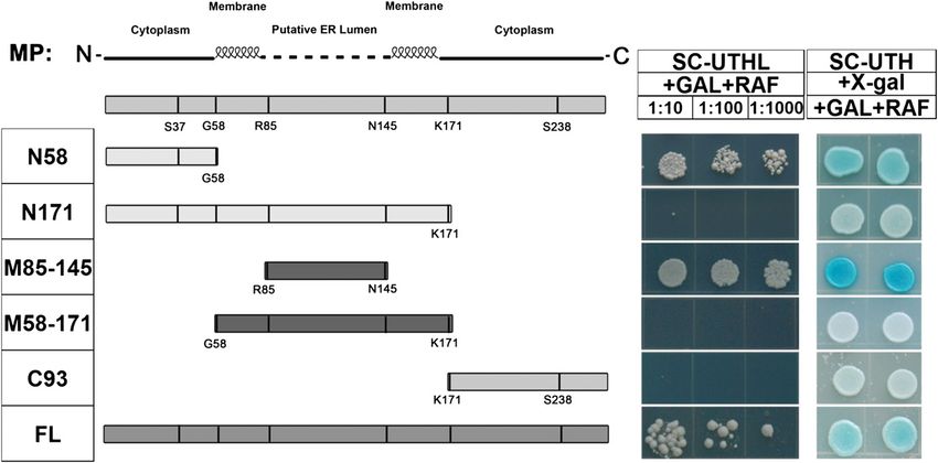
Role of the Rubisco Small Subunit in Viral Infection

mediated extreme resistance in the ToMV-host plant
interaction.

RESULTS
NbRbCS Interacts with ToMV MP

   To identify ToMV MP-interacting proteins, we used
ToMV MP as bait and conducted yeast (Saccharomyces
cerevisiae) two-hybrid (Y2H) screens of a tomato com-
plementary DNA (cDNA) library previously con-
structed in the Y2H prey vector (Liu et al., 2002b).
From the screens, we obtained a number of clones that
encode proteins that may interact with ToMV MP.
Three of these candidates were identified as tomato
RbCS cDNAs. To test whether Nicotiana benthamiana
RbCS (NbRbCS) also interacts with ToMV MP, we
cloned the full-length NbRbCS cDNA (N. benthamiana
tentative consensus annotator no. TC23245) in the Y2H
activation domain (AD) vector to produce AD::NbRbCS.
Indeed, BD::ToMV MP interacted specifically with AD::
NbRbCS, as indicated by the growth of yeast on Leu–
plates containing Gal (Fig. 1A). However, as the controls,
AD::NbRbCS and bait LexA binding domain (BD; BD
vector), AD vector and BD::ToMV MP, as well as AD
vector and BD vector showed no interaction (Fig. 1A).
   The ToMV MP-NbRbCS interaction was further con-
firmed by glutathione S-transferase (GST) pull-down
assays. GST-ToMV MP and NbRbCS-33FLAG-63His                                Figure 1. ToMV MP and NbRbCS interacted in yeast and in vitro. A,
fusion proteins were separately expressed in Escherichia                   Growth of yeast strains containing NLS-LexA BD (control) or NLS-LexA
coli. The GST-ToMV MP fusion protein was purified                           BD ToMV MP baits transformed with AD-NbRbCS or AD (control) on
                                                                           Leu-deficient medium containing Gal and raffinose (Raf; left) or Glu
using glutathione-Sepharose beads and then mixed with
                                                                           (right) at 28˚C for 3 d. Yeast cells were plated at 10- and 100-fold
total soluble proteins of E. coli expressing the NbRbCS-                   dilutions. B, GST pull-down assay to confirm the interaction between
33FLAG-63His fusion protein or 33FLAG-63His con-                           ToMV MP and NbRbCS in vitro. The total soluble proteins of E. coli
trol. Western-blot analysis using an anti-FLAG antibody                    expressing the NbRbCS-33FLAG-63His fusion protein or an empty
showed that only GST-ToMV MP specifically pulled                            vector 33FLAG-63His were incubated with purified GST-ToMV MP or
down NbRbCS-33FLAG-63His, demonstrating that                               GST immobilized on glutathione-Sepharose beads. Beads were
ToMV MP was able to bind to NbRbCS (Fig. 1B).                              washed and analyzed by SDS-PAGE and western-blot (WB) assays
   In vivo pull-down assays were performed to test if                      using anti-FLAG antibody (top panel). The bottom panel shows inputs
ToMV MP directly interacts with NbRbCS in planta.                          of purified fusion proteins in pull-down assays. Equal aliquots of glu-
ToMV MP tagged with a Myc epitope (ToMV MP-Myc)                            tathione beads loaded with GST-ToMV MP or GST were separated by
                                                                           SDS-PAGE and stained with Coomassie Brilliant Blue. Black arrows
and NbRbCS tagged with a hemagglutinin (HA) pep-
                                                                           indicate the bands corresponding to GST-ToMV MP and GST. [See
tide (HA-NbRbCS) were expressed in plants under the                        online article for color version of this figure.]
control of Cauliflower mosaic virus 35S promoter. Ex-
pression of ToMV MP-Myc was confirmed by western-                           N-Terminal and Middle Domains of ToMV MP
blot analysis with anti-Myc antibody (Fig. 2, bottom).                     Are Responsible for Its Interaction with NbRbCS
Total proteins extracted from leaves coinfiltrated with
Agrobacterium tumefaciens carrying the ToMV MP-Myc                            TMV MP can be divided into N-terminal, middle, and
expression cassette together with A. tumefaciens carrying                  C-terminal regions joined by two putative a-helical
either HA vector or HA-NbRbCS expression cassettes                         transmembrane domains (Brill et al., 2000). The
were immunoprecipitated using an anti-HA antibody.                         N-terminal and C-terminal regions are thought to be
The resulting precipitates were analyzed by western blot                   cytoplasmic, and the middle region of MP is presumed
using an anti-Myc antibody. ToMV MP-Myc was only                           to fold in the endoplasmic reticulum (ER) lumen in plant
detected in the immunoprecipitations with HA-NbRbCS                        cells (Brill et al., 2000, 2004). Moreover, the N-terminal
but not in immunoprecipitations with the control HA                        and middle regions of MPs are conserved among toba-
(Fig. 2, top), although the anti-HA antibody could pull                    moviruses (Supplemental Fig. S1). According to this
down both HA-NbRbCS and control HA proteins (Fig.                          structural model, we generated a series of ToMV MP-
2, middle). These experiments clearly demonstrate that                     deletion mutants in order to determine which amino acid
ToMV MP interacts with NbRbCS in plant cells.                              domain(s) is responsible for ToMV MP binding to
Plant Physiol. Vol. 161, 2013                                                                                                                 375

                                 Downloaded on December 22, 2020. - Published by https://plantphysiol.org
                                Copyright (c) 2020 American Society of Plant Biologists. All rights reserved.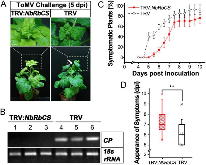
Zhao et al.
Subcellular Localization of the NbRbCS-ToMV
MP Interaction
The subcellular localization of the NbRbCS-ToMV MP
interaction was studied by citrine yellow fluorescent
protein (YFP)-based bimolecular fluorescence comple-
mentation (BiFC) assays, where YFP was split into
N-terminal (nYFP) and C-terminal (cYFP) fragments for
protein fusion (Burch-Smith et al., 2007). When cYFP-
NbRbCS was transiently coexpressed with ToMV
MP-nYFP in N. benthamiana, bright fluorescent signals
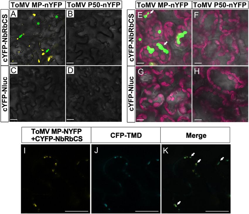
were observed in mesophyll and epidermal cells. The
YFP fluorescence was observed mainly in amorphous
aggresomes in the cytoplasm (Fig. 4, A and E, arrows),
while some fluorescence was also found in punctate
structures that were likely to be PD in the cell walls of
the epidermal cells (Fig. 4A, asterisks). Indeed, the
punctate-localized YFP fluorescence from the ToMV
MP-RbCS interaction coincided with the PD regions
labeled by the cyan fluorescent protein (CFP)-tagged
PD marker CFP-TMD (for transmembrane domain of
Arabidopsis plasmodesmata-located protein1a [PDLP1a];
Thomas et al., 2008; Fig. 4, I–K). Interestingly, the
subcellular localization of the NbRbCS-ToMV MP in-
teraction was similar to that of ToMV MP itself. ToMV
MP-YFP accumulated primarily in PD along cell walls
and partially in cytosolic inclusion bodies (referred to
as virus replication complexes [VRCs]; Supplemental
Fig. S2), as described previously (Kahn et al., 1998;
Ashby et al., 2006). RbCS is a nucleus-encoded chlo-
roplast protein (Chua and Schmidt, 1978; Smith and
Figure 2. In vivo pull-down assay for the interaction between ToMV MP Ellis, 1979); however, no signal for NbRbCS-ToMV MP
and NbRbCS. Immunoprecipitation (IP) was performed on extracts interaction was detected in chloroplasts (Fig. 4G).
containing transiently expressed 33HA-NbRbCS and ToMV MP-43Myc Moreover, no interaction signals were observed for
or extracts containing transiently expressed ToMV MP-43Myc and cYFP-NbRbCS with nYFP-ToMV P50 (Fig. 4, B and F)
33HA by using protein A/G agarose beads incubated with anti-HA or for various control combinations (Fig. 4, C, D, G, and
antibody. After immunoprecipitation, precipitates were analyzed by
H). These results demonstrate that the interaction of
western blot using anti-Myc (top panel) or anti-HA (middle panel) an-
tibody. The expression of ToMV MP-43Myc was confirmed by western-
ToMV MP with NbRbCS may occur at PD and VRCs.
blot analysis with anti-Myc antibody (bottom panel). IB, Immunoblot.
Silencing of NbRbCS Enables ToMV to Induce Local
NbRbCS. The Y2H assays demonstrated that the Necrosis But Reduces Virus Systemic Movement
N-terminal region of ToMV MP (amino acid residues
1–58) interacted with NbRbCS, as indicated by growth To examine the biological relevance of the NbRbCS-
of yeast on Leu– plates containing Gal and raffinose ToMV MP interaction to ToMV infection of plants, we
and blue yeast colonies on plates containing 5-bromo-4- used the Tobacco rattle virus (TRV) virus-induced gene
chloro-3-indolyl b-D-galactopyranoside. The middle silencing (VIGS) vector (Liu et al., 2002a) to silence
region (amino acid residues 85–145) also interacted with NbRbCS in N. benthamiana. Western-blot assays showed
NbRbCS (Fig. 3). By contrast, no interaction was ob- that silencing of NbRbCS dramatically reduced NbRbCS
served between NbRbCS and the C-terminal fragment protein levels (Fig. 5A). We also noted that silencing of
(amino acid residues 171–264) of ToMV MP. Surpris- NbRbCS caused leaf chlorosis and upward leaf curling
ingly, there was also no interaction between NbRbCS (Fig. 5B, top panels).
and a longer N-terminal polypeptide (amino acid res- To test the role of RbCS in virus infection, when
idues 1–171) or a longer middle amino acid section NbRbCS-silencing phenotypes had fully established in
(amino acid residues 58–171). These two polypeptides N. benthamiana plants, the plants were mechanically
contain the dual transmembrane domains, which may inoculated with ToMV and monitored for viral spread
disturb the NbRbCS-binding activity of ToMV MP. As and symptom development. In the NbRbCS-silenced
expected, the full-length ToMV MP protein did interact plants, ToMV induced necrosis in the inoculated leaves
with NbRbCS (Fig. 3). These results suggest that the at approximately 60 h post infection (Fig. 6, top row,
N-terminal and middle domains of ToMV MP are red arrows). However, the noninfective plant sap
responsible for its interaction with RbCS. control (Supplemental Fig. S3A) and TMV-GFP
376 Plant Physiol. Vol. 161, 2013
Downloaded on December 22, 2020. - Published by https://plantphysiol.org
Copyright (c) 2020 American Society of Plant Biologists. All rights reserved.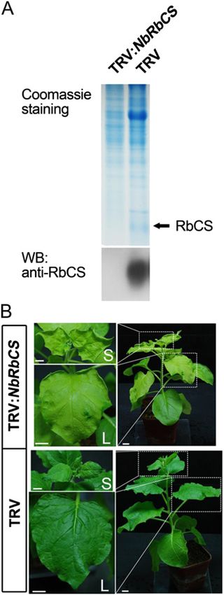
Role of the Rubisco Small Subunit in Viral Infection

          Figure 3. Analysis of NbRbCS interaction with ToMV MP deletions. The ToMV MP deletion derivatives used in Y2H assays are
          depicted in the left diagram. Boxes in the top bar represent the coding region, and the amino acid residues and relative positions in
          ToMV MP are indicated under each construct. The bars on the bottom indicate the segmental and full-length derivatives used in
          each assay. For each hybrid, dilutions of yeast cultures at 1021, 1022, and 1023 were spotted onto Leu-deficient (left) or 5-bromo-4-
          chloro-3-indolyl b-D-galactopyranoside (X-gal)-containing (right) plates and grown for 3 d at 28˚C. SC-UTH, Synthetic complete
          medium lacking uracil, Trp, and His; SC-UTHL, synthetic complete medium lacking uracil, Trp, His and Leu; RAF, raffinose.

(Supplemental Fig. S4A, top row) did not induce any                             control plants had systemic ToMV infection at 5.5 dpi
necrosis in the NbRbCS-silenced plants, suggesting that                         (Fig. 7, C and D). Furthermore, ToMV RNA can be
ToMV specifically induced necrosis. Moreover, necrosis                           detected by reverse transcription (RT)-PCR at 5 dpi in
spread out along proximate veins and resulted in the                            the upper uninoculated leaves of TRV control plants
collapse of large leaf patches or even the entire leaf                          with viral symptoms but not in the upper uninocu-
lamina. However, no necrosis was observed in NbRbCS-                            lated leaves of NbRbCS-silenced plants without viral
nonsilenced TRV control plants (Fig. 6, bottom row). In                         symptoms (Fig. 7B), supporting the idea that the viral
addition, NbRbCS silencing increased the number of                              symptoms can be used as an indicator of the presence
TMV-GFP infection foci in the inoculated leaves and the                         of ToMV. Systemic symptoms appeared at 7 dpi in
percentage of total infected leaf area (Supplemental Fig.                       almost 80% of nonsilenced control plants compared
S4), although it reduced the size of a single TMV-GFP                           with approximately 45% of NbRbCS-silenced plants.
focus (Supplemental Fig. S4). These results suggest that                        Furthermore, more than 20% of NbRbCS-silenced plants
silencing of NbRbCS may enhance host susceptibility to                          remained symptomless, but all NbRbCS nonsilencing
tobamoviruses.                                                                  plants became systemically infected with ToMV at 10 dpi
   To follow up on the local infection results, we next                         (Fig. 7C). The general tendency was that systemic in-
examined the effect of NbRbCS silencing on systemic                             fection occurred 1 to 2 d later in NbRbCS-silenced
trafficking of ToMV. Viral systemic symptom de-                                  plants than in nonsilenced plants (Fig. 7D).
velopment can be used as an indicator to monitor                                   Taken together, these findings suggest that silencing
long-distance trafficking of ToMV and other plant                                of NbRbCS enhances the local susceptibility of N.
viruses (Leisner et al., 1993; Li et al., 2005; Lewis and                       benthamiana to ToMV infection but reduces ToMV
Lazarowitz, 2010). In N. benthamiana, ToMV systemic                             long-distance movement.
infection resulted in curved apical shoots, crinkled ap-
ical leaves, chlorotic mottling of newly emerged leaves,
and backward winding of young leaves (Fig. 7A, right                            Silencing of NbRbCS Compromised Tm-22-Mediated
column). Surprisingly, we found that NbRbCS silencing                           Extreme Resistance against ToMV
delayed the appearance of ToMV systemic symptoms.
Systemic symptoms were first observed in approxi-                                   Enhanced host susceptibility to viruses in NbRbCS-
mately 5% of NbRbCS-silenced plants at 5.5 d post in-                           silenced plants suggests that NbRbCS may have a role
fection (dpi), but more than 40% of nonsilenced TRV                             in plant antiviral defense. To test this hypothesis, we
Plant Physiol. Vol. 161, 2013                                                                                                                     377

                                 Downloaded on December 22, 2020. - Published by https://plantphysiol.org
                                Copyright (c) 2020 American Society of Plant Biologists. All rights reserved.
Zhao et al.

Figure 4. In vivo subcellular localization of the
NbRbCS-ToMV MP interaction by BiFC assay. A to
H, NbRbCS and control N-terminal luciferase
(Nluc) were tagged at the N terminus with cYFP
(cYFP-NbRbCS and cYFP-nLUC) and were coex-
pressed transiently with ToMV MP or control ToMV
P50 tagged at the C terminus with nYFP (ToMV MP-
nYFP and ToMV P50-nYFP) in N. benthamiana
leaves, and any fluorescence was observed in epi-
dermal (A–D) and mesophyll (E–H) cells by confo-
cal microscopy at 2 dpi. The NbRbCS interaction
with ToMV MP was observed in cytoplasmic ag-
gregates (indicated with arrows) and in discrete
punctate sites (indicated with asterisks) along the
cell edges. I to K, BiFC constructs and the PD
marker CFP-tagged TMD (CFP-TMD) were coex-
pressed in N. benthamiana leaf epidermal cells. YFP
channel (I), CFP channel (J), and merged images (K)
show that good colocalization existed between the
subcellular localization of the ToMV MP-NbRbCS
interaction (YFP) and CFP-TMD in discrete punctate
sites along the cell border. Green fluorescence
shows sites with the two YFP and CFP fluorescence
merged. Arrows indicate the positions of overlain
YFP and CFP fluorescence. Bars = 20 mm.

used RT-PCR to examine the effect of silencing                             (data not shown). These data suggest that NbRbCS is
NbRbCS on the expression of the defense-related PR1a                       required for Tm-22-mediated local extreme resistance.
gene. As seen in Supplemental Figure S5, PR1a RNA
levels were reduced in the NbRbCS-silenced plants
compared with the nonsilenced TRV control plants                           DISCUSSION
agroinfiltrated with TRV empty VIGS vector. How-
ever, the level of 18S rRNA was not affected by                            RbCS May Interact with MPs from Many Tobamoviruses
NbRbCS gene expression. These results suggest that                            Through a range of in vitro and in vivo protein-
NbRbCS may participate in antiviral defenses in plants.                    protein interaction assays, we have identified NbRbCS
   To further test this idea, we investigated whether                      as a new ToMV MP-interacting host protein. NbRbCS
the ToMV MP-NbRbCS interaction has a role in Tm-22                         interacted with the N-terminal and middle domains
gene-mediated resistance against ToMV infection. This                      of ToMV MP. These domains are conserved among
experiment was performed in transgenic Tm-22 N.                            tobamovirus MPs, suggesting that RbCS may com-
benthamiana line T1-4, which is extremely resistant to                     monly interact with MPs from tobamoviruses. Indeed,
both ToMV and TMV (J. Zhao and Y. Liu, unpublished                         we found that NbRbCS associated and interacted with
data; Supplemental Materials and Methods S1). It                           TMV MP in planta (Supplemental Figs. S6 and S7;
should be noted that ToMV MP is the corresponding                          Supplemental Results S1). RbCS is translated in the cy-
avirulence protein that is recognized by the Tm-22 gene                    toplasm and then imported into chloroplasts (Keegstra
product. As expected, the NbRbCS-nonsilenced T1-4                          et al., 1995; Schatz and Dobberstein, 1996). However,
plants were extremely resistant to ToMV infection,                         we found that NbRbCS interacts with ToMV and TMV
and no hypersensitive response (HR) was observed in                        MPs in the cytoplasm but not in the chloroplast. It is
the inoculated leaves (Fig. 8A, bottom row). However,                      possible that both ToMV and TMV MPs may form a
ToMV induced HR necrosis in the inoculated leaves of                       protein complex with NbRbCS and, consequently, hi-
NbRbCS-silenced T1-4 plants at 4 to 5 dpi (Fig. 8A, top                    jack this protein prior to its entry into the chloroplast
row, arrows). In addition, the noninfective plant sap                      to facilitate virus infection of plants.
control did not induce any necrosis in the NbRbCS-
silenced T1-4 plants (Supplemental Fig. S3B). Further-
more, ToMV RNA was readily detectable by RT-PCR at                         Role of RbCS in Host Susceptibility to Virus Infection
4 dpi in the inoculated leaves of NbRbCS-silenced plants                   and in Plant Defense
but not in nonsilenced T1-4 plants (Fig. 8B). Moreover,
we consistently failed to detect any ToMV RNA in the                         RbCS is expressed in mesophyll cells but not in non-
upper noninoculated leaves of both NbRbCS-silenced                         photosynthetic epidermal cells (Lu et al., 2002). Removal
and control plants even at 6 weeks post ToMV infection                     of the lower RbCS-free epidermis from plant leaves
378                                                                                                                Plant Physiol. Vol. 161, 2013

                                    Downloaded on December 22, 2020. - Published by https://plantphysiol.org
                                   Copyright (c) 2020 American Society of Plant Biologists. All rights reserved.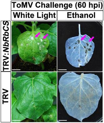
Role of the Rubisco Small Subunit in Viral Infection

                                                                           It is possible that silencing of NbRbCS completely
                                                                           disrupted Tm-22 gene-mediated systemic resistance
                                                                           against ToMV, but the virus cannot move systemi-
                                                                           cally, because the systemic movement is prevented by
                                                                           the lack of NbRbCS. However, we cannot rule out the
                                                                           possibility that Tm-22 gene-mediated local and sys-
                                                                           temic resistance can be functionally separated.
                                                                              In several cases, resistance gene-mediated HR and
                                                                           virus resistance have been found to require light
                                                                           (Chandra-Shekara et al., 2006), and the expression of
                                                                           Rubisco genes, including RbSC, is light dependent. It is
                                                                           well known that dark-treated plants are more sus-
                                                                           ceptible to TMV infection and that light treatments
                                                                           decrease plant susceptibility to TMV infection (Helms
                                                                           and McIntyre, 1967). These findings suggest that RbCS
                                                                           may play a role in plant defense. In addition, NbRbCS
                                                                           silencing could have a general effect on plant metab-
                                                                           olism, and this effect may reduce plant defenses and
                                                                           further cause an enhanced susceptibility to viral infec-
                                                                           tions. It is possible that healthy plants have sufficient
                                                                           levels of RbCS and are thus less susceptible to infection
                                                                           by tobamoviruses. However, ToMV MP may interact
                                                                           with RbCS and disturb its function to optimize cellular
                                                                           environments that are suitable for the establishment of
                                                                           ToMV infection. On the other hand, virus infection
                                                                           could be effectively established in cells in which levels
                                                                           of RbCS are reduced as in NbRbCS-silenced plants.
                                                                              Besides RbCS, the 33-kD subunit of the oxygen-
                                                                           evolving complex (OEC) and Rab GDP dissociation
                                                                           inhibitor (GDI) are involved in TMV infection (Abbink
                                                                           et al., 2002; Kramer et al., 2011). Both the OEC 33-kD
Figure 5. Silencing of NbRbCS induces leaf chlorosis in N. ben-
thamiana plants. A, Western blot (WB) to confirm the silencing of
                                                                           subunit and GDI interact with TMV 126-kD replicase
NbRbCS at the protein level. Total protein from equal weights of           and negatively regulate host susceptibility. The OEC
NbRbCS-silenced leaves and nonsilenced TRV control leaves was              33-kD subunit was suggested to have a general role in
separated by SDS-PAGE and stained with Coomassie Brilliant Blue (top       defense, and GDI was suggested to participate in
panel). Parallel samples were detected by western blotting using anti-     vesicle trafficking to enhance the establishment of an
RbCS antibodies (bottom panel). For each sample, protein fractiona-        infection (Abbink et al., 2002). TMV MP was also
tion and immunoblotting were performed with three replications. B,         suggested to function as an RNA silencing enhancer to
Silencing of NbRbCS resulted in leaf chlorosis. Expanded upper leaves      influence host cell gene expression and thus to enhance
(L) and shoot apices (S) within the dotted rectangles are shown in
magnification on the left. Photographs were taken 3 weeks after VIGS
infiltration. Bars = 1 cm.

reduces infection by TMV or Tobacco necrosis virus
(Wieringabrants, 1981), suggesting that RbSC may have
a negative impact on these viruses. Consistent with this
hypothesis, NbRbCS silencing enabled ToMV to estab-
lish severe local infections and produce necrotic lesions
in inoculated leaves. Furthermore, silencing of NbRbCS
caused a significant increase in the numbers of local
TMV infection foci (Supplemental Fig. S4B), demon-
strating that RbCS negatively affects host susceptibility
to tobamoviruses. This conclusion is further supported
by the finding that NbRbCS silencing reduced defense-
related gene expression and compromised Tm-22 gene-                        Figure 6. Silencing of NbRbCS enables ToMV to induce local necrosis.
mediated local extreme resistance against ToMV                             ToMV induced necrosis in the inoculated leaves of NbRbCS-silenced
(Fig. 8) and TMV-GFP (Supplemental Fig. S8; Supple-                        N. benthamiana plants. Leaf photographs were taken at 60 h post in-
mental Results S1). Strikingly, NbRbCS silencing only                      fection (hpi) when necrosis appeared on NbRbCS-silenced plants (left
compromised Tm-22 gene-mediated local extreme                              column). After being cleared with ethanol, leaves were photographed
resistance but not systemic resistance against ToMV.                       again (right column). Bars = 1 cm.

Plant Physiol. Vol. 161, 2013                                                                                                              379

                                 Downloaded on December 22, 2020. - Published by https://plantphysiol.org
                                Copyright (c) 2020 American Society of Plant Biologists. All rights reserved.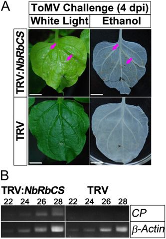
Zhao et al.

                                                                          bodies, termed X-bodies, viroplasms, or VRCs in plants
                                                                          (Heinlein et al., 1998; Más and Beachy, 1999; Szecsi
                                                                          et al., 1999). TMV-induced VRCs are ER-derived
                                                                          aggregates that contain viral RNA, 126- and 183-kD
                                                                          replicase, MP, Coat Protein (CP), and host factors (Más
                                                                          and Beachy, 1999). MP-binding NbRbCS seems to be
                                                                          one of these host factors, since we found that the
                                                                          NbRbCS interaction with MPs of ToMV and TMV
                                                                          mainly localized to irregularly shaped cytoplasmic in-
                                                                          clusion bodies (Fig. 4; Supplemental Fig. S7) and
                                                                          NbRbCS could only form irregularly shaped cytoplas-
                                                                          mic inclusion bodies when ToMV MP was coexpressed
                                                                          (Supplemental Fig. S9). Although the formation of in-
                                                                          clusion bodies from ER in infected cells might be dis-
                                                                          pensable for viral replication, VRCs may exist to
                                                                          regulate TMV RNA movement efficiency (Boyko et al.,
                                                                          2000; Waigmann et al., 2007). NbRbCS may participate
                                                                          in viral cell-to-cell movement by regulating the function
                                                                          of MP within VRCs.
Figure 7. Silencing of NbRbCS delays systemic infection by ToMV. A,          We found that silencing of NbRbCS reduces the
Silencing of NbRbCS delays the systemic symptoms induced by ToMV.         systemic movement of ToMV and TMV (Fig. 7;
Plants were photographed at 5 dpi (bottom row). The white arrow points    Supplemental Fig. S10; Supplemental Results S1). This
out the shoot curling caused by ToMV systemic infection. Shoot apices     reduction may be caused by reduced virus cell-to-cell
are shown in partially enlarged images in the top row. Bars = 1 cm. B,    movement in NbRbCS-silenced plants, because silenc-
RT-PCR detection of ToMV RNA from TRV control plants with viral
                                                                          ing of NbRbCS reduces the size of TMV infection foci
symptoms and NbRbCS-silenced plants without viral symptoms at 5 dpi.
Representative gels show RT-PCR products (32 cycles of amplification)
                                                                          (Supplemental Fig. S4, A and D). However, there is
corresponding to fragments of ToMV CP and the control 18s rRNA. C,        also a possibility that NbRbCS is involved in ToMV
Percentage of plants that displayed ToMV-induced systemic symptoms.       systemic movement through the change in flow of
All values depicted with symbols and bars in line charts are means 6 SD   photoassimilates in NbRbCS-silenced plants. It was
from six independent experiments. D, Box plots depict dpi for ToMV        proposed that plant viruses, paralleling the flow of
systemic infection. Significantly slower ToMV systemic infection was      photoassimilates, spread systemically through the
observed in NbRbCS-silenced plants compared with that in nonsilenced
TRV control plants. Asterisks above the bars denote the significance of
differences between NbRbCS-silenced plants and nonsilenced control
plants according to two-sample Student’s t test (**P , 0.01; n = 51).
Whiskers represent 5% to 95% intervals of each grouped data set, and
crosses represent maximal and minimal observations, respectively.

host susceptibility to TMV infection (Vogler et al.,
2008; Amari et al., 2012). Whether RbCS works to-
gether with the OEC 33-kD subunit and/or GDI to
affect virus infection and whether RbCS participates in
RNA silencing need to be further investigated.

Role of RbCS in Virus Movement

   Both RbCS and RbCL were reported to interact with
the P3 proteins encoded by different potyviruses (Lin
et al., 2011). Furthermore, potyvirus P3 localizes to PD
and acts in intercellular virus movement (Wei et al.,                     Figure 8. Silencing of NbRbCS compromised Tm-22-mediated local
2010; Wen and Hajimorad, 2010). These reports are                         resistance against ToMV. ToMV triggered the HR in inoculated leaves
consistent with our finding that NbRbCS interacts with                     in NbRbCS-silenced but not in nonsilenced control Tm-22 transgenic
                                                                          plants. A, Photographs were taken at 4 d post ToMV infection (left
MPs of ToMV and TMV, which are involved in viral
                                                                          column), and then the excised leaves were decolored in ethanol and
cell-to-cell movement. Indeed, silencing of NbRbCS                        photographed again (right column). Red arrows indicate the HR le-
reduced the size of TMV infection foci (Supplemental                      sions. Bars = 1 cm. B, RT-PCR detection of ToMV CP transcripts in
Fig. S4, A and D; Supplemental Results S1), suggesting                    inoculated leaves from NbRbCS-silenced and control Tm-22 transgenic
that MP-binding NbRbCS participates in cell-to-cell                       plants. The number of PCR cycles is shown above each lane. b-Actin
movement of TMV. Viral infection induces the for-                         was included as a loading control, and photographs were typical re-
mation of irregularly shaped cytoplasmic inclusion                        sults from three repeated experiments.

380                                                                                                               Plant Physiol. Vol. 161, 2013

                                   Downloaded on December 22, 2020. - Published by https://plantphysiol.org
                                  Copyright (c) 2020 American Society of Plant Biologists. All rights reserved.
Role of the Rubisco Small Subunit in Viral Infection
vascular system (Wintermantel et al., 1997; Susi et al., A. tumefaciens cultures (optical density at 600 nm = 1.0) were infiltrated into N.
benthamiana leaves. The infiltrated leaves were detached at 60 to 72 h post
1999). Silencing of NbRbCS may cause a change in the infiltration for the corresponding assays. For coexpression, equal amounts of
flow of photoassimilates because of leaf chlorosis and A. tumefaciens cultures were mixed and used for infiltration. VIGS was per-
reduced photosynthesis. Consistent with this hypothe- formed as described (Liu et al., 2002a).
sis, TMV MP does affect carbohydrate metabolism and
photoassimilate translocation during TMV infection. RNA Extraction, RT, and RT-PCR
Transgenic overexpression of TMV MP resulted in pale-
green leaves, elevated carbohydrate accumulation, and Total RNA was extracted using TRIzol reagent (Tiangen) according to the
manufacturer’s protocol. First-strand cDNA was synthesized from 2 mg of
reduced rates of photoassimilate export in source leaves RNA using a 21-nucleotide [oligo(dT) plus two anchoring nucleotides] or
(Lucas et al., 1993; Olesinski et al., 1995, 1996; Almon gene-specific primer and Moloney murine leukemia virus reverse transcriptase
et al., 1997). Furthermore, TMV MP induced alterations (Tiangen). DNA fragments corresponding to 18S rRNA, Actin, NbRbCS, ToMV
in carbon partitioning in the mesophyll and is involved MP, CP, and GFP were PCR amplified using the respective primers. The se-
quences of the primers used in PCR are available upon request.
in regulating photosynthesis. However, this process is
independent of change in the PD size-exclusion limit
(Balachandran et al., 1995; Olesinski et al., 1995). TMV Monitoring Protein Expression Levels and in Vivo
MP may also attenuate photosynthesis and carbon me- Pull-Down Assays
tabolism by affecting certain mesophyllic factors (Wolf Total proteins from N. benthamiana leaves were extracted in buffer con-
and Millatiner, 2000). Along with a decrease of Rubisco taining 150 mM Tris-HCl, 150 mM NaCl, 25 mM sodium fluoride, 0.1% Triton
during TMV infection (Itaya et al., 2002) and tobamovirus X-100, pH 7.5, 2 mM dithiothreitol, 0.5 mM phenylmethanesulfonyl fluoride
MP-RbCS physical interactions, it is possible that RbCS (PMSF), and plant protease inhibitor cocktail (Sigma-Aldrich) as described
(Serino et al., 1999). Protein extracts were separated by 10% SDS-PAGE and
may act as a potential node linking MP with photo- transferred to Immobilon-P polyvinylidene difluoride membranes (Millipore)
assimilate allocation and photosynthesis. Thus, re- for western-blot analysis using anti-RbCS, anti-HA, or anti-Myc (Santa Cruz)
duction of functional RbCS levels may account for the primary antibody. For in vivo pull-down assays, protein extracts were incu-
inability of tobamovirus to traffic over long distances. bated with the anti-HA antibody (1:200 diluted) for 4 h at 4°C following
overnight incubation with protein A/G plus agarose beads (AbMART)
equilibrated with the extraction buffer. The beads were washed three times
with the extraction buffer. Immunoprecipitated samples were used for western-
blot analysis as described above using the anti-Myc primary antibody.
MATERIALS AND METHODS
Plant Materials and Plasmids GST Pull-Down Assays
2
Transgenic line T1-4 (also called TM#1) is a Tm-2 -transformed Nicotiana GST-ToMV MP and NbRbCS-33FLAG-63His fusion proteins were pro-
benthamiana line that shows extreme resistance against ToMV and TMV duced in BL21 (DE3) cells (Stratagene) and purified using glutathione-
(J. Zhao and Y. Liu, unpublished data; Supplemental Materials and Methods S1). Sepharose beads (GE Healthcare) and nickel-nitrilotriacetic acid agarose
Vectors pTRV1 and pTRV2-LIC were described previously (Liu et al., 2002a; resins (Qiagen) according to the manufacturer’s instructions. About 1 mg of
Dong et al., 2007); pTRV2-NbRbCS was generated by cloning PCR products of purified GST fusion proteins or GST was incubated for 3 h with purified
NbRbCS cDNA into pTRV2-LIC. DNA fragments of TMV MP-nLUC, ToMV NbRbCS-33FLAG-63His or 33FLAG-63His at 4°C in 0.2 mL of buffer A (100
MP-nLUC, cLUC-NbRbCS, cYFP-NbRbCS, ToMV MP-nYFP, ToMV p50- mM NaCl, 50 mM Tris-Cl, pH 7.5, 0.1 mM EDTA, 0.1 mM EGTA, 50 mM NaF,
nYFP, and CFP-TMD were obtained by overlapping PCR. The resulting PCR 0.2% Triton X-100, 0.1% b-mercaptoethanol, 1 mM PMSF, and complete pro-
products were cloned between the duplicated Cauliflower mosaic virus 35S tease inhibitor). The beads were washed three times with ice-cold buffer B (100
promoter and Nos terminator of pJG045, a pCAMBIA1300-based T-DNA mM NaCl, 50 mM HEPES, pH 7.5, 0.1 mM EDTA, 1 mM PMSF, and complete
vector (J. Zhao and Y. Liu, unpublished data). Full-length ToMV MP was protease inhibitor) at 4°C. The washed beads were boiled in SDS sample
cloned into pGEX4T-1 vector to express GST-tagged fusion proteins in Esch- buffer, and proteins were separated by SDS-PAGE for western-blot assays
erichia coli. The NbRbCS-33FLAG-63His fusion DNA fragment was cloned using the anti-FLAG antibody.
into pET28a to express double-tagged RbCS-33FLAG-63His in E. coli.
BiFC and Fluorescence Microscopy
Y2H Screen and Interaction Assays
Citrine YFP-based BiFC was performed as described (Burch-Smith et al.,
The full-length ToMV MP gene was PCR amplified and cloned into 2007). Live plant imaging was performed on a Zeiss LSM710 confocal mi-
pYL302, in which the nuclear localization signal sequence of the LexA DNA croscope. Enhanced GFP- or citrine YFP-derived fluorescence was acquired
BD is under the control of a GAL10 promoter (Y. Liu and S.P. Dinesh-Kumar, using excitation 488-nm laser and emission 493- to 598-nm filters and excita-
unpublished data), to generate the LexA DNA BD containing bait vector BD- tion 514-nm laser and emission 519- to 587-nm filters, respectively. CFP flu-
ToMV MP. The full-length NbRbCS gene was PCR amplified and cloned into orescence was excited with a 405-nm laser line, and the emissions were
pJG4-5 (Kolonin et al., 2000) to produce the B42 DNA AD-containing prey captured at 430 to 460 nm. For colocalization assays, two fluorescent channels
vector AD-NbRbCS. The Y2H prey library containing tomato (Solanum lyco- were separately monitored in two tracks, and signals were pseudocolored in
persicum) cDNA (Liu et al., 2002b) was used to screen ToMV MP-binding digital images as described in the figure legends. Twelve-bit confocal images
proteins. The Y2H screen and interaction assays were performed as described were acquired with an EC Plan-Neofluar 103/0.30 M27 objective for 103
(Liu et al., 2002b). magnification and a Plan-Apochromat 403/0.95 Korr M27 objective for 403
magnification. Images were analyzed with ZEN 2009 Light Edition and
Image-Pro Plus 6.0 software.
Transient Expression by Agroinfiltration and VIGS
All plants were grown in pots at 22°C in a growth chamber under a 16-h- Infection and Statistical Analysis of ToMV
light/8-h-dark cycle with 120 mmol m–2 s–1 white light illumination. For
Agrobacterium tumefaciens-mediated transient expression studies, GV2260 Plants were rub inoculated with ToMV. Viral spread and symptoms were
strains containing the relevant expression vector were grown overnight, pel- monitored for at least 2 weeks. All experiments were performed at least three
leted, resuspended in infiltration buffer (10 mM MgCl2, 10 mM MES, and times, and at least three plants were used per construct each time. OriginPro 8.1
200 mM acetosyringone, pH 5.6), and kept at room temperature for 4 h. and SPSS 19.0 were used to conduct statistical analyses and to generate graphs.
Plant Physiol. Vol. 161, 2013 381
Downloaded on December 22, 2020. - Published by https://plantphysiol.org
Copyright (c) 2020 American Society of Plant Biologists. All rights reserved.Zhao et al.
Sequence data from this article can be found in the GenBank/EMBL data Brandner K, Sambade A, Boutant E, Didier P, Mély Y, Ritzenthaler C,
libraries under the following accession numbers: X02144 (ToMV); AY179605 Heinlein M (2008) Tobacco mosaic virus movement protein interacts
(Actin); JN247448 (PR1a); AF536201 (Tm22); AAF62891 (GFP); AY818369 (YFP); with green fluorescent protein-tagged microtubule end-binding protein
GU734654 (CFP); AT5G43980 (TMD); and P03583 (TMV MP). 1. Plant Physiol 147: 611–623
Brill LM, Dechongkit S, DeLaBarre B, Stroebel J, Beachy RN, Yeager M
(2004) Dimerization of recombinant tobacco mosaic virus movement
Supplemental Data protein. J Virol 78: 3372–3377
Brill LM, Nunn RS, Kahn TW, Yeager M, Beachy RN (2000) Recombinant
The following materials are available in the online version of this article. tobacco mosaic virus movement protein is an RNA-binding, alpha-
helical membrane protein. Proc Natl Acad Sci USA 97: 7112–7117
Supplemental Figure S1. Amino acid sequence alignment of tobamovirus
Brizard JP, Carapito C, Delalande F, Van Dorsselaer A, Brugidou C (2006)
MPs.
Proteome analysis of plant-virus interactome: comprehensive data for
Supplemental Figure S2. Subcellular localization of ToMV MP. virus multiplication inside their hosts. Mol Cell Proteomics 5: 2279–2297
Burch-Smith TM, Schiff M, Caplan JL, Tsao J, Czymmek K, Dinesh-
Supplemental Figure S3. Mock inoculation does not induce local necrosis Kumar SP (2007) A novel role for the TIR domain in association with
in NbRbCS-silenced plants. pathogen-derived elicitors. PLoS Biol 5: e68
Supplemental Figure S4. Silencing of NbRbCS enhances local multiplica- Chandra-Shekara AC, Gupte M, Navarre D, Raina S, Raina R, Klessig D,
tion but reduces cell-to-cell movement of TMV-GFP. Kachroo P (2006) Light-dependent hypersensitive response and resis-
tance signaling against Turnip crinkle virus in Arabidopsis. Plant J 45:
Supplemental Figure S5. Silencing of NbRbCS reduces PR1a expression. 320–334
Supplemental Figure S6. TMV MP interacts with NbRbCS in plants. Chen MH, Sheng J, Hind G, Handa AK, Citovsky V (2000) Interaction
between the tobacco mosaic virus movement protein and host cell pectin
Supplemental Figure S7. BiFC visualization of subcellular localization of methylesterases is required for viral cell-to-cell movement. EMBO J 19:
the TMV MP-NbRbCS interaction. 913–920
Supplemental Figure S8. Silencing of NbRbCS compromised Tm-2 2- Chua NH, Schmidt GW (1978) Post-translational transport into intact
mediated extreme resistance against TMV-GFP. chloroplasts of a precursor to the small subunit of ribulose-1,5-
bisphosphate carboxylase. Proc Natl Acad Sci USA 75: 6110–6114
Supplemental Figure S9. Coexpression of ToMV MP resulted in the for- Citovsky V, Knorr D, Schuster G, Zambryski P (1990) The P30 movement
mation of NbRbCS in cytosolic aggresomes and punctae along the cell protein of tobacco mosaic virus is a single-strand nucleic acid binding
wall. protein. Cell 60: 637–647
Citovsky V, Wong ML, Shaw AL, Prasad BV, Zambryski P (1992) Visu-
Supplemental Figure S10. Suppression of NbRbCS delays TMV-GFP sys-
alization and characterization of tobacco mosaic virus movement pro-
temic infection in N. benthamiana.
tein binding to single-stranded nucleic acids. Plant Cell 4: 397–411
Supplemental Materials and Methods S1. Dardick CD, Golem S, Culver JN (2000) Susceptibility and symptom de-
velopment in Arabidopsis thaliana to Tobacco mosaic virus is influenced
Supplemental Results S1.
by virus cell-to-cell movement. Mol Plant Microbe Interact 13: 1139–1144
Deom CM, Oliver MJ, Beachy RN (1987) The 30-kilodalton gene product of
tobacco mosaic virus potentiates virus movement. Science 237: 389–394
ACKNOWLEDGMENTS Deom CM, Schubert KR, Wolf S, Holt CA, Lucas WJ, Beachy RN (1990)
Molecular characterization and biological function of the movement
We thank Jian-Min Zhou for providing the luciferase complementation protein of tobacco mosaic virus in transgenic plants. Proc Natl Acad Sci
imaging assay vectors. USA 87: 3284–3288
Received October 18, 2012; accepted November 10, 2012; published November Ding B, Haudenshield JS, Hull RJ, Wolf S, Beachy RN, Lucas WJ (1992)
12, 2012. Secondary plasmodesmata are specific sites of localization of the tobacco
mosaic virus movement protein in transgenic tobacco plants. Plant Cell
4: 915–928
LITERATURE CITED Dong Y, Burch-Smith TM, Liu Y, Mamillapalli P, Dinesh-Kumar SP
(2007) A ligation-independent cloning tobacco rattle virus vector for
Abbink TE, Peart JR, Mos TN, Baulcombe DC, Bol JF, Linthorst HJ (2002) Si- high-throughput virus-induced gene silencing identifies roles for
lencing of a gene encoding a protein component of the oxygen-evolving complex NbMADS4-1 and -2 in floral development. Plant Physiol 145: 1161–1170
of photosystem II enhances virus replication in plants. Virology 295: 307–319 Feki S, Loukili MJ, Triki-Marrakchi R, Karimova G, Old I, Ounouna H,
Almon E, Horowitz M, Wang HL, Lucas WJ, Zamski E, Wolf S (1997) Nato A, Nato F, Guesdon JL, Lafaye P, et al (2005) Interaction between
Phloem-specific expression of the tobacco mosaic virus movement pro- tobacco ribulose-1,5-biphosphate carboxylase/oxygenase large subunit
tein alters carbon metabolism and partitioning in transgenic potato (Rubisco-LSU) and the PVY coat protein (PVY-CP). Eur J Plant Pathol
plants. Plant Physiol 115: 1599–1607 112: 221–234
Amari K, Vazquez F, Heinlein M (2012) Manipulation of plant host sus- Ghoshroy S, Lartey R, Sheng J, Citovsky V (1997) Transport of proteins
ceptibility: an emerging role for viral movement proteins? Front Plant and nucleic acids through plasmodesmata. Annu Rev Plant Physiol
Sci 3: 10 Plant Mol Biol 48: 27–50
Ashby J, Boutant E, Seemanpillai M, Groner A, Sambade A, Ritzenthaler Heinlein M, Epel BL, Padgett HS, Beachy RN (1995) Interaction of toba-
C, Heinlein M (2006) Tobacco mosaic virus movement protein functions movirus movement proteins with the plant cytoskeleton. Science 270:
as a structural microtubule-associated protein. J Virol 80: 8329–8344 1983–1985
Balachandran S, Hull RJ, Vaadia Y, Wolf S, Lucas WJ (1995) Alteration in Heinlein M, Padgett HS, Gens JS, Pickard BG, Casper SJ, Epel BL,
carbon partitioning induced by the movement protein of tobacco mosaic Beachy RN (1998) Changing patterns of localization of the tobacco
virus originates in the mesophyll and is independent of change in the mosaic virus movement protein and replicase to the endoplasmic re-
plasmodesmal size exclusion limit. Plant Cell Environ 18: 1301–1310 ticulum and microtubules during infection. Plant Cell 10: 1107–1120
Bazzini AA, Hopp HE, Beachy RN, Asurmendi S (2007) Infection and Helms K, McIntyre GA (1967) Light-induced susceptibility of Phaseolus
coaccumulation of tobacco mosaic virus proteins alter microRNA levels, vulgaris L. to tobacco mosaic virus infection. I. Effects of light intensity,
correlating with symptom and plant development. Proc Natl Acad Sci temperature, and the length of the preinoculation dark period. Virology
USA 104: 12157–12162 31: 191–196
Boyko V, van der Laak J, Ferralli J, Suslova E, Kwon MO, Heinlein M Itaya A, Matsuda Y, Gonzales RA, Nelson RS, Ding B (2002) Potato
(2000) Cellular targets of functional and dysfunctional mutants of spindle tuber viroid strains of different pathogenicity induces and
tobacco mosaic virus movement protein fused to green fluorescent protein. suppresses expression of common and unique genes in infected tomato.
J Virol 74: 11339–11346 Mol Plant Microbe Interact 15: 990–999
382 Plant Physiol. Vol. 161, 2013
Downloaded on December 22, 2020. - Published by https://plantphysiol.org
Copyright (c) 2020 American Society of Plant Biologists. All rights reserved.Role of the Rubisco Small Subunit in Viral Infection
Kahn TW, Lapidot M, Heinlein M, Reichel C, Cooper B, Gafny R, Beachy Meshi T, Motoyoshi F, Maeda T, Yoshiwoka S, Watanabe H, Okada Y
RN (1998) Domains of the TMV movement protein involved in subcel- (1989) Mutations in the tobacco mosaic virus 30-kD protein gene over-
lular localization. Plant J 15: 15–25 come Tm-2 resistance in tomato. Plant Cell 1: 515–522
Kawakami S, Watanabe Y, Beachy RN (2004) Tobacco mosaic virus in- Olesinski AA, Almon E, Navot N, Perl A, Galun E, Lucas WJ, Wolf S
fection spreads cell to cell as intact replication complexes. Proc Natl (1996) Tissue-specific expression of the tobacco mosaic virus movement
Acad Sci USA 101: 6291–6296 protein in transgenic potato plants alters plasmodesmal function and
Keegstra K, Bruce B, Hurley M, Li HM, Perry S (1995) Targeting of pro- carbohydrate partitioning. Plant Physiol 111: 541–550
teins into chloroplasts. Physiol Plant 93: 157–162 Olesinski AA, Lucas WJ, Galun E, Wolf S (1995) Pleiotropic effects of
Kiselyova OI, Yaminsky IV, Karger EM, Frolova OY, Dorokhov YL, tobacco-mosaic-virus movement protein on carbon metabolism in
Atabekov JG (2001) Visualization by atomic force microscopy of tobacco transgenic tobacco plants. Planta 197: 118–126
mosaic virus movement protein-RNA complexes formed in vitro. J Gen Schatz G, Dobberstein B (1996) Common principles of protein transloca-
Virol 82: 1503–1508 tion across membranes. Science 271: 1519–1526
Kolonin MG, Zhong J, Finley RL (2000) Interaction mating methods in Serino G, Tsuge T, Kwok S, Matsui M, Wei N, Deng XW (1999) Arabi-
two-hybrid systems. Methods Enzymol 328: 26–46 dopsis cop8 and fus4 mutations define the same gene that encodes sub-
Kragler F, Curin M, Trutnyeva K, Gansch A, Waigmann E (2003) MPB2C, unit 4 of the COP9 signalosome. Plant Cell 11: 1967–1980
a microtubule-associated plant protein binds to and interferes with cell- Shimizu T, Yoshii A, Sakurai K, Hamada K, Yamaji Y, Suzuki M, Namba
to-cell transport of tobacco mosaic virus movement protein. Plant S, Hibi T (2009) Identification of a novel tobacco DnaJ-like protein that
Physiol 132: 1870–1883 interacts with the movement protein of tobacco mosaic virus. Arch Virol
Kramer SR, Goregaoker SP, Culver JN (2011) Association of the Tobacco 154: 959–967
mosaic virus 126kDa replication protein with a GDI protein affects host Smith SM, Ellis RJ (1979) Processing of small subunit precursor of ribulose
susceptibility. Virology 414: 110–118 bisphosphate carboxylase and its assembly into whole enzyme are
Lanfermeijer FC, Dijkhuis J, Sturre MJ, de Haan P, Hille J (2003) Cloning stromal events. Nature 278: 662–664
and characterization of the durable tomato mosaic virus resistance gene Susi P, Pehu E, Lehto K (1999) Replication in the phloem is not necessary
Tm-2(2) from Lycopersicon esculentum. Plant Mol Biol 52: 1037–1049 for efficient vascular transport of tobacco mosaic tobamovirus. FEBS
Lanfermeijer FC, Jiang GY, Ferwerda MA, Dijkhuis J, de Haan P, Yang Lett 447: 121–123
RC, Hille J (2004) The durable resistance gene Tm-2(2) from tomato Szecsi J, Ding XS, Lim CO, Bendahmane M, Cho MJ, Nelson RS, Beachy
confers resistance against ToMV in tobacco and preserves its viral RN (1999) Development of tobacco mosaic virus infection sites in Ni-
specificity. Plant Sci 167: 687–692 cotiana benthamiana. Mol Plant Microbe Interact 12: 143–152
Leisner SM, Turgeon R, Howell SH (1993) Effects of host plant develop- Thomas CL, Bayer EM, Ritzenthaler C, Fernandez-Calvino L, Maule AJ
ment and genetic determinants on the long-distance movement of cau- (2008) Specific targeting of a plasmodesmal protein affecting cell-to-cell
liflower mosaic virus in Arabidopsis. Plant Cell 5: 191–202 communication. PLoS Biol 6: e7
Lewis JD, Lazarowitz SG (2010) Arabidopsis synaptotagmin SYTA regu- Ueki S, Spektor R, Natale DM, Citovsky V (2010) ANK, a host cytoplasmic
lates endocytosis and virus movement protein cell-to-cell transport. Proc receptor for the Tobacco mosaic virus cell-to-cell movement protein, facilitates
Natl Acad Sci USA 107: 2491–2496 intercellular transport through plasmodesmata. PLoS Pathog 6: e1001201
Li Y, Wu MY, Song HH, Hu X, Qiu BS (2005) Identification of a tobacco Vogler H, Kwon MO, Dang V, Sambade A, Fasler M, Ashby J, Heinlein
protein interacting with tomato mosaic virus coat protein and facilitat- M (2008) Tobacco mosaic virus movement protein enhances the spread
ing long-distance movement of virus. Arch Virol 150: 1993–2008 of RNA silencing. PLoS Pathog 4: e1000038
Lin L, Luo Z, Yan F, Lu Y, Zheng H, Chen J (2011) Interaction between Waigmann E, Curin M, Heinlein M (2007) Tobacco mosaic virus: a model for
potyvirus P3 and ribulose-1,5-bisphosphate carboxylase/oxygenase macromolecular cell-to-cell spread. In E Waigmann, M Heinlein, eds, Viral
(Rubisco) of host plants. Virus Genes 43: 90–92 Transport in Plants, Vol 7. Springer-Verlag, Heidelberg/Berlin, pp 29–62
Liu Y, Schiff M, Marathe R, Dinesh-Kumar SP (2002a) Tobacco Rar1, Watanabe Y, Emori Y, Ooshika I, Meshi T, Ohno T, Okada Y (1984)
EDS1 and NPR1/NIM1 like genes are required for N-mediated resis- Synthesis of TMV-specific RNAs and proteins at the early stage of in-
tance to tobacco mosaic virus. Plant J 30: 415–429 fection in tobacco protoplasts: transient expression of the 30K protein
Liu Y, Schiff M, Serino G, Deng XW, Dinesh-Kumar SP (2002b) Role of and its mRNA. Virology 133: 18–24
SCF ubiquitin-ligase and the COP9 signalosome in the N gene-mediated Wei T, Zhang C, Hong J, Xiong R, Kasschau KD, Zhou X, Carrington JC,
resistance response to Tobacco mosaic virus. Plant Cell 14: 1483–1496 Wang A (2010) Formation of complexes at plasmodesmata for potyvirus
Lu C, Koroleva OA, Farrar JF, Gallagher J, Pollock CJ, Tomos AD (2002) intercellular movement is mediated by the viral protein P3N-PIPO. PLoS
Rubisco small subunit, chlorophyll a/b-binding protein and sucrose: Pathog 6: e1000962
fructan-6-fructosyl transferase gene expression and sugar status in sin- Wen RH, Hajimorad MR (2010) Mutational analysis of the putative pipo of
gle barley leaf cells in situ: cell type specificity and induction by light. soybean mosaic virus suggests disruption of PIPO protein impedes
Plant Physiol 130: 1335–1348 movement. Virology 400: 1–7
Lucas WJ, Gilbertson RL (1994) Plasmodesmata in relation to viral Wieringabrants DH (1981) The role of the epidermis in virus-induced local
movement within leaf tissue. Annu Rev Phytopathol 32: 387–411 lesions on cowpea and tobacco leaves. J Gen Virol 54: 209–212
Lucas WJ, Olesinski A, Hull RJ, Haudenshield JS, Deom CM, Beachy RN, Wintermantel WM, Banerjee N, Oliver JC, Paolillo DJ, Zaitlin M (1997)
Wolf S (1993) Influence of the tobacco mosaic-virus 30-kda movement Cucumber mosaic virus is restricted from entering minor veins in
protein on carbon metabolism and photosynthate partitioning in trans- transgenic tobacco exhibiting replicase-mediated resistance. Virology
genic tobacco plants. Planta 190: 88–96 231: 248–257
Más P, Beachy RN (1999) Replication of tobacco mosaic virus on endo- Wolf S, Deom CM, Beachy RN, Lucas WJ (1989) Movement protein of
plasmic reticulum and role of the cytoskeleton and virus movement tobacco mosaic virus modifies plasmodesmatal size exclusion limit.
protein in intracellular distribution of viral RNA. J Cell Biol 147: 945–958 Science 246: 377–379
McLean BG, Zupan J, Zambryski PC (1995) Tobacco mosaic virus move- Wolf S, Millatiner A (2000) Effect of tobacco mosaic virus movement
ment protein associates with the cytoskeleton in tobacco cells. Plant Cell protein on photosynthesis in transgenic tobacco plants. J Plant Physiol
7: 2101–2114 156: 253–258
Plant Physiol. Vol. 161, 2013 383
Downloaded on December 22, 2020. - Published by https://plantphysiol.org
Copyright (c) 2020 American Society of Plant Biologists. All rights reserved.You can also read