Kissing of the Two Predominant Hairpin Loops in the Coxsackie B Virus 3 Untranslated Region Is the Essential Structural Feature of the Origin of ...
←
→
Page content transcription
If your browser does not render page correctly, please read the page content below
JOURNAL OF VIROLOGY, Jan. 1997, p. 686–696                                                                                    Vol. 71, No. 1
0022-538X/97/$04.0010
Copyright q 1997, American Society for Microbiology

    Kissing of the Two Predominant Hairpin Loops in the Coxsackie B
          Virus 39 Untranslated Region Is the Essential Structural
               Feature of the Origin of Replication Required
                    for Negative-Strand RNA Synthesis
                WILLEM J. G. MELCHERS,1* JOOST G. J. HOENDEROP,1 HILBERT J. BRUINS SLOT,2
                      CORNELIS W. A. PLEIJ,3 EVGENY V. PILIPENKO,4 VADIM I. AGOL,4
                                                              1
                                         AND JOEP M. D. GALAMA

      Department of Medical Microbiology, University of Nijmegen, 6500 HB Nijmegen,1 CAOS/CAMM Center, University of
           Nijmegen, 6500 GL Nijmegen,2 and Leiden Institute of Chemistry, University of Leiden, 2300 RA Leiden,3
                       The Netherlands, and Institute of Poliomyelitis and Viral Encephalitides, Russian

                                                                                                                                               Downloaded from http://jvi.asm.org/ on May 16, 2021 by guest
                               Academy of Medical Sciences, Moscow Region 142782, Russia4
                                           Received 30 January 1996/Accepted 1 October 1996

            Higher-order RNA structures in the 3* untranslated region (3*UTR) of enteroviruses are thought to play a
         pivotal role in viral negative-strand RNA synthesis. The structure of the 3*UTR was predicted by thermody-
         namic calculations using the STAR (structural analysis of RNA) computer program and experimentally
         verified using chemical and enzymatic probing of in vitro-synthesized RNA. A possible pseudoknot interaction
         between the 3D polymerase coding sequence and domain Y and a “kissing” interaction between domains X and
         Y was further studied by mutational analysis, using an infectious coxsackie B3 virus cDNA clone (do-
         main designation as proposed by E. V. Pilipenko, S. V. Maslova, A. N. Sinyakov, and V. I. Agol (Nucleic Acids
         Res. 20:1739–1745, 1992). The higher-order RNA structure of the 3*UTR appeared to be maintained by an
         intramolecular kissing interaction between the loops of the two predominant hairpin structures (X and Y)
         within the 3*UTR. Disturbing this interaction had no effect on viral translation and processing of the
         polyprotein but exerted a primary effect on viral replication, as was demonstrated in a subgenomic coxsackie
         B3 viral replicon, in which the capsid P1 region was replaced by the luciferase gene. Mutational analysis did
         not support the existence of the pseudoknot interaction between hairpin loop Y and the 3D polymerase coding
         sequence. Based on these experiments, we constructed a three-dimensional model of the 3*UTR of coxsackie B
         virus that shows the kissing interaction as the essential structural feature of the origin of replication required
         for its functional competence.

   Coxsackie B viruses are small RNA viruses with a genome of              VPg is uridylated for negative-strand RNA synthesis to form
approximately 7,500 bases (47). Replication is initiated in the            an uridylated VPg-dUU which subsequently acts as a primer
cytoplasm of the host cell by the synthesis of a complementary             for the initiation of negative-strand RNA synthesis (44).
RNA strand of negative polarity (41). This double-stranded                    Higher-order RNA structures in the 39UTR are thought to
RNA intermediary is subsequently encapsidated by vesicles                  play a role in the initiation of negative-strand RNA synthesis.
derived from the rough endoplasmatic reticulum (7) to pro-                 Several attempts have been made to elucidate the structure of
duce the replication complex, a membranous double-stranded                 the enterovirus 39UTR (13, 27). The secondary structures pre-
RNA complex containing virally encoded proteins and cellular               dicted for the enterovirus 39UTR all seem to point to a con-
factors (6). The 59 untranslated region (59UTR) contains re-               formation consisting of two (X and Y) to three (X, Y, and Z)
gions with functional domains for positive-strand RNA synthe-              hairpin structures, in which the poly(A) tract is partly included
sis (2, 35). Andino et al. (3) have recently described the exis-           (13, 27). Although the functional aspects of these secondary
tence of a ribonucleoprotein complex formed at the 59 end of               structures have not yet been elucidated, it has been suggested
poliovirus RNA which contained a cloverleaf-like structure                 that interactions occur either between the two predominant
formed by the first part of the 59UTR interacting with the virus           loops (X and Y) within the 39UTR (27) or between one hairpin
3CD precursor and a cellular factor. They proposed a trans-                loop (Y) and the flanking coding sequences of the 3D RNA
initiation model for the synthesis of positive-strand RNA in
                                                                           polymerase (13), forming two higher-order tertiary RNA struc-
which the synthesis of a newly formed positive-strand RNA
                                                                           tures. However, the actual existence of either one of these
molecule allows a new cloverleaf-like structure to form, with
                                                                           higher-order RNA structures has yet to be proven.
the subsequent formation of a new ribonucleoprotein complex
which can then catalyze the initiation of the next positive-                  To gain insight into the mechanisms involved in virus nega-
strand RNA (3). The 3Dpol catalyzes both positive-strand as                tive-strand RNA synthesis, we have further characterized the
well as negative-strand RNA synthesis, and it is thought that              enterovirus 39UTR by predicting its secondary and tertiary
                                                                           structures using thermodynamics-based structure calculations,
                                                                           verifying it by chemical and enzymatic probing, and introduc-
  * Corresponding author. Mailing address: Dept. of Medical Micro-         ing site-directed mutations in an infectious coxsackie B3 virus
biology, University of Nijmegen, P.O. Box 9101, 6500 HB Nijmegen,          cDNA clone. The effect of these mutations on virus viability
The Netherlands. Phone: 31-24-3617574. Fax: 31-24-3540216. E-mail:         and growth characteristics, RNA synthesis, and viral protein
W.Melchers@mmb.azn.nl.                                                     synthesis and processing were determined, and based on the

                                                                     686
VOL. 71, 1997 KISSING OF RNA: FOREPLAY TO VIRAL REPRODUCTION 687
TABLE 1. Mutagenesis oligonucleotides
Mutation Oligonucleotide sequence
pCB3-39UTR:ACCG7349–73523UGGC ............................5-TACCGTTATCTGGTTGCCATAGCACAGTAGGGTT-3
pCB3-39UTR:CGGU7392–73953GCCA ............................5-AAAAAAAAAAACCGCTGGCAATGCGGAGAATTTA-3
pCB3-3D:UGGU7282–72853GCCA...................................5-TCTAAAAGGAGTCCATGGTCTTCCTGCGTAGAGT-3
pCB3-39UTR:GUGC7343–73463CAGG/
GUAC7365–73683CUUG................................................5-GAATTTACCCCTACTCAAGCGTTATCTGGTTCGGTTACCTGAGTAGGGTTAAGCCA-3
pCB3-3D:UUG7285–72873CUU.........................................5-AAAAGGAGTCAAGCCACTTCCTG-3
pCB3-39UTR:C73923G......................................................5-TTTCCGCACCCAATGCGGAGA-3
pCB3-39UTR:G73523C......................................................5-TTATCTGGTTGGGTTAGCACA-3
pCB3-39UTR:C73923U......................................................5-TTTCCGCACCAAATGCGGAGA-3
pCB3-39UTR:G73523A......................................................5-TTATCTGGTTTGGTTAGCACA-3
pCB3-39UTR:U73913G .....................................................5-TTCCGCACCGCATGCGGAGAA-3
pCB3-39UTR:A73533C ......................................................5-GTTATCTGGTGCGGTTAGCAC-3
Downloaded from http://jvi.asm.org/ on May 16, 2021 by guest
structure, a three-dimensional model of the 39UTR was devel- of Vero cells using the DEAE-dextran method as previously described (45).
oped. After transfection, the cells were washed three times with phosphate-buffered
saline, overlaid with cell culture medium, and incubated in duplicate at 33 and
368C. When virus growth was observed, the cells were incubated until the cyto-
MATERIALS AND METHODS pathic effect (CPE) was complete. When no CPE was observed, the cells were
incubated until the cytopathic effect (CPE) was complete. When no CPE was
Prediction of the enterovirus 3*UTR structure. To calculate the secondary observed 5 days after transfection, the cell cultures were subjected to three cycles
structure of the coxsackie B virus 39UTR, we employed the STAR (structural of freezing and thawing, and 250 ml was subsequently passaged to fresh Vero cell
analysis of RNA) computer algorithm (1). The STAR program simulates RNA monolayers. When the CPE was complete, the cultures were subjected to three
folding by stepwise addition of stems to the structure formed at the preceding cycles of freezing and thawing and the viruses were stored in 1-ml aliquots at
steps. The predicted structure starts with the stem-loop with the lowest free 2808C. When no CPE was observed 5 days after passage, the mutations were
energy and then adds new stem-loops that are consistent with those already considered to be lethal.
incorporated, according to their stability. These include base pair interactions of Single-cycle growth curves. The kinetics of viral growth were estimated by
nucleotides in a loop with complementary nucleotides in single-stranded regions, measuring the virus yields in single-cycle infections. Vero cell monolayers (5 3
as well as nucleotides in other loops. Stabilization caused by stacking of double 106 cells) grown in 25-cm2 flasks were infected with wild-type and mutant viruses
helices is also taken into account. STAR is therefore able to predict not only at a multiplicity of infection (MOI) of 1 TCID50 per cell. After 30 min of
secondary structures but also aspects of tertiary structures (1). Part of the 3D adsorption at room temperature, the cells were washed three times with MEM,
polymerase coding region was analyzed together with the complete 39UTR and and 5 ml of cell culture medium was added. The cells were grown at 33, 36, and
the poly(A) tract (nucleotides [nt] 7271 to 7410) to develop a model of the 398C for 4, 6, and 8 h. Viruses were released by three successive cycles of freezing
structure of the coxsackie B3 virus. and thawing, and titers were determined as described above.
Oligonucleotide-directed site-specific mutagenesis. A full-length DNA copy of Sequence analysis of 3*UTR of mutant viruses. Total RNA was isolated from
coxsackie B3 virus (pCB3/T7) which was cloned behind a T7 RNA polymerase
100 ml of cellular lysates obtained from the 8-h time point of the growth curve,
promoter was used in the experiments (15). The XbaI (nt 4947) to XbaI (MCS
using a single extraction procedure with guanidinium thiocyanate-phenol-chlo-
pCB3/T7) fragment was subcloned into phagemid pALTER-1, and mutations
roform (9). Mutated RNA was PCR amplified, using a poly(T) primer and a
were introduced using the Altered Sites in vitro mutagenesis system (Promega)
primer located in the 3D coding region (59-GTTGTTTGACCCTCCCCGCG-39;
according to the recommendations of the manufacturer. Synthetic oligonucleo-
nt 7241 to 7260) as described previously (48). The resultant 179-bp reverse
tides (Isogen BioScience, Maarsen, The Netherlands) were used to introduce
transcriptase PCR products were purified by low-melting-point agarose gel elec-
site-specific mutations (Table 1).
trophoresis and sequenced as described above.
The nucleotide sequences of the mutated cDNAs were verified by dideoxy
Analysis of viral RNA synthesis. To study the effect of the mutations on RNA
chain termination sequence analysis of plasmid DNA with oligonucleotide 59-G
synthesis, the BssHII-SalI (nt 4238-MCS) fragments of the constructs were
TTGTTTGACCCTCCCCGCG-39 (complementary to nt 7241 to 7260) using the
Ampli Cycle sequencing kit according to the instructions of the manufacturer cloned in the chimeric subgenomic replicon pCB3/T7-LUC, in which the capsid
(Perkin-Elmer). The mutated fragment was cloned in pCB3/T7-XbaI for further coding P1 region is replaced by the firefly luciferase gene (45). Vero cells grown
characterization. in 25-cm2 flasks to a confluency of 75% were transfected with 4 mg of T7 RNA
Chemical and enzymatic probing of coxsackie B3 virus 3*UTR. Chemical and polymerase-generated pCB3/T7-LUC cRNA derived from SalI linearized plas-
enzymatic probing was performed on wild-type pCB3/T7 copy RNA and on the mids. The cells were washed three times with phosphate-buffered saline at 0, 4,
mutated-copy RNAs (Table 1). Plasmids were linearized by digestion with SalI. 6, and 10 h after transfection and lysed in 400 ml of lysis buffer, and the luciferase
Copy RNA transcripts were generated and purified, using a protocol described activity was measured in a liquid scintillation counter, using the Luciferase Assay
previously (26). Full-length copy RNA transcripts (1 mg) were used for the System according to the recommendations of the manufacturer (Promega).
chemical and enzymatic probing. The conditions used to treat the full-length In vitro translation reactions. Copy RNA transcripts were synthesized and
copy RNA with dimethyl sulfate (DMS), RNase T1, cobra venom nuclease V1, translated in T7 TNT Rabbit Reticulocyte Lysate (Promega), a coupled tran-
and Bacillus cereus and Phy M nucleases have been described in detail previously scription-translation system, according to the manufacturer’s recommendations.
(25–27), as have those for locating the modified nucleotides or cleavage sites by The translation reactions (20 ml) were initiated with 0.5 mg of SalI-linearized
the primer extension technique. The cDNA products were electrophoretically plasmid DNA and supplemented with 20% (vol/vol) HeLa cell initiation factors.
separated on 10 and 20% polyacrylamide–8 M urea slab gels. Proteins were synthesized and labeled with 20 mCi of Tran-35S-label (Amer-
Cells and viruses. Virus propagations and RNA transfections were performed sham), which is a mixture of [35S]cysteine and [35S]methionine with a specific
with Vero cells grown in minimal essential medium (MEM) supplemented with activity of .1,000 Ci/mmol, for 3 h at 308C. After incubation, RNA was degraded
10% fetal bovine serum (GIBCO). After infection, the cells were fed with MEM by treatment with RNase T1 (500 U) and RNase A (5 mg) for 10 min at 308C.
containing 3% fetal bovine serum. After transfection, MEM containing 10% Translation products were analyzed with 5-ml amounts on a 12.5% polyacryl-
fetal bovine serum was added to the cells (45). Virus yields were determined by amide gel (Bio-Rad) containing sodium dodecyl sulfate (17). Gels were fixed in
endpoint titrations using eight replicates of serial 10-fold dilutions in 96-well 30% methanol–10% acetic acid, rinsed in dimethyl sulfoxide, fluorographed
plates containing Vero cell monolayers (37). The 50% tissue culture infective using 20% PPO (2,5-diphenyloxazole) in dimethyl sulfoxide, dried under a vac-
dose (TCID50) was calculated according to the probit method of Reed and uum, and exposed to Kodak XAR film at 2808C.
Muench (31). Three-dimensional model generation. A three-dimensional model was con-
Transcription and transfection of cells with copy RNA transcripts. Plasmids structed using the SYBYL software (SYBYL Molecular Modelling Software,
were linearized by digestion with SalI, extracted with phenol-chloroform, and version 6.1A; Tripos, Inc., St. Louis, Mo.). Stacked domains were generated
ethanol precipitated. Copy RNA transcripts obtained from two separate clones using default parameters for an ‘‘A’’ helical model (4). The initial model was
of each mutation were generated in a 100-ml reaction mixture containing 2 mg of optimized using energy minimizations (AMBER 3.0 all-atom force field [46] and
linearized template DNA, 40 mM Tris HCl (pH 7.5), 10 mM NaCl, 6 mM MgCl2, charges, distance-dependent dielectric function, ε0 5 40, 8 Å nonbonded cutoff,
2 mM spermidine, 2.5 mM each nucleoside triphosphate, 100 U of RNase Powell minimizer with line searching) until the root mean square gradient con-
inhibitor (Pharmacia), and 30 U of T7 RNA polymerase (Pharmacia), and 2 ml verged to less than 0.05 kcal/Å2. Neither counterions nor solvent molecules were
was analyzed on a 1% agarose gel. Copy RNA (4 mg) was used for transfection added.688 MELCHERS ET AL. J. VIROL.
dicating that it is double stranded. The flanking sequence in
this hairpin loop (A7353) showed a moderate single-stranded
signal (Fig. 3A). Some subtle differences in the probing pattern
of coxsackie B3 virus domain X compared to that obtained by
the analysis described previously have been observed (27).
These differences are due to the fact that in the previous report
all single-strand-specific signals were indicated, very weak sig-
nals being marked by open symbols (27). In Fig. 3A, only
strong and moderate signals are shown.
To verify the double-strand sensitivity of the hairpin loop of
domain Y, we constructed pCB3-39UTR:ACCG7349–73523
UGGC by altering the double-strand-sensitive sequence in the
loop of domain Y (CCG7350–7352) to the complementary bases
(Fig. 3B). This construct showed sensitivities to single- and
double-stranded probes, as did the wild-type sequence, except
that the double-strand-sensitive sequence in the hairpin loop
of the Y domain now showed a very strong sensitivity for
Downloaded from http://jvi.asm.org/ on May 16, 2021 by guest
FIG. 1. Structure of coxsackie B3 virus 39UTR. Shown is a model predicted
by STAR. The 39UTR consists of three hairpin structures indicated as X, Y, and
Z. The Z hairpin is not present in the poliovirus-like enteroviruses. The structure
is closed by the interaction of the poly(A) with a U stretch overlapping the
39UTR and the 3D coding region, indicated as the S domain. The pseudoknot
(P) and kissing (K) interactions between domain Y and the 3D polymerase
coding region and domains X and Y, respectively, are represented by intercon-
necting lines. The stop codon is shown in italics.
RESULTS
Models of the 3*UTR of coxsackie B3 virus. The structure of
the 39UTR of different enteroviruses was predicted by STAR.
The 39UTR is highly conserved, and based on the size of the
39UTR, the enteroviruses can be divided into a poliovirus-like
subgroup containing 65 nt and a coxsackie B virus-like sub-
group that is approximately 100 nt long. Both enterovirus sub-
groups possess two common domains that can both form a
stem-loop structure (domains X and Y [Fig. 1]). The coxsackie
B virus-like subgroup contains a third domain (domain Z [Fig.
1]), located upstream of domain Y, that is also capable of
forming a stem-loop, resulting in a final secondary structure
that looks like a cloverleaf (Fig. 1). All enteroviruses contain a
short poly(U) stretch prior to the termination codon. This
poly(U) stretch can base pair with residues from the poly(A)
tract and, hence, form element S (Fig. 1). An interaction be-
tween domain Y (nt 7343 to 7352) and sequences within the
3D coding region (nt 7282 to 7290), forming a classical
pseudoknot structure, and another interaction between the
same region in the loop of domain Y (nt 7349 to 7354) and the
loop of domain X (nt 7390 to 7395), forming a loop-loop
intramolecular “kissing” RNA interaction, appeared to be pos-
sible (Fig. 1). In contrast to the pseudoknot, the kissing inter-
action is limited to the 39UTR. The model could be employed
for all enteroviruses sequenced so far, indicating that it is
phylogenetically conserved as well (data not shown).
FIG. 2. Chemical and enzymatic probing of the coxsackie B3 virus 39UTR.
The model was experimentally verified, using probes specific The virus RNAs were treated with B. cereus, Phy M, RNase T1 (T1), DMS, and
for single- and double-stranded RNA regions (Fig. 2). The cobra venom nuclease V1 (CV) as described in Materials and Methods, and then
results obtained with the enzymatic and chemical probing were they were used as templates for the oligonucleotide-primed cDNA synthesis by
in reasonable accord with the predicted secondary structure of reverse transcriptase. Lane RNA corresponds to the nontreated RNA samples.
Lanes G, C, A, and U correspond to the samples containing untreated RNA
the model (Fig. 3). Concerning the tertiary structural elements, templates and appropriate terminators for DNA synthesis. (A) Part of the pri-
the CCG7350–7352 stretch in the loop of domain Y indeed mary data obtained by the probing of the wild-type virus; (B) part of construct
showed a strong sensitivity for cobra venom nuclease V1, in- pCB3-39UTR:ACCG7349–73523UGGC.VOL. 71, 1997 KISSING OF RNA: FOREPLAY TO VIRAL REPRODUCTION 689
Downloaded from http://jvi.asm.org/ on May 16, 2021 by guest
FIG. 3. Chemical and enzymatic verification of the predicted structure of coxsackie B3 virus 39UTR. RNase T1 (attacking single-stranded regions), Phy M (cleaving
after A and U), and B. cereus (cleaving after U and C) as well as DMS served as probes for single strandedness, while cobra venom nuclease V1 cleaved double-stranded
RNA helical regions. The positions of cleavages or modifications induced by single- or double-stranded probes are indicated by symbols explained in the figure.
Relatively moderate signals are represented by open and bracketed symbols. Illustrated are compositions from several independent experiments. (A) Wild-type
coxsackievirus B3 structure; (B) construct pCB3-39UTR:ACCG7349–73523UGGC; (C) construct pCB3-39UTR:CGGU7392–73953GCCA; (D) construct pCB3-39UTR:
ACCG7349–73523UGGC/CGGU7392–73953GCCA; (E) construct pCB3-3D:UGGU7282–72853GCCA; (F) construct pCB3-39UTR:ACCG7349–73523UGGC/
UGGU7282–72853GCCA. Further details are described in the text.
single-stranded probes, suggesting that the tertiary interaction disturbed. This mutation, however, had no effect on the dou-
was being disturbed by this mutation. To gain insight into the ble-strand nature of the CCG7350–7352 sequence in the loop-
ori- sensitive of domain Y, arguing against a tertiary interaction
gin of the tertiary structure, we constructed pCB3-39UTR: between the loop in domain Y and the sequences within the
CGGU7392–73953GCCA, in which the CGGU nucleotide RNA polymerase coding region (Fig. 3E). The double-mutated
stretch in the loop of domain X was replaced by its comple- pCB3-39UTR:ACCG7349–73523UGGC/UGGU7282–72853GCCA
mentary sequence, disturbing the possible kissing interaction. did not further affect the probing results (Fig. 3F).
Chemical and enzymatic probing of the copy RNA of this Experimental verification of the pseudoknot interaction. To
construct gave the same results as those obtained with pCB3- verify the existence of a pseudoknot interaction between do-
39UTR:ACCG7349–73523UGGC, and the double-stranded re- main Y (G7343–G7352) and the 3D polymerase coding region
gion in the loop of domain Y became sensitive for single- (U7282–C7291), two mutations were induced and introduced in
stranded probes (Fig. 3C). The double mutation in construct the infectious cDNA clone pCB3/T7. Construct pCB3-39UTR:
pCB3-39UTR:ACCG 7349–7352 3UGGC/CGGU 7392–7395 3 GUGC7343–73463CAGG/GUAC7365–73683CUUG creates a
GCCA fully restored the possible kissing interaction and in- mirror image of the top part of the Y stem and should disturb
deed restored the double-strand-sensitive nature of the the interaction with the 39 end of the 3D polymerase coding
CCG7350–7352 sequence in the loop of domain Y (Fig. 3D). In region, and construct pCB3-3D:UUG7285–72873CUU, in which
pCB3-3D:UGGU7282–72853GCCA, the sequence in the 3D the leucine codon UUG in the 3D polymerase coding region
coding region is altered such that a possible pseudoknot is fully was replaced by the leucine codon CUU, should disturb the690 MELCHERS ET AL. J. VIROL.
specific mutations were introduced. Constructs pCB3-39UTR:
G73523C and pCB3-39UTR:C73923G disturbed the tertiary
interaction, resulting in no virus being produced after trans-
fection (and passage) of the copy RNAs of these mutations to
Vero cells at either 33 or 368C. When both mutations were
generated simultaneously to yield pCB3-39UTR:C73923G/
G73523C, the tertiary interaction was restored and the virus
obtained exhibited the growth characteristics of the wild-type
virus, as examined by single-cycle growth analysis at 33, 36, and
398C (Fig. 5). Replacing the cytosine at position 7392 with
uracil yielded pCB3-39UTR:C73923U, in which a U7392/G7352
base pair in the tertiary interaction was generated, which
proved unstable and resulted in a temperature-sensitive virus
(Fig. 5). Replacing the G7352 with adenosine in construct
pCB3-39UTR:G73523A led to an A7352/C7392 mismatch in the
tertiary interaction which proved lethal. Introducing both sub-
stitutions in construct pCB3-39UTR:C 7392 3U/G 7352 3A
Downloaded from http://jvi.asm.org/ on May 16, 2021 by guest
restored the interaction and generated a viable but tempera-
ture-sensitive virus which exhibited growth characteristics sim-
ilar to those of virus vCB3-39UTR:C73923U (Fig. 5). Two
further mutations were introduced to investigate the impor-
tance of the A7353/U7391 base pair in which the adenosine
showed a moderate single-strand sensitivity (Fig. 3). Both con-
structs pCB3-39UTR:U73913G and pCB3-39UTR:A73533C
disturbed the tertiary interaction and resulted in viable but
temperature-sensitive viruses (Fig. 5). The double mutation in
construct pCB3-39UTR:U73913G/A73533C restored the in-
teraction, and the resulting virus exhibited growth character-
istics similar to those of the wild-type virus (Fig. 5). Sequence
analysis of the 39UTR of these mutant viruses showed that the
mutations introduced by site-directed mutagenesis were re-
tained in the viral RNA and that no other mutations had
occurred. The results obtained by the mutagenic analysis are in
accordance with the probing results. Thus, an RNA-RNA ter-
tiary interaction exists between the hairpin loops X and Y,
forming an intramolecular kissing interaction which is essential
for virus reproduction.
Biological function of the kissing interaction in virus repro-
duction. In vitro translation reactions were performed to in-
vestigate whether the mutations affected translation or pro-
cessing of the polyprotein. Figure 6 shows that the protein
patterns obtained with the mutated constructs were similar to
FIG. 4. Single-cycle growth curves of the mutants involved in the pseudoknot those of the wild-type virus, indicating that disturbing the kiss-
interaction. Vero cells were infected with wild-type and mutant viruses at an ing interaction caused no defects in the synthesis and process-
MOI of 1 TCID50 per cell. The cells were grown at 33, 36, and 398C for 4, 6, and ing of the virus polyprotein.
8 h. Virus titers were determined as described in Materials and Methods. The
mutant viruses are indicated in the figure.
In a previous study (45), a chimeric subgenomic replicon,
pCB3/T7-LUC, which carries the luciferase gene in place of
the P1 capsid protein coding region, was constructed and used
to study the effects of mutations on RNA replication. After
formation of the pseudoknot while the 3D polymerase amino transfection of pCB3/T7-LUC copy RNA, a triphasic pattern
acid sequence remains intact. The effect of the mutations on of luciferase accumulation that reflects virus RNA replication
virus viability was studied by transfection of Vero cells with can be observed. First, luciferase activity increases as the result
copy RNA transcripts. A CPE was observed with both mutants of translation of the input copy RNA (phase I); then the
at 48 to 72 h after transfection, which is similar to that obtained activity remains constant until the fifth hour after transfection
with wild-type pCB3/T7. Moreover, both mutations yielded in which replication occurs (phase II); and finally, the activity
virus which exhibited the growth characteristics of wild-type shows a second increase as a result of the translation of newly
virus, as examined by single-cycle growth analysis at 33, 36 and synthesized chimeric RNA strands (phase III). Figure 7 shows
398C (Fig. 4). Sequence analysis of the 3D polymerase–39UTR this triphasic pattern after transfection of wild-type pCB3/T7-
region of these mutant viruses showed that the mutations in- LUC. A similar pattern was obtained with construct pCB3-
troduced by site-directed mutagenesis were retained in the 39UTR/T7-LUC:GUGC7343–73463CAGG/GUAC7365–73683
viral RNA and that no other mutations had occurred. These CUUG, in which the pseudoknot was disturbed although the
results are in accordance with those from the chemical and virus obtained showed wild-type growth characteristics, and
enzymatic probing, and therefore, no evidence was found for with construct pCB3-39UTR/T7-LUC:C73923G/G73523C, in
the presence of this pseudoknot interaction. which the kissing interaction was restored. To study whether
Experimental verification of the kissing interaction. To in- some of the mutations in the 39UTR were lethal because of a
vestigate the existence of a kissing interaction, further site- replication defect, the mutations were introduced in the rep-VOL. 71, 1997 KISSING OF RNA: FOREPLAY TO VIRAL REPRODUCTION 691
Downloaded from http://jvi.asm.org/ on May 16, 2021 by guest
FIG. 5. Single-cycle growth curves of the mutants involved in the kissing interaction. Vero cells were infected with wild-type and mutant viruses at an MOI of 1
TCID50 per cell. The cells were grown at 33, 36, and 398C for 4, 6, and 8 h. Virus titers were determined as described in Materials and Methods. The mutant viruses
are indicated in the figure.
licon, and copy RNA of the mutated replicons was transfected 9 shows the results after optimization using energy minimiza-
to Vero cells. Figure 7 shows that disturbing the kissing inter- tion.
action had a primary effect on virus replication, as was dem-
onstrated by the complete absence of the third phase. These DISCUSSION
data show that the kissing interaction between the X and Y
domains of the coxsackie B3 virus 39UTR plays a pivotal role The enteroviruses can be divided into two main groups ac-
in the replication of the virus. cording to serological tests and genetic analysis, one subgroup
Three-dimensional model of the coxsackie B3 virus 3*UTR. containing the polioviruses and most coxsackie A viruses and
According to the secondary structure model, the domains X the other consisting of coxsackie B viruses, enterovirus 71, the
and S can be regarded as having one common stacked helical echoviruses, and some coxsackie A viruses (5, 49). With re-
element forming a helical superdomain (Fig. 1 and 8). Since spect to the 39UTR, the poliovirus-like subgroup was found to
there are no intervening nucleotides between domains Y and have an approximately 28-nt deletion, representing a full hair-
Z, the helical characteristics of the respective domains can also pin structure, when compared to members of the coxsackie B
be stacked, forming a second helical superdomain. The kissing virus-like subgroup. As also found in this study, the secondary
interaction, or K domain, comprising 6 base pairs, can be structures predicted for the enterovirus 39UTR all seem to
stacked as a continuation of either one of the helical superdo- point to a conformation consisting of two or three stem-loops
mains. An initial model in which the kissing interaction was an (domains X, Y, and Z), in which part of the poly(A) tract is
elongation of the Y and Z domains proved unstable, and fur- included (domain S) (5, 13, 27). The model we employed was
ther efforts were made, using the model in which the K domain based on a calculation of the thermodynamics and appeared to
is an uninterrupted connection to the X-S helical superdomain be similar to that described by Pilipenko et al. (27), in which a
(Fig. 8). Residue A7391 was used to fold back from the K phylogenetic comparison was used for the RNA folding. Minor
domain to the X domain, bridging the major groove of the differences were predicted in the stem of domain Z, the cor-
extended helix S-X-K. The two stacked moieties were joined responding multibranched loop, and the size of the stem of
via residue A7348 and oriented relative to one another using the domain X. Minor differences were also predicted between the
9-nt stretch sequence C7355 to C7363 to fill the gap between the model described here and that developed by Jacobson et al.
K and Y domains. Simultaneously, constraints were satisfied to (13), who also used a thermodynamic approach. However, the
allow A7298–U7301 and G7376–A7380 to connect both helical fact that each group independently proposed the same second-
superdomains. The three-dimensional model presented in Fig. ary structure for the enterovirus 39UTR makes the actual ex-692 MELCHERS ET AL. J. VIROL.
Downloaded from http://jvi.asm.org/ on May 16, 2021 by guest
FIG. 6. In vitro translation of RNA derived from wild-type pCB3/T7 and the 39UTR mutated plasmids. In vitro translation was assayed in a T7 TNT-coupled
transcription-translation reticulocyte lysate system supplemented with HeLa S10 extract (see Materials and Methods). The mutated constructs are indicated in the
figure. An extract from wild-type infected cells, pulse-labeled with [35S]methionine, was used as a marker (lane vivo). The positions of proteins and precursors are
indicated on the left.
istence of this structure plausible. Indeed, chemical and enzy- structure of the enterovirus 39UTR. In the first one, a classical
matic probing of the 39UTR was in reasonable agreement with pseudoknot structure (29) in which bases of domain Y would
the predicted secondary structure. pair with nucleotides located upstream in the 3D polymerase
Two alternative structures were possible for the tertiary coding region is formed, whereas the second predicted a loop-
FIG. 7. Analysis of RNA replication of 39UTR mutations with a chimeric luciferase replicon pCB3/T7-LUC. Shown is the luciferase accumulation after transfection
of Vero cells with copy RNA of wild-type pCB3/T7-LUC, pCB3-39UTR/T7-LUC:GUGC7343–73463CAGG/GUAC7365–73683CUUG and pCB3-39UTR/T7-LUC:
C73923G/G73523C. No phase III luciferase accumulation was observed in the pCB3/T7-LUC constructs containing those 39UTR mutations that were lethal upon
transfections. Luciferase activities were measured as described in Materials and Methods.VOL. 71, 1997 KISSING OF RNA: FOREPLAY TO VIRAL REPRODUCTION 693
mary single-strand-specific cleavage occurs in the loop of do-
main Y, two RNA molecules may disperse. Hence, the kissing
interaction may be destabilized, resulting in the observed sin-
gle-stranded signals. Construct pCB3-3D:UGGU7282–72853
GCCA, in which the 3D coding sequence involved in a possible
pseudoknot interaction was replaced with the complementary
sequence, did not affect the double-stranded nature of the
corresponding sequence in the loop of domain Y. Also, the
double-stranded nature of the nucleotide stretch CCG7350–7352
was not restored in pCB3-39UTR:ACCG7349–73523UGGC/
UGGU7282–72853GCCA. A kissing interaction between the
hairpin loops of Y and X rather than a pseudoknot interaction
between hairpin loop Y and sequences in the 3D coding region
is therefore favored. This is in contrast to the findings of
Jacobson et al. (13), who described experimental evidence for
the existence of such a pseudoknot interaction in the poliovirus
39UTR using a temperature-sensitive mutant (3NC202), iso-
Downloaded from http://jvi.asm.org/ on May 16, 2021 by guest
lated previously by Sarnow et al. (36), which has an 8-nt inser-
tion within the loop of domain Y, directly downstream of the
stem structure. The mutant 3NC202 showed wild-type levels of
virus yields at a low temperature and decreased yield when
grown at 39.58C. They subsequently isolated eight revertants
that synthesized wild-type levels of RNA at 39.58C, of which six
could reform a wild-type pseudoknot structure. However, this
cannot be considered proof for such a structure, since mutant
3NC202 itself was capable of forming a pseudoknot structure
that was almost identical to the wild-type structure (13). There-
fore, we constructed pCB3-39UTR:GUGC7343–73463CAGG/
GUAC7365–73683CUUG to create a mirror image of the top
part of the Y stem and disturbed the interaction with the 39 end
FIG. 8. Tertiary structure model of the coxsackie B3 virus 39UTR. The spe- of the 3D polymerase coding region, and we also generated
cific domains X, Y, Z, and S are indicated as shown in Fig. 1. The K domain pCB3-3D:UUG7285–72873CUU, in which the leucine codon
represents the kissing interaction. For further details, see the text.
CUU in the 3D coding region replaced UUG and succeeded in
disturbing the formation of the pseudoknot while keeping the
3D amino acid sequence intact. Both mutants yielded a virus
loop intramolecular kissing interaction between nucleotides in which exhibited growth characteristics identical to those of the
the loop structure of domain Y and domain X. Using muta- wild-type virus. We therefore could find no evidence to support
tional analysis, Pierangeli et al. (24) also suggested the exis- the presence of this pseudoknot interaction. This is in agree-
tence of a tertiary interaction in the 39UTR, although they ment with the results described by Rohll et al. (34) and
were not able to locate this specific interaction. Although the Pierangeli et al. (24), who found that insertions (up to 1,000 nt)
nucleotide stretch (ACCG7349–7352) in domain Y is single just downstream of the termination codon, resulting in a very
stranded in the secondary structure model, it showed sensitivity low probability to form a pseudoknot, did not influence the
for the double-strand-specific cobra venom nuclease V1 or RNA replication of the virus RNA. The kissing interaction
only moderate single-stranded hits by chemical and enzymatic showed a remarkable resemblance to the tertiary structure
probing, indicating its involvement in tertiary base pairing. predicted by Pilipenko et al. (27), which was based on phylo-
When this double-strand-sensitive region was replaced in the genetic comparisons. Our experimental evidence suggest that a
loop of domain Y with the complementary sequence (construct kissing interaction between the loop structures of domains X
pCB3-39UTR:ACCG7349–73523UGGC), it showed a very and Y does indeed exist. Replacement of the double-strand-
strong sensitivity for single-stranded probes. Replac- sensitive G7352 in the loop of domain Y with a cytosine (pCB3-
ing the possible interacting bases in the loop of domain X 39UTR:G73523C) introduced a C7352/C7392 mismatch and a
with the complementary sequence (construct pCB3-39UTR: disturbance of the kissing interaction. This mutation appeared
CGGU7392–73953GCCA) also resulted in a strong sensitivity to be lethal. Replacement of the complementary C7392 in the
for single-stranded probes of the CCG7350–7352 stretch in the loop of the X domain with a guanosine (pCB3-39UTR:
loop of domain Y. In the double mutation, the kissing inter- C73923G) also resulted in a lethal mutation. However, when
action was restored, and probing this construct indeed restored both mutations were introduced simultaneously (pCB3-
the double-strand sensitivity of the CCG7350–7352 stretch in the 39UTR:C73923G/G73523C), the kissing interaction was re-
loop of domain Y. The absence of cobra venom nuclease stored and the resultant virus exhibited the growth character-
V1-induced cleavages in the loop of domain X may be due to istics of the wild-type virus. When the cytosine at position 7392
the limitations of the method. In fact, V1 RNase does not was replaced with uracil, a U7392/G7352 base pair was generated
induce cleavages in all double-stranded regions (Fig. 3A). Di- in the kissing interaction, resulting in a temperature-sensitive
gestion with single-stranded-specific probes in the loop se- virus which could produce only 5% of the wild-type virus yield
quence of domain X may be due to two reasons. (i) There is an at 398C, most probably due to the less stable U/G base pairing
equilibrium between two spatial foldings (with and without the (39). Replacement of the complementary G7352 in the loop of
kissing interaction). Thus, the single-stranded signals appeared domain Y into an adenosine, resulting in an A7352/C7392 mis-
when the tertiary structure is in the second state. (ii) These match in the kissing interaction, also proved to be lethal. The
observed signals are the secondary cleavages. When the pri- simultaneous introduction of both mutations, resulting in694 MELCHERS ET AL. J. VIROL.
Downloaded from http://jvi.asm.org/ on May 16, 2021 by guest
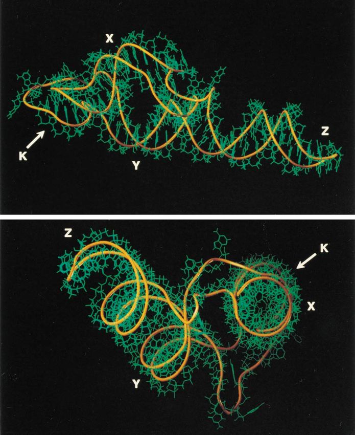
FIG. 9. Three-dimensional model of the coxsackie B3 virus 39UTR. Shown are stick diagram displays from the side view of the model (top) and from the top view
of the coxsackie B3 virus 39UTR (bottom).
pCB3-39UTR:C73923U/G73523A, produced a viable but tem- sitive viruses with virus yields of 0.002 and 0.2% of that of
perature-sensitive virus and at 398C yielded only 13% of that wild-type virus at 398C, respectively. When both mutations
produced by the wild-type virus. The growth characteristic of were introduced simultaneously, the resultant mutant virus had
this latter mutant is similar to that of mutant vCB3-39UTR: wild-type virus growth characteristics at 398C, clearly indicating
C73923U and might be accounted for by similar stabilities of that this base pair is involved in the kissing interaction as well.
U/G and A/U base pair interactions (39). The importance of The full characterization of the kissing interaction is now in
the moderately single-strand-sensitive A7353 in the loop of do- progress. We assume that it consists of six base pairs, which is
main Y was studied in pCB3-39UTR:A73533C and pCB3- also phylogenetically acceptable, since it can be applied to all
39UTR:U73913G. Both mutations produced temperature-sen- enteroviruses sequenced so far (data not shown). Furthermore,VOL. 71, 1997 KISSING OF RNA: FOREPLAY TO VIRAL REPRODUCTION 695
six residues are essential to have residue A7391 to gap the major replicon containing the hepatitis A virus 39UTR, which is also
groove of the superdomain and fold back from domain K to unable to form a kissing structure. However, these results
domain X (Fig. 8). Based on these findings, we constructed a could not be confirmed in vivo using a full-length infectious
three-dimensional model of the 39UTR using computer as- poliovirus cDNA clone containing the hepatitis A virus 39UTR
sisted molecular modelling (Fig. 9). Pilipenko et al. (27) pro- (23).
posed that the final three-dimensional structure of the 39UTRs An intermolecular kissing interaction has also been postu-
of the poliovirus-like and coxsackie B virus-like subgroups lated to describe the mechanism of antisense RNA-target
were similar in their overall organization to tRNA species. RNA duplex formation for the replication control of plasmid
Indeed, a tRNA-like conformation of the 39UTR has earlier R1 (20–22). Hitherto, the existence and biological importance
been found in some alpha-like RNA plant viruses (32, 33). of an intramolecular kissing interaction between two RNA
However, these virus RNAs can be aminoacylated (19), a fea- hairpin loops has only been postulated to take place in a
ture that is unknown for the 39UTR of enteroviruses. Com- variety of RNA molecules such as signal recognition particle
parisons of the three-dimensional structure of the enterovirus RNA (50), Escherichia coli 4.5S RNA (38), archaebacterial 7S
39UTR with the crystallographic structures of tRNA molecules RNA (14), and also in human immunodeficiency virus (8). We
(16, 40) showed that the structures were dissimilar (data not have now demonstrated that these interactions do exist and
shown), since the enterovirus 39UTR does not have the typical might even be a general feature in the higher-order structures
L-shaped formation found in tRNA (Fig. 9). Disruption of the of RNA that are essential for it to function effectively.
Downloaded from http://jvi.asm.org/ on May 16, 2021 by guest
tertiary interaction had no effect on virus translation and pro-
cessing, since wild-type protein patterns were found using an in ACKNOWLEDGMENTS
vitro translation assay. However, disturbing the kissing inter-
action resulted in a defect in virus RNA replication, as was We thank Reinhard Kandolf, University of Tübingen, Germany, for
demonstrated with a subgenomic coxsackie B3 virus replicon. his generous gift of the infectious coxsackie B3 virus clone pCB3/T7, J.
Flanagan, University of Florida, for the HeLa cell initiation factors,
Very recently, it has been demonstrated that in the poliovirus and Peter Donnelly, for critically reviewing the manuscript.
39UTR, an intramolecular kissing interaction is also essential This research was partly supported by grants from the European
for virus replication (28). Communities (INTAS/RFBR no. 01365.i96), the International Science
The importance of RNA structures in RNA-protein inter- Foundation, the Human Frontiers Science Program Organization, and
actions is generally known, and the tertiary RNA structure can the Russian Foundation for Basic Research to Vadim I. Agol.
be essential for stabilizing the structure for the subsequent
interaction with proteins (11). As in the case of other complex REFERENCES
higher-order RNA structures like pseudoknots, it is reasonable 1. Abrahams, J. P., M. van den Berg, E. van Batenburg, and C. Pleij. 1990.
to assume that the kissing interaction in the enterovirus 39UTR Prediction of RNA secondary structure, including pseudoknotting, by com-
puter simulation. Nucleic Acids Res. 18:3035–3044.
acts as a specific binding site for viral and/or cellular proteins 2. Andino, R., G. E. Rieckhof, and D. Baltimore. 1990. A functional ribonucle-
involved in the initiation of negative-strand RNA synthesis (23, oprotein complex forms around the 59-end of poliovirus RNA. Cell 63:369–
42). Indeed, Harris et al. (12) have described the formation of 380.
a 39-terminal ribonucleoprotein complex composed of a 3AB- 3. Andino, R., G. E. Rieckhof, P. L. Achacoso, and D. Baltimore. 1993. Polio-
virus RNA synthesis utilizes an RNP complex formed around the 59-end of
3CD interaction with the 39UTR. The subsequent proteolysis viral RNA. EMBO J. 12:3587–3598.
of 3CDpro releases the 3D polymerase. Protein 3AB then 4. Arnott, S., D. W. L. Hukins, and S. D. Dover. 1972. Optimised parameters for
forms a complex with protein 3D to stimulate the activity of the RNA double-helices. Biochem. Biophys. Res. Commun. 48:1392–1399.
virus polymerase (12, 18, 30), which may use the uridylated 5. Auvinen, P., G. Stanway, and T. Hyypiä. 1989. Genetic diversity of entero-
virus subgroups. Arch. Virol. 104:175–186.
VPg (44) to initiate negative-strand RNA synthesis. A similar 6. Bienz, K., D. Egger, T. Pfister, and M. Troxler. 1992. Structural and func-
protein 3D interaction with a tertiary (pseudoknot) structure tional characterization of the poliovirus replication complex. J. Virol. 66:
in the 39UTR of encephalomyocarditis virus has also been 2740–2747.
proposed (10). Our results suggest that the kissing interaction 7. Bienz, K., D. Egger, M. Troxler, and L. Pasamontes. 1990. Structural orga-
nization of poliovirus RNA replication is mediated by viral proteins of the P2
most probably forms the binding site and not the pseudoknot genomic region. J. Virol. 64:1156–1163.
structure, as suggested by Harris et al. (12). Although Harris et 8. Chang, K.-Y., and I. Tinoco. 1995. Characterization of a “kissing” hairpin
al. (12) did not consider other proteins to contribute to the complex derived from the human immunodeficiency virus genome. Proc.
complex formation, Todd et al. (43) recently provided evidence Natl. Acad. Sci. USA 91:8705–8709.
9. Chomczynski, P., and N. Sacchi. 1987. Single step method of RNA isolation
for an interaction of the 39UTR with certain unidentified cel- by acid guanidinium thiocyanate-phenol-chloroform extraction. Anal. Bio-
lular proteins as well. We propose that the kissing interaction chem. 162:156–159.
is the essential structural feature of the origin of replication 10. Cui, T., and A. G. Porter. 1995. Localization of binding site for encephalo-
required for its functional competence in virus negative-strand myocarditis virus RNA polymerase in the 39-noncoding region of the viral
RNA. Nucleic Acids Res. 23:377–382.
RNA synthesis. 11. Frankel, A. D., I. W. Mattaj, and D. C. Rio. 1991. RNA-protein interactions.
The presence of such a tertiary structure in the 39UTRs of Cell 67:1041–1046.
both the coxsackie B virus- and poliovirus-like viruses makes 12. Harris, K. S., W. Xiang, L. Alexander, W. S. Lane, A. V. Paul, and E.
the exchange of the 39UTR between these viruses and the Wimmer. 1994. Interaction of poliovirus polypeptide 3CDpro with the 59 and
39 termini of the poliovirus genome. J. Biol. Chem. 269:27004–27014.
subsequent replication of the chimeras understandable, since 13. Jacobson, S. J., D. A. M. Konings, and P. Sarnow. 1993. Biochemical and
the binding sites are identical (34). On the other hand, the genetic evidence for a pseudoknot structure at the 39 terminus of the polio-
rhinovirus genus has a 39UTR which consists of a single stem- virus RNA genome and its role in viral amplification. J. Virol. 67:2961–2971.
loop structure which cannot form a kissing interaction. How- 14. Kaine, B. P. 1990. Structure of the archaebacterial 7S RNA molecule. Mol.
Gen. Genet. 221:315–321.
ever, a poliovirus chimera containing the rhinovirus 14 39UTR 15. Klump, W. M., I. Bergmann, B. C. Muller, D. Ameis, and R. Kandolf. 1990.
was still capable of initiating poliovirus negative-strand RNA Complete nucleotide sequence of infectious coxsackievirus B3 cDNA: two
synthesis (34), which is difficult to explain. One explanation initial 59 uridine residues are regained during plus-strand RNA synthesis.
might be that this occurs because ribonucleoprotein complex J. Virol. 64:1573–1583.
16. Ladner, J. E., A. Jack, J. D. Robertus, R. Brown, D. Rhodes, B. F. C. Clark,
formation occurs differently in the rhinovirus 39UTR, although and A. Klug. 1975. Structure of yeast phenylalanine transfer RNA at 2.5 A
formation of the complex as such is sufficient to initiate repli- resolution. Proc. Natl. Acad. Sci. USA 72:4414–4418.
cation. Rohll et al. (34) also found replication in a poliovirus 17. Laemmli, U. K. 1970. Cleavage of structural proteins during the assembly of696 MELCHERS ET AL. J. VIROL.
the head of bacteriophage T4. Nature (London) 227:680–685. virus RNA. Differences and similarities with canonical tRNA. Nucleic Acids
18. Lama, J., M. A. Sanz, and P. L. Rodriguez. 1995. A role of 3AB protein in Res. 10:1929–1946.
poliovirus genome replication. J. Biol. Chem. 270:14430–14438. 34. Rohll, J. B., D. H. Moon, D. J. Evans, and J. W. Almond. 1995. The 39
19. Mans, R. M. W., C. W. A. Pleij, and L. Bosch. 1991. tRNA-like structures. untranslated region of picornavirus RNA: features required for efficient
Structure, function and evolutionary significance. Eur. J. Biochem. 201:303– genome replication. J. Virol. 69:7835–7844.
324. 35. Rohll, J. B., N. Percy, E. Ley, D. J. Evans, J. W. Almond, and W. S. Barclay.
20. Marino, J. P., R. S. Gregorian, G. Csankovski, and D. M. Crothers. 1995. 1994. The 59 untranslated regions of picornavirus RNAs contain indepen-
Bent helix formation between RNA hairpins with complementary loops. dent functional domains essential for RNA replication and translation. J. Vi-
Science 268:1448–1454. rol. 68:4384–4391.
21. Persson, C., E. Gerhart, H. Wagner, and K. Nordström. 1990. Control of 36. Sarnow, P., H. D. Bernstein, and D. Baltimore. 1986. A poliovirus temper-
replication of plasmid R1: structures and sequences of the antisense RNA, ature-sensitive RNA synthesis mutant located in a noncoding region of the
CopA, required for its binding to the target RNA, CopT. EMBO J. 9:3767– genome. Proc. Natl. Acad. Sci. USA 83:571–575.
3775. 37. Schmidt, N. J. 1979. Cell culture techniques for diagnostic virology, p. 65–
22. Persson, C., E. Gerhart, H. Wagner, and K. Nordström. 1990. Control of 139. In E. H. Lennette, and N. J. Schmidt (ed.), Diagnostic procedures for
replication of plasmid R1: formation of an initial transient complex is rate- viral, rickettsial and chlamydial infections. American Public Health Associ-
limiting for antisense RNA-target RNA pairing. EMBO J. 9:3777–3785. ation, Washington, D.C.
23. Philippe, C., F. Eyermann, L. Bernard, C. Portier, B. Ehresmann, and C. 38. Struck, J. C. R., and V. A. Erdmann. 1990. Phylogenetic and biochemical
Ehresmann. 1993. Ribosomal protein S15 from Escherichia coli modulates evidence for a secondary structure model of a small cytoplasmic RNA from
its own translation by trapping the ribosome on the mRNA initiation loading Bacilli. Eur. J. Biochem. 192:17–24.
site. Proc. Natl. Acad. Sci. USA 90:4394–4398. 39. Sugimoto, N., R. Kierzek, S. M. Freier, and D. H. Turner. 1986. Energetics
of internal GU mismatches in ribo-oligonucleotide helixes. Biochemistry
Downloaded from http://jvi.asm.org/ on May 16, 2021 by guest
24. Pierangeli, A., M. Bucci, P. Pagnotti, A. M. Degener, and R. P. Bercoff. 1995.
Mutational analysis of the 39-terminal extra-cistronic region of poliovirus 25:5755–5759.
RNA: secondary structure is not the only requirement for minus strand RNA 40. Sussman, J. L., and S.-H. Kim. 1976. Three-dimensional structure of a
transfer RNA in two crystal forms. Analysis of three sets of atomic coordi-
replication. FEBS Lett. 374:327–332.
nates of yeast phenylalanine tRNA establishes common features. Science
25. Pilipenko, E. V., V. M. Blinov, B. K. Chernov, T. M. Dmitrieva, and V. I.
192:853–858.
Agol. 1989. Conservation of the secondary structure elements of the 59-
41. Takeda, N., R. J. Kuhn, C.-F. Yang, T. Takegami, and E. Wimmer. 1986.
untranslated region of cardio- and aphtovirus RNAs. Nucleic Acids Res.
Initiation of poliovirus plus-strand RNA synthesis in a membrane complex of
17:5701–5711.
infected HeLa cells. J. Virol. 60:43–53.
26. Pilipenko, E. V., A. P. Gmyl, S. V. Maslova, A. P. Belov, A. N. Sinyakov, M.
42. Tang, C. K., and D. E. Draper. 1989. Unusual mRNA pseudoknot structure
Huang, T. D. K. Brown, and V. I. Agol. 1994. Starting window, a distinct
is recognized by a protein translation repressor. Cell 59:511–536.
element in the Cap-dependent internal initiation of translation on picorna-
43. Todd, S., J. H. C. Nguyen, and B. L. Semler. 1995. RNA-protein interactions
viral RNA. J. Mol. Biol. 241:398–414. directed by the 39 end of human rhinovirus genomic RNA. J. Virol. 69:3605–
27. Pilipenko, E. V., S. V. Maslova, A. N. Sinyakov, and V. I. Agol. 1992. Towards 3614.
identification of cis-acting elements involved in the replication of enterovirus 44. Toyoda, H., C. F. Yang, N. Takeda, A. Nomoto, and E. Wimmer. 1987.
and rhinovirus RNAs: a proposal for the existence of tRNA-like terminal Analysis of RNA synthesis of type 1 poliovirus by using an in vitro molecular
structures. Nucleic Acids Res. 20:1739–1745. genetic approach. J. Virol. 61:2816–2822.
28. Pilipenko, E. V., K. V. Poperechny, S. V. Maslova, W. J. G. Melchers, H. J. 45. van Kuppeveld, F. J. M., J. M. D. Galama, J. Zoll, and W. J. G. Melchers.
Bruins Slot, and V. I. Agol. Cis-element, oriR, involved in the initiation of 1995. Genetic analysis of a hydrophobic domain of coxsackie B3 virus protein
(2)strand poliovirus RNA: a quasi-globular multi-domain RNA structure 2B: a moderate degree of hydrophobicity is required for a cis-acting function
maintained by tertiary (“kissing”) interactions. Submitted for publication. in viral RNA synthesis. J. Virol. 69:7782–7790.
29. Pleij, C. W. A. 1990. Pseudoknots: a new motif in the RNA game. Trends 46. Weiner, S. J., P. A. Kollman, D. T. Nguyen, and D. A. Case. 1986. An all atom
Biochem. Sci. 15:143–147. force field for simulations of proteins and nucleic acids. J. Comp. Chem.
30. Plotch, S. J., and O. Palant. 1995. Poliovirus protein 3AB forms a complex 7:230–252.
with and stimulates the activity of the viral RNA polymerase, 3Dpol. J. Virol. 47. Wimmer, E., C. U. T. Hellen, and X. Cao. 1993. Genetics of poliovirus. Annu.
69:7169–7179. Rev. Genet. 27:353–436.
31. Reed, L. J., and H. Muench. 1938. A simple method of estimating fifty 48. Zoll, J., P. Jongen, J. Galama, F. van Kuppeveld, and W. Melchers. 1993.
percent endpoints. Am. J. Hyg. 27:493–497. Coxsackievirus B1-induced murine myositis: no evidence for viral persis-
32. Rietveld, K., K. Linschooten, C. W. A. Pleij, and L. Bosch. 1984. The three- tence. J. Gen. Virol. 74:2071–2076.
dimensional folding of the tRNA-like structure of tobacco mosaic virus 49. Zoll, J., J. Galama, and W. Melchers. 1994. Intratypic genome variability of
RNA. A new building principle applied twice. EMBO J. 3:2613–2619. the coxsackievirus B1 2A protease region. J. Gen. Virol. 75:687–692.
33. Rietveld, K., R. van Poelgeest, C. W. A. Pleij, J. H. van Boom, and L. Bosch. 50. Zwieb, C. 1989. Structure and function of signal recognition particle RNA.
1982. The tRNA-like structure at the 39 terminus of turnip yellow mosaic Prog. Nucleic Acid Res. Mol. Biol. 37:207–234.You can also read