JBC Papers in Press. Published on January 23, 2019 as Manuscript RA118.004396
←
→
Page content transcription
If your browser does not render page correctly, please read the page content below
JBC Papers in Press. Published on January 23, 2019 as Manuscript RA118.004396
      The latest version is at http://www.jbc.org/cgi/doi/10.1074/jbc.RA118.004396

                 Structure and flexibility of the extracellular region of the PirB receptor

                       Hedwich C. Vlieg, Eric G. Huizinga, Bert J.C. Janssen*

    From Crystal and Structural Chemistry, Bijvoet Center for Biomolecular Research, Department of
         Chemistry, Faculty of Science, Utrecht University 3584 CH Utrecht, The Netherlands

                             Running title: Structure of the PirB ectodomain

*To whom correspondence should be addressed: Bert J.C. Janssen: Crystal and Structural Chemistry,
Bijvoet Center for Biomolecular Research, Department of Chemistry, Faculty of Science, Utrecht
University 3584 CH Utrecht, The Netherlands, Tel.: +31 (0)30-2532866; E-mail: b.j.c.janssen@uu.nl

Keywords: receptor, crystal structure, surface plasmon resonance, neurobiology, cell signaling, synaptic
plasticity, immunoglobulin-like domain, transmembrane protein, immune response

                                                                                                              Downloaded from http://www.jbc.org/ by guest on April 24, 2019
                                                     interactions. Taken together, these structural
ABSTRACT                                             findings and the observed PirB-MAG
         Murine      paired      immunoglobulin      interactions are compatible with a model for
receptor B (PirB) and its human ortholog             intercellular signaling in which the PirB
leukocyte immunoglobulin-like receptor B2            extracellular domains, which point away from
(LILRB2) are widely expressed inhibitory             the cell surface, enable interaction with ligands
receptors that interact with a diverse set of        in trans.
extracellular ligands and exert functions
ranging from down regulation of immune                        Mouse       Paired    immunoglobulin-like
responses to inhibition of neuronal growth.          receptor B (PirB), also named Leukocyte
However, structural information that could           immunoglobulin-like receptor subfamily B
shed light on how PirB interacts with its ligands    member 3 (LILRB3), is a promiscuous type I
is lacking. Here, we report crystal structures of    transmembrane receptor with diverse tissue-
the PirB ectodomain; the first full ectodomain       dependent functions, ranging from immune
structure for a LILR family member, at 3.3-4.5       response modulation, hematopoietic stem cell
Å resolution. The structures reveal that PirB's      consolidation to central nervous system (CNS)
six Ig-like domains are arranged at acute            plasticity regulation. To exert its cellular functions
angles, similar to the structures of leukocyte       a diverse set of cell-surface expressed and secreted
immunoglobulin-like receptor (LILR) and              ligands interact with the PirB ectodomain.
killer-cell immunoglobulin-like receptor (KIR).               PirB is a member of the LILR family;
We observe that this regular arrangement is          LILRs are receptors for histocompatibility
followed throughout the ectodomain, resulting        complex class I (MHC-I) proteins and modulate
in an extended zigzag conformation. In two out       the strength of immune responses by stimulatory
of the five structures reported here, the            (LILRAs) or inhibitory (LILRBs) signaling (1, 2).
repeating zigzag is broken by the first domain       PirB is a functional ortholog of human LILRB2.
that can adopt two alternative orientations.         As such, it is used as a mouse model to study
Quantitative binding experiments revealed a 9        LILRB2 function.
µM dissociation constant for PirB-myelin                      PirB is expressed on various types of
associated glycoprotein (MAG) ectodomain             hematopoietic cells, where it down-regulates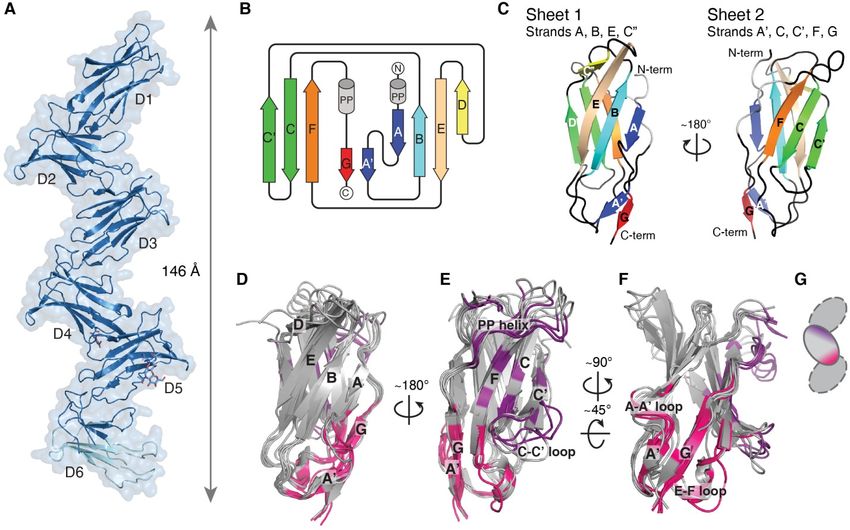
Structure of the PirB ectodomain

activation and differentiation. For example,               human ortholog LILRB2, with which it has an
through interaction with MHC-I molecules, PirB             overall sequence identity of 52% for the
inhibits B-cell (3) and mast cell activation (4).          extracellular Ig-like domains. Instead of the six Ig-
Furthermore, through interaction with secreted             like domains for mouse PirB, the human LILRB2
angiopoietin-like proteins (ANGPLTs), PirB                 ectodomain consists of only four Ig-like domains.
down-regulates blood platelet activation (5) and is        The first and third domain of PirB are most similar
involved in maintaining the stemness of                    to the first domain of LILRB2 (35% and 43%
hematopoietic stem cells (6).                              sequence identity, respectively), while the second
         Unlike the other LILRB family members,            and fourth domain of PirB are most similar to the
PirB and LILRB2 are also expressed in neurons,             second domain of LILRB2 (56% and 43%
where they are involved in restricting synaptic            sequence identity, respectively). The fifth and
plasticity and neuronal regeneration. PirB and             sixth domain of PirB are most similar to the third
MHC-I regulate synaptic plasticity in the visual           and fourth domain of LILRB2 (64% and 57%
cortex (7, 8). In addition, PirB is found to be            sequence identity, respectively). Although mouse
involved in Alzheimer's disease as a receptor for          PirB and human LILRB2 differ in domain number,

                                                                                                                   Downloaded from http://www.jbc.org/ by guest on April 24, 2019
β-amyloid (9). PirB and LILRB2 are also receptors          they interact with a similar set of ligands and have
for a group of ligands called the myelin associated        similar functions (1, 6, 10)
inhibitors (MAIs) (10). These MAIs are Nogo (11,                    Despite the functional importance of PirB,
12), myelin associated glycoprotein (MAG) (13)             there is no structural information available. To
and oligodendrocyte-myelin glycoprotein (OMgp)             date, the only known structures for LILR family
(14). In the healthy CNS this inhibitory signaling         members consist of two consecutive Ig-like
helps to balance the neuronal plasticity needed for        domains. For example, two partial ectodomain
basic brain function such as learning and memory           structures have been published for LILRB2, one of
(15). However, up on injury this inhibition                the first two domains, D1-D2 (23), and one of the
prevents neuronal regeneration, leading to                 last two domains, D3-D4 (24). Insight in the
permanent damage to neuronal circuits (16).                structural features of PirB will aid understanding
         PirB is a type I transmembrane protein            of how the protein is able to interact with such
with a domain organization that is typical for the         diverse ligands and trigger signaling to carry out
LILR protein family (17). The extracellular                its diverse range of functions.
segment is predicted to consist of six tandem Ig-                   Here we present the crystal structure of the
like domains, referred to as D1-D6. The Ig-like            PirB ectodomain and investigate its interactions
domains are connected to a transmembrane helix             with the extracellular segment of the MAI MAG.
by a short linker that is predicted to be disordered       The six Ig-like domains of PirB are arranged in an
and O-linked glycosylated. The intracellular tail          extended repeating zig-zag arrangement. The PirB
contains three immunoreceptor tyrosine-based               N-terminal domain, D1, has positional flexibility
inhibitor motives (ITIMs). Upon activation of the          and adopts three alternative orientations in which
receptor through ligand binding, tyrosines in these        the angle between domains D1 and D2 varies from
motives are phosphorylated by Src family kinases           75° to 315°. Using surface plasmon resonance
(18, 19). The phosphotyrosines recruit Src                 (SPR) experiments we show that the dissociation
homology domain 2 (SH2)-containing phosphatase             constant for the PirB ectodomain - MAG
1 or 2 (SHP-1/2), that mediate further downstream          ectodomain interaction is 9 µM. The extended
signaling (20–22).                                         conformation of PirB may enable trans-cellular
         PirB shares a high sequence homology              interaction with ligands, such as MAG and MHC-
with LILR proteins and, in particular, with its            I, to induce intercellular signaling.

                                                       2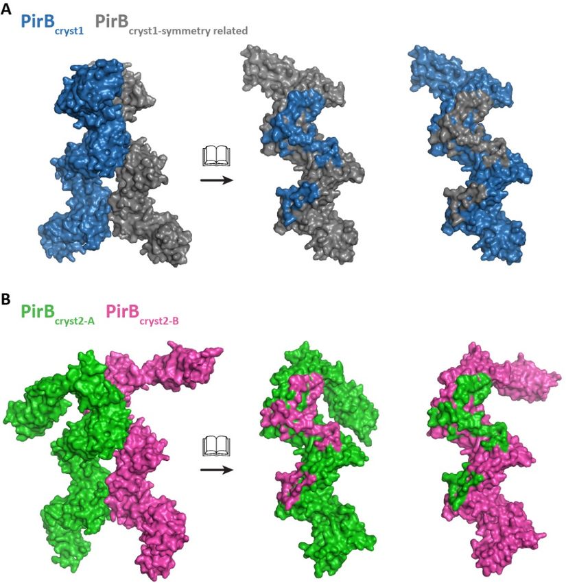
Structure of the PirB ectodomain

                                                               strands A and G and a 310-helix in the E-F loop.
RESULTS                                                        These secondary structure elements are not present
                                                               in all domains, for instance D1, D3 and D6 lack β-
PirB adopts an extended zigzag structure                       strand D, in D5 and D6 the G and A' strands are
         The crystal structure of the PirB                     unraveled, and in D2 and D6 the 310-helix is
ectodomain containing all 6 Ig-like domains                    missing. Moreover, β-strand G is very short in D2
(PirB1-6) reveals an extended zigzag conformation              and it does not interact with β-strand F, leaving a
with multiple orientations for the N-terminal Ig-              separate parallel β-sheet composed of strands A'
like domain (Figure 1 and Figure S-1). The PirB1-6             and G. Despite the differences in secondary
structure was determined in three crystal forms,               structure    elements,      the   three-dimensional
denoted PirBcryst1, PirBcryst2, PirBcryst3, with               structures of all six Ig-like domains are very
maximum resolutions of 3.3, 3.4 and 4.5 Å,                     similar; in PirBcryst1 they have an average RMSD
respectively (Table 1). In two of the crystal forms,           on Cα's of just 1.6 ± 0.3 Å (Figure 1D-F).
PirBcryst2 and PirBcryst3, the asymmetric unit
contains two monomers, resulting in a total of five            Domain interfaces

                                                                                                                     Downloaded from http://www.jbc.org/ by guest on April 24, 2019
unique structures. The six tandem Ig-like domains                       Consecutive Ig-like domains interact with
are arranged in a zigzag shape, with only minor                each other through topologically equivalent
deviations between the five structures (Figure 2A-             interfaces (Figure 1D-F), that are also observed in
C and Figure S-2). From tip to tip (Tyr104-Cα to               the two or three domain containing structures for
Ser536-Cα), the structure measures 146 Å in the                LILR and KIR proteins (23, 24, 33–35, 25–32). In
extended conformation found in PirBcryst1. Glycans             PirB this 'canonical' domain interface is repeated
are observed in four of the six predicted N-linked             five times, resulting in the observed extended
glycosylation sites (Asn 338, 479, 500 and 531), in            zigzag shape (Figure 1A). In each interface, the
at least one of the five structures. In PirBcryst1             most N-terminal domain contributes residues of
domain D6 is partially disordered; residues 528-               the A' strand, G strand and F-E loop (Figure 1F),
549 and 567-593 are not visible in the electron                while the C-terminal side of the interface (located
density and were omitted from the model. The                   on the following domain) is centered on β-sheet 2
best-defined structure for domain 6 is observed in             and comprises the F, C and C' β-strands, the C-C'
the second monomer (chain B) of PirBcryst2                     loop and the polyproline helix preceding strand G
(PirBcryst2-B), therefore this structure is used for the       (purple in Figure 1E). These interactions leave
analysis of D6 outlined below, additionally this               sheet 1 (strands A, B, D, E) and the N-terminal tip
structure is used to supplement the PirB1-6                    of the domain (strand D and connecting loops and
structure shown in Figure 1A.                                  the B-C loop) available for interactions with
         The six Ig-like domains are composed of               ligands.
two antiparallel β-sheets, linked together by a                         The domain interfaces are stabilized by
disulfide bridge. The six domains are topologically            hydrophobic interactions, hydrogen bonds and salt
similar to each other (Figure 1B-F and Figure S-               bridges. On average, the total surface area buried
1), and to the Ig-like domains of KIR and other                in an interface is 1088 ± 158 Å2. All interfaces,
LILR proteins (23, 24, 33–36, 25–32). β-sheet 1                except the D4-D5 interface, have a hydrogen
comprises strands A, B, D and E and sheet 2                    bonding network between residues in strand G or
comprises strands A', C, C', F and G. As is often              loop E-F on the N-terminal domain to residues in
seen in LILR and KIR family members, the A-A'                  loop F-G on the C-terminal domain. Additionally,
strand is shared between the two β-sheets.                     in each interface a salt bridge or hydrogen bond is
Additionally, there is a polyproline helix before β-           formed between a residue in strand G (or in loop

                                                           3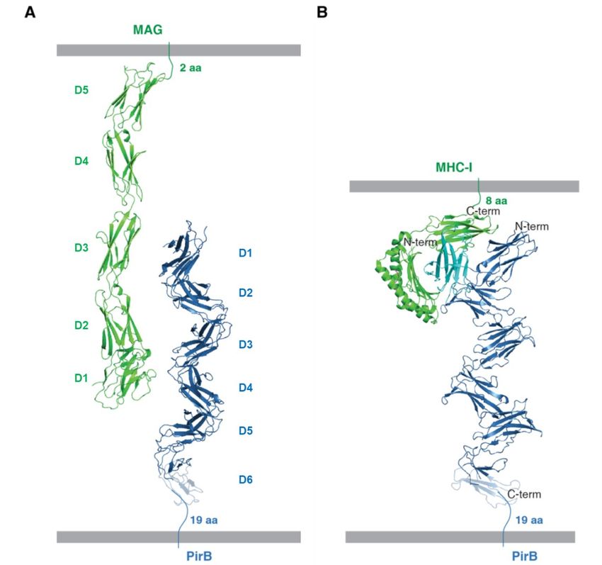
Structure of the PirB ectodomain

A-A' in D1) and a residue in strand C', located at          of Val 117 and Gly 119 in strand G of D1, and Tyr
the edge of the interface. Interaction through this         206 and Trp 208 in strand G of D2 (Figure 2D).
canonical interface results in an acute angle               The domain orientation is further stabilized by a
between consecutive domains. The orientation is             salt bridge between Asp 36 in the A-A' loop of D1,
slightly different for each domain pair, with               and Lys 162 in strand C' on D2.
interdomain angles ranging from 62° to 79°                           The two non-canonical interfaces are even
(Figure 2A-C and Figure S-2).                               smaller, with only 542 Å2 total buried surface area
         This range of observed interdomain angles          in monomer A and 436 Å2 in monomer B. The
is in line with structures of other LILR and KIR            non-canonical interfaces on D1 partially overlap
proteins (23, 24, 33–36, 25–32). Structural                 with the canonical interface. In monomer A, this
comparison of PirB with partial structures                  interface comprises strand A', loop E-F and the
available for LILRB2 (23, 24), PirB’s closest               domain linker; all but one (Val 39) of the D1
human homologue, yields RMSDs that follow the               residues are also involved in the canonical
trend expected from sequence identity. PirB                 interface (Figure 2E). In monomer B, the interface
domains 5-6 are most similar to LILRB2 domains              on D1 comprises loops A'-B and E-F, and the

                                                                                                                  Downloaded from http://www.jbc.org/ by guest on April 24, 2019
3-4 (60% sequence identity, 1.1 Å RMSD in                   domain linker (Figure 2F). On D2 the non-
PirBcryst2-B). LILRB2 domains 1 and 2 are most              canonical interfaces are very different from the
closely related to PirB domains 1-2 (46%                    canonical one. In chain A, Trp 147 in the B-C loop
sequence identity, 2.2 Å RMSD in PirBcryst1) and            replaces Trp 208 as the central residue in the
PirB domains 3-4 (44% sequence identity, 2.1 Å              hydrophobic interface (Figure 2E). Moreover,
RMSD in PirBcryst1).                                        residues in the linker region between the domains
                                                            contribute to the interface; His 120 and Tyr 121
PirB domain D1 can adopt three different                    line the interface and Trp 122 interacts through
orientations                                                ring stacking with His 148 in loop B-C on D2. In
         In two of our PirB structures D1 and D2            chain B, the hydrophobic part of the interface is
do not interact via the canonical interface,                formed by Trp 42 in loop A'-B of D1, and Trp 147
resulting in two PirB conformations that deviate            in loop B-C of D2 (Figure 2F). Both non-
from the regular zigzag described above (Figure             canonical interfaces are further stabilized by salt
2A-C and Figure S-3). In both monomers in                   bridges and hydrogen bonds, although the
PirBcryst2 D1 is bend over to the other side of D2,         hydrogen bonding network is not as extensive as
predominantly rotating around the Trp 122 C-Cα              the one seen in the canonical D1-D2 interfaces.
bond, resulting in an interdomain angle of 315o in                   In summary, our crystal structures show
chain A and an angle of 269o in chain B (Figure             three distinct orientations for PirB D1. Chain B in
2B, C).                                                     PirBcryst2 seems to represent an intermediate state
         The three distinct D1 orientations give rise       between the canonical conformation and the
to three different D1-D2 interfaces (Figure 2D-F).          conformation of PirBcryst2 chain A. On the other
The canonical D1-D2 interface, seen in PirBcryst1,          hand, the main body of the protein, D2-D6, shows
is smaller than the other canonical interfaces, it          very little conformational variation. Taken
has an average total buried surface area of                 together, the PirB structures indicate that whereas
903 ± 51 Å2 versus 1145 ± 107 Å2 for the other              there is limited flexibility between domains D2-
domain interfaces. The canonical D1-D2 interface            D6, a range of orientations is possible for D1.
consists of a hydrophobic core centered around
Trp 208 in strand G of D2, together with a                  PirB self-association
hydrogen bonding network between the backbones

                                                        4
Structure of the PirB ectodomain
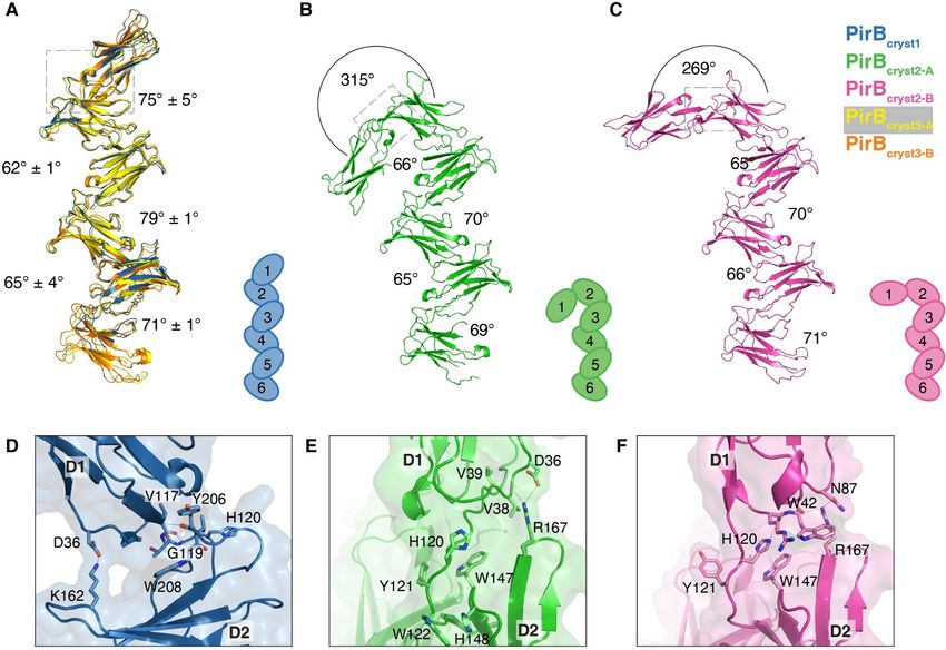
PirBcryst1 and PirBcryst2 reveal dimers with a DISCUSSION
common intermolecular interface that may PirB is a versatile receptor that binds to
represent a PirB cis dimer (Figure 3). The numerous, structurally varied, ligands. We have
interface has a total buried surface area of 3531 Å2 solved the structure of PirB's ectodomain in three
in PirBcryst1 and 3708 Å2 in PirBcryst2, whereas the conformations and performed binding experiments
area of the second largest interfaces in these to gain insight into PirB-ligand binding. What sets
crystals are 1991 Å2 and 1609 Å2, respectively. In the PirB ectodomain structure apart from other
the third crystal form this dimer is not observed; structures of LILR and KIR family members is
here the largest interface is 2087 Å2. The putative that it is the largest of its kind to be elucidated and
dimerization interface consists of two patches. The it is the only reported KIR/LILR structure with
first patch starts at the C-terminal tip of D2 and multiple orientations for one of its domains.
continues across D3 to the N-terminal tip of D4. The six Ig-like domains that make up
The second patch comprises residues at the C- PirB's ectodomain are arranged in a regular zigzag
terminal tip of D4 and the N-terminal tip of D5. repeat. Interestingly, the first domain can break
D1 is not involved in this putative dimerization this regular pattern and adopt multiple
Downloaded from http://www.jbc.org/ by guest on April 24, 2019
interface; therefore, the interface is not affected by orientations. In our crystal structures three distinct
the observed mobility of this domain. orientations are observed for PirB D1. In all three
PirB shows self-association in SPR crystal forms the D1 domain is involved in crystal
(Figure S-4). While, homo-interactions can be packing and this may stabilize conformations of
detected by SPR they cannot be quantified using PirB that are only sparsely populated in solution.
this technique as self-association of ligands The conformations in PirBcryst1 and PirBcryst2-A
coupled on the chip surface compete with analyte seem to represent the extremes in a range of
binding. Despite this, both the crystal structures conformations, while PirBcryst2-B represents an
and the SPR data indicate that PirB has propensity intermediate state between the two. To our
to self-associate. knowledge flexibility of one of the Ig-like
domains has not been reported for any of the LILR
Ligand binding or KIR family members. Furthermore, there are no
Ligand interaction is critical to PirB's role indications of such flexibility in the structures for
as an extracellular receptor. To verify the LILR and KIR family members. This is surprising,
interaction of the PirB ectodomain for ligand as, at first glance, the PirB D1-2 interface is very
binding, surface plasmon resonance (SPR) similar to the D1-2 interfaces of other LILR family
experiments were performed for PirB's known members, by comparing the sequences or the
binding partner MAG (Figure S-5 and S-6). C- available structures.
terminally biotinylated PirB1-6 was immobilized at The conformational variability in PirB
three different densities on a streptavidin coated may be explained by the size of the interface and a
SPR chip, to probe interactions with increasing number of residues unique to PirB. The canonical
concentrations of the extracellular segment of D1-D2 interface is less extensive than the other
MAG in the mobile phase. The entire extracellular canonical interfaces in PirB, with an average total
MAG segment, containing five extracellular Ig- buried surface area of 903 ± 51 Å2 instead of
like domains (MAG1-5), was used. MAG1-5-PirB1-6 1145 ± 107 Å2 for the other interfaces. However, it
binding was found to have a KD of 9 ± 2.9 M is in the same range as the D1-D2 interface in
(Figure S-6). other LILR structures, where the total buried
surface area is 839 ± 37 Å2 (23, 34–36). Moreover,
the three-dimensional organization of the interface
5Structure of the PirB ectodomain
is very well conserved between the LILRs and conclusive, oligomerization (e.g. dimerization)
PirB. On closer inspection, four residues unique to could be part of the signaling mechanism in (some
PirB, but with important roles in the three different of) PirB's roles as a receptor when expressed on
interfaces (Figure 2D-F) stand out; His 120 and the cell surface. Nonetheless, to our knowledge,
Trp 42, 122 and 147. His 120 is part of the linker dimerization has not been documented to have a
region between D1 and D2 and points outward functional role for any of the LILR family
into the solvent in the canonical structure. The members, nor for the related KIR proteins. Also,
corresponding residue in other LILR family there is no evidence for dimerization in the
members (human LILRs A1, A2, A3, A4, B1, B2, (partial) structures of LILR and KIR proteins.
B3, B4 and mouse LILRB4), is an alanine that PirB1-6 is separated from the cell surface
does not participate in the D1-2 interface, or a by a 19-amino acid linker that may confer
phenylalanine (human LILRs A5, A6, B5 and flexibility to the orientation of PirB with respect to
chimpanzee LILR A6 and B5), that engages in the membrane. Therefore, while the maximum
extensive hydrophobic interactions with domain 1. distance that the six Ig-like domains can span in
As His 120 is bulky, unlike the much smaller the extended PirB conformation is 146 Å, it is not
Downloaded from http://www.jbc.org/ by guest on April 24, 2019
alanine, and is not able to contribute to the clear if PirB projects that far from the membrane.
interface with hydrophobic interactions like If PirB adopts a dimer conformation as observed
phenylalanine, it might destabilize the PirBcryst1 in PirBcryst1 and PirBcryst2 the orientation relative to
D1-2 interface. The three tryptophans unique to the membrane becomes much more restricted. A
PirB (42, 122 and 147) are not involved in the PirB dimer in the extended zigzag conformation
canonical interface, instead they provide (PirBcryst1) would protrude 130 Å from the plane
hydrophobic patches for D1 interaction in the non- defined by the most membrane-proximal tips of
canonical orientations. Because of Trp 147's the protein (Figure S-7). For the non-canonical
location in loop BC, which is of variable length, orientations of D1, this distance is less; 100 Å in
the corresponding residues in other LILR family the fully flipped conformation (PirBcryst2-A) and
members can only be determined by structural 120 Å in the intermediate state (PirBcryst2-B).
comparison. In LILRs with published structures Neuronal PirB interacts with MAIs,
the corresponding residues are glutamine or expressed on oligodendrocytes, in trans. While
arginine, with extended hydrophilic side chains interactions of MAIs with the Nogo Receptor
that are consistent with exposure to the solvent. (NgR) have been well studied (37, 38), less is
For Trp 42 the corresponding residue in other known about binding of these proteins to PirB.
LILR family members is always an arginine and Binding affinities of 14 nM have been reported for
for Trp 122 the corresponding residues are either Fc-tagged MAG to PirB (10), but this interaction
hydrophilic (Glu, Ser, Asn) or small (Pro, Ile, is most likely enhanced by artificially dimerizing
Ala). These corresponding residues would not be MAG. Indeed, weaker interactions of 33 µM have
able to interact with D1 in the same manner as Trp been reported for binding of untagged MAG to
42, 122 or 147. In conclusion, three tryptophans PirB (39) although in their experiments the
that are unique to PirB provide alternate interfaces maximum concentration of MAG used, 3 µM, is
for D1-D2 interaction and, together with His 120, too low for accurate affinity determination. Using
may promote conformational mobility of D1. higher MAG concentrations we obtain an affinity
Our crystal structures provide evidence for of 9 µM for MAG – PirB interaction.
PirB dimerization, and SPR data also indicate that There are no crystal structures available of
PirB1-6 has a propensity for self-association. While complexes involving MAG and PirB to provide
our experimental data on PirB dimerization are not additional clues to how these proteins might
6Structure of the PirB ectodomain
interact (Figure 4a). Structures are available of For SPR experiments the original PirB1-6
LILR family members in complex with MHC-I construct was subcloned into pUPE107.62, a
loaded with a (viral) peptide. The LILR–MHC-I vector containing a C-terminal His6-tag for
trans binding mode is highly conserved amongst purification and a C-terminal biotin acceptor
all studied complexes (40). As a member of the peptide (BAP)-tag for biotinylation.
LILR family, PirB is likely to display the same
binding mode to form complexes with MHC-I Expression and purification
(Figure 4b). Interestingly, PirB would only be able The soluble ectodomain constructs (in
to bind MHC-I complexes in the extended zigzag vector pUPE107.30) were expressed by transient
conformation; in the non-canonical conformations, transfection in HEK293-ES cells (U-Protein
seen in PirBcryst2, D1 would clash with the MHC-I Express) and grown in FreeStyle 293 Expression
complex. PirB dimerization as observed in Medium (Thermo Fisher). HEK293-ES is a cell
PirBcryst1 and PirBcryst2 on the other hand would not line which lacks the N-acetylglycosaminyl
interfere with MHC-I binding. transferase I enzyme and therefore produces
In conclusion, our data reveal that the proteins with short, homogeneous oligomannose
Downloaded from http://www.jbc.org/ by guest on April 24, 2019
extracellular segment of PirB is extended and that glycans (41). After 6 days, cells are spun down (10
PirB has a propensity to self-associate. The minutes at 1000x g) and the medium is harvested.
dimerization mode revealed in the crystal The medium is concentrated 10-fold and
structures of PirB1-6 is compatible with dimer diafiltrated against IMAC binding buffer (500 mM
formation of full length transmembrane PirB on NaCl, 50 mM Hepes pH 7.8) using a Quixstand
the cell surface in cis, but the importance of PirB benchtop system (GE Healthcare). Cell debris is
dimerization for signaling has not been removed by centrifugation (30 minutes at 9500x
investigated further. In full-length PirB the mobile g). The protein was purified from the cleared
N-terminal domain D1 is likely positioned furthest concentrate by Ni-affinity chromatography on a
away from the cell-surface, poised for interaction 5 ml HiTrap column (GE Healthcare) followed by
with ligands on other cells. Possibly, the mobility size exclusion chromatography (SEC) on a
of D1 contributes to its ability to interact with a Superdex200 column (GE Healthcare) equilibrated
diverse set of binding partners. Taken together, with SEC buffer (150 mM NaCl, 25 mM Hepes
our structural and interaction data are compatible pH 7.5). The purified protein was concentrated
with a model for intercellular signaling in which using a spin concentrator with the appropriate
PirB has an extended conformation on the cell MWCO, snap frozen in liquid nitrogen and stored
surface to enable interaction with ligands in trans at -80oC until use.
(Figure 4). PirB1-6 for SPR was expressed in
HEK293-ES cells (U-Protein Express), in small 4
EXPERIMENTAL PROCEDURES ml cultures. To allow in vivo biotinylation of these
Cloning of constructs constructs, the cells were co-transfected with the
The soluble ectodomain constructs for E. coli BirA biotin ligase and the cell medium was
PirB and MAG were generated using polymerase supplemented with 25 g/ml biotin. After 6 days,
chain reaction (PCR). The templates and construct the medium was harvested as described above and
boundaries are listed in Table 2. All constructs diluted 10-fold with IMAC binding buffer. The
were subcloned into pUPE107.30 (U-Protein protein was purified from the diluted medium by
Express) (C-terminal His6-tag) using BamHI/NotI batch binding to Ni Sepharose beads (GE
restriction sites. Healthcare). After incubation at room temperature
for 2 hours, the beads were transferred to spin
7Structure of the PirB ectodomain
columns (ThermoFisher) and washed 4 times with domains of the refined PirBcryst1 structure were
binding buffer, followed by elution with 50 L of placed back in density for PirBcryst2 and PirBcryst3
binding buffer supplemented with 200 mM of followed by further refinement to yield the
imidazole. structures reported here.
The structure for PirBcryst3 was solved with
Crystallization and data collection MR in Phaser (45), using structures of LILRA5
Crystals of PirB1-6 were grown at 20oC (35) and LILRB2 (24) as MR models. The
using the hanging drop vapor diffusion method structure was refined using iterative rounds of
and a concentration of 9 to 10 mg/ml PirB1-6. manual building in Coot (46) and refinement in
PirBcryst1, in space group P4122, was obtained REFMAC 5 (47). The resulting structure was used
against a reservoir solution of 1.1 M LiCl, 14% as a model for MR to solve the structure of
PEG 6000, 0.1 M Citric acid buffer pH 5.5. A PirBcryst2 with Phaser. To prevent bias in the
dataset to 3.3 Å resolution was collected at domain orientations, single domains were used as
Diamond light source beamline I04 (wavelength MR models. The resulting structure was refined in
0.9795 Å, temperature 100 K). PirBcryst2, in space Coot and REFMAC. The same domain-by-domain
Downloaded from http://www.jbc.org/ by guest on April 24, 2019
group P21, was obtained against a reservoir approach was used to solve the structure from
solution of 1.2 M LiCl, 12% PEG 6000, 0.1 M PirBcryst1, with PirBcryst2 as a MR model. The new
Citric acid buffer pH 5.5. A dataset to 3.4 Å structure was refined in REFMAC and
resolution was collected at Swiss light source phenix.refine (48) and manual model building in
beamline PX-I (wavelength 1.0000 Å, temperature Coot.
100 K). PirBcryst-3, also in space group P21, was To improve the structure from PirBcryst2,
obtained against a reservoir solution of 14.6% D1-5 of this structure were substituted with D1-5
polyacrylate 5100 sodium salt and 0.07 M Tris pH from PirBcryst1. This was done by individually
8.0. An incomplete dataset to 4.5Å was collected superimposing the domains, followed by extensive
at Swiss light source beamline PX-I (wavelength refinement in REFMAC, phenix-refine and
1.0000 Å, temperature 100 K). The missing manual model building in Coot. A similar
degrees were collected at ESRF beamline ID23-1 approach was used to improve the structure from
(wavelength 0.9686 Å, temperature 100 K). The PirBcryst3, domains from the higher resolution
two datasets were merged in AIMLESS and PirBcryst1 (D1 and D2-5) and PirBcryst2 (D6) were
further processed as described below. used to rebuild the structure. To avoid overfitting
of the low resolution PirBcryst3 structure, refinement
Structure solution and refinement was kept to a minimum. After an initial round of
The data were integrated using iMOSFLM rigid body refinement (one body per domain) in
(42) or XDS (43), followed by scaling and phenix.refine, a limited number of adjustments
merging using the AIMLESS pipeline (44). In were made in Coot. Namely, the missing domain
brief, the strategy for solving and refinement of connections were added; sugars were added where
the structures was as follows; the three structures they were visible in the density; His 120 and Tyr
were solved in the order in which the datasets 121, that are Ramachandran outliers in the model
were collected (first PirBcryst3, then PirBcryst2 and because of a crystal contact, were fixed; and loop
finally PirBcryst1). Each time, the previously solved 170-178, that is displaced in the PirBcryst3 structure
and partially refined structure was used as a because of a crystal contact, was modeled in the
molecular replacement (MR) model for the next visible density using the corresponding region in
dataset. Resulting in the 'best' PirB structure from PirBcryst2-A as a template. After this model building
PirBcryst1. To improve the other two structures, the the structure was subjected to one round of jelly
8Structure of the PirB ectodomain
body refinement in REFMAC and one more round microspotter. This method creates multiple spots,
of rigid body refinement in phenix.refine. or regions of interest (ROIs) on the chip surface,
each with a ligand density of choice. Purified
Structure analysis analyte (MAG1-5 or PirB1-6 Figure S-4 and S-6)
The domain interfaces for all five unique was flowed over the chip at a constant temperature
monomers as well as the dimer interfaces were of 25oC in running buffer (150 mM NaCl, 20 mM
analyzed using the PISA server (49). Angles Hepes pH 7.2, 0.001% Tween-20). Equilibrium
between Ig-like domains were determined from binding experiments were performed to measure
the angles between the largest principle axes of the binding affinities of MAG to PirB. Using the
these domains. Glycosylation predictions were analyte in a concentration range of 0.8-108 M for
preformed using the NetNGlyc 1.0 Server and the MAG1-5.
NetOGlyc 4.0 Server (50). Structure RMSDs were Data processing was started in SprintX
calculated in gesamt (51), using the Cα atoms for (IBIS Technologies), where the data were blanked
alignment. All figures of the structures were once, using reference spots close to the ROIs. The
generated using Pymol (DeLano Scientific LLC). data were then zeroed before each injection of
Downloaded from http://www.jbc.org/ by guest on April 24, 2019
analyte, and exported to Scrubber (BioLogic). In
Scrubber the amount of bound MAG1-5 was
Surface plasmon resonance determined when equilibrium is reached at the end
SPR experiments were performed on an of the association phase (Figure S-6). A saturation
MX96 (IBIS Technologies), using a SensEye curve was fitted with a 1:1 Langmuir binding
Sensor (IBIS Technologies) with a streptavidin model in Graphpad Prism to determine the
coated dextran matrix. Biotinylated PirB1-6 was maximum analyte binding (Bmax) and equilibrium
coupled to the chip as the SPR-ligands using a binding constant (KD) of MAG1-5-PirB1-6
Wasatch Microfluidics continuous flow interaction.
Acknowledgements: We thank the staff of European Synchrotron Radiation Facility beamline ID23-1,
Diamond light source beamline I04 and Swiss light source beamline PX-I.
Conflict of interest: The authors declare that they have no conflicts of interest with the contents of this
article.
Author contributions: HCV and BJCJ designed the experiments. HCV performed the experiments and
analyzed the data together with EGH and BJCJ. BJCJ supervised the project. HCV, EGH and BJCJ wrote
the manuscript.
REFERENCES
1. Hudson, L. E., and Allen, R. L. (2016) Leukocyte Ig-like receptors - A Model for MHC class i
disease associations. Front. Immunol. 7, 281
2. Takai, T. (2005) Paired immunoglobulin-like receptors and their MHC class I recognition.
Immunology. 115, 433–440
3. Maeda, A., Scharenberg, A. M., Tsukada, S., Bolen, J. B., Kinet, J. P., and Kurosaki, T. (1999)
Paired immunoglobulin-like receptor B (PIR-B) inhibits BCR-induced activation of Syk and Btk
by SHP-1. Oncogene. 18, 2291–2297
4. Masuda, A., Nakamura, A., Maeda, T., Sakamoto, Y., and Takai, T. (2007) Cis binding between
9Structure of the PirB ectodomain
inhibitory receptors and MHC class I can regulate mast cell activation. J Exp Med. 204, 907–920
5. Fan, X., Shi, P., Dai, J., Lu, Y., Chen, X., Liu, X., Zhang, K., Wu, X., Sun, Y., Wang, K., Zhu, L.,
Zhang, C. C., Zhang, J., Chen, G. Q., Zheng, J., and Liu, J. (2014) Paired immunoglobulin-like
receptor B regulates platelet activation. Blood. 124, 2421–2430
6. Zheng, J., Umikawa, M., Cui, C., Li, J., Chen, X., Zhang, C., Hyunh, H., Kang, X., Silvany, R.,
Wan, X., Ye, J., Cantó, A. P., Chen, S. H., Wang, H. Y., Ward, E. S., and Zhang, C. C. (2012)
Inhibitory receptors bind ANGPTLs and support blood stem cells and leukaemia development.
Nature. 485, 656–660
7. Syken, J., Grandpre, T., Kanold, P. O., and Shatz, C. J. (2006) PirB restricts ocular-dominance
plasticity in visual cortex. Science. 313, 1795–800
8. Bochner, D. N., Sapp, R. W., Adelson, J. D., Zhang, S., Lee, H., Djurisic, M., Syken, J., Dan, Y.,
and Shatz, C. J. (2014) Blocking PirB up-regulates spines and functional synapses to unlock visual
cortical plasticity and facilitate recovery from amblyopia. Sci. Transl. Med. 6, 258ra140
9. Kim, T., Vidal, G. S., Djurisic, M., William, C. M., Birnbaum, M. E., Garcia, K. C., Hyman, B. T.,
and Shatz, C. J. (2013) Human LilrB2 is a β-amyloid receptor and its murine homolog PirB
regulates synaptic plasticity in an Alzheimer’s model. Science. 341, 1399–404
10. Atwal, J. K., Pinkston-Gosse, J., Syken, J., Stawicki, S., Wu, Y., Shatz, C., and Tessier-Lavigne,
M. (2008) PirB is a functional receptor for myelin inhibitors of axonal regeneration. Science. 322,
Downloaded from http://www.jbc.org/ by guest on April 24, 2019
967–70
11. Chen, M. S., Huber, A. B., Van Der Haar, M. E. D., Frank, M., Schnell, L., Spillmann, A. A.,
Christ, F., and Schwab, M. E. (2000) Nogo-A is a myelin-associated neurite outgrowth inhibitor
and an antigen for monoclonal antibody IN-1. Nature. 403, 434–439
12. GrandPré, T., Nakamura, F., Vartanlan, T., and Strittmatter, S. M. (2000) Identification of the
Nogo inhibitor of axon regeneration as a Reticulon protein. Nature. 403, 439–444
13. McKerracher, L., David, S., Jackson, D. L., Kottis, V., Dunn, R. J., and Braun, P. E. (1994)
Identification of myelin-associated glycoprotein as a major myelin-derived inhibitor of neurite
growth. Neuron. 13, 805–811
14. Kottis, V., Thibault, P., Mikol, D., Xiao, Z. C., Zhang, R., Dergham, P., and Braun, P. E. (2002)
Oligodendrocyte-myelin glycoprotein (OMgp) is an inhibitor of neurite outgrowth. J. Neurochem.
82, 1566–1569
15. Baldwin, K. T., and Giger, R. J. (2015) Insights into the physiological role of CNS regeneration
inhibitors. Front. Mol. Neurosci. 8, 23
16. Boghdadi, A. G., Teo, L., and Bourne, J. A. (2018) The Involvement of the Myelin-Associated
Inhibitors and Their Receptors in CNS Plasticity and Injury. Mol. Neurobiol. 55, 1831–1846
17. Kubagawa, H., Burrows, P. D., and Cooper, M. D. (1997) A novel pair of immunoglobulin-like
receptors expressed by B cells and myeloid cells. Proc. Natl. Acad. Sci. U. S. A. 94, 5261–6
18. Ho, L. H., Uehara, T., Chen, C.-C., Kubagawa, H., and Cooper, M. D. (1999) Constitutive tyrosine
phosphorylation of the inhibitory paired Ig-like receptor PIR-B. Proc. Natl. Acad. Sci. 96, 15086–
15090
19. Zhang, H., Meng, F., Chu, C. L., Takai, T., and Lowell, C. A. (2005) The Src family kinases Hck
and Fgr negatively regulate neutrophil and dendritic cell chemokine signaling via PIR-B.
Immunity. 22, 235–246
20. Bléry, M., Kubagawa, H., Chen, C. C., Vély, F., Cooper, M. D., and Vivier, E. (1998) The paired
Ig-like receptor PIR-B is an inhibitory receptor that recruits the protein-tyrosine phosphatase SHP-
1. Proc. Natl. Acad. Sci. U. S. A. 95, 2446–2451
21. Maeda, A., Kurosaki, M., Ono, M., Takai, T., and Kurosaki, T. (1998) Requirement of SH2-
containing protein tyrosine phosphatases SHP-1 and SHP-2 for paired immunoglobulin-like
receptor B (PIR-B)-mediated inhibitory signal. J. Exp. Med. 187, 1355–1360
22. Berg, K. L., Carlberg, K., Rohrschneider, L. R., Siminovitch, K. A., and Stanley, E. R. (1998) The
major SHP-1-binding, tyrosine-phosphorylated protein in macrophages is a member of the
KIR/LIR family and an SHP-1 substrate. Oncogene. 17, 2535–2541
10Structure of the PirB ectodomain
23. Shiroishi, M., Kuroki, K., Rasubala, L., Tsumoto, K., Kumagai, I., Kurimoto, E., Kato, K., Kohda,
D., and Maenaka, K. (2006) Structural basis for recognition of the nonclassical MHC molecule
HLA-G by the leukocyte Ig-like receptor B2 (LILRB2/LIR2/ILT4/CD85d). Proc Natl Acad Sci U
S A. 103, 16412–16417
24. Nam, G., Shi, Y., Ryu, M., Wang, Q., Song, H., Liu, J., Yan, J., Qi, J., and Gao, G. F. (2013)
Crystal structures of the two membrane-proximal Ig-like domains (D3D4) of LILRB1/B2:
Alternative models for their involvement in peptide-HLA binding. Protein Cell. 4, 761–770
25. Fan, Q. R., Mosyak, L., Winter, C. C., Wagtmann, N., Long, E. O., and Wiley, D. C. (1997)
Structure of the inhibitory receptor for human natural killer cells resembles haematopoietic
receptors. Nature. 389, 96–100
26. Snyder, G. a, Brooks, a G., and Sun, P. D. (1999) Crystal structure of the HLA-Cw3 allotype-
specific killer cell inhibitory receptor KIR2DL2. Proc. Natl. Acad. Sci. U. S. A. 96, 3864–9
27. Maenaka, K., Juji, T., Stuart, D. I., and Jones, E. Y. (1999) Crystal structure of the human p58
killer cell inhibitory receptor (KIR2DL3) specific for HLA-Cw3-related MHC class I. Structure. 7,
391–398
28. Moradi, S., Berry, R., Pymm, P., Hitchen, C., Beckham, S. A., Wilce, M. C. J., Walpole, N. G.,
Clements, C. S., Reid, H. H., Perugini, M. A., Brooks, A. G., Rossjohn, J., and Vivian, J. P. (2015)
The structure of the atypical killer cell immunoglobulin-like receptor, KIR2DL4. J. Biol. Chem.
Downloaded from http://www.jbc.org/ by guest on April 24, 2019
290, 10460–10471
29. Saulquin, X., Gastinel, L. N., and Vivier, E. (2003) Crystal Structure of the Human Natural Killer
Cell Activating Receptor KIR2DS2 (CD158j). J. Exp. Med. 197, 933–938
30. Graef, T., Moesta, A. K., Norman, P. J., Abi-Rached, L., Vago, L., Older Aguilar, A. M., Gleimer,
M., Hammond, J. A., Guethlein, L. A., Bushnell, D. A., Robinson, P. J., and Parham, P. (2009)
KIR2DS4 is a product of gene conversion with KIR3DL2 that introduced specificity for HLA-A *
11 while diminishing avidity for HLA-C. J. Exp. Med. 206, 2557–2572
31. Vivian, J. P., Duncan, R. C., Berry, R., O’Connor, G. M., Reid, H. H., Beddoe, T., Gras, S.,
Saunders, P. M., Olshina, M. A., Widjaja, J. M. L., Harpur, C. M., Lin, J., Maloveste, S. M., Price,
D. A., Lafont, B. A. P., McVicar, D. W., Clements, C. S., Brooks, A. G., and Rossjohn, J. (2011)
Killer cell immunoglobulin-like receptor 3DL1-mediated recognition of human leukocyte antigen
B. Nature. 479, 401–405
32. Ryu, M., Chen, Y., Qi, J., Liu, J., Fan, Z., Nam, G., Shi, Y., Cheng, H., and Gao, G. F. (2011)
Lilra3 binds both classical and non-classical HLA class i molecules but with reduced affinities
compared to lilrb1/lilrb2: Structural evidence. PLoS One. 6, e19245
33. Chapman, T. L., Heikema, A. P., West, A. P., and Bjorkman, P. J. (2000) Crystal structure and
ligand binding properties of the D1D2 region of the inhibitory receptor LIR-1(ILT2). Immunity.
13, 727–736
34. Cheng, H., Mohammed, F., Nam, G., Chen, Y., Qi, J., Garner, L. I., Allen, R. L., Yan, J., Willcox,
B. E., and Gao, G. F. (2011) Crystal structure of leukocyte Ig-like receptor LILRB4 (ILT3/LIR-
5/CD85k): A myeloid inhibitory receptor involved in immune tolerance. J. Biol. Chem. 286,
18013–18025
35. Shiroishi, M., Kajikawa, M., Kuroki, K., Ose, T., Kohda, D., and Maenaka, K. (2006) Crystal
structure of the human monocyte-activating receptor, “Group 2” leukocyte Ig-like receptor A5
(LILRA5/LIR9/ILT11). J. Biol. Chem. 281, 19536–19544
36. Yang, Z., and Bjorkman, P. J. (2008) Structure of UL18, a peptide-binding viral MHC mimic,
bound to a host inhibitory receptor. Proc. Natl. Acad. Sci. 105, 10095–10100
37. Schimmele, B., and Plückthun, A. (2005) Identification of a functional epitope of the nogo
receptor by a combinatorial approach using ribosome display. J. Mol. Biol. 352, 229–241
38. Zander, H., Reineke, U., Schneider-Mergener, J., and Skerra, A. (2007) Epitope mapping of the
neuronal growth inhibitor Nogo-A for the Nogo receptor and the cognate monoclonal antibody IN-
1 by means of the SPOT technique. J. Mol. Recognit. 20, 185–196
39. Matsushita, H., Endo, S., Kobayashi, E., Sakamoto, Y., Kobayashi, K., Kitaguchi, K., Kuroki, K.,
11Structure of the PirB ectodomain
Söderhäll, A., Maenaka, K., Nakamura, A., Strittmatter, S. M., and Takai, T. (2011) Differential
but competitive binding of Nogo protein and class I major histocompatibility complex (MHCI) to
the PIR-B ectodomain provides an inhibition of cells. J. Biol. Chem. 286, 25739–25747
40. Willcox, B. E., Thomas, L. M., and Bjorkman, P. J. (2003) Crystal structure of HLA-A2 bound to
LIR-1, a host and viral major histocompatibility complex receptor. Nat. Immunol. 4, 913–919
41. Reeves, P. J., Callewaert, N., Contreras, R., and Khorana, H. G. (2002) Structure and function in
rhodopsin: High-level expression of rhodopsin with restricted and homogeneous N-glycosylation
by a tetracycline-inducible N-acetylglucosaminyltransferase I-negative HEK293S stable
mammalian cell line. Proc. Natl. Acad. Sci. 99, 13419–13424
42. Battye, T. G. G., Kontogiannis, L., Johnson, O., Powell, H. R., and Leslie, A. G. W. (2011)
iMOSFLM: A new graphical interface for diffraction-image processing with MOSFLM. Acta
Crystallogr. Sect. D Biol. Crystallogr. 67, 271–281
43. Kabsch, W. (2010) Integration, scaling, space-group assignment and post-refinement. Acta
Crystallogr. Sect. D Biol. Crystallogr. 66, 133–144
44. Evans, P. R., and Murshudov, G. N. (2013) How good are my data and what is the resolution?
Acta Crystallogr. Sect. D Biol. Crystallogr. 69, 1204–1214
45. McCoy, A. J., Grosse-Kunstleve, R. W., Adams, P. D., Winn, M. D., Storoni, L. C., and Read, R.
J. (2007) Phaser crystallographic software. J. Appl. Crystallogr. 40, 658–674
Downloaded from http://www.jbc.org/ by guest on April 24, 2019
46. Emsley, P., Lohkamp, B., Scott, W. G., and Cowtan, K. (2010) Features and development of Coot.
Acta Crystallogr. Sect. D Biol. Crystallogr. 66, 486–501
47. Murshudov, G. N., Skubák, P., Lebedev, A. A., Pannu, N. S., Steiner, R. A., Nicholls, R. A.,
Winn, M. D., Long, F., and Vagin, A. A. (2011) REFMAC5 for the refinement of macromolecular
crystal structures. Acta Crystallogr. Sect. D Biol. Crystallogr. 67, 355–367
48. Headd, J. J., Echols, N., Afonine, P. V., Grosse-Kunstleve, R. W., Chen, V. B., Moriarty, N. W.,
Richardson, D. C., Richardson, J. S., and Adams, P. D. (2012) Use of knowledge-based restraints
in phenix.refine to improve macromolecular refinement at low resolution. Acta Crystallogr. Sect.
D Biol. Crystallogr. 68, 381–390
49. Krissinel, E., and Henrick, K. (2007) Inference of Macromolecular Assemblies from Crystalline
State. J. Mol. Biol. 372, 774–797
50. Steentoft, C., Vakhrushev, S. Y., Joshi, H. J., Kong, Y., Vester-Christensen, M. B., Schjoldager,
K. T. B. G., Lavrsen, K., Dabelsteen, S., Pedersen, N. B., Marcos-Silva, L., Gupta, R., Paul
Bennett, E., Mandel, U., Brunak, S., Wandall, H. H., Levery, S. B., and Clausen, H. (2013)
Precision mapping of the human O-GalNAc glycoproteome through SimpleCell technology.
EMBO J. 32, 1478–1488
51. Krissinel, E. (2012) Enhanced fold recognition using efficient short fragment clustering. J. Mol.
Biochem. 1, 76–85
52. Pronker, M. F., Lemstra, S., Snijder, J., Heck, A. J. R., Thies-Weesie, D. M. E., Pasterkamp, R. J.,
and Janssen, B. J. C. (2016) Structural basis of myelin-associated glycoprotein adhesion and
signalling. Nat. Commun. 7, 13584
FOOTNOTES
This work was supported by a Netherland Organization for Scientific Research (NWO) VIDI grant
(723.012.002) and by NWO grant 01.80.104.00. The atomic coordinates and structure factors have been
deposited in the Protein Data Bank (http://www.pdb.org/) with PDB ID 6GRQ, 6GRS and 6GRT for
PirBcryst1, PirBcryst2 and PirBcryst3, respectively.
12Structure of the PirB ectodomain
TABLES
Table 1 Crystallographic data collection and refinement statistics
PirBcryst1 PirBcryst2 PirBcryst3
Data collection
Beamline DLS I04 SLS PX SLS PX-I (1.0000 Å)*
Wavelength (Å) 0.9795 1.0000 ESRF ID23-1 (0.9686 Å)*
Space group P4122 P21 P21
Cell dimensions
a, b, c (Å) 106.4 106.4 217.9 54.7 185.3 99.1 67.3 127.1 144.1
α, β, γ () 90 90 90 90 105.6 90 90 103.4 90
Resolution (Å) 53.19 – 3.30 52.69 – 3.40 70.07 - 4.50
(3.56 – 3.30) (3.63 – 3.40) (4.67 - 4.50)
No. reflections 19,634 (3,949) 25,778 (4,683) 13,858 (1,273)
Rmerge 0.277 (2.767) 0.108 (0.824) 0.226 (0.907)
I / σI 8.2 (1.5) 6.9 (1.8) 4.0 (1.6)
Completeness (%) 100 (100) 98.8 (99.2) 98.9 (98.0)
Redundancy 22.3 (22.5) 3.2 (3.2) 3.5 (3.5)
Downloaded from http://www.jbc.org/ by guest on April 24, 2019
CC1/2 0.998 (0.542) 0.949 (0.586) 0.960 (0.594)
Refinement
Chains in asymmetric unit 1 2 2
Rwork / Rfree 0.248 / 0.295 0.256 / 0.307 0.313 / 0.339
No. non-H atoms
Protein 4222 9167 9264
Ligand/ion 42 42 42
B-factors (Å2)
Protein 128 139 244
Ligand/ion 182 212 254
R.m.s. deviations
Bond lengths (Å) 0.003 0.003 0.008
Bond angles () 0.79 0.68 1.22
Ramachandran most favored (%) 94.10 95.75 94.45
Ramachandran outliers (%) 0.20 0.52 1.62
Molprobity score 2.14 1.76 2.09
Protein Data Bank code 6GRQ 6GRS 6GRT
* Diffraction data for this crystal were collected at two different beamlines and combined to form one
dataset, see Methods section for details.
Table 2. Ectodomain constructs
Construct Residues (Uniprot) Uniprot entry # Template
PirB1-6 25-619 P97484 IMAGE clone 4488338
MAG1-5 20-508 P20917 IMAGE clone 40039200
13Structure of the PirB ectodomain
FIGURES
Downloaded from http://www.jbc.org/ by guest on April 24, 2019
Figure 1 Crystal structure of PirB1-6 reveals a semi-rigid zigzag of tandem Ig-like domains. (A) The
PirB ectodomain forms an elongated structure with a repeating zigzag of Ig-like domains D1 to D6.
Glycans are shown in stick representation. The model depicted is from PirBcryst1; the light blue part of D6
is missing in the density, and has been extended here for viewing purposes using D6 of PirBcryst2-B. (B)
Topology diagram for D2; β-sheets (arrows) and polyproline helices (grey cylinders) are indicated.
Topology diagrams for the other domains can be found in Figure S-1. (C) Ribbon-drawing of D2, using
the same color scheme as in panel b. (D, E, F) Overlay of all six of PirB's Ig-like domains (D1 to D5 from
PirBcryst1 and D6 from PirBcryst2) illustrates the close structural similarity between domains and domain-
interfaces. Residues involved in interactions with the previous domain (forming the C-terminal face of the
interface) are colored purple, and residues involved in interactions with the next domain (forming the N-
terminal face of the interface) are shown in pink. This color scheme is illustrated in the cartoon (G).
14Structure of the PirB ectodomain
Downloaded from http://www.jbc.org/ by guest on April 24, 2019
Figure 2 PirB D1 can deviate from the canonical zigzag and adopts three distinct orientations. (A-
C) The five unique PirB structures from three crystal forms are shown. (A) PirB chains A (yellow) and B
(orange) from PirBcryst3 are overlaid with PirBcryst1 (blue), these three structures show the same regular
zigzag conformation. (B-C) The PirBcryst2 chains A (green) and B (pink) show strikingly different angles
between D1 and D2. (D-F) Close-up of the interface between D1 and D2 for each of the three distinct
orientations of D1, the enlarged area is indicated by a dashed-line box in panels B-D. The sidechains of
selected residues are shown in stick representation. For clarity, the two structures from PirB cryst3 are not
included in panel D.
15Structure of the PirB ectodomain
Downloaded from http://www.jbc.org/ by guest on April 24, 2019
Figure 3 The same PirB dimer is observed in two crystal forms. Dimer and open book representation
with a footprint indicating the interacting residues for (A) PirBcryst1 and (B) PirBcryst2. Note that the dimer
in PirBcryst1 is generated by a crystallographic two-fold axis and the gray monomer is therefore identical to
the blue monomer.
16Structure of the PirB ectodomain
Downloaded from http://www.jbc.org/ by guest on April 24, 2019
Figure 4 Models for intercellular interaction of PirB with two ligands, MAG and MHC-I. (A)
Crystal structures for MAG (PDB code 5LF5) (52) and PirB are shown in ribbon representation. (B)
Model for the PirB MHC‐I complex based on the structure of LILRB2 in complex with HLA‐G (PDB
code 2DYP) (23). HLA‐G is composed of an α chain (green) and β2‐microglobulin (cyan) and is loaded
with a peptide (pink). In A and B the PirB structure shown is from PirBcryst1; the semi‐transparent part of
D6 is missing in the density, and has been extended here for viewing purposes using D6 of PirBcryst2‐B.
The number of amino acids between the structured part and the membrane attachment site is indicated
near each linker.
17Structure and flexibility of the extracellular region of the PirB receptor
Hedwich C. Vlieg, Eric G. Huizinga and Bert J.C. Janssen
J. Biol. Chem. published online January 23, 2019
Access the most updated version of this article at doi: 10.1074/jbc.RA118.004396
Alerts:
• When this article is cited
• When a correction for this article is posted
Click here to choose from all of JBC's e-mail alerts
Downloaded from http://www.jbc.org/ by guest on April 24, 2019You can also read