Structural insights into vesicle amine transport 1 (VAT 1) as a member of the NADPH dependent quinone oxidoreductase family - Nature
←
→
Page content transcription
If your browser does not render page correctly, please read the page content below
www.nature.com/scientificreports
OPEN Structural insights
into vesicle amine transport‑1
(VAT‑1) as a member
of the NADPH‑dependent quinone
oxidoreductase family
Sun‑Yong Kim1,3, Tomoyuki Mori1,3, Min Fey Chek1,3, Shunji Furuya1, Ken Matsumoto2,
Taisei Yajima2, Toshihiko Ogura2 & Toshio Hakoshima1*
Vesicle amine transport protein-1 (VAT-1) has been implicated in the regulation of vesicular transport,
mitochondrial fusion, phospholipid transport and cell migration, and is a potential target of anticancer
drugs. Little is known about the molecular function of VAT-1. The amino acid sequence indicates
that VAT-1 belongs to the quinone oxidoreductase subfamily, suggesting that VAT-1 may possess
enzymatic activity in unknown redox processes. To clarify the molecular function of VAT-1, we
determined the three-dimensional structure of human VAT-1 in the free state at 2.3 Å resolution and
found that VAT-1 forms a dimer with the conserved NADPH-binding cleft on each protomer. We also
determined the structure of VAT-1 in the NADP-bound state at 2.6 Å resolution and found that NADP
binds the binding cleft to create a putative active site with the nicotine ring. Substrate screening
suggested that VAT-1 possesses oxidoreductase activity against quinones such as 1,2-naphthoquinone
and 9,10-phenanthrenequinone.
Vesicle amine transport protein-1 (VAT-1) was originally isolated as a synaptic vesicle membrane protein, abun-
dantly found in cholinergic synaptic vesicles, and suggested to be involved in vesicular transport1. VAT-1 was
also identified as a mitofusin (MFN)-binding protein that is localized in the cytoplasm, with a small amount
associated with mitochondria, and modulates mitochondrial f usion2,3. MFNs are dynamin-related GTPases and
GTP-binding promotes MFN dimerization for mitochondria tethering in the process of mitochondria f usion4.
VAT-1 has also been implicated as playing a role in the phosphatidylserine (PS) transport process, which enables
mitochondria to receive PS from the endoplasmic reticulum (ER)5. Biochemical studies of VAT-1 have suggested
zinc ion-simulative NADPH affinity, putative ATPase activity, and calcium d ependency6–8. It has also been pro-
posed that VAT-1 plays a role in cancer cell motility9. Moreover, a recent study indicated that a natural polyenone,
neocarzilin A (NCA), produced by Streptomyces carzinostaticus, functions as a potent inhibitor of cancer cell
motility by targeting VAT-1-controlled p athways10. Although these investigations have suggested a variety of
VAT-1 molecular functions in cell regulation, amino acid sequence analysis has indicated that VAT-1 belongs
to the NAD(P)-dependent quinone oxidoreductase subfamily. A related protein in the subfamily is ζ-crystallin
which is commonly found in the lenses of guinea pig and camel, but not in other mammalian animals including
human11,12. In an enzyme kinetic study, guinea pig ζ-crystallin (33.9% sequence identity with VAT-1) exhibited
high specificity towards NADPH cofactor over NADH13. In addition, guinea pig ζ-crystallin13 and another
ζ-crystallin-like homolog in Saccharomyces cerevisiae, yeast Z ta114 (25.5% sequence identity with VAT-1), exhib-
ited high reactivity toward orthoquinones, such as 1,2-naphthoquinone and 9,10-phenanthrenequinone. Both
ζ-crystallin and Zta1 have been classified as enzymes in the group of NADPH:quinone reductase (EC 1.6.5.5)15. At
present, however, the potential enzymatic function of VAT-1 seems rather incongruous with the previously pro-
posed functions of VAT-1 in cell regulation. Here, we set out to determine the crystal structure of human VAT-1
and investigate the molecular functions based on the three-dimensional structure. With analysis of the crystal
1
Structural Biology Laboratory, Nara Institute of Science and Technology, 8916‑5 Takayama, Ikoma,
Nara 630‑0192, Japan. 2Department of Developmental Neurobiology, Institute of Development, Aging and
Cancer, Tohoku University, 4‑1 Seiryo, Aoba, Sendai, Miyagi 980‑8575, Japan. 3These authors contributed equally:
Sun-Yong Kim, Tomoyuki Mori and Min Fey Chek. *email: hakosima@bs.naist.jp
Scientific Reports | (2021) 11:2120 | https://doi.org/10.1038/s41598-021-81409-y 1
Vol.:(0123456789)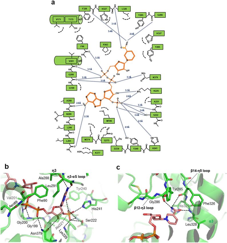
www.nature.com/scientificreports/
a 1 43 168 285 309 357 393
Human VAT-1 Switch
segment
Domain I Domain II Domain I
b N c N
β1 β2-β3 loop
β2
C
β4 C
β3
β5 β6
Domain I α1-β4 loop
β15
α1
α10 α9 α10-β16
loop
η5
α5 α5
NADP
η3 α3 η3
η4
α4
αS
β11
β14
Domain II
β10 β9
β8
α6
α6 α8 α7
α7 β12
e
d
NADP
α3
α5
η3
α4
αS
Domain I
β8
β10 β12
β9 β11
α6 β14
α8
α7
f
Domain II
Scientific Reports | (2021) 11:2120 | https://doi.org/10.1038/s41598-021-81409-y 2
Vol:.(1234567890)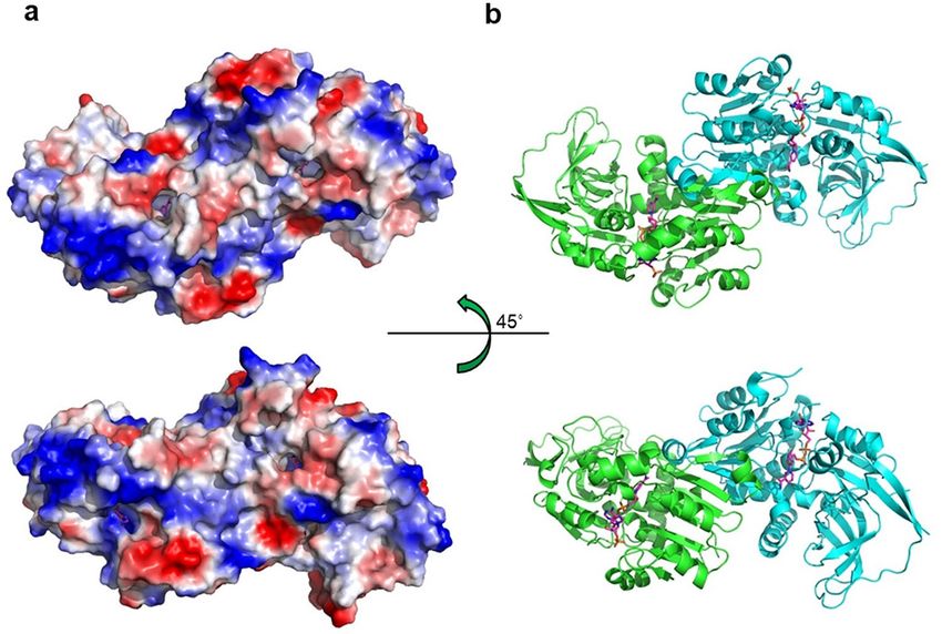
www.nature.com/scientificreports/
◂Figure 1. Structure of VAT-1. (a) Domain organization of the human VAT-1, which comprised of Domain
I and Domain II. A flexible Switch segment was displayed. (b) The VAT-1 structure in the free form. VAT-1
comprises two α/β domains, designated as Domain I and II. Water molecules found in the nucleotide-binding
site are shown as red spheres. The Switch segment (yellow) covers the binding site. To aid the visualization, α5,
α6, and α7 were aligned to NADP-bound VAT-1 in (c). (c) The VAT-1 structure in the NADP-bound form. The
bound NADP molecule is shown as a space-filling model (color codes; carbon in grey, nitrogen in blue and
oxygen in red). Large conformational changes are found in the Switch segment (Tyr285–Phe309 in yellow)
covering the binding site. Part of the Switch segment is folded into an α-helix (αS). (d) Topology of secondary
structures found in the free form of VAT-1. Secondary structures comprise α-helices (α1–α10) and 3 10-helices
(η1–η5) (pink cylinders) and β-strands (β1–β16) (blue arrows). Domain I is connected to Domain II by α3–α4
helix and the loop from β14 strand, followed by the η5–α9–β15–α10–β16 segment incorporated in Domain I.
Domain II contains the short β–α–β motif for mediating dimerization of VAT-1 that comprises β13 and β14
strands (orange arrows) and α8 helix (green cylinder). The segment between β12 and β13 strands is designated
as the Switch segment (yellow) because of its dynamic conformational changes. The Switch segment contains
two 310-helices (η3 and η4) in the free form, but in the NADP-bound form one 3 10-helix (η4) is transformed
into a longer α-helix (αS), which is also shown. Residue numbers corresponding to each secondary structure are
shown. The six-stranded Rossmann fold β8–α4–β9–α5–β10–α6–β11–α7–β12–α8–β14 provides the fundamental
framework for NADPH binding. (e) The Rossmann fold in Domain II with the bound NADP molecule found in
the crystal structure of the NADP-bound form of VAT-1. The fold comprises β8–α4–β9–α5–β10–α6–β11–α7–
β12–α8′ and β14–η5 forming a six-stranded parallel β-sheet with sequence β10–β9–β8–β11–β12–β14. (f) The
NADP molecule in the bound form of VAT-1 with omit map which was calculated by a refinement of Phenix.
refine with NADP-deleted model (contoured at 3σ).
structures of free-form VAT-1 and NADP-bound VAT-1 complex and additional enzymatic studies, we revealed
that VAT-1 contains a conserved NADPH binding cleft and binds NADPH to function as an oxidoreductase.
Results
Structure determination. Recombinant human VAT-1 was purified and crystallized. Since use of the full-
length (1–393 residues) protein yielded crystals that diffracted only to low resolution (below 8 Å), we sought to
generate crystals that could diffract to higher resolution by employing a truncated protein for crystal formation.
Truncation of the poorly conserved N-terminal region of the protein to generate N-truncated VAT-1 (43–393
residues), hereafter referred to simply as VAT-1 unless otherwise stated (Fig. 1a, Suppl. Fig. S1), yielded crystals
that diffracted to 2.3 Å resolution. The structure was determined by molecular replacement using the known
structure of a VAT-1 homolog-like protein (PDB ID 4A27) and refined (Suppl. Table S1). The obtained structure
revealed a tunnel that possesses a conserved NADPH-binding motif and is obviously expected to accommodate
an NADPH molecule. We confirmed that NADPH binds VAT-1 with low affinity using low-c isothermal titra-
tion calorimetry (ITC) method16 (Suppl. Fig. S2) and crystallized VAT-1 with bound NADP. We obtained crys-
tals that diffracted to 2.62 Å resolution and the structure was refined (Suppl. Table S1).
Overall structure. The VAT-1 molecules in the free and NADP-bound states display essentially the same
kidney-shaped structures (Fig. 1b,c). VAT-1 comprises ten α-helices (α1–α10), five 310-helices (η1–η5) and
sixteen β-strands (β1–β16), and forms two α/β domains, designated as Domain I and II (Fig. 1d). Domain
I is formed by 126 N-terminal residues (Ala43–Ala168) and 37 C-terminal residues (His357–Asn393), while
Domain II is formed by 187 residues (Leu169–Pro356) (Fig. 1a). Domain I comprises nine β-strands consisting
of an isolated two-stranded antiparallel β-sheet (β1–β2), a six-stranded mixed β-sheet (β15–β16–β3–β4–β5–
β7), which is highly twisted, and β3-strand forms an additional antiparallel β-sheet with β6-strand. Domain
II comprises seven β-strands consisting of a six-stranded parallel β-sheet (β10–β9–β8–β11–β12–β14) and one
isolated β-strand (β13). The long segment (25 residues) between β12 and β13 strands is conformationally flex-
ible and displays different conformations among the crystallographically independent molecules in the crystals
with part of the segment being disordered (Suppl. Fig. S3). We refer to the β12–β13 segment as the Switch seg-
ment (Tyr285–Phe309) because of the dynamic conformational changes that are invoked upon NADP binding
(Fig. 1b,c), which will be discussed in detail below.
The VAT-1 structure formed by the two α/β domains is closely related to that displayed by members of the
quinone oxidoreductase family (Suppl. Fig. S4). When the VAT-1 structure is superimposed on the starting
structure of human VAT-1 homolog-like protein, which exhibits 45.8% sequence identity, the root-mean-square
(rms) deviation of the Cα atoms is 1.13 Å. We found that ζ-crystallin-like quinone oxidoreductase Zta114 (PDB
ID 3QWB, with sequence identity of 25.5%) displays a closely related structure with a relatively small Cα atom
rms deviation of 1.51 Å. CurF E R17 from Lyngbya majuscule (PDB ID 5DP2, with 26.9% sequence identity) also
exhibits a small Cα atom rms deviation of 1.67 Å. It should be noted that Zta1 is an active quinone oxidoreductase
and that CurF ER is a unique enoyl-acyl carrier protein reductase that catalyzes cyclopropanation in the curacin
A biosynthesis pathway17. The similarity in structure between VAT-1 and the aforementioned enzymes led us to
investigate the potential enzymatic activity of VAT-1 (full-length) and VAT-1 (43–393) (see below).
Rossmann fold nucleotide‑binding site. The obtained structures revealed that VAT-1 conserves a
Rossmann fold18 in Domain II with the GXGXXG motif for nucleotide binding, a modified AXGXXG motif,
AAGGVG (residues 197–202), at the N-terminal end of α4-helix (Fig. 1e). We observed clear electron density
for the bound NADP molecule (Fig. 1f). The nucleotide-binding site is located at the interface between Domain
Scientific Reports | (2021) 11:2120 | https://doi.org/10.1038/s41598-021-81409-y 3
Vol.:(0123456789)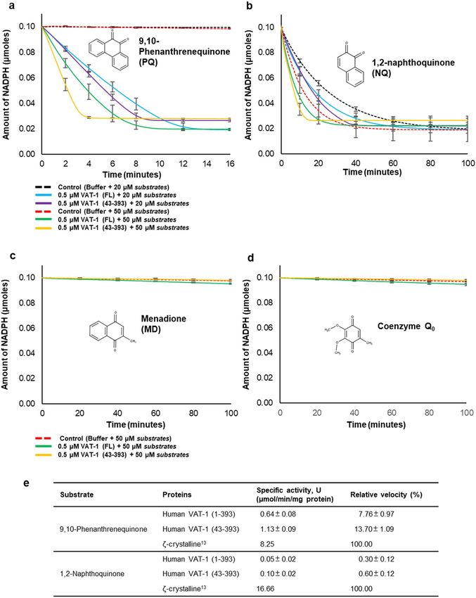
www.nature.com/scientificreports/
Figure 2. VAT-1 dimer. (a) The VAT-1 homodimer in the NADP-bound form. The bound NADP molecules are
shown as stick models (orange). The β13–α8–β14 segment of each VAT-1 protomer forms the βαβ motif. The
βαβ motif (magenta) of VAT-1 mol A (green) and the βαβ motif (orange) of mol B (cyan) mediates the dimer
interface by forming intermolecular antiparallel associations between β13 and β13′ strands, and β14 and β14′
strands. (b) The βαβ motifs from two protomers build a compact module by forming a mini hydrophobic core
with hydrocarbon side chains from Val311 (at β13 strand), Leu316 (α8 helix) and Val323 (β14 strand).
I and II, and is covered by the long 25-residue segment between β12 and β13 strands, extending from Domain II
toward Domain I (Fig. 1b,c). The nucleotide-binding site is also partly covered by Domain I, which projects α10-
helix, α10–β16 loop, α1-helix and α1–β4 loop toward the binding site. Moreover, η4-helix blocks one edge of the
binding site. As a result of being covered by Domain I and the Switch segment from Domain II, the nucleotide
binding site is embedded in the interface between Domain I and II and forms a tunnel. In the free form of VAT-
1, the tunnel is filled with solvent water molecules (Fig. 1b). In the NADP-bound form, the nucleotide-binding
tunnel is filled by the bound NADP molecule, which adopts an extended conformation and is almost completely
buried inside (Fig. 1c). Only part of the adenine ring at one entrance and part of the nicotinamide ring contain-
ing the amide group are accessible from the solvent region (Suppl. Fig. S5).
Dimeric structure. The obtained crystal structures showed that VAT-1 forms a homodimer in which each
protomer is related by a pseudo dyad axis, as observed in the structures of the related oxidoreductase family
members14,17,19 (Fig. 2a). The dimerization is mediated by Domain II. The β13–α8–β14 segment (the βαβ motif)
in Domain II is packed into a compact fold by forming a hydrophobic mini core (Val311–Leu316–Val323)
between sandwiched β13 on β14 strands and plays a key role in formation of the dimer interface (Fig. 2b).
The βαβ motifs from two protomers form an intermolecular two-stranded antiparallel β-sheet (β13–β13′) and
an intermolecular β14–β14′ antiparallel association, resulting in formation of an extended 12-stranded β-sheet
from the six-stranded parallel β-sheets of the two protomers. In addition to the β–β association, α8-helix from
each protomer forms nonpolar interfaces to mediate the dimerization. The fundamental architecture and dimer-
ization mediated by the βαβ motif are conserved in the NADP-bound VAT-1 structure, though local conforma-
tional changes occur upon nucleotide binding as described below.
NADP binding. The bound NADP molecule is tightly held in the narrow tunnel of VAT-1 with a fixed con-
formation without any residual mobility (Fig. 1c). The NADP/NADPH cofactor comprises the ADP (adenosine-
2′,5′-diphosphate) moiety and NMN (nicotinamide 5′-mononucleotide) moiety. The bound NADP molecule
has the ADP moiety in the syn conformation with the C3′-endo pucker of the ribose ring conformation and the
NMN in the anti conformation with the C2′-endo pucker of the ribose ring conformation. In the binding tunnel,
VAT-1 forms fourteen direct hydrogen bonds with the bound NADP molecule (Fig. 3a).
At one entrance of the tunnel, β9–α5 loop (Ser222), α5-helix (Lys225) and β10–α6 loop (Tyr240, His241)
from Domain II form the phosphate-binding site and tightly trap the 2′-phosphate group of ADP moiety by
forming six hydrogen bonds with the site, indicating the importance of the 2′-phosphate group of NADPH in
VAT-1 binding (Fig. 3b). The adenine ring is recognized by forming two hydrogen bonds between the adenine
2-amino group and the Switch segment (the main chains of Ala288 and Leu291 of η3-helix) from VAT-1 covering
the binding site. The 3′-hydroxyl group of the ADP ribose forms a hydrogen bond with conserved Asn379 (at
α10–β16 loop) from Domain I. Asn379 also forms a hydrogen bond with the 5′-phosphate of ADP and fixes the
ADP conformation by bridging two parts of ADP through two hydrogen bonds. The diphosphate group at the
center of the NADP molecule is fixed at the middle of the tunnel by forming five hydrogen bonds with VAT-1.
With one of the diphosphate groups, the 5′-phosphate group of the NMN moiety is trapped by the AXGXXG
motif at the N-end of α4-helix (Fig. 3c). The nicotinamide ring forms three hydrogen bonds via the amide group
of the nicotinamide with the main chains of β14–η5 loop (Phe326 and Leu328) and the N-terminal end of the
Switch segment (Tyr285), whereas the ribose of the NMN moiety has no direct hydrogen bond.
Nonpolar interactions also contribute to NADP binding. The adenine ring is sandwiched with Lys377 (at
α10–β16 loop) and Phe90 (α1-helix)/Met196 (β8–α4 loop). Phe90 and Met196 also contact with the ribose of
the NMN moiety. Thus, Phe90 and Met196 are sandwiched between two ribose rings of the NADP molecule. The
nicotinamide ring sits on Tyr285 (Switch segment), Thr175 (α3-helix), Val201 (α4-helix) and Phe326 (β14–η5
loop).
Scientific Reports | (2021) 11:2120 | https://doi.org/10.1038/s41598-021-81409-y 4
Vol:.(1234567890)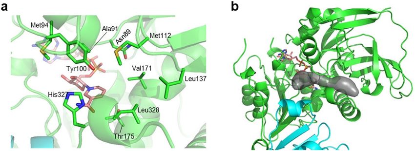
www.nature.com/scientificreports/
Figure 3. NADP bound to VAT-1. (a) Graphical summary of the NADP-VAT-1 interactions calculated by
IGPLOT33. (b) The ADP moiety of the bound NADP forms 12 hydrogen bonds with VAT-1. (c) The NMN
L
moiety of the bound NADP forms 6 hydrogen bonds with VAT-1.
Switch segment dynamics. NADP binding induces conformational changes in the VAT-1 structure. The
major conformational change is found in the Switch segment (Fig. 4a,b). As mentioned above, the Switch seg-
ment participates in recognition of both adenine and nicotinamide rings of NADP. In the NADP-bound form,
the Switch segment induces conversion of short η4-helix to longer αS-helix and approaches η3-helix to make
nonpolar helix-helix contacts.
These interactions should stabilize η3-helix to interact with the adenine ring. In addition to the dynamic
conformational change in the Switch segment, helices and loops forming the binding site are also shifted to
accommodate the NADP molecule. Among these, α1-helix and the following α1–β4 loop from Domain I is
lifted by ~ 1 Å to enlarge the binding tunnel by forming hydrogen bonds with η5-helix and η5–α5 loop from
Domain II (Fig. 4c).
The Switch segment is directly linked to β13-strand, suggesting that conformational changes in the Switch
segment affect the βαβ motif that mediates dimerization. Moreover, the Switch segment makes direct contacts
with α8-helix of the other protomer in the free form (Fig. 4d). On NADP binding, the Switch segment moves
from α8-helix of the other protomer, which results in a shift of the protomer (by maximum of ~ 6 Å).
The Switch segment contains clusters of nonpolar residues, being characterized by two tryptophan residues
(Trp305 and Trp306). Intriguingly, most of these nonpolar residues are exposed to the solvent region (Fig. 4e).
On NADP binding, αS-helix is folded, whereas most of the nonpolar residues are still exposed to the outside
Scientific Reports | (2021) 11:2120 | https://doi.org/10.1038/s41598-021-81409-y 5
Vol.:(0123456789)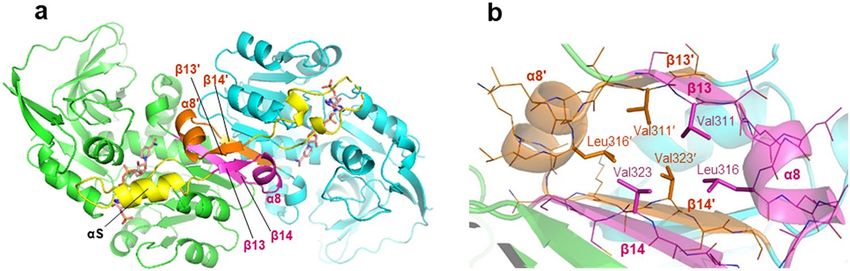
www.nature.com/scientificreports/

          Scientific Reports |   (2021) 11:2120 |   https://doi.org/10.1038/s41598-021-81409-y   6

Vol:.(1234567890)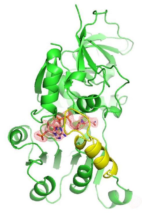
www.nature.com/scientificreports/
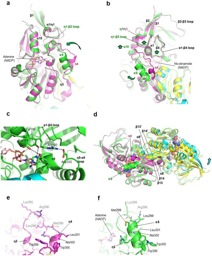
◂Figure 4. Induced fit and conformational dynamics of the switch loop on NADP binding. (a) The induced fit
conformational change on NADP binding. Structural overlay is shown for NADP-bound VAT-1 (green) on the
free form (magenta). An arrow (green) indicates the large shift of the Switch segment on NADP binding. A view
for the 2′,5′-ADP moiety of the bound NADP (stick model) is provided with the dimer partner in the NADP-
bound form (cyan) and in the free form (yellow). (b) As in (a) but a rotated view of 90° around the vertical axis
to show a view of the NMN moiety of the bound NADP. Arrows (green) indicate the large shift of the Switch
segment and small local shifts of α1–β4 loop, η1–β2 loop and α10-helix on NADP binding. (c) Formation of
the hydrogen bonding bridge widens the nucleotide-binding site. In the NADP-bound form, α1–β4 loop of
Domain I forms hydrogen bond/salt bridge interactions with η5-helix and η5–α9 loop of Domain II. Hydrogen
bonding residues comprise Tyr100–His237 and Asp101–Tyr326 and the salt bridge comprises Asp102–Asp332.
(d) Conformational changes on NADP binding affect the dimer configuration. Structural overlay is shown
for the VAT-1 dimer in the NADP-bound form (green and cyan) on the free form (magenta and yellow). The
bound NADP molecules are shown as space-filling models. One protomer (green) of the NADP-bound form is
superimposed on one protomer (magenta) of the free form to clarify the positional shifts of the other protomers.
(e) Exposure of nonpolar residues from αS-helix of the Switch segment in the free form of VAT-1. (f) Exposure
of nonpolar residues from αS-helix of the Switch segment in the NADP-bound form of VAT-1.
(Fig. 4f). These exposed nonpolar residues (Leu298, Met299, Leu301, Ala302, Trp305 and Trp306) participate
in crystal contacts with adjacent VAT-1 molecules by making nonpolar contacts with nonpolar residues of the
Switch segment (Suppl. Fig. S6). In the crystal of the NADP-bound form, nonpolar contacts between αS-helices
were found to include intermolecular tryptophan–tryptophan interactions in the crystals (Suppl. Fig. S6a). These
intermolecular interactions may contribute to mediate dimer–dimer interactions in solution. We examined
this possibility by analyzing both VAT-1 (full-length) and VAT-1 (43–393) at various concentrations using
size-exclusion chromatography (SEC) (Suppl. Fig. S7a,b). Both VAT-1 constructs displayed a similar trend in
solution as the proteins tend to form dimer at low concentration and shift to tetramer (dimer–dimer) at higher
concentrations. VAT-1 (full-length) was eluted as tetramer peak at the concentration of 300 μM, meanwhile
the VAT-1 (43–393) exists as tetramer–dimer equilibrium. The results of SEC are in agreement with the cur-
rent crystal structures, where VAT-1 exists in dimer and dimer–dimer conformations. The conformation of the
Switch segment was affected by the NADP-binding as shown in our crystal structures, we continued to analyze
the effect of its binding towards the oligomerization of VAT-1 (Suppl. Fig. S7c,d). In the presence of 10-folds
higher molar concentration of NADP, VAT-1 (full-length) remained unaffected as free forms. Surprisingly, a
broader shoulder peak was observed for 50 μM VAT-1 (43–393) in the presence of 500 μM NADP, indicating
that VAT-1 (43–393) has a higher tendency to shift to dimer by the addition of NADP in comparison to VAT-1
(full-length). However, the exact reason for the reduced tendency in tetramerization exhibited by the truncated
VAT-1 (43–393) in the presence of excess NADP was unclear.
Putative substrate binding site. Intriguingly, one side of the nicotinamide ring in the nucleotide-bind-
ing tunnel is open without any VAT-1 residues making direct contacts with the ring (Fig. 5a). The bound nico-
tinamide ring should provide the active site of the potential oxidoreductase activity of VAT-1 as with members
of the quinone oxidoreductase family14,17,19. Zta1 also has a large space on the nicotinamide ring of the bound
NADPH molecule and a small glycerol molecule was found to be located at the space14. This space is proposed
as the substrate binding site of substrates such as 1,2-naphthoquinone (NQ) and 9,10-phenanthrenequinone
(PQ). We thought that the large empty space on the nicotinamide ring plane in our NADP-bound VAT-1 struc-
ture may be the substrate binding site for potential oxidoreductase activity (Fig. 5b). The space is rather flat and
surrounded by nonpolar residues (Ala91, Met94, Met112 and Leu137 from Domain I, Val171 and Leu328 from
Domain II) and polar residues (Asn89 and Tyr100 from Domain I, and Thr175 and His327 from Domain II)
(Fig. 5a). Interestingly, Asn89 of VAT-1 seems to correspond to Asn48 of Zta1, which was proposed to partici-
pate in recognition of substrates NQ and PQ by forming a direct hydrogen bond with the carbonyl group of
each substrate14. In addition to Asn48, residues Ile50 and Leu131 of Zta1 were shown to contribute to substrate
binding. These two residues correspond to Ala91 and Val171 of VAT-1. Therefore, the putative substrate-binding
space of VAT-1 may be slightly larger than that of Zta1. As proposed for Zta1, electron transfer via π–π stacking
interactions may occur once the phenyl ring of quinone substrate compounds is aligned parallel to the nicotina-
mide ring. An increase in hydrophobicity around the positively charged nicotinamide group is likely to acceler-
ate electron transfer from NADPH to substrate.
Quinone oxidoreductase activity. Our structure of the NADP-bound form clarified that VAT-1 contains
a conserved nucleotide-binding site, which is common to members of the NADPH-dependent oxidoreductase
subfamily, and that NADP binds to this site of VAT-1 in a manner similar to the binding of NADP/NADPH
to enzymatically active members of the oxidoreductase subfamily such as Zta1. Moreover, we found a puta-
tive substrate-binding site on the nicotinamide ring of NADP. These results suggest that VAT-1 may possess
oxidoreductase activity against some quinone derivatives. We then investigated the enzymatic activity of VAT-1
against certain potential substrates including 1,2-naphthoquinone (NQ) and 9,10-phenanthrenequinone (PQ),
the substrates for Zta1 and ζ-crystallin13,14, as well as menadione (MD) and ubiquinone 0 (UQ0) (Fig. 6). MD is
vitamin K3, while UQ0 is coenzyme Q0. Among these quinones, both VAT-1 (full-length) and VAT-1 (43–393)
possessed the highest specificity toward PQ at a specific activity of 0.64 U and 1.13 U, respectively (Fig. 6a,e).
VAT-1 (full-length) and VAT-1 (43–393) reacted to NQ at a much slower rate and possessed a specific activity of
0.049 U and 0.102 U, respectively (Fig. 6b,e). Although VAT-1 (full-length) and VAT-1 (43–393) reactive towards
PQ and NQ, the activities were relative low, which were 7.8 and 13.7% for PQ, and 0.3 and 0.6% for NQ, respec-
Scientific Reports | (2021) 11:2120 | https://doi.org/10.1038/s41598-021-81409-y 7
Vol.:(0123456789)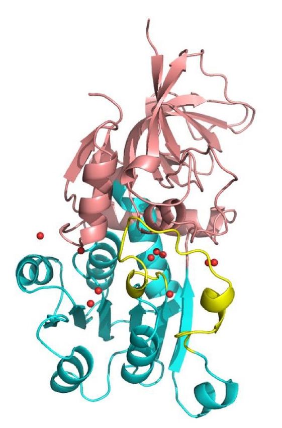
www.nature.com/scientificreports/
Figure 5. Putative substrate-binding site. (a) The putative substrate-binding site on the nicotinamide ring
plane. (b) The cavity of the putative substrate-binding site is depicted as a gray surface.
tively, compared to ζ-crystallin13 (Fig. 6e). Interestingly, VAT-1 (43–393) consumed PQ and NQ at a higher rate
and displayed nearly twofold higher activities over the VAT-1 (full-length). In contrast, both of VAT-1 constructs
did not show any significant activities towards MD and UQ0 (Fig. 6c,d). To investigate the kinetic properties of
VAT-1 (full-length) towards PQ, we calculated the kinetic constants from double reciprocal Lineweaver–Burk
plots of the reversed velocity (1/ν) versus the reversed substrate concentration (1/S) (Fig. 7a). In the presence of
PQ at various concentrations, the initial velocities exhibited by 0.5 μM VAT-1 (full-length) were measured and
plotted. The calculated Km and kcat of VAT-1 (full-length) were 29.89 μM and 1.13 s−1 respectively, indicating that
VAT-1 (full-length) is an active quinone oxidoreductase (Fig. 7b).
Discussion
We have demonstrated that VAT-1 has the α/β fold structure exhibiting essentially the same architecture as the
NADPH-dependent quinone oxidoreductase structures that are characterized by a kidney-shaped structure
comprising two domains in α/β folds, designated as Domain I and II. The interface between Domain I and II form
a deep tunnel of the nucleotide-binding site with a conserved sequence for phosphate binding on the Rossmann
fold of Domain I. Part of the nucleotide-binding site provides a small pocket for putative substrate binding as
observed in other oxidoreductases such as Z ta114.
The putative molecular function of VAT-1 has been discussed with respect to phospholipid b inding5. Reported
structures of lipid transfer and binding proteins reveal the presence of deep and long hydrophobic clefts or tun-
nels to accommodate long nonpolar hydrocarbon chains of l ipids20–23. However, the VAT-1 dimer possesses no
deep cleft or tunnel other than the nucleotide-binding site (Fig. 8). Although the nucleotide-binding site seems
to be large enough to accommodate some phospholipids, the nature or mechanism of the binding specificity
remains a serious question. The negatively charged head group of phospholipids may be trapped at the phosphate-
binding site for NADP with some specificity. Further experimentation is required to clarify the potential lipid
binding mechanism.
The Switch segment (Tyr285–Phe309) may be a specific feature for VAT-1 and its homologs, but not for other
ζ-crystallin homologs such as Zta1 (Suppl. Fig. S8a). We found that the Switch segment participates in direct
interactions with the bound NADP molecule, so that the function of the Switch segment should be coupled with
nucleotide binding. In our structure, NADP binding induced folding of the Switch helix to facilitate protrusion
of nonpolar residues such as tryptophan toward the outside, suggesting that NADP binding may trigger an inter-
molecular interaction with other binding partners. It is likely that the Switch helix may contribute to interaction
with membranes, such as mitochondrial membranes to mediate recruitment of VAT-1 toward mitochondria.
We demonstrated oxidoreductase activity of VAT-1 in the presence of NADPH in vitro. The activity was
observed with NQ and PQ, known substrates of Zta1 and ζ-crystallin13,14. In fact, the putative substrate binding
site of VAT-1 resembles that of Zta1 and ζ-crystallin (Suppl. Fig. S8b). In addition, VAT-1 also displayed a sub-
strate affinity towards PQ at a Km value of 29.9 μM, which was comparable to those of the Zta1 (Km = 4.56 μM14)
and ζ-crystallin (Km = 13 μM13) (Fig. 6b). The enzymatic studies further indicate VAT-1 is a NADPH-dependent
quinone oxidoreductase. Since some reports indicated a putative function of VAT-1 in mitochondria dynamics,
it is an interesting question whether quinone compounds participating in mitochondrial functions could act
as substrates of VAT-1. However, we detected no activity against UQ. VAT-1 may contribute to the regulation
of energy metabolism of cells by participating in some metabolic pathways but not in mitochondrial processes.
The unknown authentic substrate(s) may be other quinone compounds such as cofactors essential for other
processes in metabolic pathways. Further experiments are required to identify the substrate(s) and the role of
the oxidoreductase activity of VAT-1.
Methods
Cloning, expression and purification. The cloned cDNA of human VAT-1 was purchase from GE
Healthcare. Full-length (residues 1–393) and N-terminal truncated (residues 37–393 and 43–393) forms of the
human VAT-1 cDNA fragments were amplified with KOD-Plus-PCR kit (TOYOBO), and with human VAT-1
cDNA as template DNA. PCR-amplified cDNA fragments were inserted between Sma I and Not I in pGEXM
Scientific Reports | (2021) 11:2120 | https://doi.org/10.1038/s41598-021-81409-y 8
Vol:.(1234567890)
www.nature.com/scientificreports/
Figure 6. Potential quinone oxidoreductase activity. (a) Time-course of NADPH decrease in the reaction of VAT-1
(full-length) and VAT-1 (43–393) with 1,2-naphthoquinone (NQ). The chemical structure is shown in the panel.
Absorbance at 340 nm was monitored to detect the decrease of the NADPH in the assay. Reactions were performed in
mixtures containing 100 mM Tris–HCl (pH 7.8), 100 μM NADPH, 20 and 50 μM NQ. Control was performed without
the addition of VAT-1. All the reactions were performed in triplicates, and the error bars were indicated. (b) As in (a)
but for 9,10-phenanthrenequinone (PQ). (c) Time-course of NADPH decrease in the reaction of VAT-1 (full-length)
and VAT-1 (43–393) with 50 μM menadione (MD), which is part of vitamin K3. (d) As in (c) but for ubiquinone 0
(UQ0), which is part of coenzyme Q0. (e) Comparison of the relative activities of 0.5 μM VAT-1 (full-length)/(43–393)
to that of the ζ-crystalline, using 50 μM of 1,2-naphthoquinone and 9,10-phenanthrenequinone as substrates.
Scientific Reports | (2021) 11:2120 | https://doi.org/10.1038/s41598-021-81409-y 9
Vol.:(0123456789)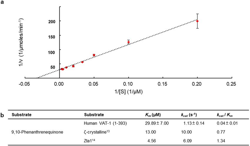
www.nature.com/scientificreports/

                                            Figure 7.  Enzyme kinetics of VAT-1. (a) The Lineweaver–Burk reciprocal plot of VAT-1 (full-length) versus PQ.
                                            The Km of VAT-1 (full-length) towards PQ was calculated as 30 μM. The initial velocities were measured at 5, 10,
                                            20, 30, 50, 100, 200, and 300 μM. All reactions were measured in triplicates. (b) The Km and kcat of VAT-1 (full-
                                            length) in compared to that of the ζ-crystalline.

                                            Figure 8.  Molecular surface of VAT-1. (a) Electrostatic potential surface of the VAT-1 dimer. (b) As in (a) but
                                            of the corresponding ribbon model.

                                            bacterial protein expression plasmid which has modified the multiple cloning site of pGEX 6P-3 (GE Health-
                                            care) (Suppl. Fig. S9), with In-Fusion HD Cloning Kit (Clontech). The recombinant proteins are expressed as
                                            N-terminal glutathione S-transferase (GST)-fused proteins. The validity of the coding sequences were verified
                                            by DNA sequencing. Escherichia coli Rosetta 2 (DE3) (Novagen) was transformed with the plasmids. The cells
                                            were initially grown in a shaking incubator at 37 °C till the cell density reached an O.D. 0.6 at 595 nm. Then, the
                                            protein expression was induced by the addition of isopropyl-β-d-thiogalactoside (IPTG) to a final concentration

          Scientific Reports |   (2021) 11:2120 |                https://doi.org/10.1038/s41598-021-81409-y                                                    10

Vol:.(1234567890)
www.nature.com/scientificreports/
of 100 µM following by incubation for 20 h at 16 °C for VAT-1 (43–393) or 20 °C for VAT-1 (full-length). The
cells were harvested by centrifugation using Beckman Avanti HP 26XPI (4000 rpm for 15 min at 4 °C). Cells
were then suspended in 2× PBS (phosphate-buffered saline) supplemented with 1 mM dithiothreitol (DTT) and
0.1% (v/v) Triton X-100 and disrupted by sonication using Qsonica (Q500) with 50% amplitude, 2 and 6 s on/
off cycles for 45 min in an ice-bath. The soluble fraction was separated by ultracentrifugation using Beckman
Optima XE-90 (30,000 rpm for 40 min at 4 °C). The supernatant containing the target protein was loaded onto
a Glutathione Sepharose 4B resin column (GE Healthcare). The column was washed with buffer containing
20 mM Tris–HCl (pH 8.0), 100 mM NaCl and 1 mM DTT, and target protein was eluted using the same buffer
containing 15 mM glutathione and cleaved using 4 units/ml HRV 3C protease (Merck) at 4 °C for overnight. The
column effluent was loaded onto a HiTrap SP cation-exchange column (GE Healthcare) and eluted using a gradi-
ent of 0–500 mM NaCl in a buffer containing 25 mM MES (pH 6.0) and 1 mM DTT. The collected fraction was
loaded onto a Superdex 200 gel filtration column (GE Healthcare) using buffer comprising 10 mM Tris–HCl (pH
7.2), 100 mM NaCl and 0.5 mM Tris(2-carboxyethyl)phosphine hydrochloride (TCEP). Analysis of the purified
samples using SDS-PAGE (Suppl. Fig. S10) and matrix-assisted laser desorption/ionization time-of-flight mass
spectrometry (MALDI-TOF MS; Bruker Daltonics) confirmed that the VAT-1 proteins (full-length and 43–393)
were successfully purified without any degradation. The purified proteins were frozen in liquid nitrogen and
stored at − 80 °C until use.
Crystallization. Preliminary crystallization screening for both of the free- (full-length and 43–393) and
NADP bound form were performed using the vapor-diffusion method at both 4 °C and 20 °C using commer-
cially available screening kits (Hampton Research and QIAGEN). Proteins were mixed at 1:1 ratio with the reser-
voir solution. Crystals of free form of VAT-1 (43–393) were observed after 2 days in equilibrium against reservoir
solution containing 100 mM BisTris (pH 6.0), 200 mM sodium nitrate and 22% (v/v) PEG 3350 (Hampton
Research) at 20 °C. Crystals of VAT-1 (full-length) were found after 6 days in equilibrium against the solution
containing 100 mM Tris–HCl (pH 7.0), 200 mM calcium acetate and 20% (v/v) PEG 3000 (QIAGEN) at 20 °C.
The best crystal of VAT-1 (43–393) and NADP complex was obtained by the mixture of 1 µl protein–ligand
solution (0.4 mM protein and 10 mM NADP in a buffer comprising 10 mM Tris–HCl (pH 7.2), 100 mM NaCl,
0.5 mM TCEP) and 1 µl reservoir solution [100 mM sodium citrate (pH 5.5) and 20% (v/v) PEG 3000 (Hampton
Research)] after incubation for 3 days at 4 °C. Crystals were cryoprotected by the addition of 25% (v/v) glycerol
for those of VAT-1 (full-length) and the NADP-bound VAT-1 (43–393). On the other hand, crystals of free form
of VAT-1 (43–393) were cryoprotected in the solution containing 100 mM BisTris (pH 6.0), 200 mM sodium
nitrate and 40% (v/v) PEG 3350. The cryoprotected crystals were flash-cooled using liquid nitrogen until use.
X‑ray data collection, phasing and refinement. All X-ray data were collected at SPring-8, Harima,
Japan. During X-ray beam exposure, crystals were flash-cooled and maintained at 100 K using a nitrogen stream.
Detailed statistics of the structure determination are shown in Supplementary Table S1. All diffraction data were
indexed and merged using the XDS program24. Molecular replacement was successfully performed using the
PHASER program25 in Phenix26, and the structure of VAT-1 homolog-like protein (PDB ID 4A27), which dis-
plays 54% sequence identity, as a reference model. The built model was refined through alternating cycles using
the Coot27,28 and phenix.refine p rograms29. Superposition of VAT-1 was performed using the program Super-
pose in Phenix . Comparison of our VAT-1 to the structures in PDB was performed with MATRAS server30.
26
The alignment of the amino acid sequences were performed using C lustalOmega31. Illustrations were prepared
using the program PyMOL Molecular Graphics System, Version 1.5.0.3 Schrödinger, LLC. The cavity calcula-
tions were performed using Caver 3.0.3 PyMol plugin32. The interaction of VAT-1 and NADP was calculated by
LIGPLOT33.
Size exclusion chromatography (SEC). Analytical gel filtration was performed with VAT-1 (full-length)
and VAT-1 (43–393) in the absence or presence of NADP by loading onto an analytical gel filtration column,
Superdex 200 (10/30) (GE Healthcare), in buffer containing 10 mM Tris–HCl (pH 7.2), 100 mM NaCl and
0.5 mM TCEP at 8 °C. In the absence of NADP, total volume 250 µl of VAT-1 protein samples were loaded onto
the column at concentrations of 10 µM, 20 µM, 50 µM, and 300 µM. For the analysis of protein–ligand complex,
various concentrations of VAT-1 proteins (10 µM, 20 µM, and 50 µM) were incubated with NADP (10-folds
higher molar concentrations) at 8 °C for 2 h prior to gel-filtration. The mixtures were then loaded onto the same
analytical gel filtration column under the same condition as previously mentioned. The elution profiles were
compared with that of Gel Filtration Standards (Bio-Rad). The molecular weights of the eluted peaks were esti-
mated using monomeric standard molecular weights, where VAT-1 (full-length) weighted 42,074 Da and VAT-1
(43–393) weighted 38,013 Da.
Binding assay using calorimetry. Binding studies utilizing isothermal titration calorimetry (ITC) were
conducted using a calorimeter (MicroCal iTC200, GE Healthcare) at 20 °C. Purified proteins were dialyzed over-
night in buffer containing 10 mM HEPES (pH 7.0), 100 mM NaCl and 0.1 mM TCEP. NADPH solution (2 mM)
was injected (0.5 μl each, 3 min pause) into VAT-1 (full-length) protein solution (0.1 mM). Details of each titra-
tion are described in the figure legends of Supplementary Fig. S2. Data fitting was performed using a 1:1 binding
model (n = 1) of the ORIGIN software program (Origin version 7.0, OriginLab Corporation, Northampton,
MA, USA) supplied with the instrument and low-c ITC method16 was used for measuring the weak interaction
between VAT-1 and NADPH.
Scientific Reports | (2021) 11:2120 | https://doi.org/10.1038/s41598-021-81409-y 11
Vol.:(0123456789)www.nature.com/scientificreports/
Enzymatic activity assay. Enzymatic activity was investigated by monitoring the decrease in NADPH
absorption at 340 nm. All assays were performed at 25 °C in a standard assay mixture containing 100 mM Tris–
HCl (pH 7.8), 100 μM NADPH, 0.5 μM VAT-1 (full-length/ 43–393) and substrates at various concentrations.
The quinone substrates used were 1,2-naphthoquinone (#346616, Sigma), 9,10-phenanthrenequinone (#156507,
Sigma), Menadione (#M5625, Sigma), and coenzyme Q0 (#D9150, Sigma). All quinone substrates were dis-
solved in ethanol, with the final concentration of alcohol in the assay being 2.5%. Reactions were initiated by the
addition of VAT-1, and the decrease in absorbance at 340 nm (ε(NADPH) = 6220 M−1 cm−1) was monitored using
a UV-1900i spectrophotometer equipped with a temperature-controlled cuvette holder TCC-100 (Shimadzu).
The decrease in absorbance resulting from the non-enzymatic reduction of quinone substrates upon addition of
NADPH was set as the background control for each assay. All reactions were measured in triplicates.
Code availability
Accession code Protein Data Bank: The atomic coordinates and structure factors for the reported crystal structures
of human VAT-1 (43–393) are deposited under accession codes 6LII (free form) and 6LHR (NADP-bound form).
Received: 17 February 2020; Accepted: 5 January 2021
References
1. Linial, M., Miller, K. & Scheller, R. H. VAT 1: An abundant membrane protein from torpedo cholinergic synaptic vesicles. Neuron
2, 1265–1273 (1989).
2. Eura, Y., Ishihara, N., Yokota, S. & Mihara, K. Two mitofusin proteins, mammalian homologues of FZO, with distinct functions
are both required for mitochondrial fusion. J. Biochem. 134, 333–344 (2003).
3. Eura, Y., Ishihara, N., Oka, T. & Mihara, K. Identification of a novel protein that regulates mitochondrial fusion by modulating
mitofusin (Mfn) protein function. J. Cell Sci. 119, 4913–4925 (2006).
4. Cao, Y.-L. et al. MFN1 structures reveal nucleotide-triggered dimerization critical for mitochondrial fusion. Nature 542, 372–376
(2017).
5. Junker, M. & Rapoport, T. A. Involvement of VAT-1 in phosphatidylserine transfer from the endoplasmic reticulum to mitochon-
dria. Traffic 16, 1306–1317 (2015).
6. Linial, M. & Levius, O. VAT-1 from Torpedo is a membranous homologue of zeta crystallin. FEBS Lett. 315, 91–94 (1993).
7. Linial, M. & Levius, O. The protein VAT-1 from Torpedo electric organ exhibits an ATPase activity. Neurosci. Lett. 152, 155–157
(1993).
8. Koch, J. et al. Human VAT-1: A calcium-regulated activation marker of human epithelial cells. Arch. Dermatol. Res. 295, 203–210
(2003).
9. Mertsch, S., Becker, M., Lichota, A., Paulus, W. & Senner, V. Vesicle amine transport protein-1 (VAT-1) is upregulated in glioblas-
tomas and promotes migration. Neuropathol. Appl. Neurobiol. 35, 342–352 (2009).
10. Gleissner, C.M.-L. et al. Neocarzilin A is a potent inhibitor of cancer cell motility targeting VAT-1 controlled pathways. ACS Cent.
Sci. 5, 1170–1178 (2019).
11. Rao, P. V. & Zigler, J. S. J. Purification and characterization of zeta-crystallin/quinone reductase from guinea pig liver. Biochim.
Biophys. Acta 1117, 315–320 (1992).
12. Duhaiman, A. S., Rabbani, N., AlJafari, A. A. & Alhomida, A. S. Purification and characterization of zeta-crystallin from the camel
lens. Biochem. Biophys. Res. Commun. 215, 632–640 (1995).
13. Rao, P. V., Krishna, C. M. & Zigler, J. S. J. Identification and characterization of the enzymatic activity of ζ-crystallin from guinea
pig lens. A novel NADPH:quinone oxidoreductase. J. Biol. Chem. 267, 96–102 (1992).
14. Guo, P.-C. et al. Structural insights into the cofactor-assisted substrate recognition of yeast quinone oxidoreductase Zta1. J. Struct.
Biol. 176, 112–118 (2011).
15. Jeske, L., Placzek, S., Schomburg, I., Chang, A. & Schomburg, D. BRENDA in 2019: A European ELIXIR core data resource. Nucleic
Acids Res. 47, D542–D549 (2019).
16. Turnbull, W. B. & Daranas, A. H. On the value of c: Can low affinity systems be studied by isothermal titration calorimetry?. J.
Am. Chem. Soc. 125, 14859–14866 (2003).
17. Khare, D. et al. Structural basis for cyclopropanation by a unique enoyl-acyl carrier protein reductase. Structure 23, 2213–2223
(2015).
18. Rao, S. T. & Rossmann, M. G. Comparison of super-secondary structures in proteins. J. Mol. Biol. 76, 241–256 (1973).
19. Porté, S. et al. Three-dimensional structure and enzymatic function of proapoptotic human p53-inducible quinone oxidoreductase
PIG3. J. Biol. Chem. 284, 17194–17205 (2009).
20. Wong, L. H., Čopič, A. & Levine, T. P. Advances on the transfer of lipids by lipid transfer proteins. Trends Biochem. Sci. 42, 516–530
(2017).
21. Ekiert, D. C. et al. Architectures of lipid transport systems for the bacterial outer membrane. Cell 169, 273-285.e17 (2017).
22. Malinverni, J. C. & Silhavy, T. J. An ABC transport system that maintains lipid asymmetry in the gram-negative outer membrane.
Proc. Natl. Acad. Sci. USA 106, 8009–8014 (2009).
23. de Saint-Jean, M. et al. Osh4p exchanges sterols for phosphatidylinositol 4-phosphate between lipid bilayers. J. Cell Biol. 195,
965–978 (2011).
24. Kabsch, W. XDS. Acta Crystallogr. D. Biol. Crystallogr. 66, 125–132 (2010).
25. McCoy, A. J. et al. Phaser crystallographic software. J. Appl. Crystallogr. 40, 658–674 (2007).
26. Adams, P. D. et al. PHENIX: A comprehensive Python-based system for macromolecular structure solution. Acta Crystallogr. Sect.
D Biol. Crystallogr. 66, 213–221 (2010).
27. Emsley, P. & Cowtan, K. Coot: Model-building tools for molecular graphics. Acta Crystallogr. Sect. D Biol. Crystallogr. 60, 2126–2132
(2004).
28. Emsley, P., Lohkamp, B., Scott, W. G. & Cowtan, K. Features and development of Coot. Acta Crystallogr. D. Biol. Crystallogr. 66,
486–501 (2010).
29. Afonine, P. V. et al. Towards automated crystallographic structure refinement with phenix.refine. Acta Crystallogr. D. Biol. Crystal-
logr. 68, 352–367 (2012).
30. Kawabata, T. & Nishikawa, K. Protein structure comparison using the markov transition model of evolution. Proteins 41, 108–122
(2000).
31. Sievers, F. et al. Fast, scalable generation of high-quality protein multiple sequence alignments using Clustal Omega. Mol. Syst.
Biol. 7, 539 (2011).
Scientific Reports | (2021) 11:2120 | https://doi.org/10.1038/s41598-021-81409-y 12
Vol:.(1234567890)www.nature.com/scientificreports/
32. Chovancova, E. et al. CAVER 3.0: A tool for the analysis of transport pathways in dynamic protein structures. PLoS Comput. Biol.
8, e1002708 (2012).
33. Wallace, A. C., Laskowski, R. A. & Thornton, J. M. LIGPLOT: A program to generate schematic diagrams of protein-ligand inter-
actions. Protein Eng. 8, 127–134 (1995).
Acknowledgements
This work was supported by a Grant-in-Aid for challenging Exploratory Research from the Ministry of Educa-
tion, Culture, Sports, Science and Technology (MEXT) of Japan (to T.H.). The synchrotron radiation experi-
ments were performed at BL41XU and BL44XU in SPring-8 with the approval of the Japan Synchrotron Radia-
tion Research Institute (JASRI) (proposal nos. 2016A2510, 2016B2510, 2016A2519, 2016B2519, 2016A6648,
2016B6648, 2017A2502, 2017A6759, 2017B6759, 2018A2503, 2018A2529, 2018A2540, 2018A6855, 2018B2503,
2018B6855, 2019A2516, 2019A2568, 2019A2576, 2019A6955, 2019B2516, 2019B2727 and 2019B6955). T.H. and
S.-Y.K. were supported in part by AMED-CREST (JP19gm1010008s0503 and JP19gm0810011h0003). T.O. was
supported by AMED-CREST (JP19gm0810001).
Author contributions
T.H. and T.O. conceived the project. T.H., S.-Y.K., T. M., and M.F.C. designed the experiments. S.-Y.K., T.M.,
M.F.C. and S.F. executed the cloning, protein biochemistry, crystallization, data collection, structure determina-
tion, enzymology and binding assays directed by T.H. K. M., T.Y. provided expertise in cell physiology. S.-Y.K.,
T.M., M.F.C. and T.H. interpreted the data. T.H. wrote the manuscript and all authors reviewed the manuscript.
Competing interests
The authors declare no competing interests.
Additional information
Supplementary Information The online version contains supplementary material available at https://doi.
org/10.1038/s41598-021-81409-y.
Correspondence and requests for materials should be addressed to T.H.
Reprints and permissions information is available at www.nature.com/reprints.
Publisher’s note Springer Nature remains neutral with regard to jurisdictional claims in published maps and
institutional affiliations.
Open Access This article is licensed under a Creative Commons Attribution 4.0 International
License, which permits use, sharing, adaptation, distribution and reproduction in any medium or
format, as long as you give appropriate credit to the original author(s) and the source, provide a link to the
Creative Commons licence, and indicate if changes were made. The images or other third party material in this
article are included in the article’s Creative Commons licence, unless indicated otherwise in a credit line to the
material. If material is not included in the article’s Creative Commons licence and your intended use is not
permitted by statutory regulation or exceeds the permitted use, you will need to obtain permission directly from
the copyright holder. To view a copy of this licence, visit http://creativecommons.org/licenses/by/4.0/.
© The Author(s) 2021
Scientific Reports | (2021) 11:2120 | https://doi.org/10.1038/s41598-021-81409-y 13
Vol.:(0123456789)You can also read