Elucidation of Teicoplanin Interactions with Drug Targets Related to COVID-19
←
→
Page content transcription
If your browser does not render page correctly, please read the page content below
antibiotics
Article
Elucidation of Teicoplanin Interactions with Drug Targets
Related to COVID-19
Faizul Azam

                                          Department of Pharmaceutical Chemistry & Pharmacognosy, Unaizah College of Pharmacy, Qassim University,
                                          Unaizah 51911, Saudi Arabia; faizulazam@gmail.com or f.azam@qu.edu.sa; Tel.: +966-50-2728652

                                          Abstract: Teicoplanin is a glycopeptide antibiotic effective against several bacterial infections, has
                                          exhibited promising therapeutic efficiency against COVID-19 in vitro, and the rationale for its use in
                                          COVID-19 is yet to be recognized. Hence, in this study a number of molecular modeling techniques
                                          were employed to decrypt the mechanistic insight of teicoplanin interaction with several COVID-19
                                          drug targets. Initially, molecular docking was employed to study the teicoplanin interaction with
                                          twenty-five SARS-CoV-2 structural and non-structural proteins which was followed by molecular
                                          mechanics/generalized Born surface area (MM/GBSA) computation for binding energy predictions
                                          of top ten models from each target. Amongst all macromolecular targets, the N-terminal domain of
                                          the nucleocapsid protein displayed the strongest affinity with teicoplanin showing binding energies of
                                          −7.4 and −102.13 kcal/mol, in docking and Prime MM/GBSA, respectively. Thermodynamic stability
                                          of the teicoplanin-nucleocapsid protein was further probed by molecular dynamics simulations
                                          of protein–ligand complex as well as unbounded protein in 100 ns trajectories. Post-simulation
                                          MM-GBSA computation of 50 frames extracted from simulated trajectories estimated an average
                                          binding energy of −62.52 ± 12.22 kcal/mol. In addition, conformational state of protein in complex
                                with docked teicoplanin displayed stable root-mean-square deviation/fluctuation. In conclusion,

                                          computational investigation of the potential targets of COVID-19 and their interaction mechanism
Citation: Azam, F. Elucidation of
                                          with teicoplanin can guide the design of novel therapeutic armamentarium for the treatment of
Teicoplanin Interactions with Drug
                                          SARS-CoV-2 infection. However, additional studies are warranted to establish the clinical use or
Targets Related to COVID-19.
                                          relapses, if any, of teicoplanin in the therapeutic management of COVID-19 patients.
Antibiotics 2021, 10, 856. https://
doi.org/10.3390/antibiotics10070856
                                          Keywords: SARS-CoV-2; teicoplanin; docking; molecular dynamics; MM/GBSA
Academic Editors: Rungroch
Sungthong and Rosa Alduina

Received: 15 March 2021                   1. Introduction
Accepted: 12 May 2021                          The outbreak of COVID-19 pandemic in China, caused by severe acute respiratory
Published: 15 July 2021                   syndrome coronavirus-2 (SARS-CoV-2) primarily spreads through close contact amongst
                                          people by sneezing, coughing, or by communicating verbally. The primary symptoms of the
Publisher’s Note: MDPI stays neutral      disease are fever, cough, fatigue, shortness of breath, and loss of smell. The complications
with regard to jurisdictional claims in
                                          of this disease include viral pneumonias, respiratory distress, and hypoxia [1]. The world
published maps and institutional affil-
                                          is suffering due to lack of effective medicine. Therefore, there is an urgent need to figure
iations.
                                          out efficient medicine through drug discovery efforts [2,3].
                                               Drug repurposing strategy is often used for recognizing novel usages of approved
                                          or investigational drugs because of several advantages such as lower risk of failure, re-
                                          duced time for drug development, and lower investment needs [4]. Therefore, numerous
Copyright: © 2021 by the author.          antimalarials, antibacterials, antiparasitic agents, and antivirals are currently prevalent in
Licensee MDPI, Basel, Switzerland.        pre-clinical as well as clinical investigations aimed at developing COVID-19 treatment by a
This article is an open access article    drug repurposing approach [5]. In particular, a widely available FDA-approved antibiotic,
distributed under the terms and
                                          teicoplanin (Figure 1), is among the molecule of interest as probable COVID-19 medicine.
conditions of the Creative Commons
                                          It belongs to the glycopeptide class of antibiotic having low toxicity profile in humans
Attribution (CC BY) license (https://
                                          and hence routinely used in clinical practice for the treatment of bacterial infections. In-
creativecommons.org/licenses/by/
                                          terestingly, it has demonstrated antiviral efficacy against several kinds of viruses such as
4.0/).

Antibiotics 2021, 10, 856. https://doi.org/10.3390/antibiotics10070856                                https://www.mdpi.com/journal/antibiotics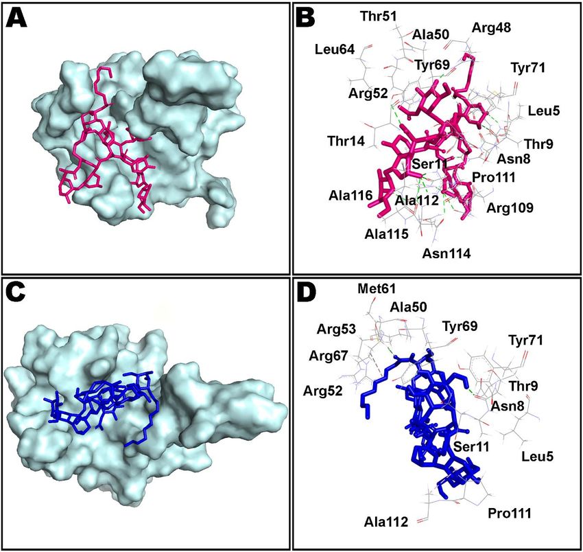
Antibiotics 2021, 10, x FOR PEER REVIEW 2 of 16
Antibiotics 2021, 10, 856 2 of 16
humans and hence routinely used in clinical practice for the treatment of bacterial infec-
tions. Interestingly, it has demonstrated antiviral efficacy against several kinds of viruses
Ebola,
such asMERS-CoV, and SARS-CoV
Ebola, MERS-CoV, [6]. Very recently,
and SARS-CoV [6]. Veryitrecently,
has beenitreported
has been that teicoplanin
reported that
can thwart can
teicoplanin the cellular entry
thwart the of SARS-CoV-2
cellular at an impressive
entry of SARS-CoV-2 1.66-µM concentration
at an impressive [7,8].
1.66-μM concen-
Furthermore,
tration IC50 value ofIC1.5
[7,8]. Furthermore, µM was
50 value recorded
of 1.5 μM was forrecorded
inhibition
forofinhibition
the SARS-CoV-2 main
of the SARS-
protease
CoV-2 by teicoplanin
main protease by[9].
teicoplanin [9].
Figure 1. Two-dimensional
Figure 1. Two-dimensionalstructural
structuralcoordinate
coordinate of teicoplanin
of teicoplanin (ACD/ChemSketch
(ACD/ChemSketch program
program version
version 2020.2.1,
2020.2.1, of Ad-
of Advanced
vanced Chemistry
Chemistry Development,
Development, Inc., Toronto,
Inc., Toronto, ON, Canada,
ON, Canada, was usedwasforused for drawing
drawing the chemical
the chemical structure).
structure).
Computer-aided
Computer-aided drug drug design
design methods
methods are
are extensively
extensively employed
employed inin drug
drug design
design and
and
discovery
discovery projects
projects owing
owing to to several
several advantages
advantages such
such asas rapid
rapid development
development process
process and
and
reduced
reduced cost
cost [10–12].
[10–12]. In
In particular,
particular, molecular
molecular docking
docking coupled
coupled with
with molecular
molecular dynamics
dynamics
simulation
simulationstudies
studiesare
areintended
intendedtoto decipher
decipherthethe
mechanism
mechanism of binding interactions
of binding at the
interactions at
molecular levels
the molecular [13].[13].
levels Rapid mechanistic
Rapid insight
mechanistic is vital
insight for for
is vital understanding structure–ac-
understanding structure–
activity
tivity relationship
relationship andand leading
leading toto theoptimization
the optimizationofofthe
thedesign
designand
anddiscovery
discoveryofofpotential
potential
molecules [14–16]. In this study, several computational techniques such as molecular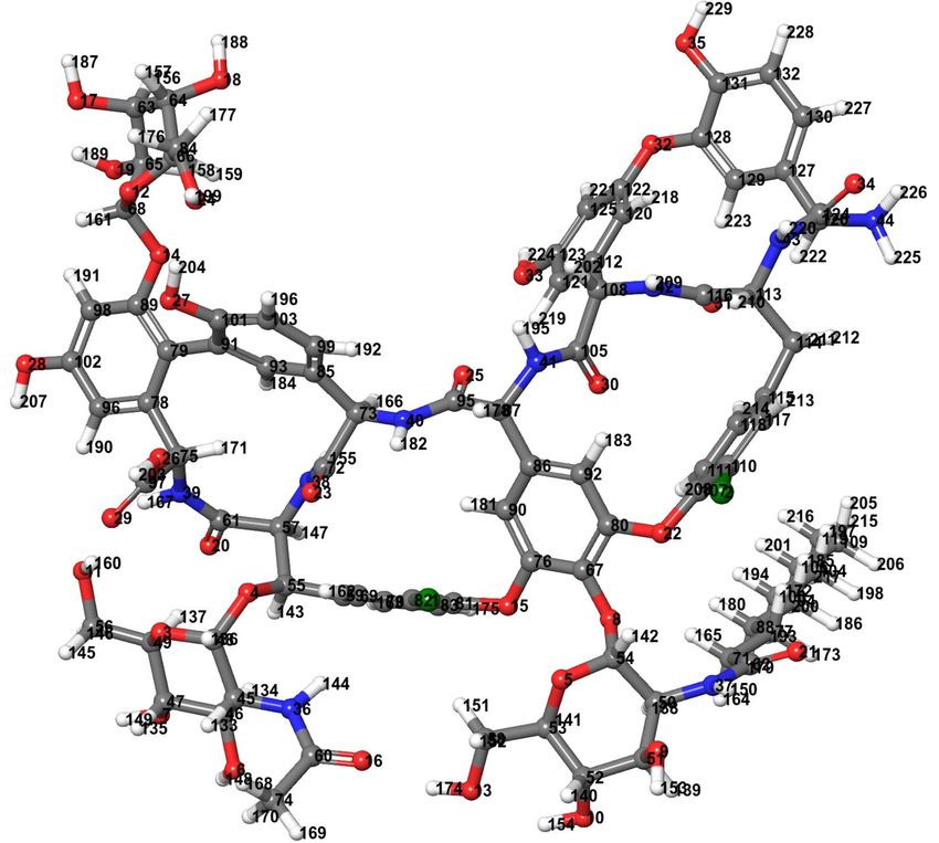
Antibiotics 2021, 10, x FOR PEER REVIEW 3 of 16
Antibiotics 2021, 10, 856 3 of 16
molecules [14–16]. In this study, several computational techniques such as molecular
docking, molecular mechanics/generalized Born surface area (MM-GBSA), and molecular
dynamics
docking, simulation were exploited toBorn
molecular mechanics/generalized inspect
surfacethe
areabinding
(MM-GBSA),interactions between
and molecular
dynamics and
teicoplanin simulation weredrug
potential exploited to inspect
targets the binding
associated withinteractions
SARS-CoV-2. between
Theteicoplanin
study is envi-
and potential
sioned to assist drug targetspotential
in finding associatedleads
with and
SARS-CoV-2. The study
accelerating drug is envisioned toprocess
development assist for
in finding potential leads and accelerating
the treatment of novel coronavirus, COVID-19. drug development process for the treatment of
novel coronavirus, COVID-19.
2. Experimental Section
2. Experimental Section
TheThe
methodology
methodologyadopted
adopted in thisstudy
in this studyhas
hasbeen
been outlined
outlined as flowchart
as flowchart in Figure
in Figure 2. 2.
FigureFigure
2. An2.outline of the
An outline of adopted methodology
the adopted methodologyininthis
thisstudy.
study.B3LYP: Becke’sthree-parameter
B3LYP: Becke’s three-parameter hybrid
hybrid model,
model, Lee–Yang–
Lee–Yang–
Parr correlation functional
Parr correlation method;
functional DFT:
method; DFT:density
density functional theory;MM/GBSA:
functional theory; MM/GBSA: molecular
molecular mechanics/generalized
mechanics/generalized Born Born
surface area; ns: nano seconds; RMSD: root-mean-square deviation; RMSF: root-mean-square fluctuation.
surface area; ns: nano seconds; RMSD: root-mean-square deviation; RMSF: root-mean-square fluctuation.
2.1.2.1. Ligand
Ligand Preparation
Preparation
TheThe molecular structure
molecular structure ofofteicoplanin
teicoplanin waswas
retrieved from PubChem
retrieved databasedatabase
from PubChem as two- as
dimensional coordinate in sdf format and converted to its three-dimensional conformation
two-dimensional coordinate in sdf format and converted to its three-dimensional confor-
by means of Open Babel program [17]. Jaguar v10.9 of Schrodinger Suites 2020-3 was used
mation by means of Open Babel program [17]. Jaguar v10.9 of Schrodinger Suites 2020-3
for density functional theory (DFT)-based optimization by Becke’s three-parameter hybrid
wasmodel,
used for
the density functional
Lee–Yang–Parr theory
(B3LYP) (DFT)-based
method optimization
at the level by Becke’s
of 6-31G [18,19]. three-param-
The optimized
eterstructure
hybrid of
model, the Lee–Yang–Parr
teicoplanin (B3LYP)
is presented in Figure method at the level
3. Prepare_ligand4.py of 6-31G
module of MGL [18,19].
Tools The
optimized
1.5.6 [20]structure of teicoplanin
was used for the preparationis presented
of ligand inin Figure
pdbqt 3. Prepare_ligand4.py
format module of
after merging all non-polar
MGL Tools 1.5.6
hydrogens, [20] was
defining used bonds/torsion
rotatable for the preparation
tree andof adding
ligand Gasteiger
in pdbqt charges.
format after merging
all non-polar hydrogens, defining rotatable bonds/torsion tree and adding Gasteiger
charges.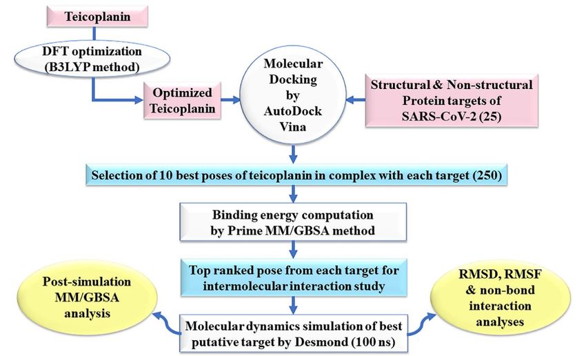
Antibiotics 2021, 10, 856 4 of 16
Antibiotics 2021, 10, x FOR PEER REVIEW 4 of 16
Figure 3.
Figure 3. Teicoplanin
Teicoplanin structure
structure optimized
optimized by
by density
density functional
functional theory
theory (DFT)
(DFT) with
with Becke’s
Becke’s three-parameter
three-parameter hybrid
hybrid model,
model,
the Lee–Yang–Parr (B3LYP) correlation functional method at 6-31G level (Jaguar v10.9 software of Schrodinger Suites
the Lee–Yang–Parr (B3LYP) correlation functional method at 6-31G level (Jaguar v10.9 software of Schrodinger Suites 2020-3
2020-3 was used). Color code: carbon—dark gray, hydrogen—light gray, nitrogen—blue, oxygen—red,
was used). Color code: carbon—dark gray, hydrogen—light gray, nitrogen—blue, oxygen—red, and chlorine—green. and chlorine—
green.
2.2. Protein Preparation
2.2. Protein Preparation
Twenty-five structural and non-structural protein targets related to SARS-CoV-2 were
Twenty-five
downloaded fromstructural
the RCSBand PDBnon-structural
repository as protein
listed intargets
Table 1.related to SARS-CoV-2
The macromolecular
were downloaded from the RCSB PDB
Pro repository as listed in Table
Pro
targets include main protease (M ), papain-like protease (PL ), RNA-dependent RNA 1. The macromolecular
targets include
polymerase main protease
(RdRp-RTP site), (M
Pro), papain-like protease (PLPro), RNA-dependent RNA
RdRp-RNA site, spike protein-receptor binding domain
polymerase (RdRp-RTP site), RdRp-RNA
(RBD), spike trimer (open state), spike monomer site, spike
(closedprotein-receptor
state), S2 protein binding domain
in post-fusion
(RBD),
state, spike trimer
N-protein (open state),
(C-domain), spike(N-domain),
N-protein monomer (closedNsp3-AMP state),site,
S2 protein
Nsp3-MES in post-fusion
site, Nsp7,
state, N-protein (C-domain), N-protein (N-domain), Nsp3-AMP site,
Nsp8, Nsp9, Nsp10, Nsp12, helicase (Nsp13-ADP site), helicase (Nsp13-NCB site), Nsp14 Nsp3-MES site,
Nsp7, Nsp8, Nsp9, Nsp10, Nsp12, helicase (Nsp13-ADP site), helicase
(N-terminal exoribonuclease; ExoN), Nsp14 (N7 methyltransferase; N7-MTase), Nsp15 (Nsp13-NCB site),
Nsp14 (N-terminal Nsp16
(exoribonuclease), exoribonuclease;
(GTA site),ExoN),Nsp16Nsp14(MGP (N7 site),methyltransferase;
and Nsp16 (SAMN7-MTase), site). All
Nsp15 (exoribonuclease), Nsp16 (GTA site), Nsp16 (MGP
the protein structures were processed in Biovia Discovery Studio site), and Nsp16 (SAM2020
Visualizer site).[21]
All
the protein
and PyMol structures were
2.4.1 [22] for processed
removing in Bioviaco-crystallized
unwanted Discovery Studio Visualizerincluding
compounds 2020 [21] and
wa-
PyMol
ter 2.4.1 [22]MGLTools
molecules. for removing unwanted
1.5.6 [20] wasco-crystallized compounds
utilized for generating including
input receptorwater mol-
files in
ecules. MGLTools
pdbqt format. 1.5.6 [20] was utilized for generating input receptor files in pdbqt for-
mat.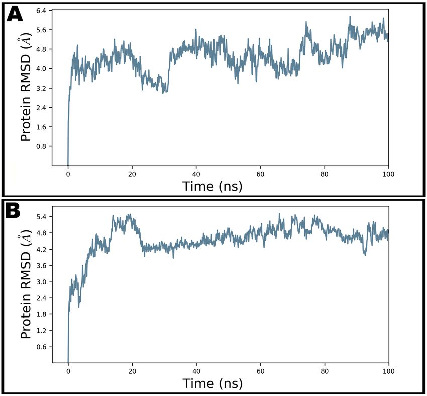
Antibiotics 2021, 10, 856                                                                                                                   5 of 16

      Table 1. Docking-predicted binding energy (∆GBind ) and grid parameters used for docking of teicoplanin with 25 potential
      COVID-19 drug targets.

     Target                                                             Grid Center for AutoDock Vina Program                  Docking
                                Targets                  PDB ID
    Number                                                                    x                y                z           ∆GBind (kcal/mol)

        1.                  Main protease                  6LU7           −9.732            11.403          68.925                 −5.4
        2.               Papain-like protease             6WUU             22.225           68.703            4.704                −5.4
        3.                 RdRp (RTP site)                 7BV2            91.776           91.560          104.863                −9.8
        4.                 RdRp (RNA site)                 7BV2            71.227           92.269          112.852                −7.7
        5.               Spike protein (RBD)               6M0J           −36.193           37.260          −5.752                 −6.1
        6.                 Spike monomer                  6VXX            219.061          220.947          261.311                −5.4
        7.                   Spike trimer                  6VYB           251.872          195.411          243.040                −6.9
        8.           S2 protein (post fusion state)        6LXT           −0.641            11.084          28.359                 −5.3
        9.              N-protein (C-domain)              6YUN            −10.288           12.683           7.740                 −5.6
       10.              N-protein (N-domain)               6YI3            16.299           11.628            6.638                −7.4
       11.                 Nsp3 (AMP site)                6W6Y              9.124          −8.677            16.220                −7.3
       12.                 Nsp3 (MES site)                6W6Y             23.830            9.255           54.812                −5.9
       13.                       Nsp7                      7BV2           104.786           80.343          127.861                −4.4
       14.                       Nsp8                      7BV2           108.168          116.454          120.901                −5.9
       15.                       Nsp9                     6WXD             53.119          −10.095          22.482                 −4.5
       16.                      Nsp10                     6WVN             64.644           15.650            9.522                −2.2
       17.                      Nsp12                      7BV2            97.382           97.966           93.920                −7.8
       18.            Nsp13 (helicase ADP site)            6JYT           405.020           47.480           62.350                −6.5
       19.            Nsp13 (helicase NCB site)            6JYT           423.816           33.797           56.132                −5.4
       20.                  Nsp14 (ExoN)                   5C8S           −39.712          −50.654          15.5594                −7.1
       21.                Nsp14 (N7mtase)                  5C8S           −10.273          −42.259          −7.644                 −3.0
       22.             Nsp15 (Exoribonuclease)            6WLC             94.134          −19.803          −25.857                −6.5
       23.                Nsp16 (GTA site)                6WVN             84.158           24.757           37.836                −6.2
       24.                Nsp16 (MGP site)                6WVN            100.029           38.995           18.481                −7.4
       25.                Nsp16 (SAM site)                6WVN             84.156           15.450           26.991                −6.5
      ADP site: adenosine diphosphate binding site; AMP: adenosine monophosphate; C-domain: carbon-terminal domain; ExoN:
      N-terminal exoribonuclease; GTA: P1-7-methylguanosine-P3-adenosine-50 ,50 -triphosphate; MES: 2-(N-morpholino)-ethanesulfonic acid;
      MGP: 7-methyl-guanosine-50 -triphosphate; N-domain: nitrogen-terminal domain; N7mtase: N7 methyltransferase; NCB site: nucleic acids
      binding site; N-protein: nucleocapsid protein; Nsp: non-structural protein; PDB: protein data bank; RBD: receptor binding domain; RdRp:
      RNA-dependent RNA polymerase; RNA: ribonucleic acid; RTP: remdesivir; SAM: S-adenosylmethionine.

                                     2.3. Molecular Docking Simulation
                                           The grid box of 30 × 30 × 30 size in x, y, and z directions with 1-Å spacing was con-
                                     structed around the active site residues as demarcated by the bound co-crystallized ligands
                                     (see Table 1 for grid points used for each receptor). However, blind docking, covering
                                     the entire macromolecular target, was opted if binding site information was unavailable.
                                     AutoDock Vina 1.1.2 [23] was used for molecular docking employing default parameters
                                     for flexible ligand and rigid protein. Moreover, the exhaustiveness parameter was set to
                                     12. Upon successful completion of docking simulation, ten best poses were individually
                                     analyzed for intermolecular interactions using PyMol 2.4.1 [22] and Biovia Discovery Stu-
                                     dio Visualizer 2020 [16,21,24] and further subjected to MM/GBSA computations in the
                                     next step.

                                     2.4. Prime MM-GBSA Calculations
                                          MM-GBSA technique are frequently used to rationalize the findings of molecular dock-
                                     ing and virtual screening. Prime MM-GBSA module of Schrödinger Suite 2020-3 [25–27]
                                     was used to compute binding energy and relevant parameters as presented in Table 2. The
                                     Prime MM-GBSA protocol amalgamates OPLS molecular mechanics energies, a VSGB
                                     solvation model for polar solvation (GSGB ), and a nonpolar solvation expression (GNP ) in-
                                     volving nonpolar solvent-accessible surface area (SASA) and van der Waals interactions [28].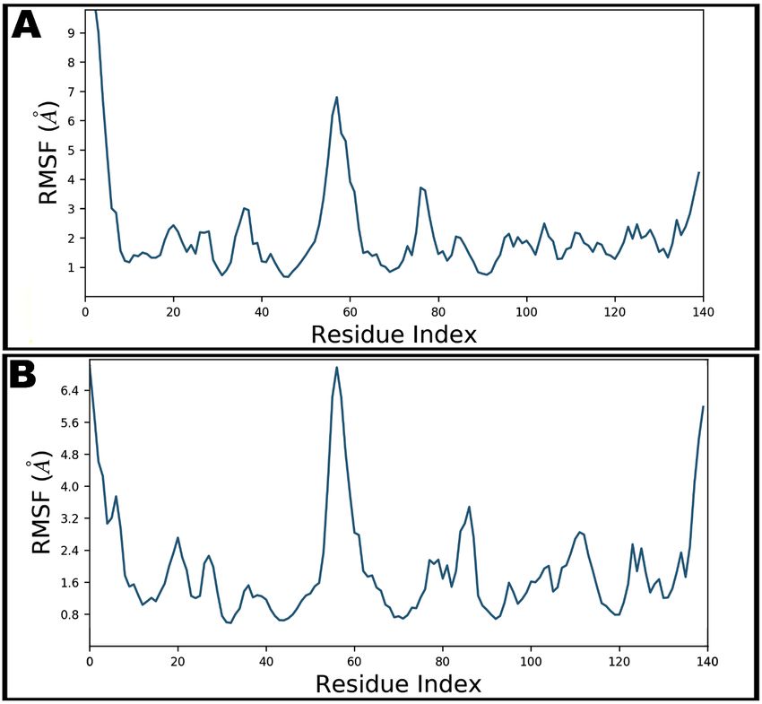
Antibiotics 2021, 10, 856                                                                                                                            6 of 16

                                      The binding-free energy (∆Gbind ) of docked teicoplanin in complex with respective proteins
                                      was calculated using the following equation [29]:

                                                                              ∆Gbind = ∆EMM + ∆Gsolv + ∆GS                                                (1)

                                      where ∆EMM is the difference in energy between the complex structure and the sum of
                                      the energies of the protein with and without teicoplanin, ∆Gsolv is the difference in the
                                      GBSA solvation energy of the teicoplanin–protein complex and the sum of the solvation
                                      energies for the teicoplanin-bound and unbound protein, and ∆GSA is the difference in the
                                      energy of surface area for the teicoplanin–protein complex and the sum of the surface area
                                      energies for the ligand and un-complexed protein.

      Table 2. Results of molecular mechanics/generalized Born surface area (MM/GBSA) computations for teicoplanin in
      complex with 25 potential COVID-19 targets.

   Target
                            Target                ∆GBind   a
                                                                 ∆GCoul   b     ∆GHBond    c
                                                                                                  ∆GLipo   d    SolvGB e        ∆Gvdw   f      Lig SE g
  Number
     1.               Main protease               −97.55         −19.11            −0.68           −59.81          44.25        −68.46             3.87
     2.            Papain-like protease           −78.75         −39.06            −3.33           −58.73          54.34        −42.74            39.85
     3.              RdRp (RTP site)               75.76         −124.58           −4.94           −91.90         209.45        −17.65           184.45
     4.              RdRp (RNA site)              −86.33         −68.97            −6.24           −13.97          78.93        −83.45            10.54
     5.            Spike protein (RBD)            −95.76         −32.67            −2.13           −54.25          48.60        −66.71            18.19
     6.              Spike monomer                −68.38         −61.51            −4.39           −13.35          55.98        −50.51            12.74
     7.                Spike trimer               −66.87         −27.37            −4.11           −21.53          53.95        −65.20            11.90
     8.        S2 protein (post fusion state)     −36.93         −23.22            −2.74           −14.11          55.19        −61.27            24.45
     9.           N-protein (C-domain)            −42.31         −30.82            −5.06           −16.05          64.88        −62.43            26.93
     10.          N-protein (N-domain)            −102.13        −40.95            −6.98           −24.95          61.07        −78.69           −0.90
     11.             Nsp3 (AMP site)              −58.33         −42.82            −4.38           −25.86          64.42        −71.09            32.93
     12.             Nsp3 (MES site)              −57.60         −12.61            −2.42           −30.13          34.62        −75.40            37.34
     13.                   Nsp7                   −66.61         −54.39            −4.15           −19.17          55.49        −60.78             8.43
     14.                   Nsp8                   −67.93         −10.55            −2.28           −25.51          28.75        −68.85            16.42
     15.                   Nsp9                   −66.83         −43.87            −3.59           −19.55          46.75        −57.02            10.15
     16.                  Nsp10                   −47.56         −29.62            −2.47           −23.97          57.87        −62.53            40.47
     17.                  Nsp12                   −87.84         −96.33            −8.48           −19.26         135.75        −108.20            8.52
     18.        Nsp13 (helicase ADP site)         −41.57         −63.94            −7.00           −19.36          88.70        −53.79            16.23
     19.        Nsp13 (helicase NCB site)         −53.99         −42.21            −4.05           −26.40          87.73        −78.57             7.39
     20.              Nsp14 (ExoN)                −49.41         −32.04            −4.56           −20.91          64.52        −80.34            28.66
     21.            Nsp14 (N7mtase)               −38.83         −21.49            −4.69           −15.49          47.57        −44.45             1.51
     22.         Nsp15 (Exoribonuclease)          −64.40         −48.67            −6.39           −23.20          47.29        −55.77            43.43
     23.            Nsp16 (GTA site)              −63.57         −55.69            −5.17           −12.18          65.46        −62.63             8.44
     24.            Nsp16 (MGP site)              −58.04         −31.42            −3.89           −22.67          59.61        −79.15            30.67
     25.            Nsp16 (SAM site)              −54.88         −34.65            −4.69           −21.69          74.67        −74.43            15.52
      All the energy values are given in kcal/mol; a : binding-free energy; b : Coulomb energy; c : hydrogen-bonding correction; d : lipophilic
      energy; e : generalized Born electrostatic solvation energy; f : Van der Waals energy; g : ligand strain energy. ADP site: adenosine diphosphate
      binding site; AMP: adenosine monophosphate; C-domain: carbon-terminal domain; ExoN: N-terminal exoribonuclease; GTA: P1-7-
      methylguanosine-P3-adenosine-50 ,50 -triphosphate; MES: 2-(N-morpholino)-ethanesulfonic acid; MGP: 7-methyl-guanosine-50 -triphosphate;
      N-domain: nitrogen-terminal domain; N7mtase: N7 methyltransferase; NCB site: nucleic acids binding site; N-protein: nucleocapsid
      protein; Nsp: non-structural protein; RBD: receptor binding domain; RdRp: RNA-dependent RNA polymerase; RNA: ribonucleic acid;
      RTP: remdesivir; SAM: S-adenosylmethionine.

                                      2.5. Molecular Dynamics Simulation
                                           The best ranked conformation of teicoplanin in complex with N-protein–N-domain
                                      furnished by MM-GBSA experiments was further assessed for their thermodynamic be-
                                      havior and stability by employing molecular dynamics (MD) simulation. Desmond 6.1
                                      program integrated with Maestro (Schrödinger, Inc. LLC, New York, NY, USA) was used for
                                      MD simulation studies [30,31]. In addition, apo form of the N-protein–N-domain was also
                                      simulated in order to understand the conformational changes upon teicoplanin binding.
                                      The teicoplanin–protein complex was placed into an orthorhombic box of 10 × 10 × 10 Å
                                      size, filled with 7210 water molecules by means of simple point charge (SPC) model
                                      implemented in system setup protocol. An OPLS3 force field was applied for the MD
                                      computations [32]. The system was neutralized using 20 and 26 Na+ and Cl− ions, respec-
                                      tively, with a salt concentration of 0.15 M that represents the physiological concentration
                                      of monovalent ions. An isothermal–isobaric (NPT) ensemble was utilized with temper-
Antibiotics 2021, 10, 856 7 of 16
ature and pressure attuned to 300 K and 1.01325 bar, respectively. A simulation time of
100 ns was adjusted, whereas trajectories were saved at every 100 ps. A cut-off radius of
9.0 Å was used for short-range van der Waals and Coulomb interactions. Nose–Hoover
thermostat [33] and Martyna–Tobias–Klein [34] methods were employed for maintaining
the system temperature and pressure, respectively. In order to integrate the equations of
motion, an RESPA integrator was used with an inner time step of 2.0 fs for bonded as well
as non-bonded interactions within the short-range cut-off [35]. The system was minimized
and equilibrated with the default protocols of the Desmond. Simulation event analysis,
simulation quality analysis, and simulation interaction diagram protocols of the Desmond
package was exercised to analyze the trajectory files.
2.6. Post-Simulation MM-GBSA Analysis
Post-simulation MM-GBSA analysis was performed by using the thermal_MMGBSA.py
script of the Prime/Desmond module of the Schrodinger suite 2020-3 [25,26]. From each
MD trajectory, every tenth frame was extracted from the last 50 ns of simulated trajectory,
averaging over 50 frames, for binding free energy calculations of teicoplanin in complex
with N-protein–N-domain. The Prime MM-GBSA method uses the rule of additivity
wherein total binding free energy (kcal/mol) represents a summation of individual energy
modules like coulombic, covalent, hydrogen bond, van der Waals, self-contact, lipophilic,
solvation, and π–π stackings of ligand and protein [36].
3. Results and Discussion
3.1. Validation of Docking Protocol
The co-crystallized ligand of main protease, N3, was redocked into the inhibitor
binding cavity of the enzyme and docked conformation of the ligand was compared with
that of the crystal structure in order to validate the docking protocol implemented in
AutoDock Vina. The root-mean-square deviation (RMSD) was less than 2 Å between
the docked and X-ray crystal structure conformations of N3, which authenticated the
scoring functions implemented in the docking program (data not shown). According to
the reported protocols, a successful docking computation should furnish the RMSD of
≤2.0 Å [10,14]. Therefore, AutoDock Vina used in this study was deemed reliable for
studying teicoplanin interaction with potential SARS-CoV-2 targets.
3.2. Molecular Docking of Teicoplanin with Potential Targets of SARS-CoV-2
Molecular docking is a computational method of accelerating the early stages of drug
discovery through unravelling the precise mechanism of intermolecular interactions of
potential drug candidates by comparing their energetic compatibility and molecular shape
with the target proteins [11,37]. With the aim of inspecting the interaction of teicoplanin
with putative drug targets of SARS-CoV-2, docking studies was performed on the recep-
tors/targets constituting both non-structural and structural proteins with prospective to be
used in the drug discovery field [38,39]. Docking-predicted binding energies of teicoplanin
with twenty-five COVID-19 drug targets are given in Table 1, whereas comprehensive
intermolecular interactions have been listed in Table S1 in the Supplementary Information.
3.3. Prime MM-GBSA Calculations of Docked Complexes
Though several docking experiments are routinely employed to emphasize the binding
mode and the affinity of a ligand relative to the protein, lack of accurate scoring function
is one of the drawbacks of these algorithms. Therefore, MM-GBSA computations are
routinely employed as a post-docking analysis in order to avoid false negatives as well as
false positives [40,41]. The top ten conformations of protein–teicoplanin-docked complexes
of each target were subjected to molecular mechanics/generalized Born surface area (MM-
GBSA) computation for estimation of more accurate free binding energies. Table 2 enlists
the MM-GBSA computed energies of best conformation of teicoplanin in complex with
each target. Several other energy components such as Coulomb energy, hydrogen-bondingAntibiotics 2021, 10, 856 8 of 16
correction, lipophilic energy, generalized Born electrostatic solvation energy, Van der Waals
energy, and ligand strain energy were also computed. Moreover, an elaborated MM-GBSA
calculation of all poses has been listed in Table S2 in the Supplementary Information.
As presented in Figure 4, nucleocapsid protein N-terminal domain ranked top among
all studied targets exhibiting ∆Gbind = −102.13 kcal/mol followed by main protease and
spike protein-RBD with binding energies of −97.55 and −95.76 kcal/mol, respectively.
However, MM-GBSA binding energy of teicoplanin-main protease complex obtained from
AutoDock 4.2 has been recently reported as -68 kcal/mol. Interestingly, RdRp-RTP site
was rendered as least promising for teicoplanin in this study, demonstrating 75.8 kcal/mol
binding energy. The most promising target also hosted ample non-bond interactions with
teicoplanin within the active site of N-domain of the N-protein. Moreover, appreciable
intermolecular forces were established with all other targets. It is evident that the N-domain
of the N-protein hosts nucleic acid substrates during processing of the viral genome into a
ribonucleoprotein unit. Therefore, targeting this protein by teicoplanin can interrupt crucial
steps of the SARS-CoV-2 life cycle such as transcription and translation processes [42].
Several proteins such as Nsp12, RdRp-RNA site, and papain-like protease were ranked as
moderately favorable targets, whereas variable affinity was noted against the rest of the
targets. The chemical structure of teicoplanin represents a unique blend of polar groups in
the form of several amino acid fragments, carbohydrate cores such as N-acetylglucosamine,
β-D-glucosamine and mannose, and non-polar fragments such as fatty acyl side-chain and
numerous aromatic rings. These moieties enabled teicoplanin to interact with diverse drug
targets of COVID-19 which warrants further experimental validation and may be useful
in drug design based on teicoplanin template. Intermolecular interactions observed after
MM-GBSA computation of all studied targets are presented in Figures S1–S4 and Table
Antibiotics 2021, 10, x FOR PEER REVIEW 9 of S1
16
in the Supplementary Information.
Figure
Figure 4.
4. Bar
Bar plot
plotshowing
showingcomparative
comparative affinity
affinity of
of teicoplanin
teicoplanin against
against drug
drug targets
targets associated
associated with
with COVID-19
COVID-19 (please
(please refer
refer
to
to Table 2 for target names against respective numbers). The analysis is based on molecular mechanics–generalized Born
Table 2 for target names against respective numbers). The analysis is based on molecular mechanics–generalized Born
surface area (MM-GBSA) computed binding energy (ΔGBind) in kcal/mol.
surface area (MM-GBSA) computed binding energy (∆GBind ) in kcal/mol.
3.4. Molecular Dynamics Simulation Studies
Dynamic and thermodynamics parameters of living systems under specific condi-
tions of physiological environments can be estimated by the application of molecular dy-
namics (MD) simulation, a widely employed computer-aided drug design technique
[11,13,14,43]. Therefore, the best docked pose of teicoplanin in complex with SARS-CoV-Antibiotics 2021, 10, 856 9 of 16
3.4. Molecular Dynamics Simulation Studies
Dynamic and thermodynamics parameters of living systems under specific conditions
of physiological environments can be estimated by the application of molecular dynamics
(MD) simulation, a widely employed computer-aided drug design technique [11,13,14,43].
Therefore, the best docked pose of teicoplanin in complex with SARS-CoV-2 nucleocapsid
protein N-terminal domain was subjected to MD simulation study in order to investigate
the stability of the ligand–protein complex as well as main intermolecular interactions
during the simulated trajectory. MM-GBSA-optimized pose bearing minimum binding
energy was selected for MD simulation study. Desmond software was employed for
the MD simulation of 100 ns in explicit solvent system. The resulting trajectories of the
simulated complex was inspected for different standard simulation parameters such as
backbone RMSDs for alpha-carbons, side chains, and heavy atoms. In addition, the root-
mean-square fluctuations (RMSFs) of individual amino acid residue and intermolecular
interactions involved were also evaluated. Figure 5A shows the RMSD of Cα atoms
of apo protein whereas the complex of teicoplanin bound to N-protein–N-domain is
presented as Figure 5B. The analysis of RMSD of teicoplanin-bound protein indicates that
the simulated system fluctuated initially during 0–22 ns but acquired stability during rest
of the simulated time. However, fluctuations can be clearly seen in the RMSD plot of the
apo protein alone (Figure 5A). Therefore, it seems logical to infer that upon teicoplanin
binding, the protein maintains its conformational stability when simulated for 100 ns
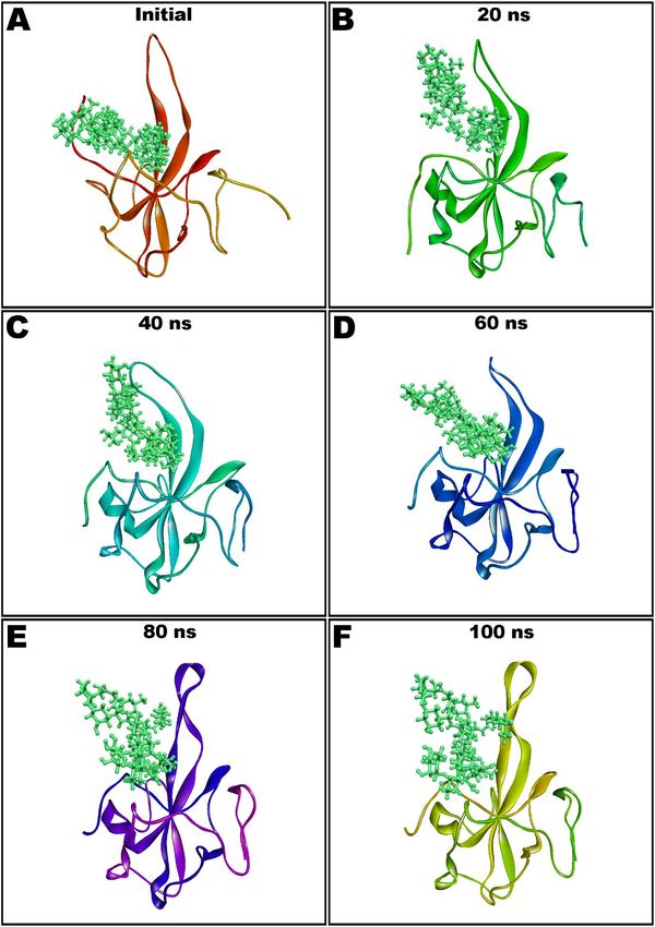
period. The conformational changes observed during simulated path has been visualized
by the snapshots taken at the intervals of every 20 ns and presented in Figure 6.
The MM-GBSA method exploits molecular mechanics, the generalized Born solvation
models and a solvent accessibility approach to estimate the free energies of binding based
on snapshots obtained from MD simulations [40,44]. MM-GBSA computation was per-
formed for the last 50 snapshots of 100 ns simulation, which estimated an average binding
energy of −62.52 ± 12.22 kcal/mol.
The local conformational alterations along SARS-CoV-2 nucleocapsid protein
N-domain were investigated by analyzing the RMSF during simulation time. Termi-
nal residues fluctuated most in both bound and unbound states. Maximum fluctuation
was noted with Gly1 as 13.29 Å which was declined upon teicoplanin binding at 7.00 Å
(Figure 7). Key residues involved in hydrogen bonding showed fluctuations at 3.74, 2.97,
1.76, 1.54, 4.12, 0.96, and 2.68 Å by Asn7, Asn8, Thr9, Ser11, Arg55, Tyr69, and Pro111,
respectively.
Simulation interactions diagrams presented in Figures 8 and 9 during entire simula-
tion time signifies a comprehensive intermolecular interaction profile of teicoplanin with
nucleocapsid protein N-domain. The modus of interaction pattern of teicoplanin illustrates
that only fewer docking-predicted main contacts were retained upon MD simulation time
of 100 ns. Figure 8 presents intermolecular interactions observed in first and last frames of
the teicoplanin bound to nucleocapsid protein N-domain. Residues participating in both
hydrogen bonding and water bridges include Asn7, Asn8, Thr9, Ser11, Thr51, Arg53, Arg55,
Tyr71, Pro111, and Asn114. In addition, Thr51, Arg53, Arg55, and Arg67 also contributed
in ionic bond interaction. However, amino acid residues Ala10, Ala50, Tyr69, and Ala112
were noted to be important for hydrophobic interaction.Antibiotics 2021, 10, x FOR PEER REVIEW 10 of 16
Antibiotics 2021, 10, 856 10 of 16
performed for the last 50 snapshots of 100 ns simulation, which estimated an average
binding energy of −62.52 ± 12.22 kcal/mol.
Figure 5.
Figure 5. The
The root-mean-square
root-mean-square deviations
deviations (RMSD)
(RMSD) ofof Cα
Cα atoms
atoms of
of N-protein–N-domain
N-protein–N-domain in
in unbound
unbound state
state (A) and
(A) and
teicoplanin-bound state (B) during 100 ns molecular dynamics simulation.
teicoplanin-bound state (B) during 100 ns molecular dynamics simulation.Antibiotics 2021, 10, 856 11 of 16
Antibiotics 2021, 10, x FOR PEER REVIEW 11 of 16
Figure 6. Molecular dynamics (MD) simulation snapshots of teicoplanin (shown as ball and stick style in green color) in
Figure 6. Molecular dynamics (MD) simulation snapshots of teicoplanin (shown as ball and stick style in green color) in
complex with nucleocapsid protein N-domain (depicted as solid ribbon) showing conformational stability (Initial: A; after
complex with nucleocapsid protein N-domain (depicted as solid ribbon) showing conformational stability (Initial: (A); after
20 ns: B; 40 ns: C; 60 ns: D; 80 ns: E; and 100 ns: F).
20 ns: (B); 40 ns: (C); 60 ns: (D); 80 ns: (E); and 100 ns: (F)).The local conformational alterations along SARS-CoV-2 nucleocapsid protein N-do-
main were investigated by analyzing the RMSF during simulation time. Terminal residues
fluctuated most in both bound and unbound states. Maximum fluctuation was noted with
Antibiotics 2021, 10, 856
Gly1 as 13.29 Å which was declined upon teicoplanin binding at 7.00 Å (Figure 7). Key
12 of 16
residues involved in hydrogen bonding showed fluctuations at 3.74, 2.97, 1.76, 1.54, 4.12,
0.96, and 2.68 Å by Asn7, Asn8, Thr9, Ser11, Arg55, Tyr69, and Pro111, respectively.
Figure 7. Root-mean-square fluctuations (RMSF) of SARS-CoV-2 nucleocapsid protein N-domain during 100 ns molecular
dynamics simulation in apo form (A) and teicoplanin
teicoplanin bound
bound form
form (B).
(B).
Simulation interactions diagrams presented in Figures 8 and 9 during entire simula-
tion time signifies a comprehensive intermolecular interaction profile of teicoplanin with
nucleocapsid protein N-domain. The modus of interaction pattern of teicoplanin illus-
trates that only fewer docking-predicted main contacts were retained upon MD simula-
tion time of 100 ns. Figure 8 presents intermolecular interactions observed in first and last
frames of the teicoplanin bound to nucleocapsid protein N-domain. Residues participat-
ing in both hydrogen bonding and water bridges include Asn7, Asn8, Thr9, Ser11, Thr51,
Arg53, Arg55, Tyr71, Pro111, and Asn114. In addition, Thr51, Arg53, Arg55, and Arg67
also contributed in ionic bond interaction. However, amino acid residues Ala10, Ala50,
Tyr69, and Ala112 were noted to be important for hydrophobic interaction.Antibiotics 2021, 10, 856 13 of 16
Antibiotics 2021, 10, x FOR PEER REVIEW 13 of 16
Figure 8. Non-bond interactions observed in first (A,B) and last (C,D) frames extracted from 100 ns molecular dynamics
Figure 8. Non-bond interactions observed in first (A,B) and last (C,D) frames extracted from 100 ns molecular dynamics
simulation of teicoplanin bound nucleocapsid protein N-domain of SARS-CoV-2. Ala: alanine; Arg: arginine; Asn:
simulation ofasparagine;
teicoplanin bound
Leu: nucleocapsid
leucine; protein
Met: methionine; N-domain
Pro: of SARS-CoV-2.
proline; Ser: Ala: alanine;
serine; Thr: threonine; Arg: arginine;
Tyr: tyrosine. Asn: asparagine;
The number
Leu: leucine;
Antibiotics Met:
of
2021, 10, xmethionine;
corresponding Pro: acid
amino
FOR PEER REVIEW proline; Ser:isserine;
residue Thr: threonine;
in accordance Tyr: tyrosine. The
with the crystallographic number obtained
information of corresponding
from the amino
14 ofacid
16
residue is in protein data bank.
accordance with the crystallographic information obtained from the protein data bank.
FigureFigure 9. Protein
9. Protein interactions
interactions withwith teicoplanin,monitored
teicoplanin, monitored throughout
throughout thethesimulation trajectory.
simulation These
trajectory. interactions
These are are
interactions
clustered by type and summarized in bar diagram including hydrogen bonds (H-bonds), hydrophobic, ionic, and water
clustered by type and summarized in bar diagram including hydrogen bonds (H-bonds), hydrophobic, ionic, and water
bridges. Ala: alanine; Arg: arginine; Asn: asparagine; Asp: aspartic acid; Cys: cysteine; Gln: glutamine; Glu:
bridges. Ala: alanine; Arg: arginine; Asn: asparagine; Asp: aspartic acid; Cys: cysteine; Gln: glutamine; Glu: glutamic acid;
glutamic acid; Gly: glycine; His: histidine; Leu: leucine; Met: methionine; Phe: phenylalanine; Pro: proline; Ser:
Gly: glycine;
serine; His: histidine; Leu:
Thr: threonine; Tyr:leucine;
tyrosine;Met:
Val:methionine; Phe: phenylalanine;
valine. The number Pro: proline;
of corresponding Ser: residue
amino acid serine; Thr: threonine; Tyr:
is according
tyrosine; Val: valine. The number of corresponding amino acid residue
to the crystallographic information obtained from the protein data bank. is according to the crystallographic information
obtained from the protein data bank.
4. Conclusions
By using computer-aided drug design techniques, the current study explained the
intermolecular interaction of the antibacterial drug, teicoplanin, with potential drug tar-
gets of SARS-CoV-2. Molecular docking studies employing AutoDock Vina highlighted
the importance of hydrophilic and hydrophobic interactions in supporting the teicoplaninAntibiotics 2021, 10, 856 14 of 16
4. Conclusions
By using computer-aided drug design techniques, the current study explained the
intermolecular interaction of the antibacterial drug, teicoplanin, with potential drug targets
of SARS-CoV-2. Molecular docking studies employing AutoDock Vina highlighted the
importance of hydrophilic and hydrophobic interactions in supporting the teicoplanin
molecule inside the nucleocapsid protein N-domain binding cavity. MM-GBSA analysis
and molecular dynamics simulation results not only reinforce the credibility of the docking
results, but also authenticate the stability of the simulated system. This study is expected
to assist lead optimization and design of COVID-19 drugs based on molecular skeleton of
teicoplanin. However, the requirement of additional experimental and clinical validation
is an obvious limitation of this study. Nevertheless, findings of this study justify further
exploration of teicoplanin repurposing.
Supplementary Materials: The following are available online at https://www.mdpi.com/article/10
.3390/antibiotics10070856/s1, Figure S1: MM/GBSA optimized complexes of teicoplanin (displayed
as stick) showing non-bond interactions with main protease (A), papain-like protease (B), RdRp-RTP
site (C), RdRp-RNA site (D), spike protein-RBD (E), and spike monomer (F). Broken green lines
enumerate hydrogen bonds whereas purple lines justify hydrophobic interactions; Figure S2. Non-
bond interactions of teicoplanin (portrayed as stick style) with spike trimer (A), S2 protein-post
fusion state (B), N-protein–C-domain (C), Nsp3-AMP site (D), Nsp3-MES site (E), and Nsp7 (F) in
MM/GBSA optimized complexes. Hydrogen bonds are shown by broken green lines and purple lines
enumerate hydrophobic interactions; Figure S3. Intermolecular interactions of teicoplanin (shown in
stick style) with Nsp8 (A), Nsp9 (B), Nsp10 (C), Nsp12 (D), Nsp13 helicase-ADP site (E), and Nsp13
helicase-NCB site (F) after MM/GBSA computations. Hydrogen bonds are represented by broken
green lines and purple lines specify non-polar interactions; Figure S4. Intermolecular complexes
of teicoplanin (shown as sticks) with Nsp14 N-exoribonuclease (A), Nsp14 N7-methyl transferase
(B), Nsp15 exoribonuclease (C), Nsp16-GTA site (D), Nsp16-MGP site (E), and Nsp16-SAM site (F)
resulted after MM-GBSA analysis showing hydrogen bonds (broken green lines) and non-polar
contacts (purple lines); Table S1: Intermolecular interactions observed in MM/GBSA optimized
complexes of teicoplanin and 25 potential COVID-19 targets (AutoDock Vina 1.1.2 was used for
molecular docking); Table S2. Computation of MM/GBSA energies of top ten docked poses of
teicoplanin in complex with 25 potential COVID-19 targets.
Funding: The author gratefully acknowledges the Deanship of Scientific Research, Qassim University,
Saudi Arabia for funding the publication of this research.
Institutional Review Board Statement: Not applicable.
Informed Consent Statement: Not applicable.
Data Availability Statement: The data presented in this study are available on request from the
corresponding author.
Acknowledgments: The author gratefully acknowledges the Deanship of Scientific Research, Qassim
University, Saudi Arabia for funding the publication of this research. The author further extends
sincere thanks to Schrodinger India for providing the access to Schrodinger Suite 2020-3. Prajwal
Nandekar and Sudharshan Pandiyan are kindly acknowledged for their help and support.
Conflicts of Interest: The author declares no conflict of interest.
References
1. Barkoff, C.M.; Mousa, S.A. Pharmacotherapy in COVID 19: Potential Impact of Targeting the Complement System. Biomedicines
2021, 9, 11. [CrossRef]
2. Raoult, D.; Hsueh, P.-R.; Stefani, S.; Rolain, J.-M. COVID-19 Therapeutic and Prevention. Int. J. Antimicrob. Agents 2020, 55, 105937.
[CrossRef]
3. Clinical Management of COVID-19. Available online: https://www.who.int/publications/i/item/clinical-management-of-
covid-19 (accessed on 31 December 2020).
4. Pushpakom, S.; Iorio, F.; Eyers, P.A.; Escott, K.J.; Hopper, S.; Wells, A.; Doig, A.; Guilliams, T.; Latimer, J.; McNamee, C.; et al.
Drug repurposing: Progress, challenges and recommendations. Nat. Rev. Drug Discov. 2019, 18, 41–58. [CrossRef] [PubMed]
5. Harrison, C. Coronavirus puts drug repurposing on the fast track. Nat. Biotechnol. 2020, 38, 379–381. [CrossRef]Antibiotics 2021, 10, 856 15 of 16
6. Zhou, N.; Pan, T.; Zhang, J.; Li, Q.; Zhang, X.; Bai, C.; Huang, F.; Peng, T.; Zhang, J.; Liu, C.; et al. Glycopeptide Antibiotics
Potently Inhibit Cathepsin L in the Late Endosome/Lysosome and Block the Entry of Ebola Virus, Middle East Respiratory
Syndrome Coronavirus (MERS-CoV), and Severe Acute Respiratory Syndrome Coronavirus (SARS-CoV). J. Biol. Chem. 2016, 291,
9218–9232. [CrossRef]
7. Baron, S.A.; Devaux, C.; Colson, P.; Raoult, D.; Rolain, J.-M. Teicoplanin: An alternative drug for the treatment of COVID-19? Int.
J. Antimicrob. Agents 2020, 55, 105944. [CrossRef]
8. Zhang, J.; Ma, X.; Yu, F.; Liu, J.; Zou, F.; Pan, T.; Zhang, H. Teicoplanin potently blocks the cell entry of 2019-nCoV. bioRxiv 2020.
[CrossRef]
9. Tripathi, P.K.; Upadhyay, S.; Singh, M.; Raghavendhar, S.; Bhardwaj, M.; Sharma, P.; Patel, A.K. Screening and evaluation of
approved drugs as inhibitors of main protease of SARS-CoV-2. Int. J. Biol. Macromol. 2020, 164, 2622–2631. [CrossRef]
10. Hussain, M.S.; Azam, F.; Ahamed, K.F.H.N.; Ravichandiran, V.; Alkskas, I. Anti-endotoxin effects of terpenoids fraction from
Hygrophila auriculata in lipopolysaccharide-induced septic shock in rats. Pharm. Biol. 2016, 54. [CrossRef] [PubMed]
11. Azam, F.; Alabdullah, N.H.; Ehmedat, H.M.; Abulifa, A.R.; Taban, I.; Upadhyayula, S. NSAIDs as potential treatment option
for preventing amyloid β toxicity in Alzheimer’s disease: An investigation by docking, molecular dynamics, and DFT studies.
J. Biomol. Struct. Dyn. 2018, 36, 2099–2117. [CrossRef]
12. Azam, F.; Taban, I.M.; Eid, E.E.M.; Iqbal, M.; Alam, O.; Khan, S.; Mahmood, D.; Anwar, M.J.; Khalilullah, H.; Khan, M.U. An
in-silico analysis of ivermectin interaction with potential SARS-CoV-2 targets and host nuclear importin α. J. Biomol. Struct. Dyn.
2020, 1–14. [CrossRef]
13. Sandomenico, A.; Di Rienzo, L.; Calvanese, L.; Iaccarino, E.; D’Auria, G.; Falcigno, L.; Chambery, A.; Russo, R.; Franzoso, G.;
Tornatore, L.; et al. Insights into the Interaction Mechanism of DTP3 with MKK7 by Using STD-NMR and Computational
Approaches. Biomedicines 2021, 9, 20. [CrossRef]
14. Azam, F.; Abodabos, H.S.; Taban, I.M.; Rfieda, A.R.; Mahmood, D.; Anwar, M.J.; Khan, S.; Sizochenko, N.; Poli, G.; Tuccinardi, T.;
et al. Rutin as promising drug for the treatment of Parkinson’s disease: An assessment of MAO-B inhibitory potential by docking,
molecular dynamics and DFT studies. Mol. Simul. 2019, 45. [CrossRef]
15. Shushni, M.A.M.; Azam, F.; Lindequist, U. Oxasetin from Lophiostoma sp. of the Baltic Sea: Identification, in silico binding mode
prediction and antibacterial evaluation against fish pathogenic bacteria. Nat. Prod. Commun. 2013, 8, 1223–1226. [CrossRef]
16. Azam, F.; Vijaya Vara Prasad, M.; Thangavel, N.; Kumar Shrivastava, A.; Mohan, G. Structure-Based Design, Synthesis and
Molecular Modeling Studies of Thiazolyl Urea Derivatives as Novel Anti-Parkinsonian Agents. Med. Chem. (Los Angeles) 2012, 8,
1057–1068.
17. O’Boyle, N.M.; Banck, M.; James, C.A.; Morley, C.; Vandermeersch, T.; Hutchison, G.R. Open Babel: An open chemical toolbox.
J. Cheminform. 2011, 3, 33. [CrossRef] [PubMed]
18. Bochevarov, A.D.; Harder, E.; Hughes, T.F.; Greenwood, J.R.; Braden, D.A.; Philipp, D.M.; Rinaldo, D.; Halls, M.D.; Zhang, J.;
Friesner, R.A. Jaguar: A high-performance quantum chemistry software program with strengths in life and materials sciences. Int.
J. Quantum Chem. 2013, 113, 2110–2142. [CrossRef]
19. Schrödinger Release 2020-3: Jaguar; Schrödinger, LLC: New York, NY, USA, 2020.
20. Morris, G.M.; Huey, R.; Lindstrom, W.; Sanner, M.F.; Belew, R.K.; Goodsell, D.S.; Olson, A.J. AutoDock4 and AutoDockTools4:
Automated docking with selective receptor flexibility. J. Comput. Chem. 2009, 30, 2785–2791. [CrossRef] [PubMed]
21. BIOVIA, Dassault Systèmes, Discovery Studio Visualizer, Version 20.1.0.19295; Dassault Systèmes: San Diego, CA, USA, 2020.
22. The PyMOL Molecular Graphics System, Version 2.0; Schrödinger, LLC: New York, NY, USA, 2020.
23. Trott, O.; Olson, A.J. AutoDock Vina: Improving the speed and accuracy of docking with a new scoring function, efficient
optimization, and multithreading. J. Comput. Chem. 2010, 31, 455–461. [CrossRef] [PubMed]
24. Fahmy, N.M.; Al-Sayed, E.; Moghannem, S.; Azam, F.; El-Shazly, M.; Singab, A.N. Breaking Down the Barriers to a Natural
Antiviral Agent: Antiviral Activity and Molecular Docking of Erythrina speciosa Extract, Fractions, and the Major Compound.
Chem. Biodivers. 2020, 17. [CrossRef]
25. Jacobson, M.P.; Pincus, D.L.; Rapp, C.S.; Day, T.J.F.; Honig, B.; Shaw, D.E.; Friesner, R.A. A hierarchical approach to all-atom
protein loop prediction. Proteins 2004, 55, 351–367. [CrossRef]
26. Jacobson, M.P.; Friesner, R.A.; Xiang, Z.; Honig, B. On the Role of the Crystal Environment in Determining Protein Side-chain
Conformations. J. Mol. Biol. 2002, 320, 597–608. [CrossRef]
27. Schrödinger Release 2020-3; Schrödinger, LLC: New York, NY, USA, 2020; pp. 19–27.
28. Vijayakumar, B.; Parasuraman, S.; Raveendran, R.; Velmurugan, D. Identification of natural inhibitors against angiotensin I
converting enzyme for cardiac safety using induced fit docking and MM-GBSA studies. Pharmacogn. Mag. 2014, 10, S639.
29. Lyne, P.D.; Lamb, M.L.; Saeh, J.C. Accurate prediction of the relative potencies of members of a series of kinase inhibitors using
molecular docking and MM-GBSA scoring. J. Med. Chem. 2006, 49, 4805–4808. [CrossRef]
30. Schrödinger Release 2020-3: Desmond Molecular Dynamics System, D. E. Shaw Research, New York, NY, 2020. Maestro-Desmond
Interoperability Tools; Schrödinger: New York, NY, USA, 2020.
31. Bowers, K.J.; Chow, D.E.; Xu, H.; Dror, R.O.; Eastwood, M.P.; Gregersen, B.A.; Klepeis, J.L.; Kolossvary, I.; Moraes, M.A.; Sacerdoti,
F.D. Scalable algorithms for molecular dynamics simulations on commodity clusters. In Proceedings of the SC’06: Proceedings of
the 2006 ACM/IEEE Conference on Supercomputing, Tampa, FL, USA, 11–17 November 2006; p. 43.Antibiotics 2021, 10, 856 16 of 16
32. Harder, E.; Damm, W.; Maple, J.; Wu, C.; Reboul, M.; Xiang, J.Y.; Wang, L.; Lupyan, D.; Dahlgren, M.K.; Knight, J.L. OPLS3:
A force field providing broad coverage of drug-like small molecules and proteins. J. Chem. Theory Comput. 2016, 12, 281–296.
[CrossRef]
33. Hoover, W.G. Canonical dynamics: Method for simulations in the canonical ensemble. Phys. Rev. A 1985, 31, 1695–1697.
[CrossRef] [PubMed]
34. Martyna, G.J.; Tobias, D.J.; Klein, M.L. Constant pressure molecular dynamics algorithms. J. Chem. Phys. 1994, 101, 4177–4189.
[CrossRef]
35. Humphreys, D.D.; Friesner, R.A.; Berne, B.J. A multiple-time-step molecular dynamics algorithm for macromolecules. J. Phys.
Chem. 1994, 98, 6885–6892. [CrossRef]
36. Dill, K.A. Additivity Principles in Biochemistry. J. Biol. Chem. 1997, 272, 701–704. [CrossRef] [PubMed]
37. Azam, F.; Amer, A.M.; Rabulifa, A.; Elzwawi, M.M. Ginger components as new leads for the design and development of novel
multi-targeted anti-Alzheimer’s drugs: A computational investigation. Drug Des. Dev. Ther. 2014, 8, 2045–2059. [CrossRef]
[PubMed]
38. Gordon, D.E.; Jang, G.M.; Bouhaddou, M.; Xu, J.; Obernier, K.; White, K.M.; O’Meara, M.J.; Rezelj, V.V.; Guo, J.Z.; Swaney, D.L.;
et al. A SARS-CoV-2 protein interaction map reveals targets for drug repurposing. Nature 2020, 583, 459–468. [CrossRef]
39. Kong, R.; Yang, G.; Xue, R.; Liu, M.; Wang, F.; Hu, J.; Guo, X.; Chang, S. COVID-19 Docking Server: A meta server for docking
small molecules, peptides and antibodies against potential targets of COVID-19. Bioinformatics 2020. [CrossRef] [PubMed]
40. Genheden, S.; Ryde, U. The MM/PBSA and MM/GBSA methods to estimate ligand-binding affinities. Expert Opin. Drug Discov.
2015, 10, 449–461. [CrossRef] [PubMed]
41. Sgobba, M.; Caporuscio, F.; Anighoro, A.; Portioli, C.; Rastelli, G. Application of a post-docking procedure based on MM-PBSA
and MM-GBSA on single and multiple protein conformations. Eur. J. Med. Chem. 2012, 58, 431–440. [CrossRef]
42. Dinesh, D.C.; Chalupska, D.; Silhan, J.; Koutna, E.; Nencka, R.; Veverka, V.; Boura, E. Structural basis of RNA recognition by the
SARS-CoV-2 nucleocapsid phosphoprotein. PLoS Pathog. 2020, 16, 1–16. [CrossRef]
43. Hospital, A.; Goñi, J.R.; Orozco, M.; Gelpí, J.L. Molecular dynamics simulations: Advances and applications. Adv. Appl. Bioinform.
Chem. 2015, 8, 37–47. [PubMed]
44. Kollman, P.A.; Massova, I.; Reyes, C.; Kuhn, B.; Huo, S.; Chong, L.; Lee, M.; Lee, T.; Duan, Y.; Wang, W.; et al. Calculating
structures and free energies of complex molecules: Combining molecular mechanics and continuum models. Acc. Chem. Res.
2000, 33, 889–897. [CrossRef] [PubMed]You can also read