Molecular architecture of the luminal ring of the Xenopus laevis nuclear pore complex - Nature
←
→
Page content transcription
If your browser does not render page correctly, please read the page content below
www.nature.com/cr
                  www.cell-research.com

                  ARTICLE                   OPEN

                  Molecular architecture of the luminal ring of the Xenopus
                  laevis nuclear pore complex
                  Yanqing Zhang1,2, Sai Li 3,4, Chao Zeng4, Gaoxingyu Huang3, Xuechen Zhu1,2, Qifan Wang1,2, Kunpeng Wang5, Qiang Zhou1,2,
                  Chuangye Yan3, Wusheng Zhang5, Guangwen Yang5, Minhao Liu3, Qinghua Tao3, Jianlin Lei 3 and Yigong Shi1,2,3,4

                      The nuclear pore complex (NPC) mediates the flow of substances between the nucleus and cytoplasm in eukaryotic cells. Here we
                      report the cryo-electron tomography (cryo-ET) structure of the luminal ring (LR) of the NPC from Xenopus laevis oocyte. The
                      observed key structural features of the LR are independently confirmed by single-particle cryo-electron microscopy (cryo-EM)
                      analysis. The LR comprises eight butterfly-shaped subunits, each containing two symmetric wings. Each wing consists of four
                      elongated, tubular protomers. Within the LR subunit, the eight protomers form a Finger domain, which directly contacts the fusion
                      between the inner and outer nuclear membranes and a Grid domain, which serves as a rigid base for the Finger domain. Two
                      neighboring LR subunits interact with each other through the lateral edges of their wings to constitute a Bumper domain, which
                      displays two major conformations and appears to cushion neighboring NPCs. Our study reveals previously unknown features of the
                      LR and potentially explains the elastic property of the NPC.
1234567890();,:

                      Cell Research (2020) 30:532–540; https://doi.org/10.1038/s41422-020-0320-y

                  INTRODUCTION                                                                                 along the nucleocytoplasmic axis and a pseudo twofold
                  A hallmark of all eukaryotic cells is the nuclear envelope (NE),                             rotational symmetry in the plane of the NE. The scaffold of an
                  which separates the nucleoplasm, where the genetic material is                               NPC is proposed to comprise a cytoplasmic ring (CR), an inner
                  stored, away from the cytoplasm, where nuclear-transcribed                                   ring (IR) and a nuclear ring (NR).5 Cytoplasmic filaments and
                  RNA is translated into protein of diverse functions. Nucleocyto-                             nuclear basket are attached to the CR and NR, respectively.5,10
                  plasmic shuttling of all substances needed for transcription and                                The luminal ring (LR), as the name indicates, resides in the
                  translation and numerous other cellular processes is mediated                                lumen of the NE and surrounds the NPC at the site of membrane
                  by the nuclear pore complex (NPC).1–3 The NPC associates with                                fusion.16,17 The LR is separated from all other components of the
                  and stabilizes a highly curved section of the NE — namely the                                NPC by the nuclear membrane and may play a role in anchoring
                  fusion between the inner (INM) and outer nuclear membranes                                   the NPC to the NE.16 Previous EM and ET studies have revealed
                  (ONM).4,5 The NPC is among the largest supramolecular                                        few describable features of the LR12,14,16–20 except that the LR of
                  complexes in cells, with a combined mass of approximately                                    the yeast NPC was found to comprise eight circumferential
                  50 MDa in yeast6,7 and 110–125 MDa in higher eukaryotes.3,8–11                               arches.6,7 At present, the overall organization, structural features
                  The protein components of the NPC are known as nucleoporin                                   and functional mechanism of the LR remain largely enigmatic.
                  (Nup). An NPC has about 34 different Nups, most of which are                                    Due to its location, the LR is speculated to be composed of
                  conserved among different organisms and each Nup is                                          integral membrane proteins. Among the vertebrate Nups, only
                  represented in multiple copies.11                                                            four have been found to be integral membrane proteins: GP210
                     X-ray structures have revealed a wealth of information on                                 (Pom152 in yeast) with a single transmembrane helix (TM),21–23
                  individual components and subcomplexes of the NPC.11 This                                    POM121 with a single TM,24 NDC1,25,26 and TMEM3327 each with
                  information, together with EM and other studies, have yielded a                              six predicted TMs. GP210 is the only Nup that contains a
                  three-dimensional model of the NPC. Cryo-ET reconstruction of                                sufficiently large luminal domain in vertebrates for the formation
                  the NPC has been achieved at average resolutions of 58 Å, 28 Å,                              of a ring scaffold in the lumen.17,28,29
                  30 Å, 20 Å and 23 Å, respectively, for Dictyostelium discoideum                                 Here we report the cryo-ET and cryo-EM structures of the NPC
                  (D. discoideum),12 Saccharomyces cerevisiae (S. cerevisiae),7                                from X. laevis oocyte, which reveal elaborate structural features of
                  Chlamydomonas reinhardtii,13 Xenopus laevis (X. laevis),14 and                               the LR. These features may define and potentially explain the
                  Homo sapiens (H. sapiens).15 The NPC has an eightfold symmetry                               functions of the LR.

                  1
                   Key Laboratory of Structural Biology of Zhejiang Province, School of Life Sciences, Westlake University, 18 Shilongshan Road, Hangzhou, Zhejiang 310024, China; 2Institute of
                  Biology, Westlake Institute for Advanced Study, 18 Shilongshan Road, Hangzhou, Zhejiang 310024, China; 3Beijing Advanced Innovation Center for Structural Biology & Frontier
                  Research Center for Biological Structure, School of Life Sciences, Tsinghua University, Beijing 100084, China; 4Tsinghua University-Peking University Joint Center for Life Sciences,
                  School of Life Sciences, Tsinghua University, Beijing 100084, China and 5Tsinghua Computing Facility & Department of Computer Science, Tsinghua University, Beijing 100084,
                  China
                  Correspondence: Yigong Shi (syg@westlake.edu.cn)
                  These authors contributed equally: Yanqing Zhang, Sai Li, Chao Zeng, Gaoxingyu Huang, Xuechen Zhu

                  Received: 27 March 2020 Accepted: 6 April 2020
                  Published online: 4 May 2020

                                                                                                                                                                               © The Author(s) 2020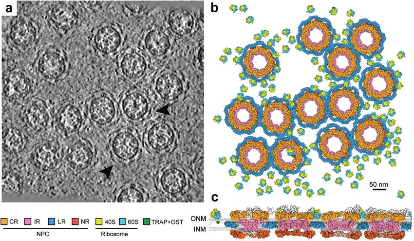
Article
                                                                                                                                                533

Fig. 1 Three-dimensional organization of the NPCs in a local region of the X. laevis NE. a Organization of the NPCs in an original tomogram
slice as viewed along the nucleocytoplasmic axis. Some of the representative array-like densities of the LR are indicated by arrowheads. The
thickness of the tomographic slice shown here is 8.9 Å. b Organization of the NPCs in the reconstructed tomogram as viewed along the
nucleocytoplasmic axis. As the outer boundary of the NPC, the LR appears to cushion the contacts among neighboring NPC particles.
Reconstructions for the individual NPC subunits (CR, IR, NR and LR) and the ribosomes associated with TRAP + OST46 were back-projected
onto the original tomograms based on the refined coordinates of the individual particles. Shown here is a section of the NE from a. Scale bar,
50 nm. c Organization of the NPCs in the reconstructed tomogram as viewed perpendicular to the nucleocytoplasmic axis. In contrast to other
ring scaffolds of the NPC, the LR resides in the lumen. 40S: Small ribosome subunit; 60S: Large ribosome subunit; TRAP: translocon-associated
protein complex; OST: oligosaccharyl-transferase.

RESULTS                                                                 Such anisotropy may reduce confidence on the interpretation of
Overall structure of the NPC from X. laevis oocyte                      detailed structural features. To address this issue and to validate
For each X. laevis oocyte, the NE was isolated and transferred onto     the cryo-ET STA reconstruction, we determined the cryo-EM
an EM grid. The grid was plunge-frozen and imaged on a Titan            structure of the LR using a completely independent data set
Krios microscope. To minimize the effect of preferred sample            through the single particle analysis (SPA) approach. During cryo-
orientation, 1575 tilt series were recorded using a combination of      EM data collection, the sample grids were tilted at fixed angles of
continuous-, bidirectional- and dose-symmetric30 schemes, each          0°, 30°, 45° and 55°, generating 12,399 good micrographs34
from −60° to 60° with a 3°-increment using SerialEM31 (Supple-          (Supplementary information, Table S2). The SPA approach resulted
mentary information, Fig. S1 and Table S1). 36,529 NPC particles        in the reconstruction of the LR subunit at an average resolution of
were averaged to generate a reconstruction with eightfold               10.7 Å (Supplementary information, Figs. S5, S6). The overall
symmetry using Dynamo.32 For these particles, individual subunits       architecture and organization of the LR are nearly identical
of the CR, IR, NR and LR were sub-boxed and subjected to three-         between the STA and SPA reconstructions (Fig. 2a, b). In particular,
dimensional (3D) classification using RELION3.033 (Supplementary         the key structural features of the STA reconstruction of the LR
information, Fig. S2). Analysis by sub-tomogram averaging (STA)         subunit can be very well superimposed to those of the SPA
led to reconstruction of the CR, IR, NR and LR at average               reconstruction (Fig. 2c).
resolutions of 9.1 Å, 13.1 Å, 13.6 Å and 15.1 Å, respectively              One representative X. laevis NPC measures 49 nm in inner
(Supplementary information, Fig. S3a–d and Table S1). For the           diameter (Fig. 2a, b). The outer diameter is approximately 122 nm
purpose of display in the original tomogram, 780 ribosomal              without the LR and 154 nm with the LR. The outer boundary of the
particles were also subjected to the STA procedure, yielding a          cylindrical NPC is defined by the LR (Fig. 2a, b), which is separated
reconstruction at 16.4 Å resolution.                                    from the other three ring scaffolds by the nuclear membrane. The
   Cryo-ET allows direct visualization of all macromolecular            overall size of the NPC described in our study resembles that
complexes on the original tomogram of the X. laevis NE. In the          reported for two vertebrate NPCs, one also from X. laevis14 and the
luminal regions that surround the periphery of the NPC, additional      other from H. sapiens,15 but contrasts with that of the S. cerevisiae
densities are clearly visible and appear to form arch-shaped            NPC7 (Supplementary information, Fig. S7). The cylindrical height
repeating structures (Fig. 1a). These densities are thought to come     of the reconstructed NPC shows some variations among the four
from the LR.16,19 Based on the positional coordinates, reconstruc-      representative organisms. Due to differences in resolution and
tions of the CR, IR, NR, LR and ribosomal subunits were individually    sample preparation, detailed structural comparison of the NPC in
projected back into the original tomograms (Fig. 1b; Supplemen-         different organisms should be performed with caution.
tary information, Video S1). Examination of four evenly-spaced
layers of the same region of a tomogram along the nucleocyto-           Structure of the LR
plasmic axis reveals spatial organization of the CR, IR, NR, LR and     Structural features of the LR subunit are reported in the main text
the ribosomes (Fig. 1c; Supplementary information, Fig. S4). Nearly     for the SPA reconstruction (Fig. 3). Nearly identical features are
every NPC is surrounded by other NPCs and the ribosomes. The LR         shown in the supplementary information for the STA reconstruc-
density encircles the NPC and fills the space between the INM and        tion (Supplementary information, Fig. S8). The LR has eight
ONM (Fig. 1b, c).                                                       subunits, each comprising two symmetric wings (Fig. 3a, b;
   Our reported average resolutions display directional anisotropy,     Supplementary information, Fig. S8a, b). The two wings span a
particularly along the Z-axis (Supplementary information, Fig. S3c).    distance of 70 nm and interact with each other through an

Cell Research (2020) 30:532 – 540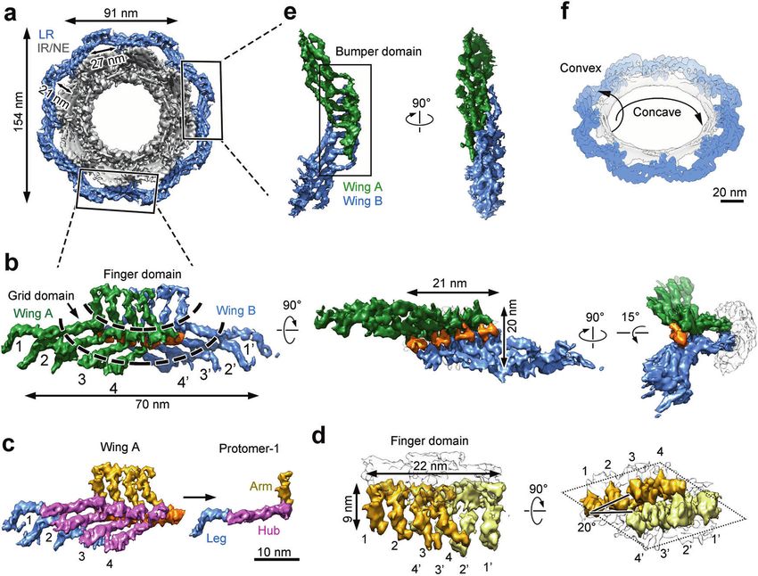
Article
534
Fig. 2 Key features of the LR in the cryo-ET reconstruction are confirmed by an independent cryo-EM reconstruction of the X. laevis NPC.
a Reconstruction of a representative NPC particle by sub-tomogram averaging (STA). A top view and a tilt-45° view are shown. The individual
subunits of the CR, IR, NR and LR were projected back into the original tomograms to allow reconstruction of a number of NPC particles.
Shown here is a representative NPC particle, which contains four ring scaffolds: CR (colored yellow), IR (pink), NR (cyan) and LR (marine).
Viewed perpendicular to the NE (left panel), the IR and LR define the inner and outer diameters, respectively, of the cylindrical NPC.
b Reconstruction of the NPC by single particle analysis (SPA). CR, IR and NR were reconstructed using the C8 symmetry. The LR was
reconstituted using the refined LR subunit and the C8 symmetry. The LR is highlighted. c The key structural features of the LR subunit are
nearly identical between the STA reconstruction (marine, top panel, accession code EMD-0983) and the SPA reconstruction (gray, bottom
panel, accession code EMD-0982). Their overlay is shown in the middle panel. Scale bar, 10 nm.
extended interface (Fig. 3b; Supplementary information, Fig. S8b). curvature of the fusion, which is defined by the diameter of the
Each wing contains four parallel, planar-arranged, elongated fusion within the equatorial plane of the NE (Fig. 3f). On the other
protomers (Fig. 3c; Supplementary information, Fig. S8c). Each hand, the Finger domain directly contacts the luminal side of the
protomer consists of an arm at one end, a central hub and a leg at fusion and may stabilize the convex curvature that connects INM
the other end. Within the same LR subunit, two wings interact to ONM. The spatial separation between INM and ONM is thought
with each other mainly through their eight hubs, generating a to be 10–30 nm.35 The thickness of the LR subunit perpendicular
rigid structure that is termed the Grid domain (Fig. 3b; Supple- to the nuclear membrane is approximately 20 nm (Supplementary
mentary information, Fig. S8b). These two wings also form an information, Fig. S8b, right panel), which may help define the
interface through their eight arms, which together constitute the thickness of the NE surrounding the NPC.
Finger domain.
The Finger domain directly contacts the fusion between the The Bumper domain of the LR
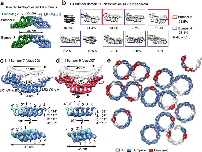
INM and ONM (Fig. 3d; Supplementary information, Fig. S8d). The Back-projection of the LR subunit reconstruction into the original
four arms 1–2–3–4 from one wing (or 1′–2′–3′–4′ from the other tomogram reveals two distinct lengths of the Bumper domains: 29
wing) constitute two pairs 1–2 and 3–4 (or 1′–2′ and 3′–4′ for the and 34 nm (Fig. 4a). The 29-nm Bumper domain has two legs from
other wing), with the tips of each pair connected to each other one wing pairing up with two legs from the other wing, producing
(Fig. 3d, left panel; Supplementary information, Fig. S8d, left six apparent legs as viewed along the nucleocytoplasmic axis
panel). Within the same Finger domain, three arms from one wing (Fig. 4a). For this reason, the 29-nm Bumper domain is hereafter
pair up with three arms from the other wing in a reciprocal order: named Bumper-6. Similarly, the 34-nm Bumper domain is named
2–4′, 3–3′ and 4–2′. Together, the eight arms display a diamond- Bumper-7 because the interface only involves one leg from each
shaped cross section, with four arms from each wing constituting wing, leading to seven apparent legs (Fig. 4a). To reveal additional
one side of the diamond (Fig. 3d, right panel; Supplementary features, we identified and subjected 33,692 candidate Bumper
information, Fig. S8d, right panel). The two distal arms, one from domains to 3D classification with application of a local mask by
each wing (1 and 1′), are placed at the opposing ends of the the STA approach (Fig. 4b). A relatively large class number of ten
diamond. was applied to ensure identification of different conformations of
Two neighboring LR subunits interact with each other through the Bumper domain. Approximately 21.5% and 38.4% of all these
their legs, producing a characteristic scaffold that is hereafter domains, representing eight classes, belong to Bumper-6 and
referred to as the Bumper domain (Fig. 3e; Supplementary Bumper-7, respectively (Fig. 4b). The remaining two classes (39.4%
information, Fig. S8e). In contrast to the Finger domain, the in total) cannot be identified and likely represent deformed or
Bumper domain is distal to the fusion and appears to cushion damaged Bumper domains, or random noise.
neighboring NPCs (Fig. 1b; Supplementary information, Fig. S4). Additional refinement of the particles that belong to Bumper-6
Both the Finger domain and the Bumper domain are visible in the and Bumper-7 reveals detailed features (Fig. 4c, d; Supplementary
original tomograms (Fig. 1a, arrowheads). Eight Grid domains and information, Fig. S9a, b). In Bumper-7, the first leg (leg-1) from one
eight Bumper domains of the LR alternate to assemble into a subunit pairs up with its corresponding leg (leg-1′) from an
closed ring scaffold, which places eight Finger domains in close adjacent subunit (Fig. 4c). In Bumper-6, two legs from one subunit
contact with the fusion (Fig. 3a; Supplementary information, form two reciprocal pairs with two legs from an adjacent subunit:
Fig. S8a). On one hand, the LR scaffold may stabilize the concave 1–2′ and 2–1′ (Fig. 4d). As a consequence of the different pairing
Cell Research (2020) 30:532 – 540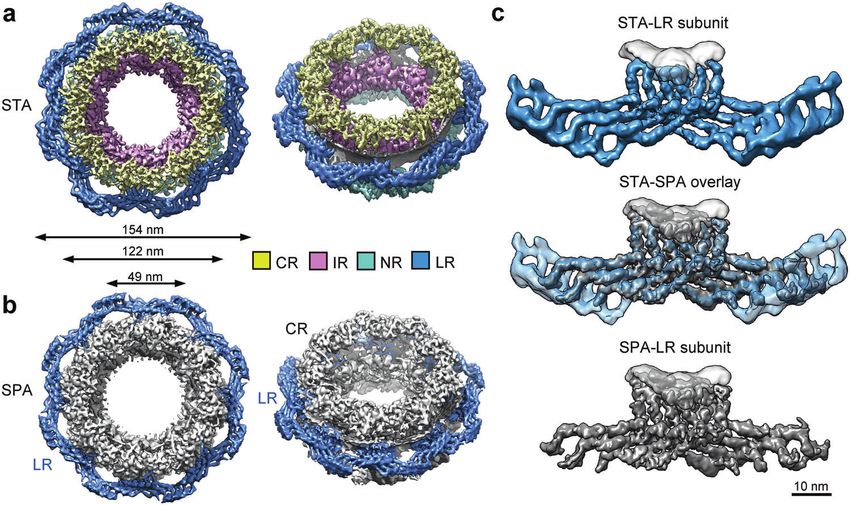
Article
                                                                                                                                                535

Fig. 3 Structure of the LR subunit by the SPA approach. a Eight LR subunits form a continuous circular scaffold. The overall structure of the
LR (colored marine) is viewed along the nucleocytoplasmic axis. b Structure of the LR subunit. Each butterfly-shaped LR subunit comprises
two symmetric wings: Wing-A (colored green) and Wing-B (blue), which interact with each other through an extended interface (orange). The
LR subunit has a Finger domain that contacts the fusion and a Grid domain that make up the bulk of the circular LR scaffold. Three mutually
perpendicular views are shown. c Each wing of the LR subunit comprises four elongated, tubular protomers. These protomers (numbered 1–4)
associate with each other in a planar fashion. Each protomer contains an arm (colored gold) at one end, a central hub (magenta) and a leg
(blue) at the other end. Scale bar, 10 nm. d The Finger domain directly contacts the fusion of nuclear membranes. The tips of the protomers
likely traverse the pore membrane and anchor the LR subunit to the pore. The cross section of the Finger domain has the shape of a diamond
as indicated by dotted lines (right panel). e The Bumper domain is formed between two neighboring LR subunits. Four legs from Wing-A of an
LR subunit interact with four legs from Wing-B of the neighboring LR subunit to form the Bumper domain. Two perpendicular views are
shown. f The LR may stabilize both the concave and the convex curvatures of the fusion. The concave curvature relates to the diameter of the
fusion within the NE, whereas the convex curvature is defined by the separation of the INM and ONM. Scale bar, 20 nm.

arrangements, the centers of the two LR subunits that form              μm.9,36 In contrast, a single X. laevis oocyte contains about 5 × 10⁷
Bumper-7 are separated by 53 nm (Fig. 4c), longer than the              NPCs, with a density of 60 NPCs per square μm.9,36 In our STA
distance of 45 nm for Bumper-6 (Fig. 4d). The pairing difference        reconstruction, NPC particles contact each other through their
also generates contrasting features in the overall structure as well    respective LRs (Figs. 1, 4e; Supplementary information, Figs S4,
as the angles extended between the hub and leg of the                   S9c, d). Judging from the cross section within the equatorial
corresponding protomers (Fig. 4c, d).                                   plane, a sizable fraction of the NPCs have been deformed into
   These refined Bumper domains were projected back onto the             elliptical appearances. At every point of contact, the Bumper
original tomograms (Fig. 4e; Supplementary information, Fig. S9c,       domains from one NPC cushion against the Bumper domains from
d). Bumper-6 and Bumper-7 are often seen in the same NPC, with          neighboring NPCs.
Bumper-7 usually representing the majority. We speculate that the          We performed a statistical analysis on deformation of the NPCs.
distinct Bumper conformations may reflect consequences of                Based on our reconstruction, a representative NPC has an outer
mechanical stress or deformation of the nuclear pore. In fact,          diameter of 154 nm, with a distance of 130 nm between two Grid
most NPCs display a slightly elliptical appearance that amounts to      domains on the opposing sides of the LR (Fig. 5a, inset). An
a small change of the pore diameter.12 Under these circumstances,       important judgment for deformation is whether the distance
the LR may help maintain the curvatures of the fusion through a         between the centers of two neighboring NPCs is shorter than the
conformational switch between Bumper-6 and Bumper-7.                    standard outer diameter of an undeformed NPC. For each NPC,
                                                                        the distance to its closest neighbor is measured and plotted
The Bumper domain cushions neighboring NPCs                             (Fig. 5a). Much to our surprise, the most frequently observed
At least two factors — the extraordinarily large molecular mass         distance is 127.5 nm, which occurs to 1208 distinct pairs of NPCs.
and the relatively loose association among the four rings (CR/IR/       The average shortest distance between two neighboring NPCs is
NR/LR) — may make the NPC susceptible to mechanical                     136 nm, which is 18 nm shorter than the outer diameter of a
deformation. There are approximately 2000–5000 NPCs in a                perfectly symmetric NPC. This analysis suggests widespread
vertebrate nucleus, with a density of 10–20 NPCs per square             deformation or crowding of the NPCs on the X. laevis oocyte.

Cell Research (2020) 30:532 – 540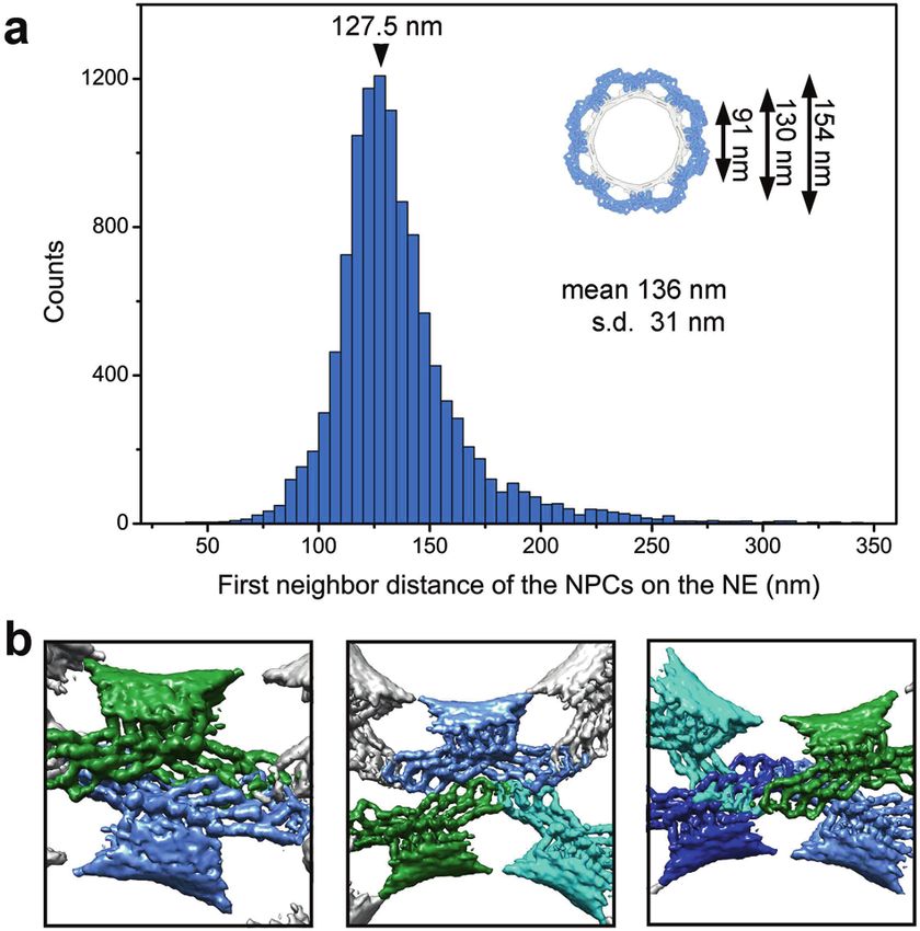
Article
536

      Fig. 4 Structure of the Bumper domain of the LR by the STA approach. a The Bumper domain adopts two major conformations. By back-
      projecting the NPC particles onto the original tomograms, two major conformations of the Bumper domain are seen and named Bumper-6
      and -7. The lengths of Bumper-6 and -7 are 29 and 34 nm, respectively. b Classification of the Bumper domains. The Bumper domains were re-
      cropped and classified. Bumper-6 and -7 represent 21.5% and 38.4% of the total particles classified. c Refined structure of Bumper-7 exhibits
      distinguishing features (upper panel). Two mutually perpendicular views are shown for an isolated Bumper-7 (middle and lower panels).
      d Refined structure of Bumper-6. In contrast to Bumper-7, the internal interface of Bumper-6 involves two pairs of promoter tips. e Mapping
      the Bumper domains onto the NPC particles. The reconstructions for Bumper-7 (marine), Bumper-6 (red) and the LR subunit (gray) were back-
      projected onto the original tomograms based on the refined coordinates of the individual particles. Shown here is a representative section of
      the nuclear membrane. The conformations of nearly all the classified Bumper domains are the same as those of the Bumper domains formed
      by independently back-projecting the LR subunits onto the original tomograms.

      Our observed distance is in excellent agreement with the reported        anisotropy has little impact on the general conclusions derived
      average pairwise distance of 135 + 5 nm for the NPCs derived             from the STA-based cryo-ET study, in part because the key
      from stage VI X. laevis oocytes.37                                       features of the LR subunit have been validated by an independent
         Close examination of the tomograms reveals striking examples          SPA-based cryo-EM study (Figs. 2b, 3). In the SPA-based
      of crowding among neighboring NPCs (Fig. 5b). The Bumper                 reconstruction, the edge of the two wings in the LR subunit is
      domain of one NPC clashes with the Bumper domain of a                    less well defined compared to the STA-based reconstruction
      neighboring NPC (Fig. 5b, left panel) or invades into the space          (Fig. 2c); this is due to application of a considerably smaller
      between two Bumper domains of a neighboring NPC (Fig. 5b,                alignment mask for SPA compared to STA. Despite differences in
      middle panel). Under extreme circumstances, the Bumper domain            the mask size, all eight legs in the LR subunit are clearly visible in
      of one NPC slides past the Bumper domain of a neighboring NPC,           both approaches (Supplementary information, Fig. S10a, b).
      allowing the Grid domains to contact each other (Fig. 5b, right          Importantly, reconstruction by SPA agrees with that by STA up
      panel). Notably, among all NPCs examined, no Bumper domain               to 13.2 Å using a common mask for the FSC (Supplementary
      reaches the membrane fusion of a neighboring NPC.                        information, Fig. S10c). The observed structural features of the LR
                                                                               subunit are previously unknown and appear to define and
                                                                               perhaps explain the function of the LR (Figs. 3–5).
      DISCUSSION                                                                  Despite its mysterious nature, existence of the LR has been
      Structures of the NPC have been reported for at least five                previously recognized. The NPC from Necturus maculosus and X.
      organisms7,12–15 (Supplementary information, Fig. S7). In this           laevis was observed to contain eight spokes that penetrate the
      study, we report the cryo-ET structure of the NPC from X. laevis         fusion into the lumen and form a “luminal ring” through radial arm
      oocyte (Supplementary information, Fig. S3), which allows                dimers or handle-like luminal domain.14,16–19,38 In contrast, the
      identification of previously unknown features of the LR (Figs. 2a,        luminal structure in the D. discoideum NPC appears to comprise
      4; Supplementary information, Fig. S8). Preferred orientation of the     eight discrete rods.12 H. sapiens NPC also contains luminal
      NPC particles led to resolution anisotropy, particularly along the Z-    connections.20 GP210 was found to form a ring around the NPC
      axis (Supplementary information, Fig. S3c, d), which may have an         from X. laevis.29 The EM structure of Pom152 has an extended,
      impact on the cylindrical height of the NPC. Nonetheless, such           tubular appearance.39,40 Reconstruction of the S. cerevisiae NPC

                                                                                                                     Cell Research (2020) 30:532 – 540
Article
537
Asymmetric variations and diameter dilation at the level of
individual NPCs were also observed in HeLa cells,41 and the algae
NPC displayed a dilated IR13; in both cases, the samples were
prepared through focused ion beam milling that was supposed to
maintain the original appearance in cells. Taken together, the NPC
may adopt a much more dynamic conformation than antici-
pated.13 The observed deformation of the NPC may reflect its
natural state on the NE and such plasticity could be indispensable
to its biological functions.18
Assuming GP210 is the primary constituent of the LR scaffold,
our finding that the Bumper domain cushions neighboring NPCs
may mechanistically explain the observation that GP210 mediates
nuclear pore formation, dilation, NPC spacing and integrity.42,43
Knock-down of GP210 in HeLa cells and Caenorhabditis elegans led
to clustering of NPCs in dying cells,43 likely due to loss of such
cushioning. The Bumper domain appears to exhibit marked
elasticity, with Bumper-7 being the default state. Any force
squeezing the NPC towards the center may cause a group of four
protomers within one wing to slide towards that within another
wing of an adjacent LR subunit, thus switching Bumper-7 to
Bumper-6 (Fig. 4c, d). This speculated property of the NPC may
serve to absorb radial shock and insulate the transport function
from movements of the nuclear membrane.
Two wings from adjacent LR subunits constitute an arch,
Fig. 5 The Bumper domain may cushion neighboring NPCs. a reminiscent of the LR in yeast.7,40 Relative to the pore membrane,
NPCs are generally deformed in the X. laevis nuclear membrane. each arch defines a passage that measures 27 nm in width (Fig. 3a;
Statistical analysis of the deformation among neighboring NPCs. For Supplementary information, Fig. S8a). As noted previously,7,40
each NPC, the distance to its nearest neighbor is measured and
plotted here. The median distance is 136 ± 31 nm. The most
these passages are aligned with the circumferential passages
frequently observed distance is 127.5 nm, which occurs to 1208 between the CR or NR and the pore membrane and between
pairs of NPC. The sizes of a symmetric LR is shown in the inset for neighboring IR subunits. Together, these passages could form
reference. b Three representative examples of close contact lateral openings between subunits of the NPC. The combination of
between neighboring NPCs. The reconstruction for the LR subunit arches and passages may outline a well-defined duct for
was back-projected onto the original tomograms based on the nucleocytoplasmic transport of INM proteins.44,45
refined coordinates of the individual LR subunit particles. Cryo-ET reconstruction also allows visualization of other
macromolecular complexes on the tomograms. Analysis of 14
shows a fusion-associated ring scaffold with eight arches, each tomograms allowed preliminary reconstruction of the ribosomal
speculated to comprise two Pom152 molecules.7,40 These previous subunits, translocon-associated protein complex (TRAP) and
studies mostly report rough overall appearance of the LR scaffold. oligosaccharyl-transferase (OST)46 (Fig. 1; Supplementary informa-
In this study, we have identified key structural features of the LR tion, Fig. S4). As observed in a previous study,41 such complexes
from X. laevis NPC using both cryo-ET and cryo-EM. are present in a large number on the cytoplasmic side of the ONM.
The lack of structural information on any of the candidate Nups In-depth examination of these tomograms may reveal additional
of the LR, together with the limited resolution of our EM molecular machineries that associate with the NPC.
reconstruction, does not allow conclusive identification of the
protein components in the density of the LR subunit. Nonetheless,
among the candidate components of the LR, only GP210 contains MATERIALS AND METHODS
a luminal domain that is large enough to constitute the LR scaffold Cryo-sample preparation
observed in our study.17,28 X. laevis GP210 contains 1898 residues Small pieces of ovary were separated from a 2-4 years old,
and was predicted to contain 15 immunoglobulin-like (Ig-like) narcotized female frog (Nasco, USA) and transferred into modified
domains39 (Supplementary information, Fig. S11a). It is likely that Barth’s saline (MBS) (10 mM HEPES, pH 7.5, 88 mM NaCl, 1 mM KCl,
GP210 constitutes the bulk of the LR protomer. Supporting this 0.82 mM MgSO4, 0.33 mM Ca(NO3)2 and 0.41 mM CaCl2) at 18 °C.
analysis, GP210 was found to form a ring around the X. laevis NPC, Stage-VI oocytes (a sphere of 1.3 mm in diameter with clear
with an eightfold symmetry and a diameter of 164 ± 7 nm.29 In separation of the black animal pole, the off-white vegetal pole and
contrast to GP210, POM121 only has its N-terminal ~30 residues in unpigmented equatorial belt) were manually sorted from the
the lumen and NDC1 only has the linker sequences between encased connective tissue membranes using forceps. For each
neighboring TMs in the lumen4 (Supplementary information, stage-VI oocyte, a small hole was generated on the side of the
Fig. S11b). Therefore, the bulk of the elongated tubular density of animal pole using forceps in MBS, and the nucleus was sucked and
the LR protomer may come from the 15 Ig-like domains of GP210 transferred into a low salt buffer (LSB) (10 mM HEPES, pH 7.5, 1
(Supplementary information, Fig. S11c). Despite these tantalizing mM KCl, 0.5 mM MgCl2, 10 μg/mL aprotinin, 5 μg/mL leupeptin)
clues, we cannot exclude the possibility that the LR is formed by using a 20-μL pipette tip. The isolated nuclei were kept in LSB for
other yet-to-be identified proteins. 10 min, and during this time the yolk was rapidly cleaned through
Few X. laevis NPC particles display a perfectly eight-fold pipetting several times.14,47 Two-to-three cleaned nuclei were
symmetry and most are slightly elliptical in appearance (Figs. 1, transferred onto a freshly glow-discharged copper EM grid (R1.2/
4e; Supplementary information, Fig. S9c, d). The shape asymmetry 1.3; Quantifoil, Jena, Germany) in an LSB liquid drop of about 5 μL.
could arise as a result of osmotic swelling or temperature The glow-discharged grids were prepared for 30 s using the
variation.17,18 Although we cannot exclude the possibility of NPC “Mid” setting of the Plasma Cleaner (Harrick, Plasma Cleaner PDC-
deformation during sample preparation, examination of the D. 32G). For each nucleus, the NE was spread onto the EM grid by
discoideum NPC on intact nuclei and H. sapiens NPC in whole cells popping a small hole on one side of the nucleus using two glass
also revealed radial displacement and elliptical distortions.12,20 needles to extrude chromatin and other nuclear contents. The NE
Cell Research (2020) 30:532 – 540Article
538
was carefully washed three times, each time using 5 μL LSB. 2 μL For reconstruction of the nuclear ring (NR), the 112,014 4×
gold fiducial beads (10 nm diameter, Aurion, The Netherlands) binned and re-centered IR subtomograms were aligned using
were applied onto the native NE samples before plunge-freezing. human NR subunit density map (accession code EMD-3107)15 as
The grids were blotted for 5 s, vitrified by plunge-freezing into the initial template and an ellipsoid at the NR location as the mask
liquid ethane using a Vitrobot Mark IV (Thermal Fisher Scientific) at (Supplementary information, Fig. S2). The NR subunit density
a temperature of 8 °C and a humidity of 100%. The quality of emerged after initial alignment, which was restricted at 60 Å
sample preparation was examined using an FEI Tecnai Arctica resolution to prevent template bias. Subsequently the boxes were
microscope (Thermo Fisher Scientific) operating at 200 kV. re-centered to the NR subunit and further aligned to 17.8 Å
resolution. 3D classification by RELION3.0 was used to select a
Cryo-ET data acquisition group of 34,068 NR subunits, which were extracted into 2× binned
The grids were imaged on a Titan Krios microscope operating at NR subtomograms with a box size of 200 × 200 × 200 voxels.
300 kV equipped with an energy filter (slit width 20 eV; GIF Subsequent alignment achieved an average resolution of 13.6 Å
Quantum LS, Gatan) and a K2 Summit direct electron detector for the NR.
(Gatan). Tilt-series were recorded in the super-resolution mode at For reconstruction of the luminal ring (LR), the 112,014 4×
a nominal magnification of 64,000×, resulting in a calibrated pixel binned and re-centered IR subtomograms were aligned using the
size of 1.111 Å. A combination of dose-symmetric,30 bi-directional aligned IR subunits as initial templates and an ellipsoid at the LR
and continuous schemes were used to collect tilt-series from −60° location as the mask (Supplementary information, Fig. S2). The LR
to 60° at a step size of 3° using SerialEM31 (Supplementary subunit density emerged after initial alignment. The boxes were
information, Fig. S1). At each tilt, a movie stack consisting of 8 re-centered to the LR subunit and further aligned to 17.8 Å
frames was recorded at 0.1 s exposure per frame, yielding a total resolution. 3D classification by RELION3.0 was used to select a
dose of ~90–150e−/Å2 per tilt-series. 1575 tilt-series were group of 34,087 LR subunits, which were extracted into 2× binned
collected using defocus values between -2 and -4 µm (Supple- LR subtomograms with a box size of 200 × 200 × 200 voxels.
mentary information, Fig. S2). A summary of data acquisition Subsequent alignment achieved an average resolution of 15.1 Å
statistics can be found in Supplementary information Table S1. for the LR. To further examine the conformation of the Bumper
domain, the boxes were re-centered to the joint region between
Cryo-ET data processing two adjacent subunits of the LR. 3D classification of the
Tilt-series were binned to a final pixel size of 2.22 Å and motion subtomograms by RELION3.0 revealed two major conformations:
corrected by averaging eight frames for each tilt using Motion- Bumper-6 and Bumper-7. Using Dynamo, 7277 Bumper-6 and
Cor2.48 Defocus of the tilt series was estimated using CTFFIND4.49 13,087 Bumper-7 particles were independently aligned and
The contrast transfer function (CTF) of each tilt-series was refined, yielding average resolutions of 17.0 Å and 15.6 Å,
corrected using NovaCTF.50 1425 tilt-series with good fiducial respectively (Supplementary information, Fig. S9a, b).
alignment were reconstructed to tomograms through weighted To illustrate the organization of the NE, ribosomes were
back projection using IMOD51 (Supplementary information, reconstructed. 780 ribosomes were manually picked from 14 4×
Fig. S2). The tomograms were 2× and 4× binned for subsequent binned tomograms and extracted into boxes of 80 × 80 × 80
processing. Subtomograms of 36,529 NPC complexes were voxels. The sub-tomograms were first aligned using EMD-431554
manually picked and extracted from the 4× binned tomograms as the template, and the resolution was restricted to 45 Å at this
into boxes of 200 × 200 × 200 voxels with the help of Dynamo stage. Next, the refined coordinates were used to extract
catalog52 for further analysis. subtomograms into boxes of 160 × 160 × 160 voxels from 2×
STA was carried out in Dynamo,32 following a published binned tomograms, which were subsequently aligned using the
protocol.53 For reconstruction of the cytoplasmic ring (CR), the FSC standard of 0.143–16.4 Å resolution. Consistent with reported
manually picked 4× binned NPC sub-tomograms were averaged ribosome reconstruction from the NE,41 some of the ribosomes
as the template for their alignment. The resolution was restricted reconstructed here are also found to be associated with
to 40 Å and a C8 symmetry was applied at this stage. Next, translocon-associated protein complex (TRAP) and
coordinates of 292,232 CR subunits were estimated by subboxing oligosaccharyl-transferase (OST).46
and the subboxes were extracted from the 4× binned tomograms The subunit maps were low passed according to the estimated
into boxes of 100 × 100 × 100 voxels for independent alignment. local resolutions of the reconstructions.55 Empirical B-factors of
To prevent overfitting, a “gold-standard” method using an -1000, -2000, -2000, -2000 and -2000 were used to sharpen the CR,
adaptive filter and an ellipsoidal mask was used to align the IR, NR, LR and ribosome reconstructions, respectively.
subboxes. The aligned CR subunits were subjected to 3D
classification (particles binned to 8×) using RELION3.0.33 Cryo-EM data acquisition
112,220 subunits survived this analysis and were further aligned Details for the acquisition of cryo-EM data are described in the
in Dynamo to 17.8 Å resolution using a criterion of 0.143 for the accompanying manuscript.34 Briefly, micrographs were recorded
Fourier shell correlation (FSC) value. Next, the refined coordinates on a Titan Krios (FEI) electron microscope, operating at 300 kV and
were used to extract and align subtomograms from 2× binned equipped with a Gatan Gif Quantum energy filter (slit width 20 eV).
tomograms. Finally, subtomograms from the unbinned tomo- A K2 Summit detector (Gatan Company) in super-resolution mode
grams were extracted into boxes of 320 × 320 × 320 voxels and with a nominal magnification of 64,000× was used, resulting in a
aligned to a final resolution of 9.1 Å. calibrated pixel size of 1.111 Å. The total dose followed a cosine
For reconstruction of the inner ring (IR), the 112,220 4× binned alpha scheme where the total dose is inversely proportional to the
CR subtomograms were aligned using the human IR subunit cosine of the tilting angle and the total dose used for the Tilt-0
density map (EMD-3106)15 as the initial template and an ellipsoid micrographs was 52e−/Å2, a summary of data acquisition statistics
at the IR location as the mask (Supplementary information, Fig. S2). can be found in Supplementary information, Table 2.
After aligning the IR subunits to 17.8 Å resolution, the boxes were
re-centered to the IR subunit and the coordinates were used to Cryo-EM data analysis
extract 2× binned IR subtomograms into boxes of 200 × 200 × 200 The single-particle cryo-EM data was mainly used to generate a
voxels. 3D classification by RELION3.0 was used to select a group reconstruction of the CR at an improved resolution.34 After
of 34,086 IR subunits. Subsequent alignment in Dynamo achieved completing this task, the same data were analyzed to generate a
an average resolution of 13.1 Å for the IR. SPA-based reconstruction of the LR. The strategy for processing of
Cell Research (2020) 30:532 – 540Article
539
the cryo-EM data toward reconstruction of the LR subunit is ADDITIONAL INFORMATION
presented in Supplementary information, Fig. S5. We first Supplementary information accompanies this paper at https://doi.org/10.1038/
attempted to reconstruct the LR subunit by simply re-centering s41422-020-0320-y.
the particles to the LR subunit and preformed image alignment
with or without any mask. This approach however failed with the Competing interests: The authors declare no competing interests.
CR subunit showing strong density at the edge of the box or the
mask that was supposed to emphasize the LR subunit. In order to
resolve this problem, two measures were implemented. REFERENCES
First, since the whole image alignment was biased toward the 1. Gorlich, D. & Kutay, U. Transport between the cell nucleus and the cytoplasm.
Annu. Rev. Cell Dev. Biol. 15, 607–660 (1999).
CR subunit, we tested whether placing a CR subunit on the
2. Ptak, C., Aitchison, J. D. & Wozniak, R. W. The multifunctional nuclear pore
opposite side could somehow neutralize this bias. Results from the complex: a platform for controlling gene expression. Curr. Opin. Cell Biol. 28,
cryo-ET study indicate a two-fold symmetry of the LR subunit at up 46–53 (2014).
to 15-Å resolution. Specifically, we updated the Euler angle and 3. Beck, M. & Hurt, E. The nuclear pore complex: understanding its function through
offsets for each particle to generate another RELION data star file. structural insight. Nat. Rev. Mol. Cell Biol. 18, 73–89 (2017).
This effort resulted in an identical reconstruction when the whole 4. Rothballer, A. & Kutay, U. Poring over pores: nuclear pore complex insertion into
reconstruction was rotated by 180°, so that another CR subunit the nuclear envelope. Trends Biochem. Sci. 38, 292–301 (2013).
would appear at the bottom of the box instead of the top 5. Hampoelz, B., Andres-Pons, A., Kastritis, P. & Beck, M. Structure and assembly of
(Supplementary information, Fig. S5). The particles within this star the nuclear pore complex. Annu. Rev. Biophys. 48, 515–536 (2019).
6. Alber, F. et al. The molecular architecture of the nuclear pore complex. Nature
file are referred to as symmetry-related particles, which were then
450, 695–701 (2007).
joined by the original particles to generate a data set with twice 7. Kim, S. J. et al. Integrative structure and functional anatomy of a nuclear pore
the number of particles as the original data set. Second, the CR complex. Nature 555, 475–482 (2018).
subunit appeared partially because the CTF parameter favored 8. Hoelz, A., Debler, E. W. & Blobel, G. The structure of the nuclear pore complex.
high resolution reconstructions of the CR subunit, this would likely Annu. Rev. Biochem. 80, 613–643 (2011).
bias the initial stages of alignment of the LR subunit toward the CR 9. Grossman, E., Medalia, O. & Zwerger, M. Functional architecture of the nuclear
side because of its strong features. To resolve this potential pore complex. Annu. Rev. Biophys. 41, 557–584 (2012).
problem, we removed all CTF parameters from previous CTF 10. Schwartz, T. U. The structure inventory of the nuclear pore complex. J. Mol. Biol.
refinements and reverted back to the CTF values from the 428, 1986–2000 (2016).
11. Lin, D. H. & Hoelz, A. The structure of the nuclear pore complex (an update). Annu.
micrograph star file.
Rev. Biochem. 88, 725–783 (2019).
These two strategies allowed us to generate an initial 12. Beck, M., Lucic, V., Forster, F., Baumeister, W. & Medalia, O. Snapshots of nuclear
reconstruction of the LR subunit. This initial reconstruction was pore complexes in action captured by cryo-electron tomography. Nature 449,
refined with application of a soft mask that covers the Finger 611–615 (2007).
domain, the Grid domain and the legs. To further improve the 13. Mosalaganti, S. et al. In situ architecture of the algal nuclear pore complex. Nat.
resolution, six rounds of 3D classifications were performed to Commun. 9, 2361 (2018).
remove empty or heterogeneous particles. This practice results in 14. Eibauer, M. et al. Structure and gating of the nuclear pore complex. Nat. Commun.
removal of 20%–30% of the particles after each round of 3D 6, 7532 (2015).
classification. The final average resolution of the reconstruction for 15. von Appen, A. et al. In situ structural analysis of the human nuclear pore complex.
Nature 526, 140–143 (2015).
the LR subunit was 10.7 Å using 311,240 particles, which includes
16. Akey, C. W. & Radermacher, M. Architecture of the xenopus nuclear pore complex
157,541 particles from the original data set and 153,699 particles revealed by three-dimensional cryoelectron microscopy. J. Cell Biol. 122, 1–19
from the symmetry-related data set. (1993).
17. Stoffler, D. et al. Cryo-electron tomography provides novel insights into nuclear
Data deposition pore architecture: implications for nucleocytoplasmic transport. J. Mol. Biol. 328,
119–130 (2003).
The Electron Microscopy Database (EMD) accession codes of the
18. Akey, C. W. Structural plasticity of the nuclear pore complex. J. Mol. Biol. 248,
LR subunit, Bumper-7, Bumper-6, the CR subunit, the IR subunit 273–293 (1995).
and the NR subunit are EMD-0983, EMD-0984, EMD-0985, EMD- 19. Frenkiel-Krispin, D., Maco, B., Aebi, U. & Medalia, O. Structural analysis of a
0986, EMD-0997 and EMD-0998, respectively, for the reconstruc- metazoan nuclear pore complex reveals a fused concentric ring architecture. J.
tions calculated by the STA approach. The EMD accession code is Mol. Biol. 395, 578–586 (2010).
EMD-0982 for the reconstruction of the LR subunit calculated by 20. Maimon, T., Elad, N., Dahan, I. & Medalia, O. The human nuclear pore complex as
the SPA approach. revealed by cryo-electron tomography. Structure 20, 998–1006 (2012).
21. Gerace, L., Ottaviano, Y. & Kondor-Koch, C. Identification of a major polypeptide
of the nuclear pore complex. J. Cell Biol. 95, 826–837 (1982).
22. Wozniak, R. W., Bartnik, E. & Blobel, G. Primary structure analysis of an integral
ACKNOWLEDGEMENTS membrane glycoprotein of the nuclear pore. J. Cell Biol. 108, 2083–2092 (1989).
We thank Westlake University for providing a Start-up fund, the Tsinghua University 23. Wozniak, R. W., Blobel, G. & Rout, M. P. Pom152 is an integral protein of the pore
Branch of China National Center for Protein Sciences (Beijing) for the cryo-EM facility membrane domain of the yeast nuclear envelope. J. Cell Biol. 125, 31–42 (1994).
and the computational facility support, and L. Zhao, X. Li and J. Wen for technical 24. Hallberg, E., Wozniak, R. W. & Blobel, G. An integral membrane protein of the pore
support. We thank X. Fu and P. Zhang at the University of Pittsburgh for advice on membrane domain of the nuclear envelope contains a nucleoporin-like region. J.
STA sample preparation and SerialEM data collection. This work was supported by Cell Biol. 122, 513–521 (1993).
funds from the National Natural Science Foundation of China (31930059, 25. Mansfeld, J. et al. The conserved transmembrane nucleoporin NDC1 is required
81920108015, 31621092 and 31430020). for nuclear pore complex assembly in vertebrate cells. Mol. Cell 22, 93–103 (2006).
26. Stavru, F. et al. NDC1: a crucial membrane-integral nucleoporin of metazoan
nuclear pore complexes. J. Cell Biol. 173, 509–519 (2006).
27. Chadrin, A. et al. Pom33, a novel transmembrane nucleoporin required for proper
AUTHOR CONTRIBUTIONS nuclear pore complex distribution. J. Cell Biol. 189, 795–811 (2010).
X.Z. and Y.Z. prepared the sample. Y.Z., C.Z., G.H., S.L., Q.W. and J.L. collected the EM 28. Greber, U. F., Senior, A. & Gerace, L. A major glycoprotein of the nuclear pore
data. Y.Z., S.L., G.H., C.Z. and Q.W. processed the EM data. Y.Z. and S.L. performed the complex is a membrane-spanning polypeptide with a large lumenal domain and
cryo-ET STA calculation. G.H. performed the cryo-EM SPA calculation. K.W., W.Z. and a small cytoplasmic tail. EMBO J. 9, 1495–1502 (1990).
G.Y. provided computing assistance. Q.Z., C.Y. and Q.T. provided critical advices. All 29. Loschberger, A. et al. Super-resolution imaging visualizes the eightfold symmetry
authors analyzed the structure. Y.Z., S.L., G.H., C.Z. and Y.S. wrote the manuscript. Y.S. of gp210 proteins around the nuclear pore complex and resolves the central
conceived and supervised the project. channel with nanometer resolution. J. Cell Sci. 125, 570–575 (2012).
Cell Research (2020) 30:532 – 540Article
540
30. Hagen, W. J. H., Wan, W. & Briggs, J. A. G. Implementation of a cryo-electron 47. Jarnik, M. & Aebi, U. Toward a more complete 3-D structure of the nuclear pore
tomography tilt-scheme optimized for high resolution subtomogram averaging. complex. J. Struct. Biol. 107, 291–308 (1991).
J. Struct. Biol. 197, 191–198 (2017). 48. Zheng, S. Q. et al. MotionCor2: anisotropic correction of beam-induced
31. Mastronarde, D. N. Automated electron microscope tomography using robust motion for improved cryo-electron microscopy. Nat. Methods 14, 331–332
prediction of specimen movements. J. Struct. Biol. 152, 36–51 (2005). (2017).
32. Castano-Diez, D., Kudryashev, M., Arheit, M. & Stahlberg, H. Dynamo: a flexible, 49. Rohou, A. & Grigorieff, N. CTFFIND4: fast and accurate defocus estimation from
user-friendly development tool for subtomogram averaging of cryo-EM data in electron micrographs. J. Struct. Biol. 192, 216–221 (2015).
high-performance computing environments. J. Struct. Biol. 178, 139–151 (2012). 50. Turonova, B., Schur, F. K. M., Wan, W. & Briggs, J. A. G. Efficient 3D-CTF correction
33. Scheres, S. H. RELION: implementation of a Bayesian approach to cryo-EM for cryo-electron tomography using NovaCTF improves subtomogram averaging
structure determination. J. Struct. Biol. 180, 519–530 (2012). resolution to 3.4A. J. Struct. Biol. 199, 187–195 (2017).
34. Huang, G. et al. Structure of the cytoplasmic ring of the Xenopus laevis nuclear 51. Kremer, J. R., Mastronarde, D. N. & McIntosh, J. R. Computer visualization of three-
pore complex. Cell Res. https://doi.org/10.1038/s41422-020-0319-4 (2020). dimensional image data using IMOD. J. Struct. Biol. 116, 71–76 (1996).
35. Watson, M. L. Further observations on the nuclear envelope of the animal cell. J. 52. Castano-Diez, D., Kudryashev, M. & Stahlberg, H. Dynamo catalogue: geometrical
Biophys. Biochem. Cytol. 6, 147–156 (1959). tools and data management for particle picking in subtomogram averaging of
36. Spector, D. L. Macromolecular domains within the cell nucleus. Annu. Rev. Cell cryo-electron tomograms. J. Struct. Biol. 197, 135–144 (2017).
Biol. 9, 265–315 (1993). 53. Li, S. et al. Acidic pH-induced conformations and LAMP1 binding of the lassa virus
37. Selles, J. et al. Nuclear pore complex plasticity during developmental process as glycoprotein spike. PLoS Pathog. 12, e1005418 (2016).
revealed by super-resolution microscopy. Sci. Rep. 7, 14732 (2017). 54. Braunger, K. et al. Structural basis for coupling protein transport and N-
38. Akey, C. W. Interactions and structure of the nuclear pore complex revealed by glycosylation at the mammalian endoplasmic reticulum. Science 360, 215–218
cryo-electron microscopy. J. Cell Biol. 109, 955–970 (1989). (2018).
39. Hao, Q., Zhang, B., Yuan, K., Shi, H. & Blobel, G. Electron microscopy of Chaeto- 55. Wan, W. et al. Structure and assembly of the Ebola virus nucleocapsid. Nature
mium pom152 shows the assembly of ten-bead string. Cell Discov. 4, 56 (2018). 551, 394–397 (2017).
40. Upla, P. et al. Molecular architecture of the major membrane ring component of
the nuclear pore complex. Structure 25, 434–445 (2017).
41. Mahamid, J. et al. Visualizing the molecular sociology at the HeLa cell nuclear Open Access This article is licensed under a Creative Commons
periphery. Science 351, 969–972 (2016). Attribution 4.0 International License, which permits use, sharing,
42. Drummond, S. P. & Wilson, K. L. Interference with the cytoplasmic tail of gp210 adaptation, distribution and reproduction in any medium or format, as long as you give
disrupts “close apposition” of nuclear membranes and blocks nuclear pore appropriate credit to the original author(s) and the source, provide a link to the Creative
dilation. J. Cell Biol. 158, 53–62 (2002). Commons license, and indicate if changes were made. The images or other third party
43. Cohen, M., Feinstein, N., Wilson, K. L. & Gruenbaum, Y. Nuclear pore protein material in this article are included in the article’s Creative Commons license, unless
gp210 is essential for viability in HeLa cells and Caenorhabditis elegans. Mol. Biol. indicated otherwise in a credit line to the material. If material is not included in the
Cell 14, 4230–4237 (2003). article’s Creative Commons license and your intended use is not permitted by statutory
44. Meinema, A. C. et al. Long unfolded linkers facilitate membrane protein import regulation or exceeds the permitted use, you will need to obtain permission directly
through the nuclear pore complex. Science 333, 90–93 (2011). from the copyright holder. To view a copy of this license, visit http://creativecommons.
45. Knockenhauer, K. E. & Schwartz, T. U. The nuclear pore complex as a flexible and org/licenses/by/4.0/.
dynamic gate. Cell 164, 1162–1171 (2016).
46. Pfeffer, S. et al. Structure of the native Sec61 protein-conducting channel. Nat.
Commun. 6, 8403 (2015). © The Author(s) 2020
Cell Research (2020) 30:532 – 540You can also read