Human tauopathy-derived tau strains determine the substrates recruited for templated amplification
←
→
Page content transcription
If your browser does not render page correctly, please read the page content below
doi:10.1093/brain/awab091                                                                                     BRAIN 2021: 144; 2333–2348              | 2333

Human tauopathy-derived tau strains
determine the substrates recruited for
templated amplification

                                                                                                                                                                   Downloaded from https://academic.oup.com/brain/article/144/8/2333/6164963 by guest on 25 November 2021
Airi Tarutani,1,2 Haruka Miyata,1 Takashi Nonaka,1 Kazuko Hasegawa,3
Mari Yoshida,4 Yuko Saito,5,6 Shigeo Murayama,5,7 Andrew C. Robinson,8
David M. A. Mann,8 Taisuke Tomita2 and Masato Hasegawa1

Tauopathies are a subset of neurodegenerative diseases characterized by abnormal tau inclusions. Specifically,
three-repeat tau and four-repeat tau in Alzheimer’s disease, three-repeat tau in Pick’s disease (PiD) and four-repeat
tau in progressive supranuclear palsy (PSP) and corticobasal degeneration (CBD) form amyloid-like fibrous struc-
tures that accumulate in neurons and/or glial cells. Amplification and cell-to-cell transmission of abnormal tau
based on the prion hypothesis are believed to explain the onset and progression of tauopathies. Recent studies
support not only the self-propagation of abnormal tau, but also the presence of conformationally distinct tau
aggregates, namely tau strains. Cryogenic electron microscopy analyses of patient-derived tau filaments have
revealed disease-specific ordered tau structures. However, it remains unclear whether the ultrastructural and bio-
chemical properties of tau strains are inherited during the amplification of abnormal tau in the brain.
In this study, we investigated template-dependent amplification of tau aggregates using a cellular model of seeded
aggregation. Tau strains extracted from human tauopathies caused strain-dependent accumulation of insoluble
filamentous tau in SH-SY5Y cells. The seeding activity towards full-length four-repeat tau substrate was highest in
CBD-tau seeds, followed by PSP-tau and Alzheimer’s disease (AD)-tau seeds, while AD-tau seeds showed higher
seeding activity than PiD-tau seeds towards three-repeat tau substrate. Abnormal tau amplified in cells inherited
the ultrastructural and biochemical properties of the original seeds.
These results strongly suggest that the structural differences of patient-derived tau strains underlie the diversity
of tauopathies, and that seeded aggregation and filament formation mimicking the pathogenesis of sporadic tau-
opathy can be reproduced in cultured cells. Our results indicate that the disease-specific conformation of tau
aggregates determines the tau isoform substrate that is recruited for templated amplification, and also influences
the prion-like seeding activity.

1 Department of Brain and Neurosciences, Tokyo Metropolitan Institute of Medical Science, Tokyo 156-8506, Japan
2 Laboratory of Neuropathology and Neuroscience, Graduate School of Pharmaceutical Sciences, The University of
  Tokyo, Tokyo 113-0033, Japan
3 Division of Neurology, Sagamihara National Hospital, Kanagawa 252-0392, Japan
4 Department of Neuropathology, Institute for Medical Science of Aging, Aichi Medical University, Aichi 480-1195,
  Japan
5 Department of Neuropathology, Tokyo Metropolitan Institute of Gerontology, Tokyo 173-0015, Japan
6 Department of Pathology and Laboratory Medicine, National Center Hospital, National Center of Neurology and
  Psychiatry, Tokyo 187-8551, Japan
7 Brain Bank for Neurodevelopmental, Neurological and Psychiatric Disorders, United Graduate School of Child
  Development, Osaka University, Osaka 565-0871, Japan

Received August 26, 2020. Revised February 14, 2021. Accepted February 25, 2021. Advance access publication March 9, 2021
C The Author(s) (2021). Published by Oxford University Press on behalf of the Guarantors of Brain.
V
This is an Open Access article distributed under the terms of the Creative Commons Attribution License (http://creativecommons.org/licenses/by/4.0/), which per-
mits unrestricted reuse, distribution, and reproduction in any medium, provided the original work is properly cited.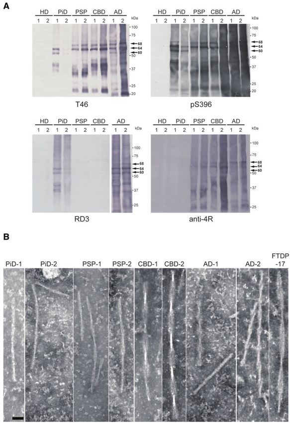
2334    | BRAIN 2021: 144; 2333–2348                                                                                     A. Tarutani et al.

8   Faculty of Biology, Medicine and Health, School of Biological Sciences, Division of Neuroscience and Experimental
    Psychology, The University of Manchester, Salford Royal Hospital, Salford M6 8HD, UK

Correspondence to: Masato Hasegawa, PhD
Department of Brain and Neuroscience
Tokyo Metropolitan Institute of Medical Science, 2-1-6 Kamikitazawa
Setagaya-ku, Tokyo 156-8506, Japan
E-mail: hasegawa-ms@igakuken.or.jp

Keywords: tau; prion-like propagation; tauopathy; strains
Abbreviations: CBD = corticobasal degeneration; CTF = C-terminal fragment; PHF = paired helical filament;
PiD = Pick’s diseasePSP = progressive supranuclear palsy

                                                                                                                                                 Downloaded from https://academic.oup.com/brain/article/144/8/2333/6164963 by guest on 25 November 2021
Introduction                                                             been reported that PiD, PSP and CBD patients present with clinical
                                                                         symptoms related to the brain region in which tau pathology is
Abnormal proteins that accumulate in the brain of patients with          observed.19 A possible mechanism to explain this intracerebral ex-
neurodegenerative diseases form amyloid-like cross-b structures,         pansion of tau pathology is prion-like propagation, in which ab-
and exhibit prion-like properties, including detergent-insolubility      normal tau self-amplifies and spreads in the brain.20,21 Although
and protease resistance, which enable them to convert normal             the molecular mechanism of cell-to-cell transmission is poorly
proteins to an abnormal form.1 Tau, which is one of these prion-         understood, numerous reports of experimental transmissions of
like proteins, is a microtubule-associated protein encoded by the        abnormal tau in vitro and in vivo support this hypothesis.22,23
MAPT gene.2 In the adult human brain, tau exists in six isoforms         Recently, it was also proposed that the variety of clinical symp-
composed of 352–441 residues with the different amino acid num-          toms and pathologies in tauopathies can be explained by the in-
bers arising from alternative splicing of exon 2, exons 2 and 3, or      volvement of distinct tau ‘strains’, which is a characteristic of
exon 10.3 These tau isoforms can be classified into three-repeat         prions. Biochemical analysis of patient-derived sarkosyl-insoluble
tau (3R tau) and four-repeat tau (4R tau) isoforms according to the      tau shows that human tauopathies are characterized by distinct
number of repeats (R1–R4) in the microtubule-binding domain,             banding patterns of the C-terminal tau fragments (tau-CTFs) and
and the two types are expressed in the adult human brain at a            trypsin-resistant tau.24,25 Moreover, tau filaments extracted from
ratio of 1:1.4 Under physiological conditions, tau is localized         the brains of patients exhibit various ultrastructural properties de-
mainly in axons as a soluble, natively unfolded protein, and func-       pending on the tauopathy involved, and cryogenic electron mi-
tions to polymerize tubulins and stabilize microtubules.5 On the         croscopy (cryo-EM) of these tau filaments has revealed disease-
other hand, accumulated pathological tau in the brains of patients       specific ordered core structures at the atomic level.26–28 In experi-
exists as filamentous insoluble forms that are hyperphosphory-           mental models of prion-like propagation, the physical and patho-
lated and partially ubiquitinated, and have lost the ability to bind     logical properties of tau strains are inherited during the
to microtubules.6                                                        amplification of seeds and the formation of tau pathology.29–34
    Neurodegenerative diseases in which tau inclusions are               Thus, it has been suggested that the diversity of ultrastructural
observed in neurons and/or glial cells are referred to as tauopathies.   and biochemical properties of tau strains is the key to the forma-
In Alzheimer’s disease and chronic traumatic encephalopathy, six         tion of disease-specific tau pathology. However, the molecular
isoforms of tau accumulate as neurofibrillary tangles in neuronal        mechanisms of formation of tau strains have not yet been eluci-
cell bodies and neuropil threads in neurites.7 Astrocytic tangles are    dated, and it is not clear how the prion-like properties of tau
also a neuropathological feature in chronic traumatic encephalop-        strains are maintained during self-templated amplification and
athy.8 In Pick’s disease (PiD), 3R tau is observed in neurons as Pick    spreading in the brain.
bodies.7 In progressive supranuclear palsy (PSP) and corticobasal de-        In this study, we investigated template-dependent amplifica-
generation (CBD), 4R tau accumulates in neurons and glial cells.9        tion of abnormal tau by introducing patient-derived tau strains
Although coiled bodies in oligodendrocytes are observed in both          into SH-SY5Y cells. Tau aggregates extracted from Alzheimer’s dis-
diseases, tufted astrocytes observed in PSP and astrocytic plaques       ease, PiD, PSP, and CBD cases caused isoform-dependent accumu-
observed in CBD are neuropathological indicators that enable each        lation of insoluble filamentous tau, and these abnormal tau
disease to be distinguished.10 Argyrophilic granular dementia and        species showed ultrastructural and biochemical properties akin to
globular glial tauopathy are also categorized as 4R tauopathies.9        those of the original seeds. Our results indicate that distinct con-
While most cases of tauopathy are sporadic, mutations in the MAPT        formations of patient-derived tau strains are involved in disease-
gene cause the onset of familial frontotemporal dementia and par-        specific tau filament formation.
kinsonism linked to chromosome 17 (FTDP-17T).11–14 Multiple mis-
sense mutations have been found in and around the microtubule-
binding domain, and affect the structure of tau and its binding to       Materials and methods
microtubules.15,16 In addition, mutations in exon 10 and intron 10
                                                                         Ethics statement
often alter the pattern of alternative splicing and lead to unequal
ratios of 3R tau and 4R tau, resulting in the onset of tauopathy, de-    Post-mortem brain tissues, which had been neuropathologically
pending on the increased tau isoform.                                    diagnosed as Huntington’s disease, Alzheimer’s disease, PiD, PSP,
    Histopathological studies indicate that the tau pathology            CBD and FTDP-17T, were obtained from the Brain Banks at the
observed in the brain of patients with Alzheimer’s disease and ar-       University of Manchester, Tokyo Metropolitan Geriatric Hospital
gyrophilic granular dementia correlates with clinical symptoms           and Institute of Gerontology, National Center of Neurology and
and spreads in the brain in a stereotypic manner.17,18 It has also       Psychiatry, and Aichi Medical University. The study protocol was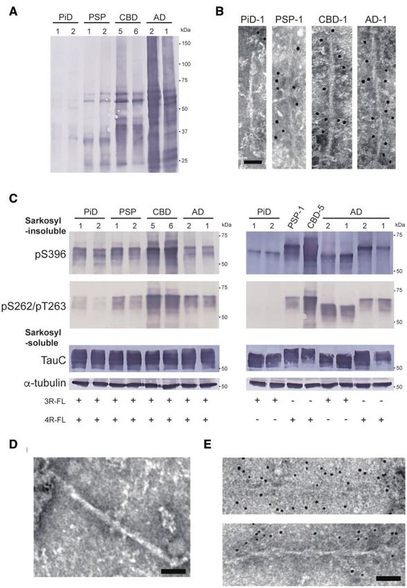
Prion-like amplification of filamentous tau                                                      BRAIN 2021: 144; 2333–2348          | 2335

approved by the ethics committees of Tokyo Metropolitan                 Cell culture, transfection of plasmids and
Institute of Medical Science (18-9) and the University of Tokyo (30-    introduction of pathogenic proteins into cells
7). All methods were performed in accordance with the relevant
guidelines and regulations. All brain tissues used in this study        Human neuroblastoma SH-SY5Y cells were maintained as
were anonymized.                                                        described.37 Cells were cultured to 40–50% confluence in 6-well or
                                                                        12-well plates and transfected with plasmids using X-
                                                                        tremeGENETM 9 (Roche Life Science) according to the manufac-
Antibodies                                                              turer’s instructions. We used non-tagged human tau 3R1N, 4R1N,
                                                                        haemagglutinin (HA)-tagged human tau 3R1N, 4R1N, and FLAG-
Anti-tau antibodies used in this study were as follows: T46 (epi-
                                                                        tagged human tau 3R1N, 4R1N in the pCDNA3.1 vector. After
tope: 404–441; Thermo Fisher Scientific), pS396 (epitope: p-Ser-396;
                                                                        transfection of plasmids, cells were incubated for 6–8 h, and patho-
Calbiochem), RD3 (epitope: 209–224; Millipore), Anti-4R (epitope:
                                                                        genic tau seeds (2 ll for 6-well plate or 1–2ll for 12-well plate)
275–291; Cosmo Bio), TauC (epitope: 429–441; Cosmo Bio), AT8 (epi-
                                                                        were introduced using MultiFectam (Promega) according to the
tope: p-Ser-202 and p-Thr-205; Thermo Fisher Scientific), tau 360–
                                                                        manufacturer’s instructions. Transfected cells were incubated for
380 (epitope: 360–380; Cosmo Bio), pS262/pT263 (epitope: p-Ser-262
                                                                        3 days.

                                                                                                                                                 Downloaded from https://academic.oup.com/brain/article/144/8/2333/6164963 by guest on 25 November 2021
and p-Thr-263; abcam). Polyclonal anti-a-synuclein (a-syn) 131–
140 antibody was obtained from Cosmo Bio. Monoclonal anti-a-
tubulin antibody (T9028), monoclonal and polyclonal anti-HA anti-
bodies (H3663 and H6908), and polyclonal anti-FLAG antibody             Preparation of sarkosyl-insoluble fractions from
(F7425) were obtained from Sigma.                                       transfected cells and immunoblotting
                                                                        Transfected SH-SY5Y cells were collected and extracted with 1 ml
Preparation of sarkosyl-insoluble fractions from                        for a 6-well plate or 0.5 ml for a 12-well plate of 1% sarkosyl in A68
                                                                        buffer (10 mM Tris-HCl pH 7.5 containing 10% sucrose, 0.8 M NaCl,
patients’ brains
                                                                        1 mM EGTA). Cell extracts were sonicated for 15 s. After incubation
Sarkosyl-insoluble fractions were prepared from patients’ brains        for 30 min at 37 C, cell extracts were ultracentrifuged at 113 000g
as described25 and resuspended in saline by sonication for 15 s and     for 20 min at 25 C. The supernatants were removed and collected
used for electron microscopy or introduction into SH-SY5Y cells.        as sarkosyl-soluble fractions, then the pellets were washed with
For immunoblotting, sarkosyl-insoluble fractions were added to          30 mM Tris-HCl (pH 7.5) and ultracentrifuged as before. The result-
SDS-sample buffer and boiled for 3 min. The samples were sepa-          ing pellets were collected as sarkosyl-insoluble fractions, resus-
rated on 4–20% gradient polyacrylamide gel (Wako) and immuno-           pended in 30 mM Tris-HCl (pH 7.5) and sonicated for 15 s. These
blotting was performed as described.35 For immunodepletion of           fractions were used for immunoelectron microscopy and as seeds
tau from the sarkosyl-insoluble fraction, a mixture of AT8, tau         for multiple passages of insoluble tau in SH-SY5Y cells. Sarkosyl-
360–380 and TauC antibodies or anti-a-syn 131–140 antibody was          insoluble and soluble fractions were added to SDS-sample buffer
coupled to PierceTM NHS-Activated Magnetic Beads (Thermo                and boiled for 3 min, and immunoblotting was performed.
Scientific) according to the manufacturer’s instructions. The sarko-    Monoclonal anti-a-tubulin was used to obtain a loading control.
syl-insoluble fraction suspended in Tris-buffered saline with           The protein concentrations of samples were determined with a
Tween-20 was incubated with tau antibody-coupled beads or a-            Pierce BCA Protein Assay Kit (Thermo Fisher Scientific). All experi-
syn antibody-coupled beads at 4 C overnight. The unbound               ments were performed at least three times. The band intensities of
sample was further incubated with new antibody-coupled beads            immunoblots were quantified using ImageQuant TL (cytiva) and
as before. The resulting supernatant was collected as an immuno-        were analysed using Prism software (GraphPad Software).
depleted sample and used for introduction into cells. a-Syn-
depleted samples were used as a control. Trypsin treatment of the
sarkosyl-insoluble fraction was performed as described.25 Trypsin
                                                                        Confocal immunofluorescence microscopy
was added at a final concentration of 0.1 mg/ml and incubated at
37 C for 30 min, and then the digestion was stopping by boiling.       Transfection of plasmids and introduction of pathogenic tau seeds
                                                                        were conducted as described above, using SH-SY5Y cells grown on
                                                                        coverslips. After incubation for 3 days, cells were fixed with 4%
Quantification of total tau by ELISA                                    paraformaldehyde and treated with AT8 and TauC antibodies and
The concentrations of total tau in sarkosyl-insoluble fractions         the secondary antibodies (anti-mouse IgG-conjugated Alexa-488
extracted from human brain samples were determined by sand-             and anti-rabbit IgG-conjugated Alexa-568, Invitrogen) as
wich ELISA. Wako human tau ELISA kit was purchased from                 described.37 The cells were mounted and analysed using a LSM780
FUJIFILM and used according to the manufacturer’s instructions.         confocal laser microscope (Carl Zeiss).

Electron microscopy                                                     Statistical analysis
Sarkosyl-insoluble fractions extracted from Alzheimer’s disease,
                                                                        Welch’s modified t-test was performed for Fig. 2B. Unpaired t-test
PiD, PSP, CBD and FTDP-17T patients’ brains were dropped onto
                                                                        was carried out for Supplementary Fig. 3D and G. In all cases, a P-
carbon-coated 300-mesh copper grids (Nissin EM) and negatively
                                                                        value 5 0.05 was regarded as statistically significant. Statistical
stained as described.36 For immunostaining, sarkosyl-insoluble
                                                                        analyses were performed using Prism software (GraphPad
fractions extracted from transfected SH-SY5Y cells or patients’
                                                                        Software).
brains were dropped onto grids and dried. The grids were immu-
nostained with appropriate primary antibodies (1:50–100) and sec-
ondary antibodies conjugated to 10 nm gold particles (BBI
Solutions, 1:50), 6 nm gold particles (abcam, 1:50) or 5 nm gold par-
                                                                        Data availability
ticles (Cytodiagnostics, 1:50) as described.36 Electron micrograph      All raw data used for figure generation in this study can be
images were recorded with a JEOL JEM-1400 electron microscope.          obtained by contacting the corresponding author.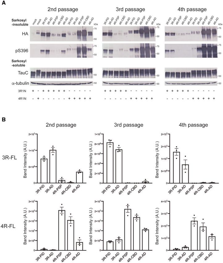
2336    | BRAIN 2021: 144; 2333–2348                                                                                   A. Tarutani et al.

Results                                                                which shows the highest expression level in the human adult
                                                                       brain.39 Three days after the introduction of the seeds, the sarko-
Biochemical and ultrastructural characterization of                    syl-insoluble fractions were extracted from transfected cells, and
patient-derived tau aggregates                                         the accumulation of insoluble tau was evaluated by immunoblot-
                                                                       ting using anti-HA and pS396 antibodies (Fig. 2A and B).
First, we prepared the sarkosyl-insoluble fractions from the brains
                                                                       Introduction of pathogenic tau seeds into SH-SY5Y cells without
of patients neuropathologically diagnosed as Alzheimer’s disease,
                                                                       transient expression of tau did not induce seeded accumulation of
PiD, PSP and CBD, and investigated the biochemical and ultrastruc-
                                                                       insoluble tau, and the added seeds themselves were also not
tural properties of tau aggregates contained in these fractions
                                                                       detected (Supplementary Fig. 2A). Alzheimer’s disease, PiD, PSP
(Supplementary Table 1). Immunoblot analysis of sarkosyl-insol-
                                                                       and CBD-derived tau seeds caused strain-dependent seeded tau
uble fractions with T46, which recognizes the C-terminal tau (404–
                                                                       aggregation in SH-SY5Y cells (Fig. 2A, B and Supplementary Fig.
441), and pS396 tau antibodies showed that different tau isoforms
                                                                       2B). PiD-tau seeds induced seeded aggregation of 3R tau in cells
accumulated in each tauopathy: there were two major bands at 60
                                                                       expressing 3R-FL, while PSP- and CBD-tau seeds caused seeded ag-
and 64 kDa in PiD, two major bands at 64 and 68 kDa in PSP and
                                                                       gregation of 4R tau only in cells expressing 4R-FL. Insoluble tau
CBD, and three major bands at 60, 64 and 68 kDa in Alzheimer’s

                                                                                                                                               Downloaded from https://academic.oup.com/brain/article/144/8/2333/6164963 by guest on 25 November 2021
                                                                       accumulated in cells expressing 3R-FL or 4R-FL after introduction
disease, which represent hyperphosphorylated full-length tau pro-
                                                                       of Alzheimer’s disease (AD)-tau seeds. Huntington’s disease seeds
teins (Fig. 1A and Supplementary Fig. 1). As reported previous-
                                                                       did not cause seeded aggregation of either 3R-FL or 4R-FL. Such
ly,24,25 disease-specific tau-CTFs, 21, 34 and 39 kDa tau in PiD, 22
                                                                       isoform-specific seeded tau aggregation induced by tau from
and 33 kDa tau in PSP, 22, 37–40 and 43 kDa tau in CBD and 19, 22,
                                                                       human tauopathies was also observed in SH-SY5Y cells expressing
25, 30, 36 and 40 kDa tau in Alzheimer’s disease were also detected
                                                                       non-tagged human full-length tau (Supplementary Fig. 2C).
(Fig. 1A and Supplementary Fig. 1). We also confirmed the identi-
                                                                       Furthermore, the amount of accumulated insoluble full-length tau
ties of the tau isoforms accumulated in each case using tau iso-
                                                                       generated by the addition of patient-derived tau seeds varied de-
form-specific antibodies, RD3 and anti-4R (Fig. 1A and
                                                                       pending on the strain. The seeding activity of AD-tau seeds for 3R-
Supplementary Fig. 1).38 Abnormal tau was not detected in the sar-
                                                                       FL was higher than that of PiD-tau seeds (Fig. 2B). In addition, CBD-
kosyl-insoluble fractions extracted from two cases with
                                                                       tau seeds tended to exhibit higher seeding activity towards 4R-FL
Huntington’s disease, and these cases were used as a control not
                                                                       as compared to PSP- and AD-tau seeds (Fig. 2B). These differences
containing abnormal tau in subsequent experiments (Fig. 1A).
                                                                       were independent of the tau concentration in seeds
    Furthermore, the sarkosyl-insoluble fractions were negatively
                                                                       (Supplementary Table 2). Unexpectedly, PiD-tau seeds from the
stained and observed by electron microscopy (Fig. 1B). In PiD cases,
                                                                       PiD-2 case caused isoform-independent tau aggregation. Although
straight filaments with 13–17 nm diameter and twisted filaments
                                                                       immunoblotting with anti-4R (Fig. 1A) did not detect 4R tau in the
with 129.8 [±5.0, mean standard deviation (SD)] nm periodicity
                                                                       PiD-2 case, PiD-tau seeds derived from this case induced the accu-
were observed. In PSP cases, thin filaments with wide regions of
                                                                       mulation of insoluble tau not only in cells expressing 3R-FL, but
14.9 (±1.7) nm diameter and narrow regions of 7 nm diameter were
                                                                       also in cells expressing 4R-FL (Fig. 2A and B). To investigate
loosely twisted with 108.7 (±4.5) nm periodicity. Tau aggregates
                                                                       whether this isoform-independent tau aggregation was caused by
derived from CBD cases are characterized by wider filaments than
                                                                       non-tau contamination in the sarkosyl-insoluble fraction, we fur-
those in PSP, and CBD filaments with wide regions of 28.9 (±3.2)
                                                                       ther purified this fraction. CBB staining of purified samples
nm diameter and narrow regions of 8.5 nm diameter were twisted
                                                                       obtained by resuspension and additional ultracentrifugation
with 138.9 (±11.7) nm periodicity. Ribbon-like filaments with wide
                                                                       showed a decrease in the a- and b-tubulin bands at 50 kDa, one of
regions of 25.2 (±6.7) nm diameter and narrow regions of 7 nm
                                                                       the major contaminants in the insoluble fraction extracted from
diameter were twisted with 236.5 (±21.2) nm periodicity in the case
                                                                       human brains (Supplementary Fig. 3A).40 Therefore, the unpurified
of FTDP-17T with intron 10 mutation + 16, which increases the ex-
                                                                       PiD-1 and PiD-2 seeds or the purified PiD-2 seeds, whose total tau
pression of 4R tau, indicating the diversity of tau filament struc-
                                                                       concentration was adjusted to 2 ng/ll, were introduced into cells
tures within 4R tauopathy. In Alzheimer’s disease cases, straight
                                                                       expressing HA-tagged 3R-FL or 4R-FL and accumulation of insol-
filaments with 14.4 (±1.6) nm diameter and paired helical filaments
                                                                       uble tau was evaluated. Although the same amount of abnormal
(PHFs) with wide regions of 19.3 (±2.1) nm diameter and narrow
                                                                       tau was introduced, the addition of the purified PiD-2 seeds signifi-
regions of 10 nm diameter, which were twisted with 81.7 (±6.6) nm
                                                                       cantly reduced the accumulation of 4R tau compared to the un-
periodicity, were observed. These ultrastructural properties of tau
                                                                       purified PiD-2 seeds (Supplementary Fig. 3B–D). These results
aggregates derived from human tauopathies are consistent with
                                                                       suggest that the accumulation of insoluble 4R tau induced by the
previous electron microscopy observations of patient-derived tau
                                                                       unpurified PiD-2 seeds was caused by some brain-derived contam-
filaments.26,27 These results indicate that tau aggregates extracted
                                                                       inant(s), not by cross-seeding. Furthermore, tau-depleted samples
from the Alzheimer’s disease, PiD, PSP, and CBD cases used in this
                                                                       from the sarkosyl-insoluble fraction were introduced into cells
study are distinct tau strains having different biochemical and
                                                                       expressing HA-tagged 3R-FL or 4R-FL (Supplementary Fig. 3E–G).
ultrastructural properties.
                                                                       Accumulation of insoluble tau in cells introduced with tau-
                                                                       depleted samples was significantly reduced compared to cells
                                                                       introduced with control samples, suggesting that insoluble tau
Seeded tau aggregation induced by patient-derived
                                                                       contained in the sarkosyl-insoluble fraction induced tau aggrega-
tau strains in SH-SY5Y cells                                           tion (Supplementary Fig. 3F and G). In addition, the C-terminal
To characterize the prion-like properties of tau strains derived       banding pattern of insoluble tau extracted from transfected cells
from human tauopathies, we investigated cellular seeded tau ag-        was compared with that of patient-derived insoluble fractions
gregation induced by the introduction of pathogenic tau seeds.         used as seeds (Fig. 2C). Immunoblotting with T46 and pS396 anti-
The sarkosyl-insoluble fractions derived from Alzheimer’s disease,     bodies of sarkosyl-insoluble fractions extracted from transfected
PiD, PSP, CBD and Huntington’s disease patients’ brains were           cells detected CTFs of 21 kDa in cells with introduced PiD-tau seeds
introduced into SH-SY5Y cells transiently expressing HA-tagged         and 22, 33 and 37 kDa in cells with PSP- and CBD-tau seeds.
human full-length 3R tau (3R-FL) and 4R tau (4R-FL) without patho-     Although these CTFs resembled patient-derived CTFs, CTFs
genic mutations. We used the 1N isoform with exon 2 inserted,          derived from transfected cells with introduced PSP- and CBD-tau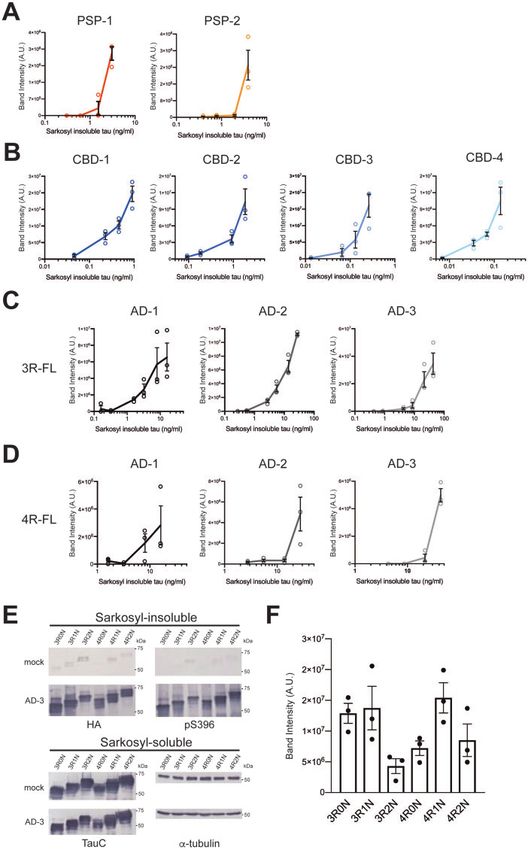
Prion-like amplification of filamentous tau BRAIN 2021: 144; 2333–2348 | 2337
Downloaded from https://academic.oup.com/brain/article/144/8/2333/6164963 by guest on 25 November 2021
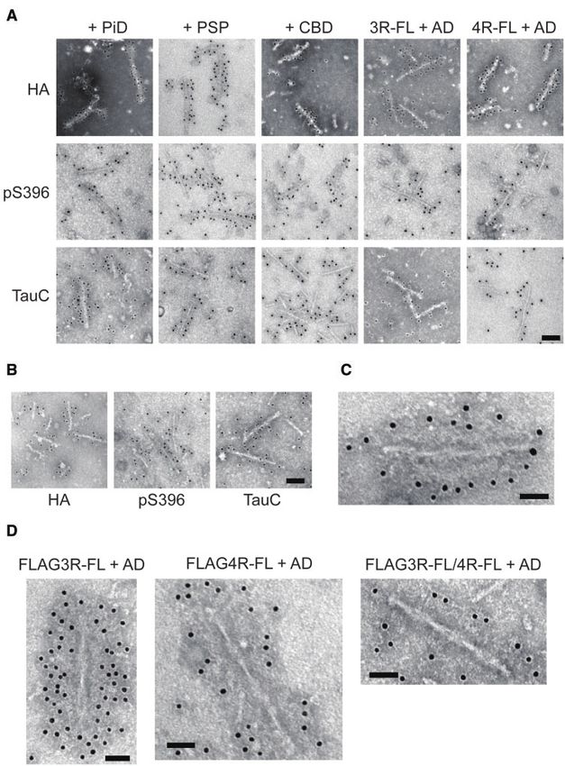
Figure 1 Biochemical and ultrastructural characterization of abnormal tau extracted from human tauopathies. (A) Immunoblot analyses of sarkosyl-
insoluble fractions prepared from brains of tauopathy and Huntington’s disease patients. Sarkosyl-insoluble full-length tau (60, 64 and 68 kDa) and
CTFs were detected with T46 (top left) and pS396 (top right) antibodies. 3R tau and 4R tau were detected with RD3 (bottom left) and anti-4R (bottom left)
antibodies, respectively. Full-length blots are presented in the Supplementary material. (B) Electron microscopy of sarkosyl-insoluble fractions
extracted from PiD, PSP, CBD, Alzheimer’s disease and FTDP-17T cases. Scale bar = 50 nm.
seeds were not clearly distinguishable. In the case of introduction pathological diagnosis of tauopathies, showed that tau aggregates
of AD-tau seeds, CTFs of 20, 22, 30, 36, 40 kDa were detected in in- were formed in TauC-positive cells with introduced patient-
soluble fractions extracted from cells expressing 3R tau, and CTFs derived tau seeds, consistent with the accumulation of insoluble
of 20, 22, 25, 36, 40 kDa were detected in insoluble fractions tau detected by immunoblotting (Fig. 2D). On the other hand, AT8-
extracted from cells expressing 4R tau. Tau-CTFs resembling pa- positive tau was not detected in cells without transient expression
tient-derived tau-CTFs were also detected in insoluble tau accu- of tau, mock cells co-expressing both 3R tau and 4R tau, or trans-
mulated in transfected cells expressing non-tagged 3R-FL or 4R-FL fected cells without transient expression of tau with introduced
(Supplementary Fig. 2D). These results indicate that the abnormal AD-tau seeds (Supplementary Fig. 4). Thus, the sarkosyl-insoluble
tau amplified in SH-SY5Y cells inherits the ultrastructural proper- fractions extracted from the brains of patients with tauopathies
ties of patient-derived tau seeds. Immunohistological staining caused strain-dependent seeded aggregation of full-length tau
with AT8, which is the most frequently used antibody in the substrates, except for the insoluble fraction derived from the PiD-2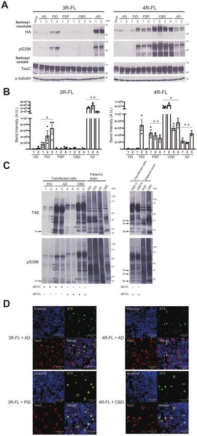
2338    | BRAIN 2021: 144; 2333–2348                                                                                            A. Tarutani et al.

                                                                               case. These results indicate that disease-specific amplification of
                                                                               tau seeds that occurs in the brains of patients with tauopathies
                                                                               was reproduced in cultured cells, and distinct patient-derived
                                                                               strains exhibited a diversity of prion-like properties, including iso-
                                                                               form specificity and seeding activity. To investigate the effect of N-
                                                                               terminal and C-terminal tau regions (fuzzy coat) that are not a part
                                                                               of the fibrous core on seeded aggregation, the sarkosyl-insoluble
                                                                               fractions extracted from the patient’s brain were treated with tryp-
                                                                               sin and the resulting trypsinized tau seeds were introduced into
                                                                               cells (Fig. 3). The trypsinized tau seeds induced seeded aggregation
                                                                               similarly to non-trypsinized tau seeds, indicating that the trypsin-
                                                                               resistant fibrous core contributes to the prion-like properties (Fig.
                                                                               3B).25

                                                                                                                                                        Downloaded from https://academic.oup.com/brain/article/144/8/2333/6164963 by guest on 25 November 2021
                                                                               Isoform-specific seeded tau aggregation in the
                                                                               presence of both 3R tau and 4R tau substrates
                                                                               We next investigated whether strain-dependent seeded tau aggre-
                                                                               gation induced by the introduction of patient-derived tau seeds
                                                                               occurs in the presence of both 3R tau and 4R tau substrates.
                                                                               Pathogenic tau seeds were introduced into SH-SY5Y cells co-
                                                                               expressing HA-tagged 3R-FL and FLAG-tagged 4R-FL, and seeded
                                                                               tau aggregation was evaluated (Fig. 4). Accumulation of insoluble
                                                                               3R tau detected with anti-HA and RD3 antibodies was observed in
                                                                               cells with introduced PiD- and AD-tau seeds. Insoluble 4R tau was
                                                                               detected with anti-FLAG and anti-4R antibodies in cells with intro-
                                                                               duced PSP-, CBD- and AD-tau seeds (Fig. 4A). Evaluation of the in-
                                                                               soluble fractions extracted from transfected cells with RD3 and
                                                                               anti-4R antibodies also showed that the tau seeds derived from
                                                                               human tauopathies caused isoform-specific seeded tau aggrega-
                                                                               tion in the presence of both 3R tau and 4R tau substrates, except
                                                                               for the accumulation of insoluble 4R tau induced by the unpurified
                                                                               PiD-2 seeds (Fig. 4B and Supplementary Fig. 5). These results indi-
                                                                               cate that pathogenic tau seeds recruit tau isoforms for seeded ag-
                                                                               gregation in a substrate-selective manner, that is, the
                                                                               conformations of patient-derived tau strains determine which tau
                                                                               isoforms are selectively accumulated.

                                                                               Evaluation of prion-like seeding activities of patient-
                                                                               derived tau strains
                                                                               Further, we examined whether pathogenic tau seeds derived from
                                                                               human tauopathies exhibit the seeding activities that characterize
                                                                               the tau strains. Total tau concentration contained in the sarkosyl-
                                                                               insoluble fraction was determined by sandwich ELISA using anti-
Figure 2 Seeded tau aggregation induced by patient-derived tau strains         body that recognizes C-terminal tau as a capture antibody. Serial
in SH-SY5Y cells expressing full-length tau. (A) Sarkosyl-insoluble frac-      dilutions of PSP- and CBD-tau seeds were introduced into SH-SY5Y
tions extracted from patients’ brains (1 ll) were introduced into SH-SY5Y      cells expressing HA-tagged 4R-FL, and the seeding activity was
cells transiently expressing HA-tagged wild-type human tau 3R1N (left) or
                                                                               evaluated (Fig. 5A, B and Supplementary Fig. 6A and B). PSP-tau
4R1N (right). Immunoblot analysis of sarkosyl-insoluble fractions and sar-
kosyl-soluble fractions extracted from mock-transfected cells, and cells
                                                                               seeds derived from PSP-1 and PSP-2 cases showed high seeding ac-
transfected with sarkosyl-insoluble fractions from two Huntington’s dis-       tivity at total tau concentrations in the seeds of 3.8 ng/ml and
ease cases, two PiD cases, two PSP cases, four CBD cases and two               5.8 ng/ml, respectively, but the seeds had almost no seeding activ-
Alzheimer’s disease cases. Insoluble tau was detected with anti-HA and         ity at the tau concentration of 1 ng/ml or less (Fig. 5A and
pS396 antibodies. Total tau was detected with TauC antibody. The tau           Supplementary Fig. 6A). On the other hand, CBD-tau seeds derived
concentrations of sarkosyl-insoluble fractions derived from human
                                                                               from Cases CBD-1–4 showed high seeding activity at 1 ng/ml or
brains are shown in Supplementary Table 2. Full-length blots are pre-
sented in the Supplementary material. (B) Quantification of the band
                                                                               higher concentration, while the seeding activity decreased at
intensities of the immunoblots with anti-HA antibody shown in A and            0.1 ng/ml or lower concentration (Fig. 5B and Supplementary Fig.
Supplementary Fig. 2B. The results are expressed as means ± standard           6B). CBD-tau seeds derived from the CBD-4 case showed the high-
error of the mean (SEM) (n = 3). *P 5 0.01; **P 5 0.001; Welch’s modified t-   est seeding activity, being effective even at 0.1 ng/ml, while the ac-
test against the value of Huntington’s disease. (C) Immunoblot analyses        tivity decreased at 0.04 ng/ml or less (Fig. 5B). Thus, tau seeds
of sarkosyl-insoluble fractions prepared from transfected SH-SY5Y cells
                                                                               derived from PSP and CBD cases vary by 10- to 100-fold in their
and patients’ brains. C-Terminal tau fragments were detected with T46
                                                                               prion-like seeding activity. Furthermore, we prepared serial dilu-
(top) and pS396 (bottom) antibodies. Full-length blots are presented in the
Supplementary material. (D) SH-SY5Y cells introduced with AD-, PiD-            tions of AD-tau seeds and introduced them into SH-SY5Y cells
and CBD-tau seeds were fixed and immunostained with AT8 (green) and            expressing HA-tagged 3R-FL or 4R-FL (Fig. 5C, D and
TauC (red) antibodies. Scale bar = 100 lm.                                     Supplementary Fig. 6C and D). In the presence of 3R tau substrate,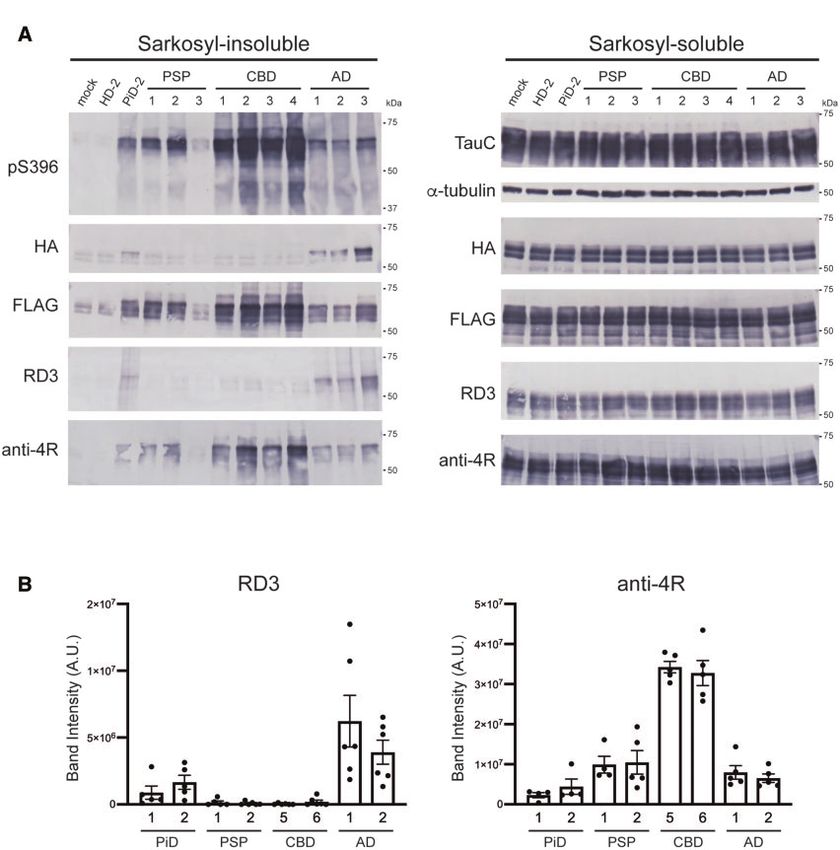
Prion-like amplification of filamentous tau                                                              BRAIN 2021: 144; 2333–2348           | 2339

                                                                                                                                                           Downloaded from https://academic.oup.com/brain/article/144/8/2333/6164963 by guest on 25 November 2021
Figure 3 Prion-like seeding activities of trypsin-treated insoluble fractions of tauopathy cases in SH-SY5Y cells. (A) Immunoblot analyses of the sarko-
syl-insoluble fractions before and after trypsin treatment. Full-length tau and C-terminal tau fragments were detected with T46 antibody. Trypsin-re-
sistant tau bands were detected with tau 360–380 antibody. Full-length blots are presented in the Supplementary material. (B) The untreated and
trypsin-treated insoluble fractions were introduced into SH-SY5Y cells transiently expressing HA-tagged human tau 3R1N or 4R1N. Immunoblot anal-
yses of sarkosyl-insoluble fractions and sarkosyl-soluble fractions extracted from mock-transfected cells, and cells with introduced untreated or
trypsin-treated tau seeds. Insoluble tau was detected with anti-HA and pS396 antibodies. Total tau was detected with TauC antibody. Full-length
blots are presented in the Supplementary material.

AD-tau seeds derived from Cases AD-1–3 showed high seeding ac-                with introduced AD-tau seeds of the same tau concentration (Figs
tivity at 1.6 ng/ml, 2.76 ng/ml and 8.62 ng/ml, respectively, while           2A, B and 5C). Furthermore, addition of AD-tau seeds derived from
the seeding activity was completely lost at 0.32 ng/ml, 0.55 ng/ml            the AD-3 case to SH-SY5Y cells expressing each of six human tau
and 4.31 ng/ml, respectively (Fig. 5C and Supplementary Fig. 6C). In          isoforms showed that AD-tau seeds have the ability to induce
the presence of 4R tau substrate, AD-tau seeds derived from the               seeded aggregation of all tau isoforms (Fig. 5E and F). Similarly, we
Cases AD-1–3 showed high seeding activity at 19.2 ng/ml, 27.57 ng/            confirmed that PiD-tau seeds derived from the PiD-2 case and
ml and 43.11 ng/ml or higher, respectively, and the seeding activity          CBD-tau seeds derived from the CBD-4 case induced seeded aggre-
was lost below 3.84 ng/ml, 13.78 ng/ml and 8.6 ng/ml, respectively            gation of the three 3R tau isoforms and three 4R tau isoforms, re-
(Fig. 5D and Supplementary Fig. 6D). Prion-like seeding activity of           spectively (Supplementary Fig. 7A and B). These results suggest
AD-tau seeds towards 3R-FL was more than 10 times higher than                 that different conformations of patient-derived tau strains may
that towards 4R-FL in the three Alzheimer’s disease cases and con-            exert distinct prion-like seeding activities. In addition, although
sequently the seeding activity of AD-tau seeds towards 4R tau sub-            the accumulation ratio of insoluble 3R tau and 4R tau induced
strate was lower than that of PSP-tau seeds. For PiD-tau seeds, the           by AD-tau seeds was almost the same in the presence of both 3R
total tau concentrations used in this study are the detection limits          tau and 4R tau (Fig. 4A and Supplementary Fig. 8B), AD-tau seeds
for induction of seeded aggregation, and the amount of insoluble              exhibited higher seeding activity in the presence of only 3R tau
tau accumulated in cells was much less than that observed in cells            substrate than in the presence of only 4R tau substrate.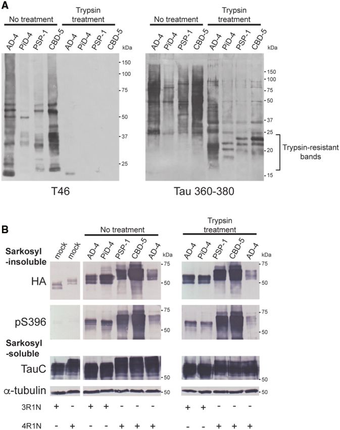
2340    | BRAIN 2021: 144; 2333–2348                                                                                             A. Tarutani et al.

                                                                                                                                                         Downloaded from https://academic.oup.com/brain/article/144/8/2333/6164963 by guest on 25 November 2021
Figure 4 Seeded tau aggregation induced by patient-derived tau strains in SH-SY5Y cells co-expressing 3R tau and 4R tau. (A) Sarkosyl-insoluble frac-
tions extracted from patients’ brains (1 ll) were introduced into SH-SY5Y cells transiently co-expressing HA-tagged human tau 3R1N and FLAG-
tagged human tau 4R1N. Immunoblot analysis of sarkosyl-insoluble fractions (left) and sarkosyl-soluble fractions (right) extracted from mock-trans-
fected cells, and cells with introduced sarkosyl-insoluble fractions from a Huntington’s disease case, a PiD case, three PSP cases, four CBD cases and
three Alzheimer’s disease cases. Insoluble tau was detected with pS396, anti-HA, anti-FLAG, RD3 and anti-4R antibodies. Total tau was detected with
TauC, anti-HA, anti-FLAG, RD3 and anti-4R antibodies. The tau concentrations of pathogenic tau seeds derived from human brains are shown in
Supplementary Table 2. Full-length blots are presented in the Supplementary material. (B) Quantification of insoluble tau accumulated in transfected
SH-SY5Y cells co-expressing human tau 3R1N and 4R1N with patient-derived tau seeds. The band intensities of the immunoblots with RD3 (left) and
anti-4R (right) antibodies shown in Supplementary Fig. 5 were evaluated. The results are expressed as means ± SEM (n = 4–6).

Template-dependent tau filament formation in
                                                                             4.6 (±0.8) nm and 5.9 (±0.6) nm diameter were observed in insoluble
transfected cells with patient-derived tau strains                           fractions from SH-SY5Y cells with introduced PiD-, PSP- and CBD-
We also examined the prion-like properties of tau aggregates                 tau seeds, respectively (Fig. 6A and Supplementary Fig. 8A). Tau fil-
amplified in SH-SY5Y cells with introduced patient-derived tau               aments extracted from cells with introduced PiD- and PSP-tau
strains. To clarify the ultrastructural features of tau aggregates           seeds resembled those extracted from the patients’ brains, but tau
accumulated in SH-SY5Y cells, immunolabelling of the sarkosyl-               filaments from cells with introduced CBD-tau seeds were thinner
insoluble fractions extracted from transfected cells using anti-HA,          than those in the brain. PHF-like structures were observed in
anti-FLAG, TauC and pS396 antibodies was performed. Abundant                 transfected cells expressing either 3R-FL or 4R-FL with AD-tau
amyloid-like filamentous structures labelled with anti-HA, pS396             seeds. These filaments showed wide regions of 17.9 (±2.6) nm and
and TauC antibodies were observed in the insoluble fractions                 19.0 (±1.8) nm diameter and narrow regions of 6.9 (±1.1) nm and 6.2
extracted from transfected cells expressing either HA-tagged 3R-FL           (±0.6) nm diameter, respectively, and were twisted with 94.9 (±9.8)
or 4R-FL with introduced PiD-, PSP-, CBD- and AD-tau seeds (Fig.             nm and 93.8 (±10.0) nm periodicity (Fig. 6A and Supplementary Fig.
6A and Supplementary Fig. 8A). HA-labelled filamentous struc-                8A). PHF-like filaments positive for anti-HA, pS396 and TauC anti-
tures with wide regions of 14.2 (±2.0, mean SD) nm, 13.0 (±1.2) nm,          bodies, which contained wide regions of 17.0 (±2.9) nm diameter
and 14.0 (±2.0) nm diameter and narrow regions of 6.7 (±1.2) nm,             and narrow regions of 7.0 (±1.3) nm diameter and were twisted
Prion-like amplification of filamentous tau BRAIN 2021: 144; 2333–2348 | 2341
Downloaded from https://academic.oup.com/brain/article/144/8/2333/6164963 by guest on 25 November 2021
Figure 5 Evaluation of prion-like seeding properties of tau aggregates derived from PSP, CBD and Alzheimer’s disease cases in SH-SY5Y cells. (A)
Serial dilutions of sarkosyl-insoluble fractions prepared from 2 PSP cases (1 ll) were introduced into SH-SY5Y cells transiently expressing HA-tagged
human tau 4R1N. The band intensities of the immunoblots with anti-HA antibody shown in Supplementary Fig. 6A were evaluated. (B) Serial dilu-
tions of sarkosyl-insoluble fractions prepared from four CBD cases (1 ll) were introduced into SH-SY5Y cells transiently expressing HA-tagged human
tau 4R1N. The band intensities of the immunoblots with anti-HA antibody shown in Supplementary Fig. 6B were evaluated. The results are expressed
as means ± SEM (n = 3). (C) Serial dilutions of sarkosyl-insoluble fractions prepared from three Alzheimer’s disease cases (1 ll) were introduced into
SH-SY5Y cells transiently expressing HA-tagged human tau 3R1N. The band intensities of the immunoblots with anti-HA antibody shown in
Supplementary Fig. 6C were quantified. (D) Serial dilutions of sarkosyl-insoluble fractions prepared from three Alzheimer’s disease cases (1 ll) were
introduced into SH-SY5Y cells transiently expressing HA-tagged human tau 4R1N. The band intensities of the immunoblots shown in
Supplementary Fig. 6D were evaluated. (E) Sarkosyl-insoluble fractions prepared from the Case AD-3 (1 ll) were introduced into SH-SY5Y cells transi-
ently expressing HA-tagged human tau 3R0N, 3R1N, 3R2N, 4R0N, 4R1N and 4R2N, respectively. Immunoblot analysis of sarkosyl-insoluble fractions
(top) and sarkosyl-soluble fractions (bottom) extracted from mock-transfected cells, and cells introduced with sarkosyl-insoluble fractions from Case
AD-3. Insoluble tau was detected with pS396 and anti-HA antibodies. Total tau was detected with TauC antibody. Full-length blots are presented in
the Supplementary material. (F) Quantification of the band intensities of the immunoblots with anti-HA antibody shown in E. The results are
expressed as means ± SEM (n = 3).2342 | BRAIN 2021: 144; 2333–2348 A. Tarutani et al.
Downloaded from https://academic.oup.com/brain/article/144/8/2333/6164963 by guest on 25 November 2021
Figure 6 Electron microscopic analysis of insoluble fractions extracted from SH-SY5Y cells transfected with patient-derived tau strains. (A)
Immunoelectron microscopy of sarkosyl-insoluble fractions extracted from transfected cells expressing HA-tagged human tau 3R1N or 4R1N with
PiD-, PSP-, CBD- and AD-tau seeds. Electron micrographs show fibrous structures positive for anti-HA (top), pS396 (middle) and TauC (bottom) antibod-
ies, that were labelled with secondary antibody conjugated to 10 nm gold particles. Scale bar = 100 nm. (B) Immunoelectron microscopy of sarkosyl-
insoluble fractions extracted from transfected cells co-expressing HA-tagged human tau 3R1N and 4R1N with AD-tau seeds. Electron micrographs
show fibrous structures positive for anti-HA antibody (left), pS396 antibody (middle) and TauC (right), that were labelled with the secondary antibody
conjugated to 10 nm gold particles. Scale bar = 100 nm. (C) PHF-like tau filament positive for anti-HA antibody in sarkosyl-insoluble fractions
extracted from transfected cells co-expressing HA-tagged human tau 3R1N and 4R1N with AD-tau seeds was labelled with secondary antibody conju-
gated to 10 nm gold particles. Scale bar = 50 nm. (D) Immunoelectron microscopy of sarkosyl-insoluble fractions extracted from transfected cells
expressing FLAG-tagged human tau 3R1N (left), 4R1N (middle) and co-expressing FLAG-tagged human tau 3R1N and 4R1N (right) with AD-tau seeds.
PHF-like tau structures positive for anti-FLAG antibody were labelled with secondary antibody conjugated to 10 nm gold particles. Scale bar = 50 nm.
with 83.7 (±3.7) nm periodicity, were also observed in the insoluble not only in cells expressing HA-tagged tau, but also cells express-
fraction extracted from transfected cells expressing both HA- ing FLAG-tagged tau. Introduction of AD-tau seeds into cells
tagged 3R-FL and 4R-FL (Fig. 6B and C). These HA-labelled tau fila- expressing FLAG-tagged 3R-FL or 4R-FL, or both resulted in the ac-
ments are akin to PHFs observed in the brain of patients with cumulation of phosphorylated insoluble FLAG-tagged tau
Alzheimer’s disease, suggesting that AD-tau seeds could work as a (Supplementary Fig. 8B). Immunolabelling of these insoluble frac-
template to form PHF-like filaments in SH-SY5Y cells. tions revealed PHF-like filaments labelled with anti-FLAG antibody
Furthermore, strain-dependent seeded tau aggregation is induced (Fig. 6D and Supplementary Fig. 9C). PHF-like filaments were seenPrion-like amplification of filamentous tau BRAIN 2021: 144; 2333–2348 | 2343
in all cases of expression of FLAG-tagged tau, as observed in cells induced seeded aggregation of only 3R tau, and accumulation of
expressing HA-tagged tau. These fibrils showed wide regions of 4R tau was not detected (Fig. 8). This supports the idea that accu-
20.6 (±2.1, mean SD) nm, 17.5 (±2.6) nm and 22.4 (±2.1) diameter mulation of insoluble 4R tau induced by tau seeds derived from
and narrow regions of 6.4 (±0.7) nm, 6.3 (±1.8) nm and 7.9 (±1.2) nm the PiD-2 case shown in Figs 2–4 was due to contamination. In add-
diameter, respectively, and were twisted with 86.6 (±8.3) nm, 89.9 ition, the strain-specific tau aggregation induced by cell-derived
(±5.0) nm and 88.2 (±8.9) nm periodicity. Similarly, PiD-, PSP-, and insoluble tau was maintained after two additional passages (Fig.
CBD-tau seeds were added to cells expressing either FLAG-tagged 8). These results indicate that the tau seeds amplified in the cells
3R-FL or 4R-FL, and isoform-dependent seeded tau aggregation inherit the prion-like seeding activity of the original seeds, and the
was detected, as observed in cells expressing HA-tagged tau isoform specificity is also retained in subsequent seeding.
(Supplementary Fig. 9A). No amyloid-like fibrous structure was
observed in the insoluble fraction extracted from cells with intro-
duced Huntington’s disease seeds (Supplementary Fig. 9B). FLAG- Discussion
labelled amyloid-like filaments with wide regions of 14.7 (±1.9) nm, We investigated seeded tau aggregation caused by tau strains
13.8 (±1.3) nm and 14.1 (±1.1) nm diameter and narrow regions of derived from the brains of patients with tauopathy and the prion-
Downloaded from https://academic.oup.com/brain/article/144/8/2333/6164963 by guest on 25 November 2021
6.5 (±0.7) nm, 5.4 (±0.5) nm and 6.5 (±0.5) nm diameter were like properties of patient-derived tau strains in cellular model.
observed in cells with introduced PiD-, PSP- and CBD-tau seeds, re-
Pathogenic tau seeds induced disease-specific seeded tau aggrega-
spectively (Supplementary Fig. 9D). These results indicate that pa-
tion and filament formation in SH-SY5Y cells. The 3R and 4R tau
tient-derived tau seeds act as a template, and intracellular HA- or isoforms, which form the frameworks of tau filaments accumu-
FLAG-tagged tau forms amyloid-like filaments similar to those lated in the brains of patients with tauopathies, are key determi-
derived from patients’ brains, which accumulate in the cells. nants of the neuropathology. PiD-, PSP- and CBD-tau seeds used in
this study induced isoform-dependent tau aggregation in SH-SY5Y
Absence of phosphorylation at Ser262 of abnormal cells expressing 3R-FL or 4R-FL individually, or co-expressing both
3R-FL and 4R-FL (Figs 2A, B and 4). This isoform-dependent tau ag-
tau amplified in transfected cells with PiD-tau seeds
gregation is also reproduced in SH-SY5Y cells with introduced syn-
The tau aggregates derived from the brains of tauopathy patients thetic 3R1N and 4R1N fibrils.43 On the other hand, AD-tau seeds
contain diverse post-translational modifications. It has been composed of six tau isoforms recruited both 3R tau and 4R tau into
reported that Ser262 is not phosphorylated in abnormal tau accu- abnormal tau, in contrast to a previous report that AD-tau seeds
mulated in the brain of PiD patients, probably because it is located were unable to infect HEK293 cells expressing either 3R tau or 4R
in the core structure of tau filaments, where it is inaccessible.41,42 tau (Figs 2A, B, 4 and 5C–F).44 These results indicate that the patho-
We performed immunoblotting and immunolabelling with pS262/ genic tau seeds extracted from patients’ brains retain structural in-
pT263 antibody of patient-derived tau seeds used in this study, formation as a template for tau substrates. Furthermore, template-
and confirmed that the PiD-tau seeds are pS262-negative (Fig. 7A dependent tau filament formation was observed in SH-SY5Y cells
and B). To examine whether insoluble tau extracted from cells with introduced patient-derived tau strains (Fig. 6 and
with introduced PiD-tau seeds inherits pS262-negativity, immuno- Supplementary Figs 8 and 9). Interestingly, AD-tau seeds showed
blotting and immunolabelling were performed using the insoluble different recruitment efficiencies for 3R-FL and 4R-FL substrates
fractions extracted from cells with introduced patient-derived tau (Fig. 5C and D). These differences in seeding activity may be due to
strains. Immunoblotting of insoluble fractions extracted from the weak interaction of 3R tau with microtubules compared to that
transfected cells showed that insoluble tau from cells treated with of 4R tau, which affects the rate and amount of intracellular accu-
PSP-, CBD-, and AD-tau seeds was pS262-positive, while that from mulation of insoluble tau (Fig. 2A and B).45 However, high seeding
cells treated with PiD-tau seeds was pS262-negative (Fig. 7C and activity of AD-tau seeds towards 3R tau has also been reported in
Supplementary Fig. 7C). Furthermore, PHF-like filaments derived an in vitro assay using real-time quaking-induced conversion (RT-
from cells with introduced AD-tau seeds were labelled with pS262/ QuIC).46 This assay is an experimental model that is not affected
pT263 antibody, while filament structures derived from cells with by binding to microtubules or other cellular environments, and the
introduced PiD-tau seeds were not (Fig. 7D and E). These results high recruitment efficiency of AD-tau seeds for 3R tau substrate in
suggested that the abnormal tau amplified and accumulated in our cellular model indicates that the difference in conformation
the transfected cells inherited the biochemical properties of the between normal 3R tau and 4R tau alters the interaction with AD-
patient-derived tau strains. tau seeds.
What drives the isoform specificity of pathogenic tau seeds
derived from human tauopathies? Atomic-level structural studies
Inheritance of prion-like seeding properties through
of tau filaments extracted from human tauopathies strongly sup-
multiple passages of insoluble tau in SH-SY5Y cells port the strain-dependent nature of seeded tau aggregation
Lastly, we investigated whether abnormal tau amplified in cells observed in our cellular model. Cryo-EM analysis revealed that tau
introduced with patient-derived tau seeds possesses seeding activ- filaments extracted from brains of Alzheimer’s disease, chronic
ity and inherits the prion-like properties of the original seeds. The traumatic encephalopathy, PiD and CBD patients had disease-spe-
insoluble fraction extracted from the transfected cells was intro- cific core structures, and patients in the same disease group
duced as secondary seeds into SH-SY5Y cells expressing HA- showed identical folds.42,47–50 The core region of Alzheimer’s dis-
tagged 3R-FL or 4R-FL. Secondary seeds derived from cells intro- ease filaments is G273-E380 in 3R tau and G304-E380 in 4R tau,
duced with patient-derived tau seeds caused the strain-dependent which covers the microtubule-binding domain including V306-
accumulation of insoluble tau in the same manner as the original K311, that is known to be important for tau aggregation.51 The
seeds (Fig. 8). Secondary PiD-tau (3R-PiD) and AD-tau (3R-AD) seeds structural evidence that the Alzheimer’s disease fold involves the
derived from cells expressing 3R-FL induced seeded aggregation of common parts of 3R tau and 4R tau may explain why PHF-like fila-
3R tau. Secondary PSP-tau (4R-PSP), CBD-tau (4R-CBD) and AD-tau ments induced by AD-tau seeds were observed not only in cells
(4R-AD) seeds derived from cells expressing 4R-FL involved only 4R expressing both 3R tau and 4R tau, but also in cells expressing 3R
tau in seeded aggregation. Intriguingly, secondary 3R-PiD seeds tau or 4R tau individually (Fig. 6C, D and Supplementary Fig. 9C). In
extracted from cells with introduced unpurified PiD-2 seeds vitro PHF-like filament formation of truncated tau C291-P397 in the2344 | BRAIN 2021: 144; 2333–2348 A. Tarutani et al.
Downloaded from https://academic.oup.com/brain/article/144/8/2333/6164963 by guest on 25 November 2021
Figure 7 Phosphorylation at Ser262 in insoluble tau extracted from SH-SY5Y cells transfected with patient-derived tau strains. (A) Immunoblot ana-
lysis with pS262/pT263 antibody of sarkosyl-insoluble fractions prepared from brains of tauopathy patients. Insoluble tau derived from PSP, CBD, and
Alzheimer’s disease cases shows pS262-positive bands, while insoluble tau derived from PiD cases is pS262-negative. Full-length blots are presented
in the Supplementary material. (B) Immunoelectron microscopy of sarkosyl-insoluble fractions extracted from PiD, PSP, CBD and Alzheimer’s disease
patients’ brains. Electron micrographs show fibrous structures derived from PiD negative for pSer262/pT263 antibody and fibrous structures derived
from PSP, CBD and Alzheimer’s disease positive for pSer262/pT263 antibody, labelled with secondary antibody conjugated to 10 nm gold particles.
Scale bar = 50 nm. (C) Sarkosyl-insoluble fractions extracted from patients’ brains (1 ll) were introduced into SH-SY5Y cells transiently co-expressing
HA-tagged human tau 3R1N and 4R1N (left), and expressing HA-tagged human tau 3R1N or 4R1N (right). Immunoblot analysis of sarkosyl-insoluble
fractions and sarkosyl-soluble fractions extracted from mock-transfected cells, and cells with introduced sarkosyl-insoluble fractions from two PiD
cases, two PSP cases, two CBD cases and two Alzheimer’s disease cases. Insoluble tau was detected with pS396 and pS262/pT263 antibodies. Total tau
was detected with TauC antibody. Full-length blots are presented in the Supplementary material. (D) Immunoelectron microscopy of sarkosyl-insol-
uble fractions extracted from transfected cells with PiD-tau seeds. Fibrous structure negative for pS262/pT263 antibody is shown. Scale bar = 50 nm.
(E) Immunoelectron microscopy of sarkosyl-insoluble fractions extracted from transfected cells with AD-tau seeds. PHF-like tau structures positive
for pS262/pT263 antibody, labelled with secondary antibody conjugated to 10 nm gold particles. Scale bar = 50 nm.
absence of heparin also supports the idea that PHF-like filament last single amino acid of R1 and the whole region of R2, respective-
formation induced by AD-tau seeds is not restricted by the sub- ly, and both cover the core region of the Alzheimer’s disease fold
strate tau isoforms.52,53 The PiD and CBD folds have the core consisting of R3 and R4. These folds can account for the isoform
regions of K254-F378 containing R1, and K274-E380 containing the specificity of patient-derived tau strains, i.e. that AD-tau seedsPrion-like amplification of filamentous tau BRAIN 2021: 144; 2333–2348 | 2345
Downloaded from https://academic.oup.com/brain/article/144/8/2333/6164963 by guest on 25 November 2021
Figure 8 Serial passages of insoluble tau extracted from transfected cells. (A) Sarkosyl-insoluble fractions extracted from cells transfected with pa-
tient-derived tau strains were introduced into SH-SY5Y cells transiently expressing HA-tagged human tau 3R1N or 4R1N (second passage). The insol-
uble tau obtained after the second passage was further passed into SH-SY5Y cells twice more (third passage and fourth passage). Immunoblot
analyses of sarkosyl-insoluble fractions and sarkosyl-soluble fractions extracted from cells transfected with 3R-PiD, 3R-AD, 4R-PSP, 4R-CBD and 4R-
AD are shown. Insoluble tau was detected with anti-HA and pS396 antibodies. Total tau was detected with TauC antibody. Full-length blots are pre-
sented in the Supplementary material. (B) The band intensities of the anti-HA antibody immunoblots shown in A were quantified. The results are
expressed as means ± SEM (n = 3).
recruit both 3R tau and 4R tau for seeded aggregation, while PiD- sequence is insufficient. Although the core region of the PSP fold
and CBD-tau seeds selectively recruit 3R tau or 4R tau. It is import- has not yet been determined, most of the core regions identified
ant for the isoform specificity that the core structures of the PiD by cryo-EM analysis correspond to the trypsin-resistant regions of
and CBD folds contain K254-G272 and K274-G303, respectively. sarkosyl-insoluble tau derived from the brains of patients with
This is supported by the ability of trypsinized patient-derived tau tauopathy. Different trypsin-resistant core regions have been iden-
seeds to induce strain-specific tau aggregation (Fig. 3). Template- tified between CBD and PSP, indicating that the conformational
substrate mismatches caused by a lack or an excess of micro- difference between PSP- and CBD-tau seeds causes the observed
tubule-binding repeats in substrates would lead to structural in- distinct seeding activity.25
stability and would not promote polymerization. A substrate that Pathogenic tau seeds derived from human tauopathies showed
covers the entire region of the template is essential for prion-like disease-specific seeding activities towards full-length tau sub-
templated amplification, and partial identity of the amino acid strates (Fig. 2A and B). In our cellular model, PiD-tau seedsYou can also read