Childhood Multisystem Inflammatory Syndrome: An Emerging Disease with Prominent Cardiovascular Involvement-A Scoping Review
←
→
Page content transcription
If your browser does not render page correctly, please read the page content below
SN Comprehensive Clinical Medicine (2021) 3:48–59
https://doi.org/10.1007/s42399-020-00650-0

 COVID-19

Childhood Multisystem Inflammatory Syndrome: An Emerging
Disease with Prominent Cardiovascular Involvement—A Scoping
Review
Amit Malviya 1        &   Animesh Mishra 1

Accepted: 16 November 2020 / Published online: 7 January 2021
# Springer Nature Switzerland AG 2021

Abstract
Multisystem inflammatory syndrome in children (MIS-C) or paediatric inflammatory multisystem syndrome temporally associ-
ated with SARS-CoV-2 (PIMS-TS) is an emerging disease in children affected with severe acute respiratory syndrome
coronavirus-2 (SARS-CoV-2) infection and thought to be an immune-mediated post-infectious complication of SARS-CoV-2.
The disease presentation is similar to Kawasaki disease but has certain distinguishing features. The exact pathogenesis is still not
clear but an aberrant immune response, antibody-mediated vascular damage and virus-mediated abnormal type I and III
interferon-gamma response are thought to be responsible. Most children who are previously healthy present after 2–4 weeks
of SARS-CoV-2 infections with febrile illness of short duration with prominent gastrointestinal, cardiac and hematologic
manifestations, progressing to vasoplegic shock, requiring vasopressor therapy. Cardiovascular involvement is prominently
marked by acute myocardial injury/myocarditis and the development of coronary artery aneurysms. Laboratory markers of
inflammation are elevated uniformly. Most children require intensive care, and few need invasive ventilation. The treatment
mainly consists of anti-inflammatory and immunomodulatory therapy like intravenous immunoglobulins and steroids. The
overall prognosis is good and reported mortality rates are 0–4%.

Keywords Multisystem inflammation . Paediatric . Covid-19 . Cardiovascular . Coronary aneurysm

List of Abbreviations                                                       CDC               Centers for Disease Control and Prevention
ACE 2            Angiotensin-converting enzyme 2                            Covid-19          Coronavirus disease 2019
BNP              B-type natriuretic peptide                                 CRP               C-reactive protein

This article is part of the Topical Collection on Covid-19
What is known
• Multisystem inflammatory syndrome in children (MIS-C) is thought to
be an immune-mediated post-infectious complication of SARS-CoV -2.
• The disease presentation is similar to Kawasaki disease but has certain
distinguishing features.
What is new?
• An aberrant immune response, antibody-mediated vascular damage
and virus-mediated abnormal type I and III interferon-gamma response
are thought to be responsible.
• Unlike KD ,MIS-C is reported to be affecting children predominantly
older than 5 years, more frequent cardiovascular involvement, and if
proven ,SARS-CoV-2 will be the first known virus to cause coronary
aneurysms directly .

* Amit Malviya                                                              1
                                                                                Department of Cardiology, North Eastern Indira Gandhi Regional
  dramit_malviya@rediffmail.com                                                 Institute of Health And Medical Sciences, Mawdiangdiang,
                                                                                Shillong, Meghalaya, India
    Animesh Mishra
    animesh.shillong@gmail.com
SN Compr. Clin. Med. (2021) 3:48–59 49
ESR Erythrocyte sedimentation rate Case Definition
IL-6 Interleukin 6
KD Kawasaki disease The Centers for Disease Control and Prevention (CDC), USA,
LDH Lactic acid dehydrogenase provided a case definition [20] for MIS-C on May 14, 2020. “An
MIS-C Multisystem inflammatory individual aged 2) organ involve-
natriuretic peptide ment (cardiac, renal, respiratory, hematologic, gastrointestinal,
PIMS-TS Paediatric inflammatory multisystem dermatologic or neurological); AND no alternative plausible di-
syndrome temporally associated agnoses; AND positive for current or recent SARS-CoV-2 infec-
with SARS-CoV-2 tion by Reverse Transcriptase -Polymerase Chain Reaction (RT-
RT-PCR Reverse transcriptase-polymerase PCR), serology, or antigen test; or Covid-19 exposure within the
chain reaction 4 weeks before the onset of symptoms. Fever >38.0°C for ≥24
SARS-CoV-2 Severe acute respiratory hours, or report of subjective fever lasting ≥24 hours. Including,
syndrome coronavirus-2 but not limited to, one or more of the following: an elevated C-
reactive protein (CRP), erythrocyte sedimentation rate (ESR),
fibrinogen, procalcitonin, d-dimer, ferritin, lactic acid dehydro-
genase (LDH), or interleukin 6 (IL-6), elevated neutrophils, re-
Introduction duced lymphocytes and low albumin Some individuals may ful-
fil full or partial criteria for Kawasaki disease but should be
The ongoing pandemic of severe acute respiratory syndrome reported if they meet the case definition for MIS-C. Consider
coronavirus-2 (SARS-CoV-2)-related coronavirus disease MIS-C in any pediatric death with evidence of SARS-CoV-2
2019 (Covid-19) is evolving at a rapid pace, and reports of infection.”
newer manifestations of the disease are being reported world- While the WHO case definition [14] is concurrent with
wide. Children and adolescents affected by Covid-19 appear CDC case definition, the case definition provided by the
to have milder symptoms in the majority, less frequent severe Royal College of Paediatrics and Child Health [13] differs
disease and fewer hospitalizations as compared with adults from the CDC case definition as it does not require SARS-
[1–3] except for the infants and children with underlying co- CoV-2 PCR testing to be positive as an essential criterion.
morbidities including congenital heart disease, who are at Furthermore, the CDC definition requires evidence of
highest risk of complications of Covid-19 [4–6]. Recently SARS-CoV-2 infection or exposure, which may not be always
several reports have described previously asymptomatic chil- possible as many infected children may be asymptomatic ini-
dren affected with SARS-CoV-2 infection manifesting as a tially and might not be tested; also the antibody testing is not
systemic hyperinflammatory status with multiorgan involve- so routinely available in many countries. It must be kept in
ment (sometimes features reminiscent of Kawasaki disease) mind that above case definitions are meant to be sensitive and
and prominent cardiogenic shock with myocardial dysfunc- in clinical practice, another diagnosis (discussed below in dif-
tion often requiring intensive care support. In Europe, it was ferential diagnosis section) should be conclusively ruled out
termed as paediatric inflammatory multisystem syndrome before labelling the case as MIS-C.
temporally associated with SARS-CoV-2 or PIMS-TS. The
condition has now rapidly evolved into a clinically well-
recognized syndrome distinct from Kawasaki disease and is Epidemiology
also termed as multisystem inflammatory syndrome in chil-
dren (MIS-C) by the Centers for Disease Control and The actual incidence of MIS-C is currently not known because
Prevention (CDC), USA [7–19]. Until now children were most children with acute Covid-19 have mild symptoms or
thought to have been largely spared from severe Covid-19 may have asymptomatic SARS-CoV-2 infection and MIS-C
disease, but the emergence of this serious condition has impli- may follow either Covid-19 or asymptomatic infection.
cations on balance of healthcare resources and counselling of Furthermore, children are tested less frequently than adults
parents. The notable absence of severe pulmonary, renal and [2]. However, some recent surveillance studies [18, 19] have
coagulation system involvement with prominent cardiac in- indicated that it is not a common complication of
volvement is some of the differentiating features from severe Covid-19. The initial cluster of children suffering from
Covid-19 in the adult population. MIS-C is being associated hyperimmune response to SARS-CoV-2 infection was
with the development of coronary aneurysm in affected chil- reported from London [7] and subsequently from vari-
dren, and if it is proven then SARS-Cov-2 will be the first ous countries like the USA, Canada and other parts of
virus to be proven to do so. the European Union [9, 13, 14, 20, 21].50 SN Compr. Clin. Med. (2021) 3:48–59
The median age of affected children varies from 8 to respiratory involvement in children admitted to hospital
10 years (range 1–17 years) in various reported studies with with acute Covid illness and having prominent respira-
slight male predilection, and proportionally more black and tory illness [1–3].
Hispanic children are affected. Out of all the reported cases, In the course of illness majority of these children, go on to
almost three fourths of the children were previously healthy develop vasodilatory/distributive shock which is often not re-
although some studies have reported more prevalence of obe- sponsive to fluid resuscitation requiring vasopressors and in
sity, overweight and asthma in their observational cohorts. minority extracorporeal membrane oxygenation support.
This pattern is in stark contrast with acute Covid-19 illness Around 20% of the patients go on to develop a severe critical
in children where infants and children with underlying medi- illness requiring mechanical ventilation support. The reported
cal conditions are prone to severe disease [4]. There is a def- incidence of complications are as follows: shock (50–80%),
inite lag period observed between the peaks of infection in myocardial dysfunction (51–100%), acute kidney injury (22–
community and peak of admissions for MIS-C cases [10, 70%), acute hepatic failure (20–30%) and acute respiratory
18–20, 22]. The time from onset of symptoms of Covid-19 failure (mostly secondary to cardiogenic causes) requiring
to hospitalization for MIS-C varies widely among studies, but invasive/non-invasive ventilation (40–50%) [15–19, 23].
it ranges from 6 to 51 days [18, 19]. Majority of the children In one of the largest reported series of MIS-C cases
exhibit evidence of SARS-CoV-2 infection, and the most [18], the organ system involvement included the gastro-
common pattern is positivity with serological tests for anti- intestinal system in 92%, cardiovascular in 80%, hema-
body detection with negative reverse transcriptase- tologic in 76%, mucocutaneous in 74% and respiratory
polymerase chain reaction (RT-PCR) for viral detection in 70%. The median duration of hospitalization was
(Table 1) [10–12, 18, 19, 22, 23]. 7 days. Almost 80% received intensive care and half
of them require vasopressor support. Coronary artery
aneurysms (z scores ≥ 2.5) were documented in one-
Clinical Manifestations tenth of the patients, and Kawasaki’s disease-like fea-
tures were documented in 40%. The comparative fea-
MIS-C is a recently recognized disease with a wide spectrum tures of all major reported studies are presented in
of manifestations. Initially, the most severe forms were report- Table 2. Notably, some studies have reported variations
ed, but now it is becoming increasingly clear that disease in symptoms and organ involvement according to age
severity varies from milder forms to very severe illness man- [19]. Dermatologic manifestations were more common
ifesting with shock and multiorgan failure. Review of avail- in children less than five years of age, the prevalence
able cases and data suggests that the syndrome of MIS-C may of myocarditis was highest in children above 10 years
follow one of the following three common patterns: firstly, a of age. Prevalence of gastrointestinal involvement was
persistent febrile illness with elevated biomarkers for inflam- high in all age groups across all studies. The acute
mation but no major organ dysfunction; secondly, acute myo- myocardial injury/myocarditis associated with MIS-C,
carditis like presentation with shock and myocardial dysfunc- in contrast to adults with SARS-CoV-2 related myocar-
tion and consequent renal or respiratory failure; and thirdly, ditis, is reported to be milder, associated with mild to
very similar to Kawasaki disease with coronary aneurysms moderate troponin elevations and rapid restoration of
and some of which progress to shock requiring vasopressors. left ventricular function.
The second and third patterns are most commonly reported.
However, the manifestations are overlapping, and the patterns
of presentations are mutually not exclusive. Laboratory Parameters
MIS-C occurs 2 to 4 weeks after infection with
SARS-CoV-2 infection. Majority of the children present The most common findings are abnormal blood cell counts
with persistent fever of more than 4 days duration with (lymphocytopenia, neutrophilia, thrombocytopenia and low
gastrointestinal symptoms like abdominal pain, vomiting red blood cell counts), elevated markers of inflammation l
and diarrhoea. Other common manifestations include cu- (C-reactive protein, erythrocyte sedimentation rate, D-dimer,
taneous rashes, conjunctivitis and mucus membrane in- fibrinogen, ferritin, procalcitonin and interleukin −6), elevated
volvement in more than half of children. Neurocognitive markers of cardiac injury (cardiac troponins, NT-Pro-BNP,
symptoms, respiratory symptoms and features of conges- BNP), abnormal liver function tests and hypertriglyceridemia
tive heart failure are present in variable frequencies at (Table 3). The severity of disease correlates well with the
presentation. Pulmonary involvement at presentation elevation in inflammatory biomarkers [12]. During the course
(e.g. cough) is not a common presentation, and usually of illness, these inflammatory makers tend to normalize over
respiratory symptoms are mild at presentation [10–19, 4–5 days of admission [23]. Imaging studies like X-ray, com-
23]. This pattern is in contrast to the pattern of puterized tomography and ultrasound usually reveal theTable 1 Major demographic characteristics of MIS-C cases
Parameters Grimaud et al. Verdoni et al. Whittaker et al. Feldstein et al. Dufort et al. Capone et al. Davies et al.
Type of study Retrospective Retrospective Case series Retrospective Descriptive Retrospective Observational
observation- and analysis study
al prospective
surveillance
Country of Paris Italy United United States USA USA United
origin Kingdom of America Kingdom
Sample 15th April to 1st Jan to 20th 23rd March to 15th March to 1st March to 17th April to 1st April to
SN Compr. Clin. Med. (2021) 3:48–59
collection 27th April 2020 16th 20th 10th 13th 10th
duration April,2020 May 2020 May 2020 May 2020 May 2020 May 2020
Inclusion Fever, shock, Suspected case Children Children Children Febrile Case definition
criteria suspected of Kawasaki fulfilling fulfilling fulfilling patients of PIMS-TS
Covid-19 disease PIMS-TS MIS-C MIS-C with
criteria criteria criteria inflammato-
ry illness
fulfilling
MIS-C
criteria
Total sample 20 10 58 186 95 33 78
size (n)
Race/ethnicity NA NA Asian 31% Asian 4% Asian 5% Asian 9%
Black 38% Black 17% Black 40% Black 24% Afro-Carib-
White 21% White 13% White 37% White 9% bean 47%
Other 10% Hispanic 29% Hispanic 36% Hispanic 27% Asian 28%
White 22%
Other 2%
Age(years) Median 10 Mean 7.5 Median 9 Median 8.3 0–5 yr: 31(31) Median 8.6 Median
(2.9–15) (2.9–16) (5.7–14) (3.3–12.5) 6-12 yr: (2.2–17) 11(8–14)
42(42)
13-20 yr:
26(26)
Sex M:50% M:70% M:43% M:62% M:54% M:61% M-67%
F: 50% F:30% F: 57% F: 48% F: 46% F:39% F: 33%
Identified microorganism
Bacterial 0 0 NA NA 0 0 3%
SARS CoV-2
NSP swab 10 (50%) 2 (20%) 15 (26%) 73 (39%) 19(20%) 33(100%) 22%
Faeces 2 (10%)
Covid-19 15 (75%) 8 (80%) 40/46 (87%) 58(31%) 45 (47%) 30(91%) 94% (43/78)
Serology patients not
tested
Total number 19 (95%) 8 (80%) 45/58 (78%) 131 (70%) 64 (67%) 33(100%) 52%
of Covid-
5152
Table 1 (continued)
Parameters Grimaud et al. Verdoni et al. Whittaker et al. Feldstein et al. Dufort et al. Capone et al. Davies et al.
positive
(RT-PCR or
Serology)
Previously 100% NA 87% 73% 64% 79% 78%
healthy
(without
any
comorbidi-
ty)
Median time 6(1–10) 6(4–8) 3–19 (range) 6(5–8) 4(3–6) 4(3–5) NA
from
symptom
onset to
admission
(days)
Median NA NA NA 25(6–51) NA NA NA
interval
from
Covid-19
symptom
to MIS-C
symptom-
s(days)
Median NA NA NA 7(4–10) 6(4–9) 4(4–8) 5(3–6.5)
duration of
hospitaliza-
tion (days)
NA—data not available
SN Compr. Clin. Med. (2021) 3:48–59SN Compr. Clin. Med. (2021) 3:48–59 53
Table 2 Clinical characteristics of MIS-C
Parameters Grimaud et al. Verdoni Whittaker Feldstein Dufort Capone Davies et al.
et al. et al. et al. et al. et al.
Clinical features (%)
Fever 100% 100% 100% 100% 100% 100% 100%
Any Gastrointestinal
Involvement 100% 60% 53% 92% 80% 97% 90%
Abdominal pain 100% NA 53% NA 61% NA 62%
Diarrhoea NA 60% 52% NA 49% NA 64%
Vomiting 100% NA 45% NA 58% NA 63%
Any Mucocutaneous 50% 70% 52% 74% 62% 64% NA
Involvement
Skin rash 50% 30% 52% 59% 60% NA 45%
Conjunctivitis 30% 30% 45% 55% 56% NA 29%
Chelitis 25% 60% 29% 74% NA NA
Adenitis (> 1.5 cm) 10% 10% 16% 18% 6% NA NA
Any respiratory symptoms NA NA 21% 70% 67% 52% NA
Any neurologic involvement NA 50% 26% 6% 30% 58% NA
Headache NA NA 26% NA 29% NA
Meningeal signs NA 40% NA 2% NA NA
GCS median (range) 15(4–15) Nil NA NA NA NA
Any cardiac involvement 100% 60% 50% 80% 97% 58% 87%
Myocarditis 100% NA 50% NA 53% 58% NA
Aneurysm 0 20% 14% 8% 9% 15% 23% (additional
12.8% had
unusually
echogenic
coronaries)
Any renal involvement 70% NA 22% 7.5% 10% 70%
AKI 70% NA 22% 5% 10% 70%
Any hematologic involvement 100% 100% 100% 76% 66% 82% 100%
Any musculoskeletal NA NA NA 23% 20% NA NA
involvement
Major systemic 100% 100% 100% 100% 100% 100% 100%
inflammation
Hypotension 100% 50% 50% 48% 32% 76% 87%
Fulfilled Kawasaki 0 (at least one 70% 22% 40% 36% 64%
diagnostic criteria feature of KD
found in all
children)
NA—data not available
typical markers polyserositis like pleural effusion, patches in General Prognosis
the lung parenchyma, free fluid in the abdomen, bowel inflam-
mation and mesenteric adenopathy. The echocardiography At present limited information is there to comment upon the
has an important role to play as many patients exhibit de- prognosis and markers of prognosis in MIS-C. Almost 1000
pressed left ventricular ejection fraction, coronary artery an- cases have been reported cumulatively worldwide till date,
eurysm, dilated coronary arteries, abnormally echogenic cor- and most of the children have recovered with anti-
onary arteries, pericardial effusion and mitral regurgitation. inflammatory treatment with very few fatal outcomes. The
The frequency of cardiac involvement varies from 30 to reported death rates vary from 0 to 4%. However, the major
70% in various studies and in children presenting with shock chunk of patients required intensive care, and some of them
initially had the highest chances of having cardiovascular in- were managed with invasive management like ventilatory
volvement [17]. support, extracorporeal membrane oxygenation and dialysis54
Table 3 Major laboratory investigations in MIS-C
Parameters Grimaud et al. Verdoni et al. Whittaker et al. Feldstein et al. Dufort et al. Capone et al.
Abnormal chest radiography NA 50% NA 42% 39% NA
/computerized tomography
LVEF Normal: 0 Normal: 50% NA Normal: 62% Normal: 48% Normal: 43%
EF < 55: 100% EF < 55: 50% EF 30–55: 33% EF < 55: 52% EF 45–54: 33%
Median: 35 (25–55%) EF < 30: 5% EF 35–44: 24%
EF < 30: 0
Coronary artery aneurysm Nil 20% 14% 8% 9% Coronary artery:
Aneurysm = 15%
Dilation = 9%
Pericardial effusion 20% 40% NA 26% 32% NA
Any abnormal echo 100% 60% 62% 38% NA NA
Elevated troponins 100% 55% 68% 50% 71% NA
Median value: 31 ng/L
Elevated BNP 100% 100% 83% 73% 90% NA
(Median value of
Pro-BNP = 3325 pg/mL)
Any abnormal blood counts 100% 100% 100% 100% 100% 100%
CRP 251 (94–458) mg/L NA 229 (156–338) mg/L NA 21.9 (15–30) mg/dl 206 (112–291) mg/L
(median level) Elevated > 10 Elevated > 3 mg/dl -:91% Elevated >3 mg/dl -:91%
mg/dl in 80%
Procalcitonin 46 (16–448) ng/ml NA NA NA 6.2 (2.2–19.7) ng/dl 12.05 (2.87–24.96) ng/mL
(median level) Elevated: 92%
Fibrinogen (median level) 7.2 (3.9–9) g/l 621 + 182 mg/dl 5.7 (4.4–7) g/l NA 624 (506–764) mg/dl 736 (619–870) mg/dl
(Mean) > 400 mg/dl in 80% > 400 mg/dl in 86%
Elevated in 90%
Ferritin (median level) NA 1176 + 1032 ng/ml (mean) 610 (359–1280) μg/L NA 522 (305–820) ng/ml 640 (313–1192) ng/ml
> 500 ng/ml-in 61% > 300 ng/ml in 75%
D dimer(median level) NA 3798 + 1318 ng/ml 3578 (2085–8235) ng/ml NA 2.4 (1.2–3.7) mg/ml 1700(958–2410) ng/ml
(mean) > 3000 ng/ml in 67% > 0.55 mg/ml in 91%
Elevated in
80%
Any presence of raised 100% 100% 100% 92% 100% 100%
inflammatory markers
NA—data not available
SN Compr. Clin. Med. (2021) 3:48–59SN Compr. Clin. Med. (2021) 3:48–59 55
(Table 4). Apart from a few studies which correlate severe Differential Diagnosis
disease with inflammatory markers, at present, we do not have
enough information on how the disease progress from mild to This rare syndrome shares common features with other paedi-
severe and what are the factors responsible for poor outcomes. atric inflammatory conditions including Kawasaki disease,
None of the available studies has any clear correlations of staphylococcal and streptococcal toxic shock syndromes, bac-
presenting features, laboratory tests and treatments, with the terial sepsis and macrophage activation syndromes which can
risk of having coronary artery abnormalities or being also present with unusual abdominal symptoms with exces-
invasively ventilated. sive inflammatory markers.
The disease has a lot of similarity with Kawasaki
disease, and in fact, in various studies, 30–70% of pa-
tients had met partial or full criteria, for diagnosing
Cardiovascular Prognosis Kawasaki disease. However, there are certain clear dif-
ferences from Kawasaki disease, like MIS-C is reported
MIS-C-related myocarditis appears to be less severe as com- to be affecting children predominantly older than 5
pared with other childhood myocarditis and better response to years, more frequent cardiovascular involvement and
treatment [11, 21, 24]. the majority have direct or indirect evidence (suggesting
The prognosis and natural history of coronary aneu- strong temporal relationship) of SARS-CoV-2 infection
rysm associated with MIS-C are unknown at present. or exposure. The nature and extent of cardiovascular
However, a recent study reported that they are largely involvement also differ in both diseases. Firstly, only
present at the time of admission, and only a few devel- 5% of Kawasaki patients require vasopressor support
op it during the course of hospitalization. Up to one- [26], while up to 20–95% of MIS-C patients require
third of patients may have coronary abnormalities like inotropic support in various reports [17–19] [23, 24].
dilated or abnormally echogenic coronaries, the signifi- Secondly, the incidence of the coronary aneurysm in
cance of which is unknown at present, half of such Kawasaki disease is more as compared with MIS-C
abnormalities disappear before discharge. The bio- [27]. No data is available about the long-term conse-
markers peak on the second day of admission and show quences of this aneurysm of MIS-C as compared with
a gradual decline towards normalcy within median 4– Kawasaki disease, where it is well known to cause
5 days. Importantly this decline precedes the normaliza- myocardial infarction [28]. There are several other clin-
tion of electrocardiographic changes and recovery of left ical and laboratory parameters which can help differen-
ventricular function [25]. Thus, the biomarkers may be tiate Kawasaki disease from MIS-C. The presence of
a useful clinical indicator of recovery. More than half of significant abdominal pain, lymphocytopenia, very high
the children recover their left ventricular ejection frac- NT-Pro-BNP levels (> 10,000 pg/ml) and severe myo-
tion before discharge. cardial dysfunction favours MIS-C [29, 30].
Table 4 Treatment and outcomes in MIS-C
Parameters Grimaud et al. Verdoni et al. Whittaker et al. Feldstein et al. Dufort et al. Capone et al Davies et al.
Intensive care 100% NA 50% 80% 80% 79% NA
High flow nasal O2 5 NA NA 26% 16% 52% 17%
Invasive mechanical ventilation 40 NA 43% 20% 10% 18% 46%
Vasopressor support 95% 20% 47% 48% 62% 76% 83%
ECMO Nil NA 5% 4% 4% NA 4%
Dialysis Nil NA NA 3% NA NA 1%
IVIG 100% 100% 71% 77% 70% 100% 76%
Any anticoagulation/antiplatelet NA 20% NA 47% NA 88% 58%
Corticosteroids 10% 80% 64% 49% 64% 70% 73%
Anti-IL-6 receptor antagonist 5% Nil NA 8% NA 9% 4%
Anti-IL-1 receptor antagonist 5% Nil 5% 13% NA 12% 10%
TNF alpha antagonist Nil Nil 14% Nil NA 3% 9%
Mortality Nil (all survived) Nil (all survived) 2% 2% 2% Nil (all survived) 3%
NA—data not available56 SN Compr. Clin. Med. (2021) 3:48–59
Pathogenesis of Cardiovascular Involvement neutrophil activation; similar mechanisms have been shown
to operate in closely linked Kawasaki disease [34–37].
There is much speculation regarding the relationship of MIS- However there is a certain limitation to this hypothesis. The
C to complete or incomplete Kawasaki disease, Kawasaki antibodies may arise during the second week of infection, and
shock syndrome, bacterial infection related to toxic shock there is a lack of information regarding the specificity of the
syndrome and macrophage activation syndrome [7, 8, 29, antibody assays carried out in patients with MIS-C; no control
30]. Combined with the fact that clinical manifestation of population has been studied to establish an association be-
MIS-C appears after a lag phase of 2–3 weeks, the time when tween SARS-CoV-2, and an MIS-C and finally worsening
viral titres are going down and antibody titres are escalating of illness has not been reported with patients treated with
upwards and most manifestation is related to convalescent plasma. Another theory of severe inflammatory
hyperinflammation similar to the adult population with severe response is proposed based on the ability of coronaviruses to
disease [31, 32]. It is hypothesised that MIS-C is a post- block type I and type III interferon responses leading to de-
infectious immune-mediated injury consequent to the SARS- layed cytokine storm [38].
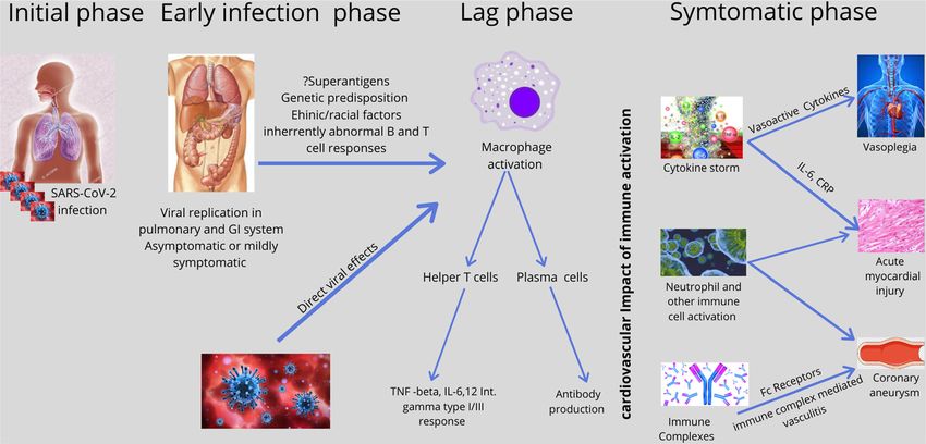
CoV-2 infection (Fig. 1). Firstly, this hypothesis is supported Myocardial affection is also a common feature in MIS-C
by the fact that the peak of MIS-C cases occurred after a delay and may be related to high circulating levels of C-reactive
of weeks from community peak [10, 18, 19] and frequent protein and Interleukin−6, which is known to be related to
finding of antibody positivity and negative RT-PCR results myocardial dysfunction in bacterial infections [39]. The dis-
[10, 11, 18, 19, 23, 30] and secondly, the children with proportionately high elevation of ventricular natriuretic pep-
MIS-C have a predominantly gastrointestinal presentation of tides and mild to moderate elevation of troponins have led
their illness and enterocytes be readily infected by SARS- some investigators to believe that acute heart failure of MIS-
CoV-2 and there is evidence of gastric adenitis in children C is related more to myocardial oedema and vasoplegia with
affected with MIS-C. Similar to the SARS-CoV-1, the IgG variable contribution from mild to moderate myocarditis [11,
antibodies may enhance organ damage and cause a heightened 24]. In this context it is important to note that at present there
immune response by altering the macrophage activity in acute are no studies available where myocardial biopsy has been
SARS-CoV-2 infection [33]. Antibodies may accentuate the done to elucidate the true mechanism of myocardial injury
disease process by facilitating viral entry or replication, the and to rule out active viral replication as the cause of damage
formation of antibody complexes and cellular activation to myocytes rather than immune-mediated damage. As for
[34]. The pathogenesis of coronary vasculature involvement other immune-mediated pathologies in children and closely
in MIS-C may also be explained by immune complex-induced related Kawasaki disease, various host factors come into play
vasculitis. Immune complexes evoke inflammatory responses like genetic susceptibility [40]. The hypothesis of genetic sus-
and vascular damage through Fc gamma receptors or ceptibility is supported by the fact that there are no reports of
Fig. 1 The possible mechanism o cardiovascular involvement in multi-system inflammatory syndrome in children (MIS-C) following SARS-CoV-2
InfectionSN Compr. Clin. Med. (2021) 3:48–59 57
MIS-C like illness from China and Japan and most of their strategies. Of note genetic and immunologic studies to mea-
paediatric cases are mild [41]. Interestingly the prevalence of sure the immune response, studies with control arms including
Kawasaki disease in these countries is one of the highest in the children who are seropositive for SARS-CoV-2 antibodies but
world. The more frequent development of MIS-C in older no disease and research related to the vaccine will be crucial.
children and adolescents and black race is likely due to the Theoretically, SARS-CoV-2 vaccine-related immune re-
variability in expression of angiotensin-converting enzyme 2 sponse may precipitate MIS-C. Research is also needed in
(ACE 2) and genetically determined aberrant T cell and B cell direction of the exact cause of heart failure and mechanisms
responses to SARS-CoV-2 infection. Children’s immune sys- of myocarditis, especially histopathology studies, to deter-
tems are in the developing stage and may respond to chal- mine the presence or absence of active viral replication in
lenges differently from adult immune systems. The immature myocardium and type of immune damage. Longitudinal stud-
respiratory tract and immune system in children contribute to ies will be the answer to the natural history of coronary aneu-
severe illness in the case of other viral respiratory diseases like rysm and their propensity to cause myocardial infarction.
influenza or respiratory syncytial virus [42]. It is also specu-
lated that children respond differently to SARS-CoV-2 infec-
tion because the maturity and function (e.g. binding ability) of Conclusion
ACE2 in children may be lower and circulating levels of the
enzyme may be higher than that in adults [43]. We are early in experience with the this new entity and there
are certain unanswered questions relating to effective treat-
ments to prevent progression to severe disease, treatments to
Management prevent the formation of coronary aneurysms and most impor-
tantly what is the natural history of such aneurysms and how
The primary aim of therapy is to reduce the systemic inflam- to follow-up such patients? To answer all these questions, we
mation, protect the organs and decrease or prevent complica- need studies with wider case definitions which include milder
tions like coronary aneurysms and death. The clinical man- forms of disease and control arms with positive serology for
agement should be informed by clinical presentation, and the SARS-CoV-2 but no disease. In coming time, the most chal-
management of such cases requires multi-disciplinary team lenging task will be to define the actual pathophysiological
(general paediatrics, infectious diseases, cardiology, intensive mechanisms and accordingly develop early diagnostic tools
care, haematology, immunology, pharmacy and rheumatolo- and effective therapies. Due to the lack of longitudinal data,
gy). Children with moderate to severe symptoms need inten- at present, it will be difficult to plan for surveillance and com-
sive care support with appropriate therapies according to or- munity care following recovery from severe illness.
gan involvement including antimicrobial drugs. The role of
antivirals is not clear at present [24]. The choice of treatment Authors’ Contribution Both the authors were involved in conceptualisa-
in any viral disease depends upon the stage of the disease and tion, drafting and final editing of the manuscript.
perceived/actual reasons for deterioration. If the clinical dete-
Funding This study received no funding.
rioration is due to replication of virus, antivirals may be ben-
eficial, or if it is due to cytokine response, then immunomod-
Data Availability The data underlying this article are available in the
ulatory therapy may be useful. It is proposed that MIS-C is article.
immune-mediated multiorgan damage; thus immunomodula-
tory therapy is preferred. The most common therapies tried for Compliance with Ethical Standards
MIS-C are intravenous immunoglobulins, systemic glucocor-
ticoids, anti-IL-1 receptor antagonist, anti-IL-6 receptor antag- Ethical Approval This article does not contain any studies with human
onist and TNF alpha antagonists with positive outcomes participants or animals performed by any of the authors.
(Table 4). At present, there is no randomized controlled trial
available to judge the relative efficacies of these medications, Consent for Publication N/A
but certain management guidelines are available based on ex-
Conflict of Interest Both authors declare no conflict of interest.
pert opinion [44].
Informed Consent Not Applicable.
Future Direction
References
In future as more clinical data and exact pathogenetic mecha-
nisms need to be elucidated, to determine the risk factors, 1. Huang C, Wang Y, Li X, Ren L, Zhao J, Hu Y, et al. Clinical
prognostic markers, rational management and prevention features of patients infected with 2019 novel coronavirus in58 SN Compr. Clin. Med. (2021) 3:48–59
Wuhan, China. Lancet. 2020;395(10223):497–506. https://doi.org/ Child Adolesc Health. 2020;4(7):e21–3. https://doi.org/10.1016/
10.1016/S0140-6736(20)30183-5 Erratum in: Lancet. 2020 Jan 30. S2352-4642(20)30164-4.
2. Coronavirus Disease 2019 in Children — United States, February 17. Toubiana J, Poirault C, Corsia A, Bajolle F, Fourgeaud J,
12–April 2, 2020. MMWR Morb Mortal Wkly Rep. 2020;69:422– Angoulvant F, et al. Kawasaki-like multisystem inflammatory syn-
6. https://doi.org/10.15585/mmwr.mm6914e4. drome in children during the covid-19 pandemic in Paris, France:
3. Castagnoli R, Votto M, Licari A, Brambilla I, Bruno R, Perlini S, prospective observational study. BMJ. 2020;369:m2094. https://
et al. Severe acute respiratory syndrome coronavirus 2 (SARS- doi.org/10.1136/bmj.m2094.
CoV-2) infection in children and adolescents: a systematic review. 18. Feldstein LR, Rose EB, Horwitz SM, Collins JP, Newhams MM,
JAMA Pediatr. 2020;174:882–9. https://doi.org/10.1001/ Son MBF, et al. Multisystem Inflammatory Syndrome in U.S.
jamapediatrics.2020.1467. Children and Adolescents. N Engl J Med. 2020:
4. Malviya A, Yadav R. COVID −19 pandemic and paediatric popu- NEJMoa2021680. https://doi.org/10.1056/NEJMoa2021680.
lation with special reference to congenital heart disease. Indian 19. Dufort EM, Koumans EH, Chow EJ, Rosenthal EM, Muse A,
Heart J. 2020;72:141–4. https://doi.org/10.1016/j.ihj.2020.06.001. Rowlands J, et al. Multisystem Inflammatory Syndrome in
5. Dong Y, Mo X, Hu Y, Qi X, Jiang F, Jiang Z, et al. Epidemiological C h i l d r e n i n N e w Yo r k St a t e . N E n g l J Me d . 2 0 20 :
characteristics of 2143 pediatric patients with 2019 coronavirus NEJMoa2021756. https://doi.org/10.1056/NEJMoa2021756.
disease in China. Paediatrics. 2020. https://doi.org/10.1542/peds. 20. Centres for Disease Control and Prevention. Emergency prepared-
2020-0702. ness and response: a multisystem inflammatory syndrome in chil-
6. Xia W, Shao J, Guo Y, Peng X, Li Z, Hu D. Clinical and CT dren (MIS-C) associated with coronavirus disease 2019 (COVID-
features in pediatric patients with COVID-19 infection: different 19). Health advisory https://emergency.cdc.gov/han/2020/
points from adults. Pediatr Pulmonol. 2020;55(5):1169–74. han00432.asp. Accessed on May 17, 2020.
https://doi.org/10.1002/ppul.24718. 21. Capone CA, Subramony A, Sweberg T, Schneider J, Shah S, Rubin
7. Riphagen S, Gomez X, Gonzalez-Martinez C, Wilkinson N, L, et al. Characteristics, cardiac involvement, and outcomes of mul-
Theocharis P. Hyperinflammatory shock in children during tisystem inflammatory disease of childhood (MIS-C) associated
COVID-19 pandemic. Lancet. 2020;395(10237):1607–8. https:// with SARS-CoV-2 infection. J Pediatr. 2020;S0022–3476(20):
doi.org/10.1016/S0140-6736(20)31094-1. 30746. https://doi.org/10.1016/j.jpeds.2020.06.044.
8. Jones VG, Mills M, Suarez D, Hogan CA, Yeh D, Segal JB, et al. 22. Levin M. Childhood multisystem inflammatory syndrome - a new
COVID-19 and Kawasaki disease: novel virus and novel case. challenge in the pandemic [published online ahead of print, 2020
Hosp Pediatr. 2020;10(6):537–40. https://doi.org/10.1542/hpeds. Jun 29]. N Engl J Med. 2020. https://doi.org/10.1056/
2020-0123. NEJMe2023158.
9. Paediatric Intensive Care Society (PICS) Statement: increased num- 23. Davies P, Evans C, Kanthimathinathan HK, Lillie J, Brierley J,
ber of reported cases of novel presentation of multisystem inflam- Waters G, et al. Intensive care admissions of children with paediat-
matory disease. Available at https://picsociety.uk/wp-content/ ric inflammatory multisystem syndrome temporally associated with
uploads/2020/04/PICS-statement-re-novel-KD-C19-presentation- SARS-CoV-2 (PIMS-TS) in the UK: a multicentre observational
v2-27042020.pdf. Accessed on May 15, 2020. study. Lancet Child Adolesc Health. 2020;4:669–77. https://doi.
10. Verdoni L, Mazza A, Gervasoni A, Martelli L, Ruggeri M, org/10.1016/S2352-4642(20)30215-7.
Ciuffreda M, et al. An outbreak of severe Kawasaki-like disease 24. Belhadjer Z, Méot M, Bajolle F, Khraiche D, Legendre A, Abakka
at the Italian epicentre of the SARS-CoV-2 epidemic: an observa- S, et al. Acute heart failure in multisystem inflammatory syndrome
tional cohort study. Lancet. 2020;395(10239):1771–8. https://doi. in children (MIS-C) in the context of global SARS-CoV-2 pandem-
org/10.1016/S0140-6736(20)31103-X. ic. Circulation. 2020. https://doi.org/10.1161/
11. Grimaud M, Starck J, Levy M, Marais C, Chareyre J, Khraiche D, CIRCULATIONAHA.120.048360.
et al. Acute myocarditis and multisystem inflammatory emerging 25. Ramcharan T, Nolan O, Lai CY, Prabhu N, Krishnamurthy R,
disease following SARS-CoV-2 infection in critically ill children. Richter AG, et al. Paediatric inflammatory multisystem syndrome:
Version 2. Ann Intensive Care. 2020;10(1):69. https://doi.org/10. temporally associated with SARS-CoV-2 (PIMS-TS): cardiac fea-
1186/s13613-020-00690-8. tures, management and short-term outcomes at a UK tertiary pae-
12. Whittaker E, Bamford A, Kenny J, Kaforou M, Jones CE, Shah P, diatric hospital. Pediatr Cardiol. 2020:1–11. https://doi.org/10.
et al. Clinical characteristics of 58 children with a pediatric inflam- 1007/s00246-020-02391-2.
matory multisystem syndrome temporally associated with SARS- 26. McCrindle BW, Rowley AH, Newburger JW, et al. Diagnosis,
CoV-2. JAMA. 2020:e2010369. https://doi.org/10.1001/jama. treatment, and long-term management of Kawasaki Disease: a sci-
2020.10369. entific statement for health professionals from the American Heart
13. Royal College of Paediatrics and Child Health. Guidance — pae- Association [published correction appears in circulation. 2019
diatric multisystem inflammatory syndrome temporally associated Jul 30;140(5):e181-e184]. Circulation. 2017;135(17):e927–99.
with COVID-19. https://www.rcpch.ac.uk/resources/guidance- https://doi.org/10.1161/CIR.0000000000000484.
paediatric-multisystem-inflammatory-syndrome-temporally- 27. Newburger JW, Sleeper LA, McCrindle BW, Minich LL, Gersony
associated-covid-19. Accessed 19th May 2020 W, Vetter VL, et al. Randomized trial of pulsed corticosteroid ther-
14. World Health Organization Scientific Brief: Multisystem inflam- apy for primary treatment of Kawasaki disease. N Engl J Med.
matory syndrome in children and adolescents with COVID-19. 2007;356(7):663–75. https://doi.org/10.1056/NEJMoa061235.
Available at: World Health Organization Scientific Brief: 28. Burns JC, Shike H, Gordon JB, Malhotra A, Schoenwetter M,
Multisystem inflammatory syndrome in children and adolescents Kawasaki T. Sequelae of Kawasaki disease in adolescents and
with COVID-19. Accessed on May 17, 2020) young adults. J Am Coll Cardiol. 1996;28:253–7.
15. Cheung EW, Zachariah P, Gorelik M, Boneparth A, Kernie SG, 29. Rowley AH. Multisystem inflammatory syndrome in children
Orange JS, et al. Multisystem inflammatory syndrome related to (MIS-C) and Kawasaki disease: two different illnesses with over-
covid-19 in previously healthy children and adolescents in New lapping clinical features. J Pediatr. 2020;224:129–32. https://doi.
York City. JAMA. 2020:e2010374. https://doi.org/10.1001/jama. org/10.1016/j.jpeds.2020.06.057.
2020.10374. 30. Shulman ST. Pediatric COVID-associated multi-system
16. Dallan C, Romano F, Siebert J, Politi S, Lacroix L, Sahyoun C. Inflammatory Syndrome (PMIS). J Pediatr Infect Dis Soc.
Septic shock presentation in adolescents with COVID-19. Lancet 2020;9:285–6. https://doi.org/10.1093/jpids/piaa062.SN Compr. Clin. Med. (2021) 3:48–59 59
31. Pan Y, Zhang D, Yang P, Poon LLM, Wang Q. Viral load of 38. Rowley AH. Understanding SARS-CoV-2-related multisystem in-
SARS-CoV-2 in clinical samples. Lancet Infect Dis. 2020;20: flammatory syndrome in children. Nat Rev Immunol. 2020;20:
411–2. 453–4. https://doi.org/10.1038/s41577-020-0367-5.
32. Zhou F, Yu T, Du R, Fan G, Liu Y, Liu Z, et al. Clinical course and 39. Pathan N, Hemingway CA, Alizadeh AA, Stephens AC, Boldrick
risk factors for mortality of adult inpatients with COVID-19 in JC, Oragui EE, et al. Role of interleukin 6 in myocardial dysfunc-
Wuhan, China: a retrospective cohort study. Lancet. tion of meningococcal septic shock. Lancet. 2004;363(9404):203–
2020;395(10229):1054–62. https://doi.org/10.1016/S0140- 9. https://doi.org/10.1016/S0140-6736(03)15326-3.
6736(20)30566-3 Erratum in: Lancet. 2020 Mar 28;395(10229): 40. Onouchi Y, Ozaki K, Burns JC, Shimizu C, Terai M, Hamada H,
1038. et al. A genome-wide association study identifies three new risk loci
33. Liu L, Wei Q, Lin Q, Fang J, Wang H, Kwok H, et al. Anti-spike for Kawasaki disease. Nat Genet. 2012;44(5):517–21. https://doi.
IgG causes severe acute lung injury by skewing macrophage re- org/10.1038/ng.2220.
sponses during acute SARS-CoV infection. JCI Insight. 41. Xu S, Chen M, Weng J. COVID-19 and Kawasaki disease in
2019;4(4):e123158. https://doi.org/10.1172/jci.insight.123158. Children. Pharmacol Res. 2020;159:104951. https://doi.org/10.
34. Katzelnick LC, Gresh L, Halloran ME, Mercado JC, Kuan G, 1016/j.phrs.2020.104951.
Gordon A, et al. Antibody-dependent enhancement of severe den- 42. Hong L, Luo Y. Respiratory viral infections in infants: causes,
gue disease in humans. Version 2. Science. 2017;358(6365):929– clinical symptoms, virology, and immunology. Clin Microbiol
32. https://doi.org/10.1126/science.aan6836. Rev. 2010;23:74–98.
35. Mayadas TN, Tsokos GC, Tsuboi N. Mechanisms of immune 43. Elena C, Carmine V, Alessandro PA. COVID-19 infection and
complex-mediated neutrophil recruitment and tissue injury. circulating ACE2 Levels: protective role in women and children.
Circulation. 2009;120(20):2012–24. https://doi.org/10.1161/ Front Pediatr. 2020;8:206. https://doi.org/10.3389/fped.2020.
CIRCULATIONAHA.108.771170. 00206;2296-2360.
44. Nakra NA, Blumberg DA, Herrera-Guerra A, Lakshminrusimha S.
36. Nagelkerke SQ, Kuijpers TW. Immunomodulation by IVIg and the
Multi-System Inflammatory Syndrome in Children (MIS-C)
Role of Fc-gamma Receptors: Classic Mechanisms of Action after
Following SARS-CoV-2 infection: review of clinical presentation,
all? Front Immunol. 2015;5:674. Published 2015 Jan 21. https://doi.
hypothetical pathogenesis, and proposed management. Children
org/10.3389/fimmu.2014.00674.
(Basel). 2020;7(7):E69. Published 2020 Jul 1. https://doi.org/10.
37. Menikou S, Langford PR, Levin M. Kawasaki disease: the role of 3390/children7070069.
immune complexes revisited. Front Immunol. 2019;10:1156.
Published 2019 Jun 12. https://doi.org/10.3389/fimmu.2019.
01156. Publisher’s Note Springer Nature remains neutral with regard to jurisdic-
tional claims in published maps and institutional affiliations.You can also read