Loss of H3K27 trimethylation is frequent in IDH1-R132H but not in non-canonical IDH1/2 mutated and 1p/19q codeleted oligodendroglioma: a Japanese ...
←
→
Page content transcription
If your browser does not render page correctly, please read the page content below
Habiba et al. acta neuropathol commun (2021) 9:95
https://doi.org/10.1186/s40478-021-01194-7
RESEARCH Open Access
Loss of H3K27 trimethylation is frequent
in IDH1‑R132H but not in non‑canonical
IDH1/2 mutated and 1p/19q codeleted
oligodendroglioma: a Japanese cohort study
Umma Habiba1,2, Hirokazu Sugino1, Roumyana Yordanova3,4, Koki Ise5, Zen‑ichi Tanei1, Yusuke Ishida1,
Satoshi Tanikawa1,6, Shunsuke Terasaka7, Ken‑ichi Sato8, Yuuta Kamoshima9, Masahiko Katoh10,
Motoo Nagane11, Junji Shibahara12, Masumi Tsuda1,6,13 and Shinya Tanaka1,6,13*
Abstract
Oligodendrogliomas are defined by mutation in isocitrate dehydrogenase (NADP(+)) (IDH)1/2 genes and chromo‑
some 1p/19q codeletion. World Health Organisation diagnosis endorses testing for 1p/19q codeletion to distinguish
IDH mutant (Mut) oligodendrogliomas from astrocytomas because these gliomas require different treatments and
they have different outcomes. Several methods have been used to identify 1p/19q status; however, these techniques
are not routinely available and require substantial infrastructure investment. Two recent studies reported reduced
immunostaining for trimethylation at lysine 27 on histone H3 (H3K27me3) in IDH Mut 1p/19q codeleted oligodendro‑
glioma. However, the specificity of H3K27me3 immunostaining in this setting is controversial. Therefore, we devel‑
oped an easy-to-implement immunohistochemical surrogate for IDH Mut glioma subclassification and evaluated a
validated adult glioma cohort. We screened 145 adult glioma cases, consisting of 45 IDH Mut and 1p/19q codeleted
oligodendrogliomas, 30 IDH Mut astrocytomas, 16 IDH wild-type (Wt) astrocytomas, and 54 IDH Wt glioblastomas
(GBMs). We compared immunostaining with DNA sequencing and fluorescent in situ hybridization analysis and
assessed differences in H3K27me3 staining between oligodendroglial and astrocytic lineages and between IDH1-
R132H and non-canonical (non-R132H) IDH1/2 Mut oligodendroglioma. A loss of H3K27me3 was observed in 36/40
(90%) of IDH1-R132H Mut oligodendroglioma. In contrast, loss of H3K27me3 was never seen in IDH1-R132L or
IDH2-mutated 1p/19q codeleted oligodendrogliomas. IDH Mut astrocytoma, IDH Wt astrocytoma and GBM showed
preserved nuclear staining in 87%, 94%, and 91% of cases, respectively. A high recursive partitioning model predicted
probability score (0.9835) indicated that the loss of H3K27me3 is frequent to IDH1-R132H Mut oligodendroglioma.
Our results demonstrate H3K27me3 immunohistochemical evaluation to be a cost-effective and reliable method
for defining 1p/19q codeletion along with IDH1-R132H and ATRX immunostaining, even in the absence of 1p/19q
testing.
Keyword: Mutation, Wild type, Trimethylation at lysine 27 of histone 3, Glioblastoma
Introduction
The current World Health Organisation classifica-
*Correspondence: tanaka@med.hokudai.ac.jp
1 tion for CNS tumors recommends integrated diagnosis
Department of Cancer Pathology, Faculty of Medicine, Hokkaido
University, N15, W7, Kita‑Ku, Sapporo 060‑8638, Japan based on combined phenotypic and genotypic findings
Full list of author information is available at the end of the article [1] Although originating from common progenitor cells
© The Author(s) 2021. Open Access This article is licensed under a Creative Commons Attribution 4.0 International License, which
permits use, sharing, adaptation, distribution and reproduction in any medium or format, as long as you give appropriate credit to the
original author(s) and the source, provide a link to the Creative Commons licence, and indicate if changes were made. The images or
other third party material in this article are included in the article’s Creative Commons licence, unless indicated otherwise in a credit line
to the material. If material is not included in the article’s Creative Commons licence and your intended use is not permitted by statutory
regulation or exceeds the permitted use, you will need to obtain permission directly from the copyright holder. To view a copy of this
licence, visit http://creativecommons.org/licenses/by/4.0/. The Creative Commons Public Domain Dedication waiver (http://creativeco
mmons.org/publicdomain/zero/1.0/) applies to the data made available in this article, unless otherwise stated in a credit line to the data.
Habiba et al. acta neuropathol commun   (2021) 9:95                                                           Page 2 of 11

harboring Isocitrate Dehydrogenase (NADP(+)) (IDH)            Materials and methods
mutations, oligodendrogliomas differ from diffuse astro-      Tumor samples
cytomas by combined whole-arm losses of chromosome            Formalin-fixed paraffin-embedded (FFPE) glioma tissues
1p and 19q (1p/19q codeletion) and frequent Telomerase        from 145 adult patients were used, including 45 IDH Mut
Reverse Transcriptase (TERT) promoter mutations. In           and 1p/19q codeleted oligodendrogliomas, 30 IDH Mut,
contrast, astrocytoma typically exhibits Tumor Protein        and 16 IDH Wt astrocytoma, and 54 IDH Wt GBM. The
P53 (TP53) and ATRX Chromatin Remodeler (ATRX)                Department of Cancer Pathology, Hokkaido University,
mutations [2–7]. From a clinical perspective, these glio-     diagnosed all cases between January 2008 and November
mas require different treatments and have different out-      2020. Tissue samples were obtained from the Nakamura
comes; therefore, the distinction of oligodendroglioma        Memorial Hospital, Kashiwaba Neurosurgical Hospital,
and astrocytoma is crucial. Subclassification of IDH          Sapporo Asabu Neurosurgical Hospital, Keiwakai Ebetsu
mutant (Mut) glioma into astrocytomas and oligoden-           Hospital, Hokkaido Neurosurgical Memorial Hospital,
drogliomas requires testing for 1p/19q codeletion. Sev-       Sapporo Shuyukai Hospital, Shinsapporo Neurosurgical
eral different methods have been used to identify 1p/19q      Hospital, Iwamizawa General Hospital, and Tomako-
status, but clear consensus guidelines or standard proto-     mai Neurosurgical Hospital. Diagnosis was performed
cols for practical use have not been established [8]. Fluo-   according to the 2016 World Health Organisation clas-
rescent in situ hybridization (FISH) is a commonly used       sification of Tumours of the Central Nervous System
method for detecting 1p/19q codeletion. PCR-based loss        (revised 4th edition). The cases prior to 2016 that were
of heterozygosity analysis, multiplex ligation-dependent      diagnosed based on previous versions of classification
probe amplification, and array comparative genomic            were reviewed according to the new integrated diagnos-
hybridization can also test 1p/19q status with high reli-     tic approach by three certified pathologists. Tissue and
ability [9–12]. However, these techniques are labor-inten-    data collection was approved by and performed accord-
sive and require substantial infrastructure investment,       ing to the regulations of the ethics committee of Hok-
making their global application difficult in countries with   kaido University Faculty of Medicine (ethics approval
less developed healthcare systems.                            number: 16-017).
   Trimethylation at lysine 27 on histone H3 (H3K27me3)
is an epigenetic modification that mediates gene silencing    Immunohistochemistry and evaluation
by Enhancer Of Zeste 2 Polycomb Repressive Complex 2          Immunohistochemistry (IHC) was performed on 4-µm
Subunit (EZH2), a component of the Polycomb complex           FFPE tissue sections. Heat-mediated antigen retrieval
(PcG) [13–15]. Loss of H3K27me3 has been reported in          was performed in Tris/EDTA buffer (pH 9.0) at 97 °C for
pediatric ependymoma with a poor prognosis, breast,           20 min. The antibodies used in this study were a mouse
ovarian and pancreatic cancer, and highly recurrent men-      monoclonal to anti-human IDH1-R132H (clone H09,
ingiomas [16–19]. Loss of H3K27me3 was also seen in           1:200, Dianova, Hamburg, Germany), a rabbit poly-
malignant peripheral nerve sheath tumors and is consid-       clonal to anti-ATRX (HPA001906, 1:700, Sigma Aldrich,
ered a useful diagnostic marker [20, 21]. Another study       St.Louis, MO, USA), a mouse monoclonal to p53 (clone
reported diagnostic relevance of decreased H3K27me3 in        DO7, original concentration, Agilent (Dako), Santa
H3.3 Histone A (H3-3A) K27M-mutant GBM [22].                  Clara, CA, USA), and a rabbit monoclonal to H3K27me3
   Although H3K27me3 has been reported to be involved         (clone EPR18607, 1:150, Abcam, Cambridge, UK). IHC
in several brain tumor entities, comprehensive data           of IDH1-R132H, ATRX, and p53 were conducted using
about H3K27me3 in IDH Mut gliomas are controver-              an Autostainer Link 48, Agilent (Dako), and IHC of
sial. Recently, Filipski et al. [23] reported that loss of    H3K27me3 was conducted manually according to the
H3K27me3 staining can potentially discriminate between        manufacturer’s instructions. Light microscopy (Olympus
oligodendroglial and astrocytic tumor lineages. Similarly,    BX53, Japan) observation was performed for histological
Feller et al. [24] and Kitahama et al. [25] reported lower    and immunohistochemical evaluation.
H3K27me3 in oligodendroglioma by data-independent                All immuno-positive cases for IDH1-R132H were clas-
acquisition (DIA)-based mass spectrometry and immu-           sified as IDH1 Mut. Negative immunostaining of ATRX
nostaining, respectively. However, using sequential IHC,      in neoplastic cells in the presence of an internal positive
Pekmezci et al. [26] did not consider H3K27me3 to be a        control was considered to indicate a loss of ATRX expres-
specific marker for the classification of diffuse gliomas.    sion. Immunohistochemistry for p53 was positive when
Therefore, we have assembled a cohort of adult diffuse        more than 50% of tumor nuclei showed intense staining.
gliomas to determine whether simple H3K27me3 immu-               Scoring of H3K27me3. Human colonic mucosa was
nostaining can be a reliable method to triage cases for       used as a positive control according to the antibody
1p/19q testing.                                               datasheet. Preserved H3K27me3 in endothelial cells
Habiba et al. acta neuropathol commun (2021) 9:95 Page 3 of 11
and immune cells served as an internal positive control. DNA was extracted from FFPE tumor tissue using a
H3K27me3 immunostaining was assessed as H3K27me3- DNA tissue extraction kit (Qiagen; Cat: 56404). The
positive (nuclear retention) or -negative (nuclear loss) extracted DNA was quantified using a NanoDrop 1000
in a blinded manner. Complete nuclear loss or dot-like (Thermo Scientific). A fragment of 129 bp spanning the
H3K27me3 staining in neoplastic cells was regarded R132 codon of IDH1 was amplified using forward primer
as nuclear loss, as described previously [23]. Each slide 5′-CGGTCTTCAGAGAAGCCATT-3′ and reverse
was scanned using a Nanozoomer XR Scanner (Hama- primer 5′-GCAAAATCACATTATTGCCAAC-3′. Like-
matsu, Japan) and viewed using NDP. Scan version 3.2.4 wise, a fragment of 293 bp spanning the R172 codon of
software. JPEG images for each case were captured from IDH2 was amplified using forward primer 5′-GCTGCA
three randomly selected areas at 20× magnification using GTGGGACCACTATT-3′ and reverse primer 5′-TGT
NDP.view 2 software. At first, using the PatholoCount GGCCTTGTACTGCAGAG-3′.
software Ver 1.0 (Mitani Corporation, Tokyo, Japan), we
scored H3K27me3 immunostaining positive when more Fluorescence in situ hybridization
than 25% of cells show diffuse staining and negative when Fluorescence in situ hybridization (FISH) was performed
more than 75% of cells show loss of staining. Later, an on 3-µm thick FFPE tissue sections to assess the chro-
automated, blinded quantification was performed based mosome 1p/19q status using the Vysis 1p36/19q13 Dual
on the previously described methodology [22]. Quanti- Color Probe Kit as described previously (Abbott Labora-
fication of immunostaining in each JPEG was conducted tories, Abbott Park, IL, USA) [8]. Briefly, paraffin sections
using Matlab’s image processing toolbox. The algorithm were deparaffinized, permeabilized, and hybridized using
used background-foreground separation with a global a probe kit. Changes in the 1p and 19q probe signals com-
threshold set using Otsu’s method. We recorded the aver- pared with controls were used to determine the presence
age intensity of extracted pixels of each area. A case’s of 1p/19q codeletion. For each sample, approximately
final score was calculated by averaging three random 100 well-defined nuclei were scored for signals from the
areas chosen from a section. H3K27me3 staining patterns probes 1p36 (red)/1q25 (green) and 19q13 (red)/19p13
in different glioma subtypes are illustrated in Fig. 1a–l. (green) under fluorescence microscopy at 1000× mag-
To assess the variability between PatholoCount scoring nification. FISH results are expressed as a percentage
and automated quantification, we evaluated the scoring of tumor cells with a deleted signal. Established criteria
results obtained from the same section. This compari- for deletion (1)(p36)/deletion(19)(q13) were consid-
son showed that the results were identical for all cases in ered when 50% of nuclei or more displayed only one red
terms of positive or negative. We used the scoring value (n × red signal) and two green signals (2n × green signal).
of automated quantification for the analysis of our data.
Statistical analysis was performed using JMP®Pro 15.2.0
Statistical analysis
DNA sequencing
DNA sequencing of IDH1 codon 132 and IDH2 codon (SAS) software (Cary, North Carolina, USA). The associa-
172 was performed in IDH1-R132H immuno-negative tions among 1p/19q deletion with H3K27me3 and ATRX
cases using an Applied Biosystems 3130 Genetic Ana- staining, IDH1/2 mutation, and histopathological param-
lyzer and Sequencing Analysis Finch TV 1.4.0 software. eters were determined using the chi-squared test/Fisher’s
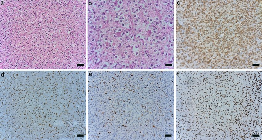
(See figure on next page.)
Fig. 1 Immunostaining and molecular analysis patterns in different glioma subtypes. Complete nuclear loss of H3K27me3 in tumor cells with
retained staining in endothelial cells in IDH1 Mut 1p/19q codeleted oligodendroglioma (a, b). c Dot-like H3K27me3 staining in negative tumor
cell nuclei was considered loss of H3K27me3 expression in IDH1 Mut 1p/19q codeleted oligodendroglioma. Arrows point to retained nuclear
staining in endothelial cells and infiltrating lymphocytes (a–c). Retained nuclear H3K27me3 staining was observed in IDH1 Mut astrocytoma (d–f),
IDH Wt Astrocytoma (g–i), and IDH Wt GBM (j–l). a–c 40× magnification (Scale bar = 20 μm); d–l 20× magnification (Scale bar = 50 μm). Mosaic
plot analysis comparing the correlation between H3K27me3 (m, n) and ATRX immunoreactivity (o) among glioma subclasses. m IDH Mut 1p/19q
codeleted oligodendrogliomas showed significantly lower H3K27me3 staining compared with other glioma subtypes. n Significant differential
expression of H3K27me3 was seen between IDH1 and IDH2 Mut 1p/19q codeleted oligodendrogliomas. o Retained ATRX staining showing a
statistically significant difference between the two IDH Mut glioma lineages. P ≤ 0.05 was considered significant. p–u Mutational analysis patterns
among glioma subtypes. IDH1-R132H Mut oligodendroglioma cases showing a single amino acid transition from p arginine to histidine (R132H), q
arginine to serine (R132S), and r arginine to leucine (R132L). IDH2-R172 Mut oligodendroglioma cases showing a single amino acid transition from
s arginine to lysine (R172K), t arginine to serine (R172S), and u arginine to tryptophan (R172W). v–y Representative FISH images of IDH Mut glioma
subtypes. A case of IDH Mut oligodendroglioma showing both 1p (v) and 19q (w) deletion. A case of IDH Mut astrocytoma showing intact 1p (x)
and 19q (y). 1p/19q deleted cases show one red signal (target) and two green signals (control). NR nuclear retention, NL nuclear loss, Mut mutated,
Wt wild type, GBM glioblastoma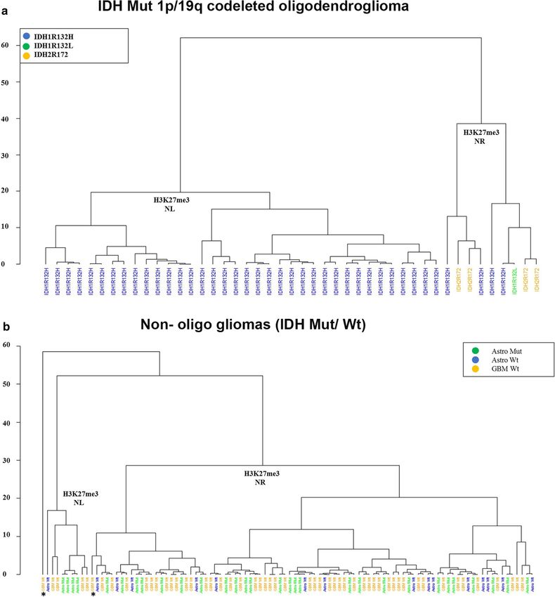
Habiba et al. acta neuropathol commun (2021) 9:95 Page 4 of 11
Habiba et al. acta neuropathol commun (2021) 9:95 Page 5 of 11
Table 1 Demographics of cases
Diagnosis Number of cases (n = 145) Gender Age
Male Female Range Mean Median
Oligodendroglioma 45 23 22 23–72 46.6 46
IDH Mut. astrocytoma (n = 30) DA (II) 16 10 06 25–68 43.7 41
AA (III) 14 08 06 25–86 47.8 46
IDH Wt. astrocytoma (n = 16) DA (II) 04 01 03 57–84 70.2 70
AA (III) 12 07 05 31–84 60 60.5
GBM IDH Wt 54 29 25 28–86 63.6 70
Mut mutated, Wt wild type, DA diffuse astrocytoma, AA anaplastic astrocytoma, GBM glioblastoma
Table 2 Types of IDH mutation in glioma cases
Diagnosis IDH1-R132H Mut IDH Mut other than R132H
Oligodendroglioma (n = 45) 40/45 IDH1 Mut (1/45) IDH1-R132L
IDH2 Mut (4/45) R172K (n = 2)
R172S (n = 1)
R172W (n = 1)
Astrocytoma (n = 30) 29/30 1/30 IDH1-R132S
Mut mutated
exact test. Association with age and gender for IDH Mut and IDH2 Mut groups (P ≤ 0.05) (Fig. 1n). Likewise, 96%
gliomas and IDH Wt gliomas was determined using the (43/45) of oligodendrogliomas and 37% (11/30) of astro-
chi-squared test. A partitioning model was deployed to cytomas had retained ATRX staining, with a statistically
predict H3K27me3 expression in IDH Mut 1p/19q code- significant differential expression between the two IDH
leted gliomas. Hierarchical clustering based on the aver- Mut glioma lineages (P ≤ 0.05) (Fig. 1o). The correlation
age intensity score was performed in R 3.6.3 (https:// between H3K27me3 and ATRX immunoreactivity among
cran.r-project.org/) to visualize the relationship between gliomas is summarized in Additional file 1: Table S1.
IDH Mut 1p/19q codeleted gliomas and non-oligo glio-
mas (IDH Mut and Wt) with H3K27me3 staining. H3K27me3 absence is prevalent in IDH1‑R132H Mut
1p/19q codeleted oligodendrogliomas
Results All 45 cases of oligodendroglioma showing 1p/19q code-
Clinical information and immunoreactivity of gliomas letion also presented with an IDH gene mutation (40/45
Patients with an IDH mutation, in either oligodendro- IDH1-R132H, 1/45 IDH1-R132L, 4/45 IDH2). The
glioma or astrocytoma, were younger (mean ages 46.6 most common mutation identified in astrocytomas was
and 45.7 years, respectively) than patients with IDH Wt IDH1-R132H (29/30 IDH1-R132H, 1/30 IDH1-R132S).
astrocytoma or GBM (mean age 65.1 and 63.6 years, H3K27me3 has reduced in 90% (36/40) of IDH1-R132H
respectively) (P ≤ 0.05). No specific differences in sex Mut oligodendrogliomas (Additional file 1: Table S1).
were observed among the groups. The demographics of Interestingly, in non-canonical IDH1-mutated (IDH1-
cases are summarized in Table 1. Among IDH Mut glio- R132L) or IDH2-mutated (IDH2-R172K, IDH2-R172W,
mas, 80% (36/45) of oligodendrogliomas and 13% (4/30) IDH2-R172S) oligodendrogliomas with 1p/19q codele-
of astrocytomas exhibited a loss of H3K27me3, with a tion, loss of H3K27me3 was never observed. H3K27me3
statistically significant association between 1p/19q code- retention was observed in 87% (26/30) of IDH1 Mut
letion and H3K27me3 loss (P ≤ 0.05). Retained nuclear astrocytomas (25/29 IDH1-R132H, 1/1 IDH1-R132S)
H3K27me3 staining was observed in 94% (15/16) and regardless of the mutation type. Mutational analysis pat-
91% (49/54) of IDH Wt astrocytoma and GBM cases, terns in different glioma subtypes are shown in Fig. 1p–u
respectively (Fig. 1m). However, all IDH2 Mut oligoden- and Table 2. Representative FISH images of IDH1 Mut
drogliomas showed retained H3K27me3 staining, indi- 1p/19q codeleted oligodendroglioma and 1p/19q intact
cating a differential methylation status between IDH1 astrocytoma are shown in Fig. 1v–y.Habiba et al. acta neuropathol commun (2021) 9:95 Page 6 of 11 Fig. 2 Hierarchical clustering based on the average intensity score. a Hierarchical clustering visualizes the relationship between IDH Mut 1p/19q codeleted oligodendrogliomas and H3K27me3 staining. b Hierarchical clustering visualizes the relationship between non-oligo gliomas (IDH Mut/ Wt) and H3K27me3 staining. *Denotes one outlier NR sample with a low score (65) and another NL sample with a borderline score (152) grouped with NR samples. NR nuclear retention, NL nuclear loss, Mut mutated, Wt wild type, GBM glioblastoma This phenomenon was confirmed by hierarchical 1p/19q codeleted oligodendroglioma (Fig. 2a), the clus- clustering based on the average intensity score, which ter patterns were not different among non-oligo gliomas showed two clusters as H3K27me3 nuclear loss (NL) and regardless of IDH mutation type (Fig. 2b). nuclear retention (NR). Although H3K27me3 expression was significantly different between IDH1 and IDH2 Mut
Habiba et al. acta neuropathol commun (2021) 9:95 Page 7 of 11
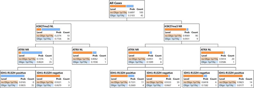
Fig. 3 Decision tree of recursive partitioning model providing the best split of the immunostaining. Blue bars correspond to IDH Mut 1p/19q
codeleted oligodendrogliomas, and orange bars correspond to not IDH Mut 1p/19q codeleted gliomas. We considered IDH Mut 1p/19q codeleted
oligodendroglioma as a dependent variable, and immunostaining (H3K27me3, ATRX, and IDH1-R132H) as predictors. NR nuclear retention, NL
nuclear loss, Mut mutated
Assessment of the predictive value of H3K27me3 in diffuse H3K27me3 staining was always present in non-canonical
gliomas IDH1/IDH2 Mut oligodendrogliomas (Fig. 1n; Additional
To explore possible implications for clinical practice, we file 1: Table S1). Unsupervised hierarchical clustering
employed a recursive partitioning model to assess the showed two primary clusters, H3K27me3 nuclear loss
value of H3K27me3 expression in diffuse gliomas to pre- (NL) and nuclear retention (NR), for both IDH Mut
dict IDH Mut, and 1p/19q codeleted oligodendroglioma. 1p/19q codeleted oligodendroglioma and non-oligo glio-
Immunohistochemical analysis for H3K27me3, ATRX, mas. Although complete differentiation was observed
and IDH1-R132H revealed that diffuse gliomas with between NL and NR in IDH1 Mut 1p/19q codeleted oli-
a loss of nuclear H3K27me3 staining, retained ATRX godendroglioma, the cluster patterns show no difference
staining, and IDH1-R132H positivity can be predicted as in non-oligo gliomas between the groups (Fig. 2a, b).
1p/19q codeleted oligodendrogliomas with a probability Therefore, H3K27me3 staining in non-oligo gliomas did
score of 0.9835. In addition, glioma with retained nuclear not provide additional information between subgroups.
H3K27me3, loss of ATRX staining, and IDH1-R132H 1p/19q codeletion is mutually exclusive with ATRX
positivity can be predicted as 1p/19q non-codeleted gli- mutation, which characterizes glial tumors of astrocytic
oma with a probability score of 0.9823. Five of nine glio- lineage. ATRX immunostaining tends to be positive for
mas with preserved H3K27me3 were oligodendrogliomas oligodendrogliomas and is useful to distinguish between
that harbor non-canonical IDH1-R132L or IDH2-R172 IDH Mut oligodendrogliomas and astrocytomas [27–30].
mutations. Among 20 cases of 1p/19q, non-codeleted gli- However, we observed a loss of ATRX staining in 4%
omas with preserved H3K27me3 staining, IDH1-R132H (2/45) of oligodendrogliomas and retained ATRX stain-
immunostaining did not provide additional information ing in 37% (11/30) of IDH Mut astrocytomas. Therefore,
beyond that of ATRX (Fig. 3). classification by ATRX IHC alone might mislead the
diagnosis of this tumor lineage (Fig. 1o).
Discussion We applied a recursive partitioning model to assess the
Here we present an approach for H3K27me3 immu- clinical utility of H3K27me3 immunostaining to predict
nostaining for adult diffuse glioma and demonstrate its IDH Mut 1p/19q codeleted oligodendroglioma. Our pre-
application in a routine diagnostic procedure. We show diction models indicate the clinical utility of H3K27me3
differences in H3K27me3 staining between oligodendro- IHC for the prediction of IDH1-R132H Mut 1p/19q
glial and astrocytic lineages and between IDH1-R132H codeleted oligodendroglioma along with IDH1-R132H
and non-canonical IDH1/2 Mut oligodendrogliomas. and ATRX IHC. Consistent with a previous report [23],
While the loss of nuclear H3K27me3 was predominantly the high predicted probability score (0.9835) indicated
seen in IDH1-R132H Mut oligodendrogliomas, retained that the loss of H3K27me3 with ATRX positivity is fre-
nuclear staining was mostly observed in IDH1 Mut quent to IDH1-R132H Mut 1p/19q codeleted oligo-
astrocytoma regardless of the mutation type. However, dendroglioma (Fig. 3). However, our data contradictHabiba et al. acta neuropathol commun (2021) 9:95 Page 8 of 11 Fig. 4 Histological and immunohistochemical features of IDH2-R172K Mut and 1p/19q codeleted anaplastic oligodendroglioma. Hematoxylin and eosin stained section at 20× (Scale bar = 50 μm) (a) and 40× magnification (Scale bar = 20 μm) (b). c IDH1-R132H staining is negative. d Retained nuclear ATRX staining. e p53 staining is negative. f Retained nuclear H3K27me3 staining. Magnification is 20× for c–f the previous report, which suggested that the retained affirmation against 1p/19q codeletion would cause mis- nuclear expression of H3K27me3 is only seen in astrocy- classification of five oligodendrogliomas as astrocytomas toma [23]. Moreover, using the same sequential immu- (Additional file 1: Figure S1). nostaining, we found a discrepancy over the sensitivity When the integrated diagnosis approach was used to of H3K27me3 immunostaining, as reported previously assess previous histological diagnoses, among 45 oligo- [26]. This discrepancy may be because our cut-off point dendrogliomas, 39 showed oligodendrogliomas, and six to decide the loss of H3K27me3 staining was 20% lower showed mixed morphology. Thirty-five out of 39 oligo- than Pekmezci et al. Moreover, Pekmezi considered dendroglioma cases that were positive for IDH1-R132H only complete loss as significant and patchy/mosaic and ATRX, and reduced H3K27me3, exhibited 1p/19q staining as a retained expression [26]. However, follow- codeletion. However, oligodendrogliomas with IDH2 ing Filipski et al., [23], complete nuclear loss or dot-like mutations, retained ATRX, and preserved H3K27me3 nuclear retention was combined as the nuclear loss in expression showed classical oligodendroglial morphol- our study. Filipski and Kitahama et al. [23, 25] mentioned ogy and did not provide additional information about that dot-like nuclear staining corresponds to the inacti- 1p/19q codeletion (Fig. 4). Thirty-three out of 46 cases vated X chromosome, which presumes to label the Barr showing astrocytic morphology were astrocytomas, and body in the female subgroup of oligodendrogliomas. We nine cases showed mixed features (formerly oligoastrocy- also observed dot-like staining in eleven female cases of toma). Four IDH Mut astrocytoma cases exhibited classic oligodendrogliomas. oligodendroglial morphology, and the integrated diagno- An alternative decision tree starting with IDH1-R132H sis was confirmed by intact 1p/19q chromosome status staining identified 50/69 IDH1-R132H-positive gliomas by FISH staining (Fig. 5). that showed ATRX nuclear retention and that required Three IDH mutations (IDH1-R132x, IDH2-R172K, and 1p/19q testing to identify 39 oligodendrogliomas. In IDH2-R140Q) occur predominantly in subsets of cancers addition, five of 45 IDH1-R132H-negative gliomas and regulate central circuitry metabolism by producing were oligodendrogliomas, which carried non-canon- the oncometabolite, 2-hydroxyglutarate (2-HG) [31]. Lu ical IDH1/2 mutations (1 IDH1-R132L, 4 IDH2-R172 et al. [32] reported that 2-HG in IDH Mut tumors pre- Mut). Therefore, H3K27me3-positive staining as a single vents the demethylation of repressive histone marks,
Habiba et al. acta neuropathol commun (2021) 9:95 Page 9 of 11
Fig. 5 Histological and immunohistochemical features of IDH1-R132H Mut astrocytoma. The hematoxylin and eosin-stained section demonstrate
a low-grade glioma with classic oligodendroglioma morphology 20× (Scale bar = 50 μm) (a) and 40× (Scale bar = 20 μm) (b) magnification.
c Positive IDH1-R132H staining. d Retained ATRX staining. e Scattered positive nuclear p53 staining. f Retained H3K27me3 nuclear staining.
Chromosome 1p and 19q were confirmed by FISH. Magnification is 20× for c–f
such as H3K9me3 and H3K27me3, resulting in increased for defining IDH1-R132H Mut 1p/19q codeleted oligo-
histone methylation. While IDH1 mutation causes a dendroglioma in the absence of molecular testing.
marked increase in hypermethylation at many genes, a
small group of hypomethylated genes was also reported Limitation of this study
[33]. Papaemmanuil et al. [34] reported that IDH2- The number of non-canonical IDH1/2 mutated 1p/19q
R172K-mutated acute myeloid leukemia (AML) showed codeleted oligodendrogliomas is small (n = 5). Thus,
severe disruption to central metabolism and was associ- further investigations of the differential expression of
ated with different gene expression and DNA methyla- H3K27me3 between IDH1-R132H and non-canonical
tion compared with other IDH1 or IDH2 mutated AML. IDH1/2 mutant oligodendrogliomas are required for
Although IDH1-R132H is the most frequent IDH muta- prognostic and therapeutic application.
tion, other IDH mutations found in oligodendrogliomas
have received less attention. Moreover, it is unknown Conclusion
whether IDH1-R132H and non-canonical IDH1/2- Our study revealed that loss of H3K27me3 nuclear stain-
mutated oligodendrogliomas have different prognostic ing among 1p/19q codeleted oligodendrogliomas is fre-
and therapeutic characteristics. Genome-wide analyses quent in cases harboring IDH1-R132H mutation. We
would help to determine the underlying mechanism. consider that H3K27me3 immunoreactivity could predict
Immunohistochemistry is a cost-effective and acces- the 1p/19q codeletion status along with IDH1-R132H
sible technique that can be readily adapted for detecting and ATRX immunostaining.
molecular surrogates [17]. Immunohistochemistry for
the mutant specific IDH1-R132H is routine for diffuse Supplementary Information
adult glioma [35]. Moreover, H3K27me3 immunohis- The online version contains supplementary material available at https://doi.
tochemistry is used as a molecular surrogate to iden- org/10.1186/s40478-021-01194-7.
tify pediatric midline gliomas [1], malignant peripheral
nerve sheath tumors [20], and H3K27M mutant glio- Additional file 1: Table S1. Correlation between H3K27me3 and ATRX
mas [22]. Therefore, H3K27me3 immunostaining can be immunoreactivity among gliomas; Figure S1. Decision tree of recur‑
sive partitioning model starting with IDH1-R132H staining followed by
regarded as a sensitive and specific molecular surrogateHabiba et al. acta neuropathol commun (2021) 9:95 Page 10 of 11
classification of tumors of the central nervous system: a summary. Acta
ATRX and H3K27me3 staining. Blue bars correspond to IDH Mut 1p/19q Neuropathol 131:803–820. https://doi.org/10.1007/s00401-016-1545-1
codeleted oligodendrogliomas, and orange bars correspond to not IDH 2. Abedalthagafi M, Phillips JJ, Kim GE, Mueller S, Haas-Kogen DA, Marshall
Mut 1p/19q codeleted gliomas. We considered IDH Mut 1p/19q code‑ RE et al (2013) The alternative lengthening of telomere phenotype is sig‑
leted oligodendroglioma as a dependent variable and immunostaining nificantly associated with loss of ATRX expression in high-grade pediatric
(H3K27me3, ATRX, and IDH1-R132H) as predictors. NR nuclear retention, and adult astrocytomas: a multi-institutional study of 214 astrocytomas.
NL nuclear loss, Mut mutated. Mod Pathol 26:1425–1432. https://doi.org/10.1038/modpathol.2013.90
3. Arita H, Narita Y, Fukushima S, Tateishi K, Matsushita Y, Yoshida A et al
(2013) Upregulating mutations in the TERT promoter commonly occur
Acknowledgements
in adult malignant gliomas and are strongly associated with total
We would like to thank Mami S, Kyoko F, and Mieko H for excellent technical
1p19q loss. Acta Neuropathol 126:267–276. https://doi.org/10.1007/
assistance. We thank Jeremy Allen, PhD, from Edanz Group (https://en-author-
s00401-013-1141-6
services.edanz.com/ac) for editing a draft of this manuscript.
4. Cancer Genome Atlas Research Network (2015) Comprehensive, integra‑
tive genomic analysis of diffuse lower-grade gliomas. N Engl J Med
Authors’ contributions
372:2481–2498. https://doi.org/10.1056/nejmoa1402121
All authors contributed to the study’s conception and design. Material
5. Koelsche C, Sahm F, Capper D, Reuss D, Sturm D, Jones DTW et al (2013)
preparation, data collection, and analysis were performed by UH, RY, IK, ST (S.
Distribution of TERT promoter mutations in pediatric and adult tumors of
Terasaka), KS, YK, MN, JS, and MK. Pathological diagnosis were made by ST (S.
the nervous system. Acta Neuropathol 126:907–915. https://doi.org/10.
Tanaka), HS, IY, ST (S. Tanikawa), and ZT. The first draft of the manuscript was
1007/s00401-013-1195-5
written by UH and revised by UH, HS, ZT, MS, and ST (S. Tanaka), and all authors
6. Lovejoy CA, Li W, Reisenweber S, Thongthip S, Bruno J, de Lange T et al
commented on previous versions of the manuscript. All authors read and
(2012) Loss of ATRX, genome instability, and an altered DNA damage
approved the final manuscript.
response are hallmarks of the alternative lengthening of Telomeres path‑
way. PLoS Genet 8:12–15. https://doi.org/10.1371/journal.pgen.1002772
Funding
7. Ichimura K (2012) Molecular pathogenesis of IDH mutations in
This work was supported by MEXT, Grant Number 19H01171 to S.T.
gliomas. Brain Tumor Pathol 29:131–139. https://doi.org/10.1007/
(S. Tanaka) and AMED under Grant Number 20cm0106571h0001, and
s10014-012-0090-4
21cm0106571h0002 to S.T.
8. Woehrer A, Sander P, Haberler C, Kern S, Maier H, Preusser M et al (2011)
FISH-based detection of 1p 19q codeletion in oligodendroglial tumors:
Availability of data and materials
Procedures and protocols for neuropathological practice—a publication
Data used in this study are available from the corresponding author on
under the auspices of the Research Committee of the European Con‑
reasonable request.
federation of Neuropathological Societies (Euro-CNS). Clin Neuropathol
30:47–55. https://doi.org/10.5414/NPP30047
Declarations 9. Smith JS, Alderete B, Minn Y, Borell TJ, Perry A, Mohapatra G et al (1999)
Localization of common deletion regions on 1p and 19q in human
Ethics approval and consent to participate gliomas and their association with histological subtype. Oncogene
Research using tissue and data collection was performed following the regula‑ 18:4144–4152. https://doi.org/10.1038/sj.onc.1202759
tions and approval by the ethics committee of Hokkaido University Faculty of 10. Jeuken JWM, Comelissen S, Boots-Sprenger S, Gijsen S, Wesseling P et al
Medicine (ethics approval number is 16-017). (2006) Multiplex ligation-dependent probe amplification: a diagnostic
tool for simultaneous identification of different genetic markers in glial
Consent for publication tumors. J Mol Diagn 8:433–443. https://doi.org/10.2353/jmoldx.2006.
Not applicable. 060012
11. Franco-Hernández C, Martínez-Glez V, de Campos JM, Isla A, Vaquero J,
Competing of interests Gutiérrez M et al (2009) Allelic status of 1p and 19q in oligodendroglio‑
The authors declare that they have no competing interest. mas and glioblastomas: multiplex ligation-dependent probe amplifica‑
tion versus loss of heterozygosity. Cancer Genet Cytogenet 190:93–96.
Author details https://doi.org/10.1016/j.cancergencyto.2008.09.017
1
Department of Cancer Pathology, Faculty of Medicine, Hokkaido University, 12. Idbaih A, Kouwenhoven M, Jeuken J, Carpentier C, Gorlia T, Kros JM et al
N15, W7, Kita‑Ku, Sapporo 060‑8638, Japan. 2 Department of Oral Pathology (2008) Chromosome 1p loss evaluation in anaplastic oligodendroglio‑
and Periodontology, Sapporo Dental College and Hospital, Dhaka, Bangla‑ mas. Neuropathology 28:440–443. https://doi.org/10.1111/j.1440-1789.
desh. 3 Department of Mathematics, Faculty of Science, Hokkaido University, 2008.00863.x
Sapporo, Japan. 4 Institute of Mathematics and Informatics, Bulgarian Academy 13. Czermin B, Melfi R, McCabe D, Seitz V, Imhof A, Pirrotta V (2002) Dros‑
of Sciences, Sofia, Bulgaria. 5 School of Medicine, Hokkaido University, Sapporo, ophila enhancer of Zeste/ESC complexes have a histone H3 methyltrans‑
Japan. 6 Institute for Chemical Reaction Design and Discovery (WPI‑ICReDD), ferase activity that marks chromosomal Polycomb sites. Cell 111:185–196.
Hokkaido University, Sapporo, Japan. 7 Kashiwaba Neurosurgical Hospital, https://doi.org/10.1016/S0092-8674(02)00975-3
Sapporo, Japan. 8 Nakamura Memorial Hospital, Sapporo, Japan. 9 Asabu 14. Margueron R, Reinberg D (2011) The Polycomb complex PRC2 and its
Neurosurgical Hospital, Sapporo, Japan. 10 Hokkaido Neurosurgical Memorial mark in life. Nature 469:343–349. https://doi.org/10.1038/nature09784
Hospital, Sapporo, Japan. 11 Department of Neurosurgery, Kyorin University 15. Müller J, Hart CM, Francis NJ, Vargas ML, Sengupta A, Wild B et al (2002)
School of Medicine, Tokyo, Japan. 12 Department of Pathology, Kyorin Univer‑ Histone methyltransferase activity of a Drosophila Polycomb group
sity School of Medicine, Tokyo, Japan. 13 Global Institution for Collaborative repressor complex. Cell 111:197–208. https://doi.org/10.1016/S0092-
Research and Education (GI‑CoRE), Hokkaido University, Sapporo, Japan. 8674(02)00976-5
16. Bayliss J, Mukherjee P, Lu C, Jain SU, Chung C, Martinez D et al (2016)
Received: 31 March 2021 Accepted: 6 May 2021 Lowered H3K27me3 and DNA hypomethylation define poorly prognostic
pediatric posterior fossa ependymomas. Sci Transl Med 8:366ra161.
https://doi.org/10.1126/scitranslmed.aah6904
17. Panwalkar P, Clark J, Ramaswamy V, Hawes D, Yang F, Dunham C et al
(2017) Immunohistochemical analysis of H3K27me3 demonstrates global
reduction in group-A childhood posterior fossa ependymoma and is a
References powerful predictor of outcome. Acta Neuropathol 134:705–714. https://
1. Louis DN, Perry A, Reifenberger G, von Deimling A, Figarella-Branger doi.org/10.1007/s00401-017-1752-4
D, Cavenee WK et al (2016) The 2016 World Health Organization 18. Wei Y, Xia W, Zhang Z, Liu J, Wang H, Adsay NV et al (2008) Loss of
trimethylation at lysine 27 of histone H3 is a predictor of poor outcomeHabiba et al. acta neuropathol commun (2021) 9:95 Page 11 of 11
in breast, ovarian, and pancreatic cancers. Mol Carcinog 47:701–706. gliomas. Acta Neuropathol Commun 4:60. https://doi.org/10.1186/
https://doi.org/10.1002/mc.20413 s40478-016-0331-6
19. Katz LM, Hielscher T, Liechty B, Silverman J, Zagzag D, Sen R et al (2018) 28. Jiao Y, Killela PJ, Reitman ZJ, Rasheed AB, Heaphy CM, de Wilde RF et al
Loss of histone H3K27me3 identifies a subset of meningiomas with (2012) Frequent ATRX, CIC, FUBP1 and IDH1 mutations refine the clas‑
increased risk of recurrence. Acta Neuropathol 135:955–963. https://doi. sification of malignant gliomas. Oncotarget 3:709–722. https://doi.org/10.
org/10.1007/s00401-018-1844-9 18632/oncotarget.588
20. Prieto-Granada CN, Wiesner T, Messina JL, Jungbluth AA, Chi P, Antonescu 29. Liu XY, Gerges N, Korshunov A, Sabha N, Khuong-Quang DA, Fontebasso
CR (2016) Loss of H3K27me3 expression is a highly sensitive marker for AM et al (2012) Frequent ATRX mutations and loss of expression in adult
sporadic and radiation-induced MPNST. Am J Surg Pathol 40:479–489. diffuse astrocytic tumors carrying IDH1/IDH2 and TP53 mutations. Acta
https://doi.org/10.1097/PAS.0000000000000564 Neuropathol 124:615–625. https://doi.org/10.1007/s00401-012-1031-3
21. Röhrich M, Koelsche C, Schrimpf D, Capper D, Sahm F, Kratz A et al (2016) 30. Reuss DE, Sahm F, Schrimpf D, Wiestler B, Capper D, Koelsche C et al
Methylation-based classification of benign and malignant peripheral (2015) ATRX and IDH1-R132H immunohistochemistry with subsequent
nerve sheath tumors. Acta Neuropathol 131:877–887. https://doi.org/10. copy number analysis and IDH sequencing as a basis for an “integrated”
1007/s00401-016-1540-6 diagnostic approach for adult astrocytoma, oligodendroglioma and
22. Venneti S, Garimella MT, Sullivan LM, Martinez D, Huse JT, Heguy A et al glioblastoma. Acta Neuropathol 129:133–146. https://doi.org/10.1007/
(2013) Evaluation of histone 3 lysine 27 trimethylation (H3K27me3) and s00401-014-1370-3
enhancer of Zest 2 (EZH2) in pediatric glial and glioneuronal tumors 31. Dang L, Su SSM (2017) Isocitrate dehydrogenase mutation and (R)-
shows decreased H3K27me3 in H3F3A K27M mutant glioblastomas. Brain 2-hydroxyglutarate: from basic discovery to therapeutics development.
Pathol 23:558–564. https://doi.org/10.1111/bpa.12042 Annu Rev Biochem 86:305–331. https://doi.org/10.1146/annurev-bioch
23. Filipski K, Braun Y, Zinke J, Roller B, Baumgarten P, Wagner M et al (2019) em-061516-044732
Lack of H3K27 trimethylation is associated with 1p/19q codeletion in 32. Lu C, Ward PS, Kapoor GS, Rohle D, Turcan S, Abdel-Wahab O et al (2012)
diffuse gliomas. Acta Neuropathol 138:331–334. https://doi.org/10.1007/ IDH mutation impairs histone demethylation and results in a block to cell
s00401-019-02025-9 differentiation. Nature 483:474–478. https://doi.org/10.1038/nature10860
24. Feller C, Felix M, Weiss T, Herold-Mende C, Zhang F, Kockmann T et al 33. Turcan S, Rohle D, Goenka A, Walsh LA, Fang F, Yilmaz E et al (2012) IDH1
(2020) Histone epiproteomic profiling distinguishes oligodendroglioma, mutation is sufficient to establish the glioma hypermethylator pheno‑
IDH-mutant and 1p/19q co-deleted from IDH-mutant astrocytoma and type. Nature 483:479–483. https://doi.org/10.1038/nature10866
reveals less tri-methylation of H3K27 in oligodendrogliomas. Acta Neuro‑ 34. Papaemmanuil E, Gerstung M, Bullinger L, Gaidzik VI, Paschka P, Roberts
pathol 139:211–213. https://doi.org/10.1007/s00401-019-02096-8 ND et al (2016) Genomic classification and prognosis in acute myeloid
25. Kitahama K, Iijima S, Sumiishi A, Hayashi A, Nagahama K, Saito K et al leukemia. N Engl J Med 374:2209–2221. https://doi.org/10.1056/NEJMo
(2021) Reduced H3K27me3 levels in diffuse gliomas: association with a1516192
1p/19q codeletion and difference from H3K27me3 loss in malignant 35. Capper D, Weißert S, Balss J, Habel A, Meyer J, Jäger D et al (2010) Char‑
peripheral nerve sheath tumors. Brain Tumor Pathol 38:23–29. https://doi. acterization of r132h mutation-specific idh1 antibody binding in brain
org/10.1007/s10014-020-00382-y tumors. Brain Pathol 20:245–254. https://doi.org/10.1111/j.1750-3639.
26. Pekmezci M, Phillips JJ, Dirilenoglu F, Atasever-Rezanko T, Tihan T, Solo‑ 2009.00352.x
mon D et al (2020) Loss of H3K27 trimethylation by immunohistochemis‑
try is frequent in oligodendroglioma, IDH-mutant and 1p/19q-codeleted,
but is neither a sensitive nor a specific marker. Acta Neuropathol Publisher’s Note
139:597–600. https://doi.org/10.1007/s00401-019-02123-8 Springer Nature remains neutral with regard to jurisdictional claims in pub‑
27. Ebrahimi A, Skardelly M, Bonzheim I, Ott I, Mühleisen H, Eckert F lished maps and institutional affiliations.
et al (2016) ATRX immunostaining predicts IDH and H3F3A status in
Ready to submit your research ? Choose BMC and benefit from:
• fast, convenient online submission
• thorough peer review by experienced researchers in your field
• rapid publication on acceptance
• support for research data, including large and complex data types
• gold Open Access which fosters wider collaboration and increased citations
• maximum visibility for your research: over 100M website views per year
At BMC, research is always in progress.
Learn more biomedcentral.com/submissionsYou can also read