Functional Consequences of PDK4 Deficiency in Doberman Pinscher Fibroblasts
←
→
Page content transcription
If your browser does not render page correctly, please read the page content below
www.nature.com/scientificreports

            OPEN           Functional Consequences of PDK4
                           Deficiency in Doberman Pinscher
                           Fibroblasts
                           Luiz Bolfer1, Amara H. Estrada1, Chelsea Larkin1,3, Thomas J. Conlon2, Francisco Lourenco1,
                           Kathryn Taggart1, Silveli Suzuki-Hatano3 & Christina A. Pacak 3,4*
                           A splice site mutation in the canine pyruvate dehydrogenase kinase 4 (PDK4) gene has been shown to
                           be associated with the development of dilated cardiomyopathy (DCM) in Doberman Pinchers (DPs).
                           Subsequent studies have successfully demonstrated the use of dermal fibroblasts isolated from DPs
                           as models for PDK4 deficiency and have shown activation of the intrinsic (mitochondrial mediated)
                           apoptosis pathway in these cells under starvation conditions. For this study, we sought to further
                           explore the functional consequences of PDK4 deficiency in DP fibroblasts representing PDK4wt/wt,
                           PDK4wt/del, and PDK4del/del genotypes. Our results show that starvation conditions cause increased
                           perinuclear localization of mitochondria and decreased cell proliferation, altered expression levels
                           of pyruvate dehydrogenase phosphatase (PDP) and pyruvate dehydrogenase (PDH), dramatically
                           increased PDH activity, and an impaired response to mitochondrial stress in affected cells. In sum,
                           these results show the broad impact of PDK4 deficiency and reveal mechanistic pathways used by
                           these cells in an attempt to compensate for the condition. Our data help to elucidate the mechanisms
                           at play in this extremely prevalent DP disorder and provide further support demonstrating the general
                           importance of metabolic flexibility in cell health.

                           Dilated Cardiomyopathy (DCM) is one of the most prevalent causes of heart disease in humans and canines.
                           More than 30 genetic mutations and transcriptional alterations related to various molecular and biochemical
                           pathways have been linked to human and canine DCM1–17. Specifically, the Doberman Pinscher (DP) canine
                           breed is highly susceptible to the development of non-ischemic DCM with high mortality rates.
                               Recent studies have shown that for many DPs, DCM can be attributed to either 1) a splice site deletion in the
                           pyruvate dehydrogenase kinase 4 (PDK4) gene, 2) a missense variant in the titin (TTN) gene, or 3) an unfortunate
                           combination of both mutations in the same dog6,8. The newly described titin variant is predicted to change the
                           protein’s structure and has been shown to decrease active tension in myofibers from affected DPs. As titin is the
                           gene most commonly associated with DCM in humans, its functional importance as a molecular spring in muscle
                           and its roles in biochemical sensing and signaling have been well described18–23.
                               In this study, we aimed to evaluate the biochemical and molecular mechanisms that are involved in PDK4
                           deficiency in DPs. Primary dermal fibroblasts from healthy DPs (control - PDK4wt/wt) and DPs carrying the PDK4
                           mutation (both heterozygous - PDK4wt/del, and homozygous - PDK4del/del) have been previously examined and
                           shown to have an increased susceptibility to apoptosis activation under starvation conditions that was mitigated
                           with adeno-associated virus (AAV) mediated gene delivery of healthy PDK4 to affected cells24,25. In the present
                           study, we sought to perform comparative analyses between fibroblasts representing the three genotypes to gain
                           a more complete understanding of the mechanistic pathways involved in the cellular response to this deficiency.
                           Previous studies have shown that PDK4 can be upregulated by lipids, for example, those derived from decanoic
                           acid and that PDK4 expression can be regulated by both the PPARβ/δ and TGFβ signaling pathways26–28. These
                           studies demonstrate that fibroblasts are excellent models for the assessment of PDK4 function. To evaluate PDK4
                           deficiency in DP fibroblasts, cells were evaluated for cell morphology and mitochondrial localization within cells,
                           PDK expression levels, pyruvate dehydrogenase (PDH) activity and abundance, and response to mitochondrial
                           stress based upon oxygen consumption analyses. Through phosphorylation of PDH, PDK4 serves as an important

                           1
                            Department of Small Animal Clinical Sciences, University of Florida College of Veterinary Medicine, Gainesville, FL,
                           32610, USA. 2CR Scientific and Compliance Consulting, LLC, Gainesville, FL, 32608, USA. 3Department of Pediatrics,
                           University of Florida College of Medicine, Gainesville, FL, 32610, USA. 4Department of Molecular Genetics and
                           Microbiology, University of Florida College of Medicine, Gainesville, FL, 32610, USA. *email: pacakc@peds.ufl.edu

Scientific Reports |   (2020) 10:3930 | https://doi.org/10.1038/s41598-020-60879-6                                                            1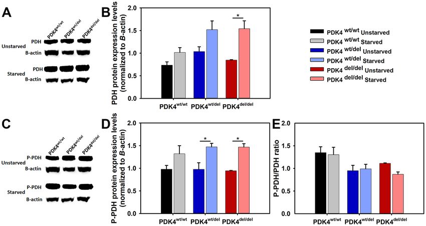
www.nature.com/scientificreports/ www.nature.com/scientificreports
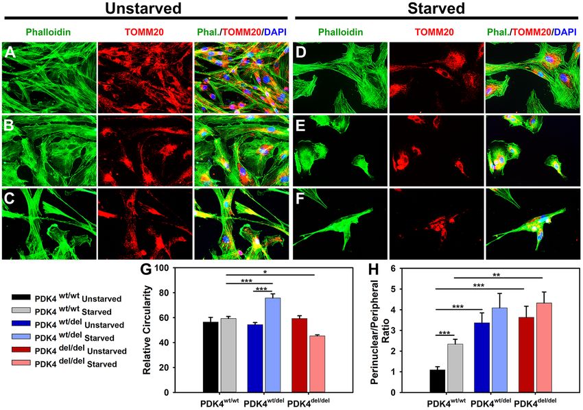
Figure 1. DP fibroblast cellular morphology and mitochondrial localization. (A–C) Primary dermal fibroblasts
from healthy controls PDK4wt/wt, heterozygous PDK4wt/del, or homozygous PDK4del/del, DPs were evaluated with
IF staining of phalloidin (green) to reveal overall cellular architecture and the mitochondrial outer membrane
protein TOMM20 (red) to locate mitochondria within cells. (D–F) Cells representing the three different
genotypes were exposed to 24 hours of starvation conditions. (G) A graph indicating how relative circularity
changed in both PDK4wt/del (increased) and PDK4del/del (decreased) cells in response to starvation conditions
as compared to healthy controls under the same conditions. (H) Perinuclear localization of mitochondria was
increased in both PDK4wt/del and PDK4del/del cells as compared to controls under the same condition except there
was no significant difference between starved PDK4wt/del and PDK4wt/wt cells. (Data presented as mean + std. err.
*p < 0.05, **p < 0.01, ***p < 0.001).
regulator of mitochondrial fuel usage by switching away from glucose oxidation under conditions of low glucose
availability. To better understand this disorder at the molecular level, the present study evaluated healthy and defi-
cient cells in their response to glucose starvation. In doing so, we have helped to identify the broader mechanistic
consequences and compensatory pathways at play in PDK4 deficiency.
Results
Starved primary dermal fibroblasts require PDK4 to survive. In order to evaluate the effects of glu-
cose deprivation in the context of PDK4 deficiency, we placed robust primary dermal fibroblast cultures repre-
senting PDK4wt/wt, PDK4wt/del, and PDK4del/del genotypes into culture medium that lacked glucose. After 24 hours
of starvation, cells were evaluated for differences in general cell morphology and mitochondrial localization as
compared to unstarved cells representing the same genotypes (Fig. 1). Immunofluorescence staining of f-actin
(phalloidin) in fixed cells revealed that PDK4wt/wt fibroblasts exhibit no significant changes in morphology after
24 hours of starvation (Fig. 1A,D,G). In contrast, fibroblasts representing the PDK4wt/del (Fig. 1B,E) and PDK4del/del
(Fig. 1C,F) genotypes displayed significant changes in cellular circularity as compared to controls under star-
vation conditions with a significant increase observed in PDK4wt/del cells and a significant decrease observed in
PDK4del/del cells (Fig. 1D–F,G).
These evaluations also showed that general cellular abundance was significantly reduced in response to starva-
tion in both PDK4wt/del and PDK4del/del cells as compared to PDK4wt/wt controls (PDK4wt/del 36% ± 2 and PDK4del/del
25% ± 1; p < 0.05 [% of PDK4wt/wt]) while there was no significant difference between cells in unstarved condi-
tions. Assessment of ratios of perinuclear to peripheral mitochondrial localization showed a significant increase
in both PDK4wt/del and PDK4del/del cells as compared to healthy controls in unstarved conditions (Fig. 1H). Under
starvation conditions, only PDK4del/del cells showed significantly more perinuclear localization of mitochondria as
Scientific Reports | (2020) 10:3930 | https://doi.org/10.1038/s41598-020-60879-6 2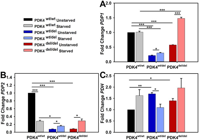
www.nature.com/scientificreports/                                                             www.nature.com/scientificreports

                           Figure 2. PDK isoform transcription levels in response to starvation. Relative transcription levels of (A) PDK1,
                           (B) PDK2, (C) PDK3, and (D) PDK4 in cells representing each of the three groups PDK4wt/wt, PDK4wt/del, and
                           PDK4del/del cells in unstarved and starved conditions. (Data presented as mean + std. err. *p < 0.05, **p < 0.01,
                           ***p < 0.001).

                           compared to controls but PDK4wt/del cells also showed an increasing trend. These observations support previous
                           findings that PDK4 function is required for healthy cell morphology and viability in response to starvation.

                           PDK4 deficiency alters PDK transcription profiles. As there are a total of 4 different PDK isoforms, we
                           sought to determine how cells representing the three different genotypes differed in their PDK transcription levels
                           under typical culture conditions and how this profile may be altered under glucose-free (starvation) conditions.
                           PDK1 transcript levels were similar across all three genotypes under typical culture conditions and remained
                           unchanged in response to 24 hours of starvation in PDK4wt/wt cells. In contrast, PDK1 transcript levels were sig-
                           nificantly reduced following starvation in both PDK4wt/del and PDK4del/del cells as compared to unstarved controls
                           (Fig. 2A). PDK2 transcript levels were similar across all three genotypes under typical culture conditions and
                           were significantly increased in response to 24 hours of starvation in all fibroblasts as compared to unstarved con-
                           ditions (Fig. 2B). PDK3 transcript levels were significantly reduced in both PDK4wt/del and PDK4del/del cells under
                           typical culture conditions as compared to PDK4wt/wt cells. In response to 24 hours of starvation, PDK3 remained
                           significantly reduced in both PDK4wt/del and PDK4del/del cells as compared to PDK4wt/wt cells. When compared
                           to corresponding unstarved culture conditions, PDK3 transcript levels were significantly reduced in fibroblasts
                           representing PDK4wt/wt and PDK4del/del genotypes. In contrast, PDK3 transcript levels were significantly increased
                           in the PDK4wt/del cells as compared to unstarved controls (Fig. 2C).
                               Finally, PDK4 transcript levels were examined using a primer-probe set that would recognize the exon 8–9
                           boundary of PDK4 that should be present in transcripts derived from all three genotypes as this area is located
                           upstream of the splice site deletion present in the affected cells between PDK4 exons 10 and 11. PDK4 was signifi-
                           cantly decreased in the PDK4wt/del and PDK4del/del cells under normal culture conditions as compared to PDK4wt/wt
                           cells. In contrast, PDK4 transcription was significantly increased in response to 24 hours of starvation in fibro-
                           blasts representing all three genotypes as compared to unstarved conditions but this effect was far more profound
                           in the PDK4wt/wt cells than either of the PDK4 deficient lines (Fig. 2D).

                           PDK4 deficiency alters PDP and PDH transcription levels. To determine the impact of PDK4 defi-
                           ciency on pyruvate dehydrogenase phosphatase (PDP) and pyruvate dehydrogenase (PDH) transcription under
                           normal and 24 hours of starvation culture conditions, gene expression assays were performed to evaluate rela-
                           tive levels of PDP1 and PDP2 in cells representing all three genotypes. PDP1 expression levels were significantly
                           higher in PDK4wt/wt cells under normal culture conditions as compared to the deficient lines. PDP1 levels were
                           unchanged in PDK4wt/wt cells under starvation conditions as compared to unstarved controls but significantly
                           increased in both PDK4wt/del and PDK4del/del cells as compared to their respective PDP1 levels in normal culture
                           conditions (Fig. 3A). In contrast, PDP2 expression levels were significantly increased in PDK4wt/wt cells under
                           normal culture conditions as compared to starvation conditions. PDP2 expression was increased in starved
                           PDK4wt/del and PDK4del/del cells as compared to unstarved controls but still remained significantly reduced as
                           compared to levels in unstarved PDK4wt/wt cells (Fig. 3B). PDH transcription levels were significantly higher in
                           both PDK4wt/del and PDK4del/del cells as compared to PDK4wt/wt cells in normal culture conditions (Fig. 3C). Only
                           the PDK4wt/del displayed a significant difference (decrease) between unstarved and starved conditions.

Scientific Reports |   (2020) 10:3930 | https://doi.org/10.1038/s41598-020-60879-6                                                          3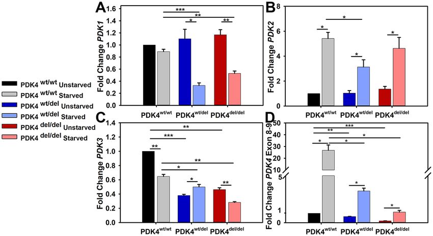
www.nature.com/scientificreports/                                                               www.nature.com/scientificreports

                           Figure 3. PDP isoforms 1 and 2 and PDH transcription levels in response to starvation. Relative transcription
                           levels of (A) PDP1, (B) PDP2, and (C) PDH, in cells representing each of the three groups PDK4wt/wt, PDK4wt/del,
                           and PDK4del/del cells in unstarved and starved conditions. (Data presented as mean + std. err. *p < 0.05,
                           **p < 0.01, ***p < 0.001).

                           Variable PDH protein expression and phosphorylation in PDK4 deficiency.                          Using a PDH anti-
                           body that recognizes the protein regardless of phosphorylation status western blots were performed to compare
                           PDH protein levels between the different genotypes in unstarved and starved conditions (Fig. 4A). The quantified
                           results showed a significant increase in PDH protein expression in the 24 hour starved PDK4del/del cells as com-
                           pared to unstarved controls but no other differences were observed in cells from the other genotypes (Fig. 4B).
                           Next, an antibody that recognizes phosphorylated PDH was used to perform western blots to determine levels
                           of the inactivated (phosphorylated) form of PDH (P-PDH) (Fig. 4C). When quantified, the results showed that
                           while all genotypes showed a trend towards more P-PDH under starvation conditions, this phenomenon was sig-
                           nificant in only the PDK4wt/del and PDK4del/del cells (Fig. 4D). Finally, we compared ratios of P-PDH to total PDH
                           in cells from all genotypes under both normal and starved conditions and found that while not significant there is
                           a trend that suggests PDK4wt/wt cells may have higher levels of the inactivated (phosphorylated) form of PDH than
                           the PDK4wt/del and PDK4del/del cells in both normal and starvation culture conditions (Fig. 4E).

                           PDH activity and expression levels are upregulated in PDK4 deficiency.                      To evaluate the effects of
                           PDK4 dysfunction on PDH activity levels, we first evaluated PDK4 expression levels by ELISA and confirmed
                           a significant deficiency in both the PDK4wt/del and PDK4del/del cells (Fig. 5A). Next, PDH activity was evaluated
                           in cells representing each genotype using an ELISA assay. At baseline conditions, PDH activity was modest in
                           PDK4wt/wt cells, significantly elevated in PDK4wt/del cells, and dramatically elevated in PDK4del/del cells (Fig. 5B).
                           We performed a rescue experiment to confirm that this dramatically high level was a result of PDK4 deficiency.
                           Purified PDK4 was added to all wells and resulted in a reduction of PDH activity that was significant for all cells
                           regardless of mutation status (Fig. 5C). The average percent decrease of PDH activity following PDK4 addition
                           was 10.67% in PDK4wt/wt cells, 31.52% in PDK4wt/del cells, and 67.58% in PDK4del/del cells. The subsequent addition
                           of glutamate then restored PDH activity to baseline levels for all three groups (Fig. 5B,C).

                           PDK4 deficient cells display altered oxygen consumption rates. To compare oxygen consumption
                           rates between the three groups of cells, the Seahorse Extracellular Flux Analyzer was used in conjunction with
                           a Mitochondrial Stress Test assay. The results showed an overall reduced oxygen consumption rate profile for
                           PDK4 deficient cells as compared to healthy controls (Fig. 6A). While the basal oxygen consumption rate (OCR)
                           was significantly increased in PDK4del/del cells, non-mitochondrial OCR was significantly decreased (Fig. 6B).
                           Proton leak was also significantly increased in PDK4del/del cells as compared to the other two groups (Fig. 6B).
                           Mitochondrial spare capacity, or the ability of cells to overcome increased stress is significantly decreased in the
                           PDK4del/del cells as compared to both other groups (Fig. 6B). In sum, these data indicate that PDK4 deficiency
                           significantly alters oxygen utilization by cells.

Scientific Reports |   (2020) 10:3930 | https://doi.org/10.1038/s41598-020-60879-6                                                             4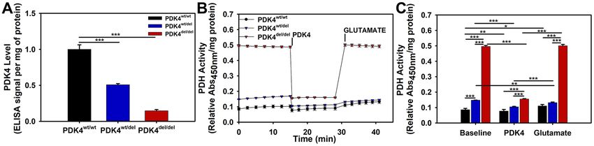
www.nature.com/scientificreports/                                                                www.nature.com/scientificreports

                           Figure 4. PDH levels and phosphorylation status in response to starvation. (A) WB image using an antibody
                           that recognizes all PDH and (B) quantified PDH data normalized to B-actin, and (C) WB image using an
                           antibody that recognizes PDH in its phosphorylated state P-PDH and (D) corresponding quantified data
                           normalized to B-actin. Quantified data represent 3 WBs of cell lysates representing each of the three groups
                           PDK4wt/wt, PDK4wt/del, and PDK4del/del cells in unstarved and starved conditions. (E) Bar graph showing the ratio
                           of P-PDH to PDH for each sample and condition. Full WB images are provided in supplemental data. (Data
                           presented as mean + std. err. *p < 0.05).

                           Figure 5. Relative PDK4 expression and PDH activities. (A) Relative PDK4 expression levels as determined by
                           an ELISA, (B) PDH activity levels between cells representing the three different genotypes and their responses
                           to PDK4 and subsequently, glutamate additions (x-axis indicates minutes time). (C) Quantified PDH activity
                           data showing significant differences between cells and conditions. PDK4wt/wt, PDK4wt/del, and PDK4del/del cells.
                           (Data presented as mean + std. err. *p < 0.05, **p < 0.01, ***p < 0.001).

                           Discussion
                           While PDK4’s crucial role in the maintenance of metabolic flexibility through phosphorylation and suppres-
                           sion of PDH activity is well described, the consequences of PDK4 deficiency, particularly in DPs, are less well
                           understood. We sought to explore the mechanisms involved in DP PDK4 deficiency through characterization of
                           primary dermal fibroblasts representing healthy, partial, and complete PDK4 deficiency. Specifically, we wanted
                           to define differences in how the cells responded to starvation, measure relative compensatory behavior between
                           genotypes based upon expression levels, and evaluate relevant enzymatic activities.
                                Our initial experiment focused on the impact of starvation on cell morphology and mitochondrial subcellular
                           localization in DP fibroblasts representing each of the three genotypes. We observed overall altered cell morphol-
                           ogy and increased perinuclear clustering of mitochondria in both PDK4wt/del and PDK4del/del cells as compared to
                           healthy PDK4wt/wt controls. These alterations suggest that PDK4 is required to maintain a regular cell shape and
                           cytoskeletal structure. Interestingly, while cellular circularity was unchanged in PDK4wt/wt cells following starva-
                           tion, PDK4wt/del cells significantly increase circularity, while PDK4del/del cells become elongated and less circular. In
                           all, these data demonstrate the impact of PDK4 deficiency on cell structure during starvation conditions.

Scientific Reports |   (2020) 10:3930 | https://doi.org/10.1038/s41598-020-60879-6                                                                5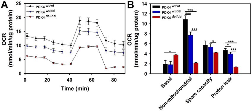
www.nature.com/scientificreports/                                                               www.nature.com/scientificreports

                           Figure 6. Extracellular Flux – Mitochondrial Stress Test. (A) Seahorse extracellular flux assay results profile
                           showing relative responses to the addition of oligomycin, FCCP, and antimycin A/rotenone. (B) Quantified
                           results representing relative OCR levels in PDK4wt/wt, PDK4wt/del, and PDK4del/del cells (Data presented as mean
                           + std. err. *p < 0.05, **p < 0.01, ***p < 0.001).

                                Further support for the relationship between altered cellular structure and apoptosis is provided by our data
                           showing increased perinuclear clustering of mitochondria in PDK4 deficient cells. In general, perinuclear local-
                           ization of mitochondria is a sign of cell stress under various disease states or environmental conditions and this
                           translocation has been shown to be physiologically important and may facilitate mitochondrial stress signaling
                           mechanisms25,29,30. Indeed, these data correlate well with a previous study designed to assess PDK4 deficient DP
                           fibroblasts where conditions causing perinuclear localization of mitochondria resulted in the activation of intrinsic
                           apoptosis25. These results support the notion that PDK4wt/del and PDK4del/del cells react differently than PDK4wt/wt
                           cells to adverse environmental conditions.
                                As PDK4 both regulates the entry of substrates into the citrate cycle and is regulated by its products, and
                           as all four PDKs regulate PDH, we reasoned that there may also be an indirect cross-regulation of the various
                           PDK isoforms. We were therefore curious to see how the presence of a PDK4 splice site mutation impacted
                           transcription levels of 3 other PDK isozymes (PDK1, PDK2, and PDK3) as up or down regulation of these may
                           indicate attempted cellular adaptations to stress through compensatory pathways. We observed an interesting
                           decrease in PDK1 transcription under starvation conditions in only the PDK4wt/del and PDK4del/del cells which
                           counterintuitively suggests that instead of upregulation of another PDK, there is perhaps an exaggerated impact
                           on general PDK expression when one is deficient. This may be due to feed-back loop information as when PDK4
                           is functional, a highly active PDH suggests abundant glucose availability. In contrast, PDK2 isozyme levels were
                           significantly increased in all three cell types under starvation conditions which does support the idea that these
                           cells are perhaps making some attempt to activate compensatory mechanisms. PDK3 transcription was unique in
                           that while both deficient lines displayed lower overall levels as compared to healthy controls, both PDK4wt/wt and
                           PDK4del/del cells showed similar profiles with significant decreases under starvation conditions. Finally, PDK4wt/del
                           cells showed a significant increase in PDK3 transcription which may represent a subtle but key compensatory
                           pathway available to that genotype which enhances its ability to adapt to starvation as compared to PDK4del/del
                           cells.
                                We also assessed PDK4 transcription across the three genotypes using a primer/probe set lying upstream of
                           the splice site mutation that would identify transcripts regardless of mutation status. Unsurprisingly, there was a
                           decrease in both affected genotypes as compared to healthy controls. However, the decrease in PDK4 transcripts
                           in heterozygous cells even under starvation conditions was more dramatic than expected and again, likely sug-
                           gests the deficient cells react to feed-back loop information that enhances the problem.
                                Our evaluations of PDP1 and PDP2 transcription levels revealed a significant increase in both the PDK4wt/del
                           and PDK4del/del cells under starvation conditions and in fact, the PDK4del/del cells display not only a switch in pro-
                           file direction upon starvation but also a significant increase in PDP1 transcription as compared to both PDK4wt/wt
                           cells. Such conditions further exacerbate the underlying PDK4 deficiency by creating a situation in which the
                           PDH is even more likely to become dephosphorylated and thus activated, than usual.
                                To complement these data, we also assessed PDH transcription levels in cells representing the three genotypes
                           and found that while each line behaved differently, in typical culture conditions there is a significant increase in
                           PDH transcripts in both deficient lines as compared to healthy controls. This too, enhances the opportunity for
                           either various PDKs or PDPs to influence PDH function within the milieu of PDK4 deficiency. Further eval-
                           uation of PDH protein expression and phosphorylation status were inconclusive but ratiometric comparisons
                           of phosphorylated PDH (inactive) to dephosphorylated PDH (activated) suggest a trend where the is less acti-
                           vated PDH in healthy controls as compared to either of the PDK4 deficient lines. The specific antibody used
                           Phospho-PDHA1/PDHA2 detects PDHA1/PDHA2 only when phosphorylated at Ser293 and/or Ser291. These

Scientific Reports |   (2020) 10:3930 | https://doi.org/10.1038/s41598-020-60879-6                                                             6
www.nature.com/scientificreports/ www.nature.com/scientificreports
data also indicate that something (likely another PDK isozyme) is managing to phosphorylate PDH in PDK4
deficient cells to some extent or perhaps the PDH remains phosphorylated longer due to decreased PDP2 levels.
It is important to note that different PDK isoforms have specific affinities for different PDH phosphorylation
sites. The three phosphorylation sites are located in the PDH E1-alpha subunit, and the four PDK isozymes
allosterically inhibit protein structure in different ways31. Site 1 phosphorylation interferes with E1’s interaction
with dihydrolipoamide acetyltransferase, while site 3 phosphorylation likely impedes interaction of E1 with the
coenzyme thiamine pyrophosphate (TPP). While other in vitro studies have shown that all PDK isoforms phos-
phorylate sites 1 and 2, site 3 is only phosphorylated by PDK131. Thus, for proper evaluation of the effects of PDK4
deficiency on the PDH complex, it was important that the antibody we used recognized phosphorylation sites 1
and 2.
While PDK4 protein levels were found to be significantly reduced in PDK4 deficient lines, the relative impact
of all these factors on actual PDH activity in cells representing the three different genotypes helped to elucidate
the full impact of this disorder. PDH activity is significantly increased in PDK4del/del cells at baseline, following
addition of PDK4, and subsequently, in the presence of additional glutamate. While the PDK4wt/del cells display
some increase in PDH activity as compared to healthy controls, it is a far more subtle response.
Finally, as oxygen consumption measurements are useful indicators to assess overall mitochondrial function,
we performed mitochondrial stress tests using the Seahorse Extracellular Flux system to evaluate cells represent-
ing all three genotypes. While our results show an overall lower O2 consumption profile for both PDK4wt/del and
PDK4del/del cells as compared to healthy controls, the data also show dramatically decreased non-mitochondrial
O2 consumption in PDK4del/del cells. Taken together, these two outcomes suggest a general decrease in cellular
metabolism in PDK4 deficiency. Spare capacity is a good general indicator of overall mitochondrial health and
was found to be decreased in PDK4del/del cells as compared to PDK4wt/del cells revealing yet another difference in
how these two genotypes respond to stress. Additionally, proton leak was found to be significantly reduced in
PDK4del/del cells as compared to the other two genotypes. While there is some debate as to whether proton leak is a
positive or negative, the current prevailing theory is that when present at appropriate levels, proton leak decreases
mitochondrial reactive oxygen species (ROS) production32. As ROS is a necessary by-product of healthy ETC
function, there may be a greater need for this protective role in cells with more robust ETC activities such as those
observed in the PDK4wt/wt and PDK4wt/del cells evaluated in this study.
In sum, this study links PDK4 deficiency to the constitutive activation of PDH and strongly suggests that at the
cellular level, PDK4 deficient DPs face impaired metabolic flexibility. Strategies to mitigate this deficiency in DPs
are currently in early research stages and include modified diets and nutritional supplements designed precisely
for these dogs. For example, as PDK4 functions at the interface of glucose and lipid metabolism, there may be
potential benefits from a ketogenic diet for PDK4 deficient DPs. Alternatively, we may find that a high carbohy-
drate diet would sufficiently provide the fuel necessary to support a constantly active PDH and avoid fuel source
depletion that may contribute in part to the eventual development of DCM in these DPs.
Importantly, these evaluations need to be tested in translational models to confirm efficacy and support clin-
ical translation prior to placing these canine patients at potential risk for unintended adverse events. Our study
using fibroblasts supports the inclusion of PDK4 deficiency as an early biomarker for the detection of DP pop-
ulations that are at risk for the development of DCM. While dermal fibroblasts and the cardiomyocytes that are
implicated in DCM are very different cell types, a previous report demonstrated altered mitochondrial structure
in heart muscle from PDK4 deficient dogs, we found that all four PDK isoforms found in the heart are also
expressed in our dermal fibroblasts, and starvation has previously been shown to specifically upregulate PDK2
and PDK4 in the heart and we observed this same response in our fibroblasts8,33–35. Thus, while fibroblasts may
not accurately model mechanisms such as contractility, ion handling, and metabolic flexibility, that are crucial to
healthy cardiomyocyte function, we have demonstrated that these cells can be useful for exploring the impact of
PDK4 deficiency on pyruvate dehydrogenase activity.
Although assessments of the impact of different fuel source supplements (fatty acids, carbohydrates, etc.)
would ideally be evaluated in cardiomyocytes isolated directly from DPs, these cells are extremely difficult to
procure due to the often sudden nature of death in this breed. Our future studies include plans to reprogram cells
from DPs into induced pluripotent stem cells (iPSCs) which can then be differentiated into cardiomyocytes. Such
a model would provide a useful platform for more thorough investigations into disease mechanisms of PDK4
deficiency as well as the testing of nutritional supplements, pharmaceuticals, and potentially CRISPR-Cas based
genome editing approaches to mitigate the onset and progression of DCM in DPs.
Materials and Methods
Approval and accordance. This study was conducted in accordance with the guidelines of the Animal Care
and Use Committee (IACUC) at the University of Florida. The study was approved by the IACUC committee
#201405165. Written informed consent authorizing study participation was obtained from each owner of the
DPs used in this study. Doberman Pinchers deemed healthy on physical examination were enrolled in this study;
there were a total of 18 canines, including 6 PDK4wt/wt, 6 PDK4wt/del and 6 PDK4del/del animals used for the study
and averaged where appropriate. They were all client-owned dogs returned to their family shortly after the biopsy
procedure was performed.
Skin biopsy procedure. The inguinal area was clipped and a local anesthetic (0.3 mL lidocaine) was injected,
followed by three alternating chlorhexidine and alcohol scrubs disinfection. A skin biopsy at approximately five
centimeters caudal to the femoral artery was performed. The skin surrounding the biopsy site was stretched and a
3-4 mm punch biopsy instrument (Paramount Biopsy Punch) was held vertically over the skin and rotated down-
ward using a twirling motion. The cylindrical skin specimen was elevated and the specimen cut with scissors from
®
the subcutaneous tissues. The small biopsy site was closed with surgical tissue glue (Glustitch PeriAcryl Tissue
Scientific Reports | (2020) 10:3930 | https://doi.org/10.1038/s41598-020-60879-6 7www.nature.com/scientificreports/ www.nature.com/scientificreports
Primer Sequence (5′-3′) Location
HPRT1 Forward AGC/TTGCTGGTGAAAAGGAC exon 5/6
HPRT1 Reverse TTATAGTCAAGGGCATATCC exon 7
PDK4 Exon 8 Forward AATGCAATGAGGGCAACAGTTGAA 5′-end of exon 8
PDK4 Exon 9 Reverse GTTTCCTCGTAAGGCCCTTAATAG 3′-end of exon 9
PDK4 Exon 10 Forward GCTGGTTTTGGTTATGGCTTACCA 5′-end of exon 10
PDK4 Exon 11 Reverse AAAGGACAACATTATTTTATAA 3′-end of exon 11
Table 1. Custom made primers used for quantitative real-time PCR. Sequences are shown in 5′-3′ direction.
Primers for the housekeeping gene HPRT are taken from Brinkhof et al.36 PDK4 primers were always taken
from the 5′-end of the first exon to the 3′-end of the second exon. A forward slash marks a boundary between
two exons.
Primer Company
PDK1 Thermo Scientific #Cf02700627_g1
PDK2 Thermo Scientific #Cf02700630_g1
PDK3 Thermo Scientific #Cf01017871_m1
PDP1 Thermo Scientific #Cf02711767_s1
PDP2 Thermo Scientific #Cf02712806_s1
PDH Thermo Scientific #Cf02701742_m1
Table 2. Designed primers based on previous studies used for quantitative real-time PCR.
Antibody Company
Anti-TOMM20 Sigma Aldrich #HPA011562
Anti-Phalloidin Life Tech #A12379
Table 3. Antibodies used in this study for immunohistochemistry.
Adhesive). A telfa pad was applied over the skin biopsy site after the procedure. No complications were noted on
any of the subjects.
Fibroblast cell culture. Primary dermal fibroblasts were harvested from skin biopsies of DPs and cultured
in DMEM with 10% fetal bovine serum (FBS), 1% Penicillin-Streptomycin and Amphotericin-B at 37 °C and 5%
CO2. For starvation experiments, fibroblasts were cultured in medium depleted of glucose and FBS for 24 and
48 hours. Viability assays were performed using Trypan blue assays.
Taqman gene expression analysis. Total RNA was extracted from fibroblasts using Quick-RNA
Mini-prep kit (Zymo Research, Irvine, CA, USA) according to the manufacturer’s protocol. Total RNA was con-
verted to cDNA using the High Capacity RNA-to-cDNA Kit (Applied Biosystems, Carlsbad, CA, USA). Real
Time qPCR was performed using custom made primers (Table 1) and primers designed based upon previous
™ ™
studies (Table 2) on a StepOne /StepOnePlus Real-Time PCR System (Applied Biosystems). The relative fold
change expression levels were calculated by the ΔΔCT method.
Immunofluorescence and microscopy. Representative fibroblasts from each genotype (PDK4wt/wt,
PDK4wt/del, and PDK4del/del) were seeded onto coverslips, grown for 24 hr, rinsed three times with 1x PBS, and
fixed with 4% paraformaldehyde. All conditions were stained with antibodies (Table 3) in the following dilutions:
1) anti-TOMM20 (1:200), 2) anti-Phalloidin (1:50) and mounted on microscope slides for imaging with medium
containing DAPI.
Protein quantification and Western blot analyses. Fibroblasts were lysed in RIPA lysis buffer contain-
ing protease and phosphatase inhibitors. Protein concentrations were determined using the DC Protein Assay
kit (BioRad). Samples were resolved on 5–10% bis-tris polyacrylamide denaturing gels and transferred to nitro-
cellulose membranes. Membranes were incubated overnight at 4 °C with primary antibodies (Table 4) in the
following dilutions: (1) anti-PDH (1:1000), (2) anti-P-PDH (1:1000), and (3) anti-β-actin (1:5000). Membranes
were washed with 1xTris-buffered saline with Tween (TBS-T), incubated with secondary antibodies for 1 hour,
and washed with TBS-T. The bands detection were performed using Amershan ECL Prime Western Blotting
Detection Reagent (GE Healthcare Life Science). Representative images from cropped blots are presented in
(Fig. 4). Full blots used to generate bar graph data are provided in (Supplemental Fig. 1).
PDH activity. PDH activity was measured with an ELISA kit following manufacturer’s instructions (Abcam,
#ab110671). Protein extracts were incubated for 3 hours at room temperature on plates pre-coated with the PDH
antibody. Following incubation, plates were emptied, washed twice with Stabilizer Buffer and incubated for
Scientific Reports | (2020) 10:3930 | https://doi.org/10.1038/s41598-020-60879-6 8www.nature.com/scientificreports/ www.nature.com/scientificreports
Antibody Company
Anti-PDH Invitrogen #459400
Anti-P-PDH Invitrogen #PA5-64845
Anti-β-actin Santa Cruz #sc47778
Table 4. Antibodies used in this study for Western blot.
1 hour at room temperature with Detector Antibody. PDH activity was determined by colorimetric measurement
every 36 seconds at 450 nm, and normalized to the total protein. The measurements were acquired following
steps: (1) Without substrate addition (Baseline), (2) With subsequent addition of human recombinant PDK4
(OriGene-TP301656), and (3) With subsequent addition of glutamate.
PDK4 level. Relative PDK4 expression levels were measured with a PDK4 ELISA kit per manufacturer’s
instructions (Abcam, #ab126582).
Extracellular flux assay - mitochondrial stress test. Cells were seeded at a density of 50,000 cells
per well in XF96-well microplate (Seahorse Bioscience). Cells were incubated for 24 hr into standard growth
medium in a humidified incubator at 37 °C with 5% CO2. After 24 h, the standard medium was exchanged by
Krebs-Henseleit buffer with 0.5% Bovine Serum Albumine (BSA), 50 mM carnitine, 200 µm palmitate, 1 mM
pyruvate, 2 mM glutamine, and 10 mM glucose. Subsequently, fibroblasts were incubated for 1 hour at 37 °C with-
out CO2. OCR and ECAR were determined using XF Cell Mito Stress Assay (Seahorse Bioscience) following
additions of: oligomycin (1 µM), carbonylcyanide-p-trifluoromethoxyphenylhydrazone (FCCP) (1.5 µM) and
rotenone/antimycin A (1 µM). Measurements were repeated in triplicate. Data were analyzed using Wave Desktop
Software (Seahorse Bioscience) following the manufacturer’s instructions and normalized to protein levels.
Morphology analysis. Analysis of cell shape and mitochondrial distribution was performed using ImageJ
(NIH, Bethesda). To measure cellular circularity we used the ratio between a cell’s area and the square of its
perimeter, multiplied by 4π: Circularity = 4π · area/(perimeter)2. For a circle, the circularity is ‘1’, while it is ‘0’ for
a line. To analyze mitochondrial distribution (ratios of perinuclear to peripheral), a circle was drawn at a distance
of 2 µm from every nucleus. For each cell, the amount of TOMM20 signal was assessed within (perinuclear) and
outside of (peripheral) the circle surrounding the nucleus. For each measurement, ≥20 cells were assessed for
each condition and genotype.
Statistical analysis. Data analyses and graph creations were performed using Sigma Plot Software. There
were 3–6 biological replicates for each condition and experiment and plate-based and blot assays were run in
triplicate. Descriptive and inferential statistics, such as paired t-tests, were performed where appropriate.
Data availability
The data and reagents will be available upon request to senior author C.A.P.
Received: 11 November 2019; Accepted: 17 February 2020;
Published: xx xx xxxx
References
1. Hazebroek, M., Dennert, R. & Heymans, S. Idiopathic dilated cardiomyopathy: possible triggers and treatment strategies. Neth.
Heart J. 20, 332–335, https://doi.org/10.1007/s12471-012-0285-7 (2012).
2. Hazlett, M. J., Maxie, M. G., Allen, D. G. & Wilcock, B. P. A retrospective study of heart disease in doberman pinscher dogs. Can.
Vet. J. 24, 205–210 (1983).
3. Lopes, R., Solter, P. F., Sisson, D. D., Oyama, M. A. & Prosek, R. Characterization of canine mitochondrial protein expression in
natural and induced forms of idiopathic dilated cardiomyopathy. Am. J. Vet. Res. 67, 963–970, https://doi.org/10.2460/ajvr.67.6.963
(2006).
4. Lopes, R., Solter, P. F., Sisson, D. D., Oyama, M. A. & Prosek, R. Correlation of mitochondrial protein expression in complexes I to
V with natural and induced forms of canine idiopathic dilated cardiomyopathy. Am. J. Vet. Res. 67, 971–977, https://doi.org/10.2460/
ajvr.67.6.971 (2006).
5. Meurs, K. M. et al. A prospective genetic evaluation of familial dilated cardiomyopathy in the Doberman pinscher. J. Vet. Intern.
Med. 21, 1016–1020 (2007).
6. Meurs, K. M. et al. A missense variant in the titin gene in Doberman pinscher dogs with familial dilated cardiomyopathy and sudden
cardiac death. Hum Genet, https://doi.org/10.1007/s00439-019-01973-2 (2019).
7. Meurs, K. M., Hendrix, K. P. & Norgard, M. M. Molecular evaluation of five cardiac genes in Doberman Pinschers with dilated
cardiomyopathy. Am. J. Vet. Res. 69, 1050–1053, https://doi.org/10.2460/ajvr.69.8.1050 (2008).
8. Meurs, K. M. et al. A splice site mutation in a gene encoding for PDK4, a mitochondrial protein, is associated with the development
of dilated cardiomyopathy in the Doberman pinscher. Hum. Genet. 131, 1319–1325, https://doi.org/10.1007/s00439-012-1158-2
(2012).
9. Meurs, K. M. et al. Evaluation of the cardiac actin gene in Doberman Pinschers with dilated cardiomyopathy. Am. J. Vet. Res. 62,
33–36 (2001).
10. Monnet, E., Orton, E. C., Salman, M. & Boon, J. Idiopathic dilated cardiomyopathy in dogs: survival and prognostic indicators. J.
Vet. Intern. Med. 9, 12–17 (1995).
11. O’Grady, M. R., Minors, S. L., O’Sullivan, M. L. & Horne, R. Effect of pimobendan on case fatality rate in Doberman Pinschers with
congestive heart failure caused by dilated cardiomyopathy. J. Vet. Intern. Med. 22, 897–904, https://doi.org/10.1111/j.1939-1676.2008.0116.x
(2008).
12. O’Sullivan, M. L., O’Grady, M. R., Pyle, W. G. & Dawson, J. F. Evaluation of 10 genes encoding cardiac proteins in Doberman
Pinschers with dilated cardiomyopathy. Am. J. Vet. Res. 72, 932–939, https://doi.org/10.2460/ajvr.72.7.932 (2011).
Scientific Reports | (2020) 10:3930 | https://doi.org/10.1038/s41598-020-60879-6 9www.nature.com/scientificreports/ www.nature.com/scientificreports
13. Oyama, M. A. & Chittur, S. Genomic expression patterns of cardiac tissues from dogs with dilated cardiomyopathy. Am. J. Vet. Res.
66, 1140–1155 (2005).
14. Smucker, M. L. et al. Naturally occurring cardiomyopathy in the Doberman pinscher: a possible large animal model of human
cardiomyopathy? J. Am. Coll. Cardiol. 16, 200–206 (1990).
15. Stabej, P. et al. Characterization of the canine desmin (DES) gene and evaluation as a candidate gene for dilated cardiomyopathy in
the Dobermann. Gene 340, 241–249, https://doi.org/10.1016/j.gene.2004.06.050 (2004).
16. Stabej, P. et al. The canine sarcoglycan delta gene: BAC clone contig assembly, chromosome assignment and interrogation as a candidate
gene for dilated cardiomyopathy in Dobermann dogs. Cytogenet. Genome Res. 111, 140–146, https://doi.org/10.1159/000086383 (2005).
17. Summerfield, N. J. et al. Efficacy of pimobendan in the prevention of congestive heart failure or sudden death in Doberman Pinschers with
preclinical dilated cardiomyopathy (the PROTECT Study). J. Vet. Intern. Med. 26, 1337–1349, https://doi.org/10.1111/j.1939-1676.2012.
01026.x (2012).
18. de Gonzalo-Calvo, D. et al. Familial dilated cardiomyopathy: A multidisciplinary entity, from basic screening to novel circulating
biomarkers. Int. J. Cardiol. 228, 870–880, https://doi.org/10.1016/j.ijcard.2016.11.045 (2017).
19. Freundt, J. K. & Linke, W. A. Titin as a force-generating muscle protein under regulatory control. J. Appl. Physiol. 126, 1474–1482,
https://doi.org/10.1152/japplphysiol.00865.2018 (2019).
20. Granzier, H. L. et al. Truncation of titin’s elastic PEVK region leads to cardiomyopathy with diastolic dysfunction. Circ. Res. 105,
557–564, https://doi.org/10.1161/CIRCRESAHA.109.200964 (2009).
21. Herman, D. S. et al. Truncations of titin causing dilated cardiomyopathy. N. Engl. J. Med. 366, 619–628, https://doi.org/10.1056/
NEJMoa1110186 (2012).
22. Kimura, A. Molecular genetics and pathogenesis of cardiomyopathy. J. Hum. Genet. 61, 41–50, https://doi.org/10.1038/jhg.2015.83
(2016).
23. Linke, W. A. T. Gene and Protein Functions in Passive and Active Muscle. Annu. Rev. Physiol. 80, 389–411, https://doi.org/10.1146/
annurev-physiol-021317-121234 (2018).
24. Sosa, I., Estrada, A. H., Winter, B. D., Erger, K. E. & Conlon, T. J. In vitro evaluation of mitochondrial dysfunction and treatment with
adeno-associated virus vector in fibroblasts from Doberman Pinschers with dilated cardiomyopathy and a pyruvate dehydrogenase
kinase 4 mutation. Am. J. Vet. Res. 77, 156–161, https://doi.org/10.2460/ajvr.77.2.156 (2016).
25. Taggart, K. et al. PDK4 Deficiency Induces Intrinsic Apoptosis in Response to Starvation in Fibroblasts from Doberman Pinschers
with Dilated Cardiomyopathy. Biores Open. Access. 6, 182–191, https://doi.org/10.1089/biores.2017.0023 (2017).
26. Degenhardt, T. et al. Three members of the human pyruvate dehydrogenase kinase gene family are direct targets of the peroxisome
proliferator-activated receptor beta/delta. J. Mol. Biol. 372, 341–355, https://doi.org/10.1016/j.jmb.2007.06.091 (2007).
27. Kanabus, M. et al. The pleiotropic effects of decanoic acid treatment on mitochondrial function in fibroblasts from patients with
complex I deficient Leigh syndrome. J. Inherit. Metab. Dis. 39, 415–426, https://doi.org/10.1007/s10545-016-9930-4 (2016).
28. Stockert, J. et al. Reverse crosstalk of TGFbeta and PPARbeta/delta signaling identified by transcriptional profiling. Nucleic Acids
Res. 39, 119–131, https://doi.org/10.1093/nar/gkq773 (2011).
29. Suzuki-Hatano, S. et al. Increased mtDNA Abundance and Improved Function in Human Barth Syndrome Patient Fibroblasts
Following AAV-TAZ Gene Delivery. Int J Mol Sci 20, https://doi.org/10.3390/ijms20143416 (2019).
30. Al-Mehdi, A. B. et al. Perinuclear mitochondrial clustering creates an oxidant-rich nuclear domain required for hypoxia-induced
transcription. Sci. Signal. 5, ra47, https://doi.org/10.1126/scisignal.2002712 (2012).
31. Patel, M. S. & Korotchkina, L. G. Regulation of mammalian pyruvate dehydrogenase complex by phosphorylation: complexity of
multiple phosphorylation sites and kinases. Exp. Mol. Med. 33, 191–197, https://doi.org/10.1038/emm.2001.32 (2001).
32. Cheng, J. et al. Mitochondrial Proton Leak Plays a Critical Role in Pathogenesis of Cardiovascular Diseases. Adv. Exp. Med. Biol. 982,
359–370, https://doi.org/10.1007/978-3-319-55330-6_20 (2017).
33. Bowker-Kinley, M. M., Davis, W. I., Wu, P., Harris, R. A. & Popov, K. M. Evidence for existence of tissue-specific regulation of the
mammalian pyruvate dehydrogenase complex. Biochem. J. 329(Pt 1), 191–196 (1998).
34. Wu, P. et al. Starvation increases the amount of pyruvate dehydrogenase kinase in several mammalian tissues. Arch. Biochem.
Biophys. 381, 1–7, https://doi.org/10.1006/abbi.2000.1946 (2000).
35. Wu, P. et al. Starvation and diabetes increase the amount of pyruvate dehydrogenase kinase isoenzyme 4 in rat heart. Biochem. J.
329(Pt 1), 197–201 (1998).
36. Brinkhof, B., Spee, B., Rothuizen, J. & Penning, L. C. Development and evaluation of canine reference genes for accurate
quantification of gene expression. Anal. Biochem. 356, 36–43, https://doi.org/10.1016/j.ab.2006.06.001 (2006).
Acknowledgements
Materials and data will be made available upon request to the corresponding author. The authors acknowledge
The Doberman Pinscher Club of America for funds to support this study.
Author contributions
L.B., A.H.E., C.A.P., F.L., K.T, S.S.H. and C.L. designed and conducted the molecular and cellular biology
experiments. L.B., A.H.E., T.J.C. and C.A.P. wrote and edited the manuscript. All authors read and approved the
final manuscript.
Competing interests
The authors declare no competing interests.
Additional information
Supplementary information is available for this paper at https://doi.org/10.1038/s41598-020-60879-6.
Correspondence and requests for materials should be addressed to C.A.P.
Reprints and permissions information is available at www.nature.com/reprints.
Publisher’s note Springer Nature remains neutral with regard to jurisdictional claims in published maps and
institutional affiliations.
Scientific Reports | (2020) 10:3930 | https://doi.org/10.1038/s41598-020-60879-6 10www.nature.com/scientificreports/ www.nature.com/scientificreports
Open Access This article is licensed under a Creative Commons Attribution 4.0 International
License, which permits use, sharing, adaptation, distribution and reproduction in any medium or
format, as long as you give appropriate credit to the original author(s) and the source, provide a link to the Cre-
ative Commons license, and indicate if changes were made. The images or other third party material in this
article are included in the article’s Creative Commons license, unless indicated otherwise in a credit line to the
material. If material is not included in the article’s Creative Commons license and your intended use is not per-
mitted by statutory regulation or exceeds the permitted use, you will need to obtain permission directly from the
copyright holder. To view a copy of this license, visit http://creativecommons.org/licenses/by/4.0/.
© The Author(s) 2020
Scientific Reports | (2020) 10:3930 | https://doi.org/10.1038/s41598-020-60879-6 11You can also read


