Comparative Effectiveness of Intracerebroventricular, Intrathecal, and Intranasal Routes of AAV9 Vector Administration for Genetic Therapy of ...
←
→
Page content transcription
If your browser does not render page correctly, please read the page content below
ORIGINAL RESEARCH
                                                                                                                                            published: 10 May 2021
                                                                                                                                   doi: 10.3389/fnmol.2021.618360

                                            Comparative Effectiveness of
                                            Intracerebroventricular, Intrathecal,
                                            and Intranasal Routes of AAV9 Vector
                                            Administration for Genetic Therapy
                                            of Neurologic Disease in Murine
                                            Mucopolysaccharidosis Type I
                                            Lalitha R. Belur 1* , Megan Romero 1 , Junggu Lee 1 , Kelly M. Podetz-Pedersen 1 ,
                                            Zhenhong Nan 2 , Maureen S. Riedl 3 , Lucy Vulchanova 3 , Kelley F. Kitto 4 ,
                                            Carolyn A. Fairbanks 4 , Karen F. Kozarsky 5† , Paul J. Orchard 6 , William H. Frey II 7 ,
                                            Walter C. Low 2 and R. Scott McIvor 1
                          Edited by:        1
                                             Department of Genetics, Cell Biology and Development, Center for Genome Engineering, University of Minnesota,
               Casper René Gøtzsche,        Minneapolis, MN, United States, 2 Department of Neurosurgery and Graduate Program in Neuroscience, University
  University of Copenhagen, Denmark         of Minnesota, Minneapolis, MN, United States, 3 Department of Neuroscience, University of Minnesota, Minneapolis, MN,
                        Reviewed by:        United States, 4 Department of Pharmaceutics, University of Minnesota, Minneapolis, MN, United States, 5 REGENXBIO Inc.,
                      Guilherme Baldo,      Rockville, MD, United States, 6 Division of Blood and Marrow Transplantation, Department of Pediatrics, University
  Federal University of Rio Grande do       of Minnesota, Minneapolis, MN, United States, 7 HealthPartners Neurosciences, Regions Hospital, St. Paul, MN,
                              Sul, Brazil   United States
                           Barry Byrne,
   University of Florida, United States
                                            Mucopolysaccharidosis type I (MPS I) is an inherited metabolic disorder caused by
                  *Correspondence:
                       Lalitha R. Belur     deficiency of the lysosomal enzyme alpha-L-iduronidase (IDUA). The two current
                   belur001@umn.edu         treatments [hematopoietic stem cell transplantation (HSCT) and enzyme replacement
                   † Present address:       therapy (ERT)], are insufficiently effective in addressing neurologic disease, in part due
                    Karen F. Kozarsky,
           SwanBio Therapeutics Inc.,
                                            to the inability of lysosomal enzyme to cross the blood brain barrier. With a goal to
       Philadelphia, PA, United States      more effectively treat neurologic disease, we have investigated the effectiveness of AAV-
                                            mediated IDUA gene delivery to the brain using several different routes of administration.
         Received: 16 October 2020
          Accepted: 30 March 2021
                                            Animals were treated by either direct intracerebroventricular (ICV) injection, by intrathecal
           Published: 10 May 2021           (IT) infusion into the cerebrospinal fluid, or by intranasal (IN) instillation of AAV9-
                              Citation:     IDUA vector. AAV9-IDUA was administered to IDUA-deficient mice that were either
           Belur LR, Romero M, Lee J,
                                            immunosuppressed with cyclophosphamide (CP), or immunotolerized at birth by weekly
         Podetz-Pedersen KM, Nan Z,
    Riedl MS, Vulchanova L, Kitto KF,       injections of human iduronidase. In animals treated by ICV or IT administration, levels of
           Fairbanks CA, Kozarsky KF,       IDUA enzyme ranged from 3- to 1000-fold that of wild type levels in all parts of the
Orchard PJ, Frey WH II, Low WC and
       McIvor RS (2021) Comparative
                                            microdissected brain. In animals administered vector intranasally, enzyme levels were
                         Effectiveness      100-fold that of wild type in the olfactory bulb, but enzyme expression was close to
of Intracerebroventricular, Intrathecal,
                                            wild type levels in other parts of the brain. Glycosaminoglycan levels were reduced to
and Intranasal Routes of AAV9 Vector
   Administration for Genetic Therapy       normal in ICV and IT treated mice, and in IN treated mice they were normalized in the
      of Neurologic Disease in Murine       olfactory bulb, or reduced in other parts of the brain. Immunohistochemical analysis
       Mucopolysaccharidosis Type I.
    Front. Mol. Neurosci. 14:618360.
                                            showed extensive IDUA expression in all parts of the brain of ICV treated mice, while
     doi: 10.3389/fnmol.2021.618360         IT treated animals showed transduction that was primarily restricted to the hind brain

Frontiers in Molecular Neuroscience | www.frontiersin.org                         1                                           May 2021 | Volume 14 | Article 618360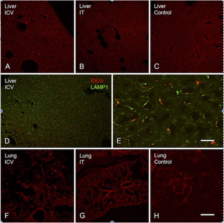
Belur et al.                                                                                                          AAV9-IDUA Gene Therapy for MPS I

                                            with some sporadic labeling seen in the mid- and fore brain. At 6 months of age,
                                            animals were tested for spatial navigation, memory, and neurocognitive function in
                                            the Barnes maze; all treated animals were indistinguishable from normal heterozygous
                                            control animals, while untreated IDUA deficient animals exhibited significant learning
                                            and spatial navigation deficits. We conclude that IT and IN routes are acceptable and
                                            alternate routes of administration, respectively, of AAV vector delivery to the brain with
                                            effective IDUA expression, while all three routes of administration prevent the emergence
                                            of neurocognitive deficiency in a mouse MPS I model.
                                            Keywords: MPS I, IDUA, AAV9, gene therapy, intracerebroventricular administration, intrathecal injection,
                                            intranasal infusion

INTRODUCTION                                                                     IT administration into the cisterna magna or the lumbar area
                                                                                 (Watson et al., 2006; Hinderer et al., 2014a, 2015; Hordeaux
The mucopolysaccharidoses are a group of rare inherited                          et al., 2019), intravenous injection (Hinderer et al., 2014b;
lysosomal disorders caused by a deficiency in the activity                       Belur et al., 2020), and intranasal (IN) administration (Wolf
of specific lysosomal enzymes, leading to aberrant                               et al., 2012; Belur et al., 2017). Site specific in vivo genome
glycosaminoglycan (GAG) catabolism (Muenzer, 2011; Wraith                        editing using engineered zinc finger nucleases delivered via AAV8
and Jones, 2014). This results in abnormal GAG accumulation in                   targeted to the liver, leads to prevention of neurobehavioral
lysosomes and leads to progressive cellular damage in multiple                   deficits in MPS I mice (Ou et al., 2019). Direct vector injection
organ systems. Mucopolysaccharidosis type I (MPS I) is caused                    into the CNS is invasive, but it is also the most effective
by deficiency of the enzyme α-L-iduronidase (IDUA) and has                       means of transducing large areas of the brain in comparison
a disease spectrum that ranges from mild to severe. The severe                   to other routes of administration, especially when administered
form of the disease (Hurler syndrome) is the most prevalent of                   intracerebroventricularly.
MPS I, with an incidence of 1:100,000. Accumulation of heparan                      We       previously     reported    the     effectiveness    of
and dermatan sulfate leads to systemic disease including growth                  intracerebroventricular (ICV) IDUA-transducing AAV8 vector
impairment, hepatosplenomegaly, cardiac disease, skeletal                        in the prevention of neurocognitive dysfunction in neonatally
dysplasia, severe neurocognitive impairment, and if untreated                    treated MPS I mice (Wolf et al., 2011). We also demonstrated
generally death is observed by age 10. Current treatments include                the effectiveness of intranasally administered IDUA transducing
allogeneic hematopoietic stem cell transplantation (HSCT)                        AAV9 (Belur et al., 2017) and the high level of systemic IDUA
and enzyme replacement therapy (ERT). HSCT is effective in                       achieved in adult MPS I mice intravenously administered IDUA-
treatment of peripheral disease, with improvement and partial                    expressing AAV9 or AAVrh10 vector (Belur et al., 2020). Results
restoration of several symptoms such as growth, mobility, and                    from these studies show the potential for achieving high-level
hepatosplenomegaly. Although HSCT impedes neurological                           expression of IDUA and delivery to the CNS using a less invasive
decline, post-transplant patients continue to exhibit below                      route of AAV vector administration.
normal IQ and impaired cognitive ability (Krivit, 2004; Hess                        In further pursuit of this goal, here we report a direct
et al., 2004; Orchard et al., 2007). Recombinant enzyme is                       comparison of intracerebroventricular (ICV), intrathecal (IT),
used in patients immediately upon diagnosis and is effective                     and intranasal (IN) routes of IDUA-transducing AAV9 vector in
in the treatment of systemic disease (Rohrbach and Clarke,                       adult MPS I mice. Supraphysiological levels (1000 times higher
2007). However, ERT has limited effect on neurologic disease                     than wt) of IDUA were widespread in different parts of the brain
due to inability of the enzyme to cross the blood brain barrier                  after ICV injection of IDUA-expressing AAV9. IDUA levels in
(Begley et al., 2008).                                                           the brain were comparatively reduced (about 10-fold) after IT
    A primary goal of genetic therapy for MPS I is delivery of                   administration as opposed to the ICV route, although relatively
enzyme to the CNS, in order to address neurologic manifestations                 higher in the hindbrain than in the forebrain or the midbrain.
of the disease. AAV vectors, especially AAV serotype 9, are                      Minimally invasive IN instillations restored wild-type or near
particularly effective in transducing a wide variety of tissues in the           wild-type levels of enzyme in all parts of the brain, with a much
body, including tissues of the CNS. AAV9 has also been shown                     higher level of enzyme observed in the olfactory lobe. Despite
to cross the blood brain barrier, which makes it particularly                    the varying levels of enzyme found in different parts of the
useful for systemic delivery with access to the brain (Duque                     brain, all 3 routes of administration prevented neurocognitive
et al., 2009; Foust et al., 2009; Gray et al., 2011; Zhang et al.,               deficit in treated animals as determined in the Barnes maze. We
2011). Different routes of administration can thus be used to                    conclude that while ICV infusion of IDUA-transducing AAV9
access the CNS, including direct injection into the parenchyma                   achieves the highest level of IDUA expression in the CNS, the
of the brain (Desmaris et al., 2004; Ciron et al., 2009; Ellinwood               lower levels of IDUA observed after less invasive IT or IN
et al., 2011), injection of vector into cerebroventricular space                 infusion are nonetheless sufficient to ameliorate neurocognitive
(Wolf et al., 2011; Janson et al., 2014; Hordeaux et al., 2018),                 deficit in MPS I mice.

Frontiers in Molecular Neuroscience | www.frontiersin.org                    2                                      May 2021 | Volume 14 | Article 618360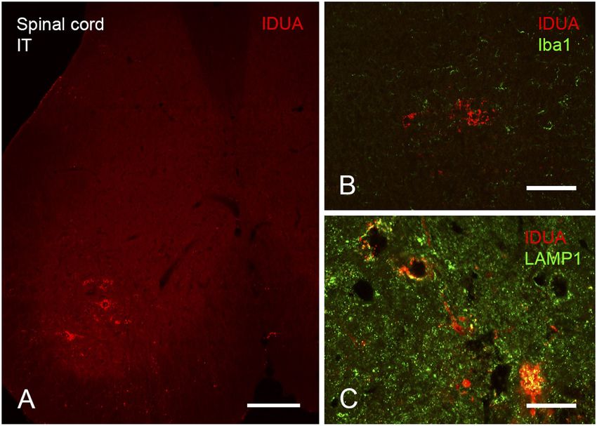
Belur et al.                                                                                                  AAV9-IDUA Gene Therapy for MPS I

MATERIALS AND METHODS                                                    positions the needle such that it slips between the vertebrae and
                                                                         makes contact with the dura mater. A reflexive flick of the tail
Vector Construct                                                         indicated puncture of the dura mater. The injector depresses the
Generation of the miniCAGS regulated IDUA (AAV-MCI)                      Hamilton syringe and introduces the injectate into the CSF of
expression cassette (pTR-MCI) has been described previously              the subarachnoid space (Hylden and Wilcox, 1980; Fairbanks,
(Wolf et al., 2011). This vector was packaged into AAV9                  2003). For IN administration (immunosuppressed, n = 7), mice
virions at the University of Pennsylvania vector core, generating        were anesthetized and placed supine. Vector was administered
recombinant (r) AAV9-IDUA. Vector titer was 1 × 10¹³                     by applying a series of four 3 µl drops with a micropipette to
genome copies/ml.                                                        the nasal cavity of each mouse, alternating between right and left
                                                                         nostrils, at 1 min intervals between each nostril, for a total of 12
                                                                         µl and a full dose of 1 × 10¹¹ vector genomes.
Animals and Immunomodulation
The MPS I mouse strain was generously provided by
Dr. E. Neufeld and IDUA−/− offspring were generated
                                                                         IDUA Enzyme Assay
from homozygous IDUA−/− and homozygous−/− by                             Animals were sacrificed at 3 months post-vector infusion,
heterozygous+/− breeding pairs. Animals were maintained                  transcardiacally perfused with 50 ml PBS, and brains
under specific pathogen-free conditions in AAALAC-accredited             dissected into right and left hemispheres. Each hemisphere
facilities. Animal work was reviewed and approved by the                 was microdissected on ice into olfactory bulb, cortex, striatum,
Institutional Animal Care and Use Committee of the University            hippocampus, cerebellum, thalamus, and brainstem. Tissues
of Minnesota. In order to avoid immune responses, MPS I                  were frozen on dry ice and stored at −20◦ C until processed.
IDUA-deficient animals were immunotolerized starting at                  Tissues were homogenized in 0.9% saline in a bullet bead
birth with an intravenous injection of 5.8 µg/g Aldurazyme               blender, and homogenates were clarified by centrifugation.
(supplied by Dr. P. Orchard), followed by 5 subsequent weekly            Tissue lysates were assayed for IDUA activity in a fluorometric
intraperitoneal injections. A second group of animals was                assay using 4-MU iduronide as substrate (Glycosynth, England),
immunosuppressed with cyclophosphamide (CP) at a dose of                 as previously described (Garcia-Rivera et al., 2007). Emitted
120 mg/kg, administered weekly by intraperitoneal injection,             fluorescence was measured in a BioTek Synergy Mx plate reader.
starting at 1–3 days after vector infusions.                             Protein was measured using the Pierce assay. Enzyme activity
                                                                         is expressed as nmol 4-methylumbelliferone released per mg
                                                                         protein per hour (nmol/mg/h).
Vector Infusions
Immunotolerized and immunosuppressed MPS I animals were
                                                                         GAG Assay
administered vector through the ICV, IT, and IN routes with
                                                                         Tissue lysates were assayed using the Blyscan Sulfated
AAV9-MCI vector at 3 months of age. For ICV delivery,
                                                                         Glycosaminoglycan Assay kit (Accurate Chemical, NY) based on
(immunotolerized, n = 9; immunosuppressed, n = 4; no
                                                                         the manufacturer’s protocol. Tissue GAGs were normalized to
immunomodulation, n = 4) mice were anesthetized with
                                                                         protein and expressed as µg GAG/mg protein.
100 mg/kg ketamine and 16 mg/kg xylazine. The animal was
secured in a Kopf stereotactic frame, and the lateral ventricle
was targeted with a Hamilton syringe (AP, +0.4 mm anterior to            Quantitative Polymerase Chain Reaction
bregma; ML, +0.8 mm right from midline; depth, 2.4 mm deeper             Genomic DNA was extracted from tissue homogenates using
from dura) using standard surgical techniques. Ten microliters           the GeneJET Genomic DNA Purification kit (Thermo Fisher
(1 × 10¹¹ vector genomes) of AAV9-MCI was infused into                   Scientific). Reaction mixtures contained 200 ng of DNA, 2×
the right lateral ventricle by hand using a 10 µl Hamilton               IQ SYBR Green Supermix (Bio-Rad), and 200 nM each of
701 N syringe (Hamilton Chromatography). Briefly, once the               forward and reverse primer. IDUA primers used were forward
syringe was inserted to the designated coordinates, infusion of          primer: 50 -AGGAGATACATCGGTACG-30 and reverse primer:
the AAV vector was begun after a 1 min break. One microliter             50 -TGTCAAAGTCGTGGTGGT-30 . PCR conditions were: 95◦ C
of vector was infused per minute for a period of 10 min. After           for 2 min, followed by 40 cycles of 95◦ C for 40 s, 58◦ C for 30 s,
completion of the infusion, the syringe was left in place for two        and 72◦ C for 1 min. The standard curve for IDUA consisted of
additional minutes before removal of the syringe and suturing of         serial dilutions of plasmid pTR-MCI.
the scalp. The animals were returned to their cages on heating
pads for recovery. For IT injections (immunotolerized, n = 9;            Immunohistochemistry
immunosuppressed, n = 5; no immunomodulation, n = 3),                    At 3 months post-vector infusion, mice were deeply anesthetized
10 µl containing 1 × 10¹¹ vector genomes was injected. The               and perfused via the heart with calcium-free Tyrode’s solution
needle (30-gauge, 0.5-inch) was connected to a length of PE10            (in mM: NaCl 116, KCl 5.4, MgCl2 ·6H2 0 1.6, MgSO4 ·7H2 O
tubing, which was then connected to a second needle that was             0.4, NaH2 PO4 1.4, glucose 5.6, and NaHCO3 26) followed by
attached to a 50 µl Luer-hub Hamilton syringe. The injection was         fixative (4% paraformaldehyde and 0.2% picric acid in 0.1 M
administered to conscious mice by gently gripping the iliac crest        phosphate buffer, pH 6.9). Tissues were dissected and stored
of the rodent and inserting the needle (bevel side up) at about          in PBS containing 10% sucrose and 0.05% sodium azide at
a 45◦ angle centered at the level of the iliac crest. The injector       4◦ C for a minimum of 24 h before being frozen and sectioned

Frontiers in Molecular Neuroscience | www.frontiersin.org            3                                      May 2021 | Volume 14 | Article 618360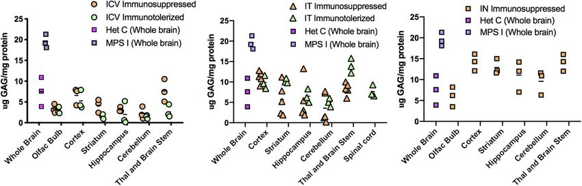
Belur et al. AAV9-IDUA Gene Therapy for MPS I
Statistical Analyses
GraphPad Prism (GraphPad software) was used for all statistical
analyses. For IDUA plasma activity and Barnes maze, data were
compared to normal heterozygote levels and untreated MPS I
mice, respectively, using two-way ANOVA, followed by Dunnett’s
multiple comparisons test. Tissue IDUA activity and GAG levels
were compared to heterozygote levels using the Kruskal Wallis
test. Significance cutoff of < 0.05 was used.
FIGURE 1 | Routes of AAV9-IDUA administration to access the brain. ICV,
Intracerebroventricular; IT, Intrathecal; IN, Intranasal.
RESULTS
at 14 µm thickness using a cryostat. Sections were mounted High Levels of Enzyme Activity in the
onto gel-coated slides and stored at −20◦ C until further use. Brain After CNS-Directed Delivery of
For immunohistochemical staining, sections were incubated in IDUA Transducing AAV9
diluent (PBS containing 0.3% Triton-X100; 1% bovine serum Intracerebroventricular (ICV), intrathecal (IT), and intranasal
albumin, 1% normal donkey serum) for 1 h at room temperature (IN) routes of AAV9 delivery to the CNS were comparatively
followed by incubation in primary antisera overnight at 4◦ C. evaluated for IDUA expression in an IDUA deficient mouse
Primary antisera included sheep anti-IDUA (specific for human model of MPS I (Figure 1). Expression of human IDUA in
IDUA; R & D Systems, Minneapolis, MN, 1:500), rabbit anti- C57BL/6 mice can be compromised by immune response,
Iba1 (Wako, 1:1,000), rabbit anti -LAMP1 (Abcam 1:500), so in our studies animals were either immunotolerized with
rabbit anti-NeuN (Abcam, 1:500). For IDUA immunostaining, Aldurazyme or immunosuppressed with cyclophosphamide
n = 3(ICV), n = 2 (IT), n = 1 (MPS I, Het). Sections were rinsed (CP) as described in Methods, with subsequent administration
in PBS, incubated in species appropriate secondary antisera of AAV9-IDUA vector at 3 months of age. Experimental
(Cy2 1:100, Cy3 1:300, Cy5 1:300; Jackson ImmunoResearch, animals were euthanized at 12 months of age and tissues
West Grove, CA) for 1 h at room temperature, rinsed again harvested for analysis of IDUA expression, storage material and
using PBS, and coverslipped using glycerol and PBS containing vector biodistribution.
p-phenylenediamine (Sigma). Images were collected using an ICV administration of AAV9-IDUA into MPSI animals
Olympus Fluoview 1000 confocal microscope and adjusted for resulted in supraphysiological levels of IDUA in all areas of
brightness and color using Adobe Photoshop software. the micro-dissected brain (Figure 2). Enzyme activity in tissue
extracts from IDUA deficient control animals was undetectable,
Barnes Maze while in Aldurazyme tolerized animals, enzyme levels ranged
At 6 months of age (3 months post-vector infusion), mice from 4- to 1000-fold that of control heterozygous animals
(n = 9–10 animals per group) were analyzed for neurocognitive (Figure 2A). In animals administered CP, enzyme levels
deficits and spatial navigation using the Barnes maze as described ranged from about 100–1,000-fold above normal (Figure 2B).
previously (Belur et al., 2017). Animals were administered 6 trials Surprisingly, animals that did not receive CP also showed very
a day for 4 days. Latency to escape was recorded and analyzed. high levels of enzyme activity that were similar to those of
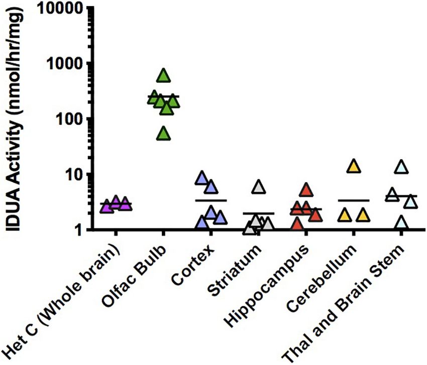
FIGURE 2 | IDUA activity in the brain after ICV administration of AAV9-IDUA vector. Brains were microdissected and assayed for IDUA enzyme activity. Each data
point indicates a value from a single animal with the mean indicated by the short horizontal line. (A) Animals were immunotolerized with Aldurazyme (laronidase)
(n = 9). (B). Animals were immunosuppressed with cyclophosphamide (n = 4). (C) Animals were not immunomodulated (n = 4). Widespread enzyme activity was
seen in all ICV treated groups compared to heterozygote normal controls (n = 3) regardless of whether they were immunomodulated or not. Enzyme was not
detected in untreated MPS I animals (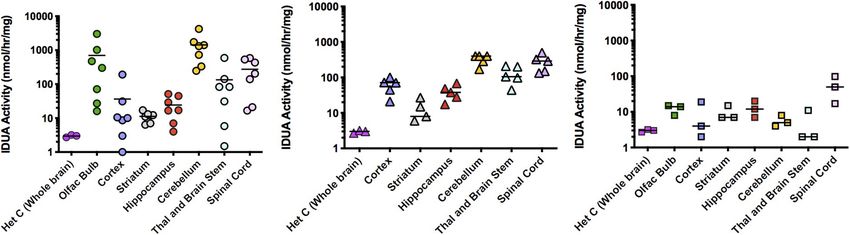
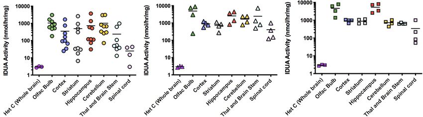
Belur et al.                                                                                                                         AAV9-IDUA Gene Therapy for MPS I

  FIGURE 3 | IDUA Activity in the brain after IT administration of AAV9-IDUA vector. Brains were microdissected and assayed for IDUA enzyme activity. Each data
  point indicates a value from a single animal with the mean indicated by the short horizontal line. (A) Animals were immunotolerized with Aldurazyme (laronidase)
  (n = 9). Widespread enzyme activity was seen in the immunotolerized IT treated group compared to heterozygote normal controls (n = 3). (B) Animals were
  immunosuppressed with cyclophosphamide (n = 5). Animals that were immunosuppressed had lower levels of activity in the cerebellum, compared to
  immunotolerized animals, although the difference was not significant. P-values for treated animals were < 0.01 compared to untreated controls. (C) Animals were
  not immunomodulated (n = 3). Activities in these animals were lower for several areas of the brain, notably the olfactory bulb, cortex, cerebellum, thalamus and brain
  stem. Levels of enzyme activity were close to that of normal heterozygote controls. Enzyme was not detected in untreated MPS I animals (
Belur et al.                                                                                                                        AAV9-IDUA Gene Therapy for MPS I

  FIGURE 5 | Glycosaminoglycan (GAG) storage in brain post-AAV administration. Tissue lysates from different parts of the brain were assayed for GAG storage. The
  levels of GAG found in whole brain of untreated MPS I mice averaged around 20 µg GAG/mg protein, and in normal heterozygotes ranged from 4 to 12 µg GAG/mg
  protein. Each data point indicates a value from a single animal with the mean values represented by horizontal lines. (A) GAG accumulation in brain following ICV
  administration. GAG levels from both immunosuppressed and immunotolerized animals were normalized across the brain. There was no significant difference
  between the 2 immunomodulated groups. P-values were
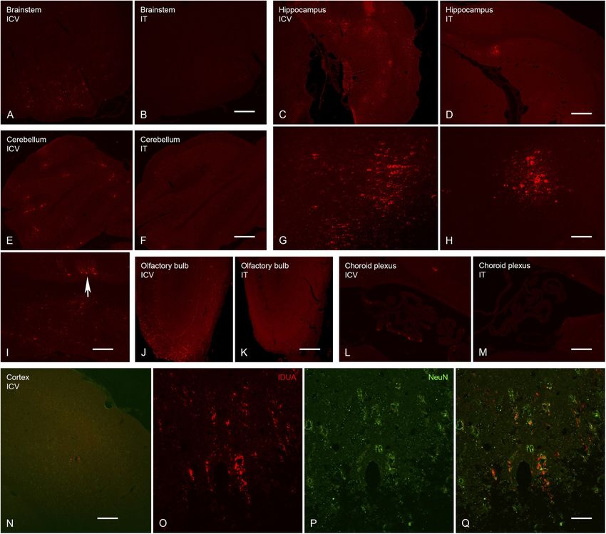
Belur et al.                                                                                                                            AAV9-IDUA Gene Therapy for MPS I

distribution of enzyme activity, scattered IDUA labeling was                              to the systemic circulation as indicated by IDUA expression
observed throughout the brain following ICV administration,                               in the liver (Figure 9). Limited IDUA labeling was also seen
while labeling following IT delivery was more limited (Figure 7).                         in lung.
We observed colocalization of IDUA and NeuN labeling in cortex
of ICV-treated mice, which demonstrated expression of IDUA                                Prevention of Neurocognitive Deficit
within neurons. In addition to brain parenchyma, IDUA labeling                            At 10 months of age, all experimental and control animals
was also observed in cells of the choroid plexus. In spinal cord,                         were evaluated for neurocognitive function using the Barnes
IDUA labeling was observed following IT but not ICV delivery                              maze, a test of spatial navigation and memory (Figure 10).
and colocalized with labeling for LAMP-1 (Figure 8), suggesting                           Animals were evaluated in 6 trials a day for a total of 4 days.
lysosomal subcellular localization of IDUA. Consistent with our                           During this time, normal animals showed an improvement in
previous observations, AAV delivery within cerebrospinal fluid                            spatial navigation, requiring an average of ∼30 s to locate
via ICV or IT injection resulted in redistribution of viral particles                     the escape hole by day 4, while untreated animals showed

  FIGURE 7 | Localization of IDUA immunolabeling in brain following ICV and IT delivery. (A,B) Select neurons in the brainstem showed IDUA-ir following ICV (A) but
  not IT (B) delivery of the vector. (C,D,G,H) Cells of the hippocampus showed IDUA-ir following both ICV (C,G) and IT (D,H) delivery, although many more
  hippocampal neurons were labeled after ICV delivery. (E,F,I) Cells of the cerebellum showed IDUA-ir following ICV (E,I) but not IT (F) delivery of the virus. The majority
  of the labeled cells in the cerebellum were Purkinje cells (arrow in I). (J,K) Many IDUA-ir cells of the olfactory bulb were seen following ICV (J) delivery as opposed to
  IT (K) delivery. Most transduction was seen in the glomerular layer. (L,M) A subset of cells of the choroid plexus showed IDUA-ir after ICV (L) delivery but not IT
  delivery (M). (N–Q) IDUA-ir was seen in the cortex of animals following ICV (N,O,Q) but not IT (not shown) delivery. Colocalization of IDUA (red) and NeuN (green, P
  and Q) labeling in cortex suggests that IDUA-ir could be seen in neurons. Scale bars: (A–F,J–N), 150 µm; (G,H,I) 75 µm; O, P, Q 25 µm.

Frontiers in Molecular Neuroscience | www.frontiersin.org                             7                                              May 2021 | Volume 14 | Article 618360
Belur et al.                                                                                                                   AAV9-IDUA Gene Therapy for MPS I

                                                                                         cells either infused into the host or generated in vivo after
                                                                                         vector infusion into the host. Previous studies have demonstrated
                                                                                         the effectiveness of in vivo and ex vivo IDUA gene transfer
                                                                                         using retroviral (Ponder et al., 2006; Herati et al., 2008a,b;
                                                                                         Metcalf et al., 2010), lentiviral (Visigalli et al., 2010, 2016), AAV
                                                                                         (see below), and Sleeping Beauty transposon vectors (Aronovich
                                                                                         et al., 2007, 2009) in mouse, dog and cat models of MPS I.
                                                                                         This has resulted in the initiation of human clinical trials
                                                                                         testing in vivo AAV mediated IDUA transduction targeting
                                                                                         the liver (Sangamo Therapeutics, SB-318, ClinicalTrials.gov
                                                                                         Identifier: NCT02702115), targeting the CNS (REGENXBIO,
                                                                                         RGX-111, ClinicalTrials.gov Identifier: NCT03580083), and
                                                                                         ex vivo lentiviral transduction of autologous hematopoietic stem
                                                                                         cells (Orchard Therapeutics, OTL-203).
                                                                                             The blood brain barrier sequesters the brain from systemically
                                                                                         administered enzyme replacement therapy (ERT), resulting in
  FIGURE 8 | Localization of IDUA immunolabeling in spinal cord following IT             challenges for the treatment of neurologic disease in MPS
  delivery. (A) Motor neurons of the ventral horn of sacral spinal cord show IDUA        I patients. These challenges can potentially be overcome by
  labeling. (B) IDUA staining is restricted to neurons and is not colocalized with
                                                                                         gene delivery with the use of AAV vector serotypes that are
  the microglial marker Iba1. (C) IDUA is colocalized within neurons with the
  lysosomal marker LAMP1. Scale bars: (A) 150 µm; (B) 25 µm; (C) 75 µm.                  capable of crossing the blood brain barrier. While we have
                                                                                         demonstrated that systemic intravenous delivery of AAV9 and
                                                                                         AAVrh10 vector delivers substantial IDUA enzyme activity to
a significant deficit in this test, requiring an average of ∼90                          the CNS (Belur et al., 2020), direct administration to the
s. In contrast, animals treated with AAV9-IDUA vector by                                 CNS ensures vector delivery and subsequent IDUA expression
either ICV or IT routes of administration exhibited significantly                        in the brain. There are several strategies by which vector
improved neurocognitive skills that were similar to normal                               can be delivered directly to the brain. For MPS I, correction
heterozygous controls, with an escape time of about 30 s                                 of neuropathology after intraparenchymal injection of AAV2
by day 4. Additionally, we have previously demonstrated                                  and AAV5 has been reported in murine, feline, and NHP
improved neurocognitive function in animals treated intranasally                         animal models (Desmaris et al., 2004; Ciron et al., 2009;
with AAV9-IDUA (Belur et al., 2017). We conclude that                                    Ellinwood et al., 2011). Delivery directly into the CSF via
emergence of neurocognitive dysfunction in MPS I mice                                    intracerebroventricular (ICV) injection (Wolf et al., 2011; Janson
is prevented by ICV, IT, or IN administration of AAV9-                                   et al., 2014; Hordeaux et al., 2018) or intrathecal (IT) injection
IDUA.                                                                                    either into lumbar or cisternal spaces in murine, feline, canine,
                                                                                         and NHP models has been reported with varying levels of success
                                                                                         for MPS I (Watson et al., 2006; Hinderer et al., 2014a, 2015;
DISCUSSION                                                                               Hordeaux et al., 2019).
                                                                                             In order to suppress the immune response in animals
In the present study, we compared and evaluated the effectiveness                        elicited by the human IDUA protein (Aronovich et al., 2007,
of different routes of vector administration for IDUA gene                               2009), we explored two immunomodulation approaches. The
delivery to the CNS. We observed supraphysiological and                                  first was immune suppression with cyclophosphamide, while in
highest levels of enzyme expression in the brain following                               the second we immunotolerized animals beginning at birth by
ICV injection, lower levels with IT administration, and lowest                           administering IDUA enzyme protein, Aldurazyme (laronidase).
(wild type) levels after IN delivery of vector. GAG levels were                          In animals that received ICV injection of vector, we found that
normalized following ICV and IT delivery, while after IN                                 levels of enzyme expression in the brain were roughly equivalent,
delivery GAG levels were normalized in the olfactory bulb and                            regardless of whether the animals were immunosuppressed or
close to normal in other parts of the brain. All 3 routes of                             immunotolerized. Surprisingly, control ICV injected animals
administration prevented neurocognitive defect as assessed by                            with no immunomodulation did not show a decrease in enzyme
the Barnes maze.                                                                         activity but exhibited IDUA levels in the brain that were
   The effectiveness of ERT and HSCT in the treatment of MPS I                           equivalent to the other two groups. Animals that received IT
and other lysosomal diseases is based on the concept of metabolic                        injections showed similar results in comparision to animals either
cross-correction, whereby enzyme that is either directly infused                         immunosuppressed or immunotolerized. However, control IT
or expressed by donor cells is taken up by host cells and                                injected animals that received no immunomodulation showed a
trafficked to lysosomes, subsequently contributing to lysosomal                          significant decrease in enzyme activity, with enzyme levels that
metabolism (Fratantoni et al., 1968). The concept of metabolic                           were similar to or slightly higher than normal heterozygotes.
cross-correction also underlies the anticipated effectiveness of                         This difference in response between the non-immunosuppressed
genetic therapies for MPS I and other MPS diseases, wherein                              ICV and IT groups could be explained by a greater amount
the missing enzyme is expressed from genetically transduced                              of vector released into the circulation after IT injection

Frontiers in Molecular Neuroscience | www.frontiersin.org                            8                                       May 2021 | Volume 14 | Article 618360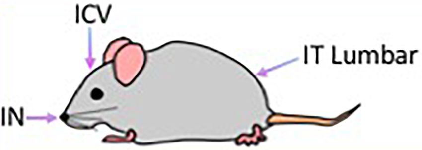
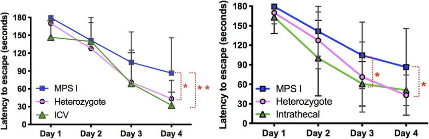
Belur et al.                                                                                                                        AAV9-IDUA Gene Therapy for MPS I

  FIGURE 9 | Localization of IDUA immunolabeling in liver and lung. (A–C) IDUA labeling was seen in the liver of ICV-treated (A) and to a lesser extent IT-treated (B)
  mice, compared to control (C). (D,E) IDUA labeling in liver colocalized with LAMP-1 labeling. (F–H) Sparse IDUA labeling was seen in lung of ICV-treated (F) and to a
  lesser extent IT-treated (G) mice, compared to control (H). Scale bars: (A–D,F–H) 150 µm; (E) 25 µm.

  FIGURE 10 | Assessment of neurocognitive improvement. The Barnes Maze was used to assess spatial learning and memory in immunomodulated animals treated
  with vector using ICV and IT routes (n = 9 in both groups). We have previously demonstrated improvement of neurocognitive deficit in IN administered animals (Belur
  et al., 2017) (*p < 0.05, **p < 0.01). (A) ICV treated animals were significantly improved based on the Barnes maze. Latency to escape in ICV treated animals was
  not significantly different from heterozygote controls, and was significantly improved compared to untreated MPS I animals. (B) IT treated animals showed the same
  pattern of cognitive improvement in the Barnes maze exhibiting significantly better performance compared to untreated MPS I controls.

compared to ICV administration. We did not test for anti-IDUA                          dose when tested soon after (Baldo et al., 2013) or during
antibody levels.                                                                       ERT administration (Ou et al., 2014). However, Schneider et al.
   Improved neurologic outcomes have been reported in MPS                              (2016), reported that in MPS I mice undergoing ERT from
I mice administered aldurazyme starting early and/or at high                           birth, interruption of treatment for a period from 2 to 4

Frontiers in Molecular Neuroscience | www.frontiersin.org                          9                                             May 2021 | Volume 14 | Article 618360
Belur et al. AAV9-IDUA Gene Therapy for MPS I
months of age compromised neurobehavioral outcome at 6 RGX-111 delivered through the CSF to the CNS for treatment
months of age, indicating deterioration in brain function. Had of MPS I1 .
ERT not been restored from 4 to 6 months in that study, Our results thus support the prospect of developing a non-
the brain and associated neurocognition would likely have invasive approach for IDUA gene delivery to the CNS for
deteriorated further. In our study, we observed normalized time high level enzyme expression and prevention of neurologic
to escape for immunotolerized, AAV9-IDUA treated animals disease in human MPS I.
evaluated in the Barnes maze 5 months after the withdrawal
of enzyme at 5 weeks of age. We did not include a control
group of MPS I mice that were administered Aldurazyme DATA AVAILABILITY STATEMENT
but not administered AAV9-IDUA, so it is formally possible
that some of the improvement seen in the Barnes maze is The original contributions presented in the study are
attributable to enzyme alone. However, the fact that performance included in the, further inquiries can be directed to the
in the Barnes maze was not reduced in comparison with corresponding author/s.
wild-type animals argues that this normalized neurocognitive
function is at least partially attributable to AAV9-IDUA
treatment of MPS I mice at 3 months of age. Moreover,
we have recently carried out experiments in MPS I animals ETHICS STATEMENT
immunosuppressed with CP and treated with AAV9-IDUA
Animal work reported in this study was reviewed and approved
intrathecally and intravenously. We observed complete retention
by the Institutional Animal Care and Use Committee of the
of cognitive function in these animals, thus demonstrating
University of Minnesota.
that treatment with AAV9-IDUA in the absence of enzyme
therapy prevents neurodegeneration in this animal model
(Belur et al., 2021).
Results from IDUA immunofluorescence analysis were AUTHOR CONTRIBUTIONS
consistent with the biochemical data obtained from animals
administered AAV9-IDUA via the different routes of LB planned and carried out experiments, collected, analyzed,
administration. ICV administered animals demonstrated and interpreted data, and wrote the manuscript. MR and
the highest levels of IDUA expression, with a high percentage JL carried out biochemical assays. KP-P performed animal
of IDUA positive cells widespread throughout the brain. immunomodulation and assisted with neurobehavioral testing.
IT injected animals showed IDUA positive cells scattered ZN performed ICV administrations. MR and LV performed
throughout the brain, with most of the labeling confined to IHC staining, image collection, analysis, and interpretation.
the hindbrain. Intranasal administration led to IDUA positive KKi carried out IT injections. CF, KFK, PO, WF, WL, and
cells localized exclusively in the olfactory bulb (Belur et al., RM provided expertise and critical feedback. RM conceived
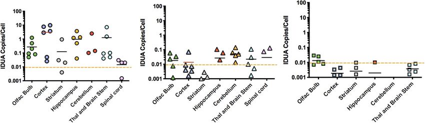
2017). The pattern of vector copy biodistribution by qPCR and designed the experiments, provided input on data analysis
also reflected that of IDUA enzyme activities observed in and interpretation, and supervised the project. All authors
different parts of the brain after vector infusion via the different contributed to the article and approved the submitted version.
routes of administration, with ICV administered animals
showing the highest vector copy number, followed by IT
injected animals, followed by IN administered animals. Results FUNDING
from the Barnes maze indicated that the levels of enzyme
in the brain after vector treatment were sufficient to rescue This study was funded by the NIH grant P01 HD032652 to
animals from the neurocognitive deficit observed in untreated the University of Minnesota and NIH grant R41 DK094538
affected animals. to REGENXBIO Inc. Behavioral studies were performed in the
This is the first study comparing different routes of vector Mouse Behavior Core at the University of Minnesota (supported
delivery at the same dose directly to the brain in adult MPS I mice. by NIH grant NS062158).
Our data demonstrate that ICV injection of vector, although
invasive, results in very high and widespread distribution of
enzyme in the brain. Lumbar IT injections result in high levels
ACKNOWLEDGMENTS
of enzyme in the hindbrain that are comparable to ICV levels,
but enzyme levels were lower in the forebrain and midbrain We thank core director Dr. Benneyworth for assistance with
compared to ICV administration. Intranasal administration the Barnes maze testing. This study was previously presented
showed the lowest enzyme levels of the three routes of delivery, as a poster at an American Society of Gene and Cell Therapy
but nevertheless, resulted in enzyme levels that were sufficient conference, and has been published in abstract form in
to reverse neurocognitive deficit (Belur et al., 2017). These conference proceedings.
results have considerable clinical implications for the treatment
of MPS diseases using AAV 9 vectors: REGENXBIO is currently
enrolling patients in a Phase I/II gene therapy clinical trial of 1
https://clinicaltrials.gov/ct2/show/NCT03580083?term=RGX-111&rank=1
Frontiers in Molecular Neuroscience | www.frontiersin.org 10 May 2021 | Volume 14 | Article 618360Belur et al. AAV9-IDUA Gene Therapy for MPS I
REFERENCES mucopolysaccharidosis I and VII dogs after neonatal gene therapy. Mol. Genet.
Metab. 95, 142–151. doi: 10.1016/j.ymgme.2008.07.003
Aronovich, E. L., Bell, J. B., Belur, L. R., Gunther, R., Koniar, B., Erickson, D. C., Herati, R. S., Ma, X., Tittiger, M., Ohlemiller, K. K., Kovacs, A., and Ponder, K. P.
et al. (2007). Prolonged expression of a lysosomal enzyme in mouse liver after (2008b). Improved retroviral vector design results in sustained expression after
sleeping beauty transposon-mediated gene delivery: implications for non-viral adult gene therapy in mucopolysaccharidosis I mice. J. Gene Med. 10, 972–982.
gene therapy of mucopolysaccharidoses. J. Gene Med. 9, 403–415. doi: 10.1002/ doi: 10.1002/jgm.1229
jgm.1028 Hess, D. C., Abe, T., Hill, W. D., Studdard, A. M., Carothers, J., Masuya, M., et al.
Aronovich, E. L., Bell, J. B., Khan, S. A., Belur, L. R., Gunther, R., Koniar, B., (2004). Hematopoietic origin of microglial and perivascular cells in brain. Exp.
et al. (2009). Systemic correction of storage disease in MPS I NOD/SCID Neurol. 186, 134–144. doi: 10.1016/j.expneurol.2003.11.005
mice using the sleeping beauty transposon system. Mol. Ther. 17, 1136–1144. Hinderer, C., Bell, P., Gurda, B. L., Wang, Q., Louboutin, J. P., Zhu, Y., et al.
doi: 10.1038/mt.2009.87 (2014a). Intrathecal gene therapy corrects CNS pathology in a feline model of
Baldo, G., Mayer, F. Q., Martinelli, B. Z., de Carvalho, T. G., Meyer, F. S., de mucopolysaccharidosis I. Mol. Ther. 22, 2018–2027. doi: 10.1038/mt.2014.135
Oliveira, P. G., et al. (2013). Enzyme replacement therapy started at birth Hinderer, C., Bell, P., Gurda, B. L., Wang, Q., Louboutin, J. P., Zhu, Y., et al.
improves outcome in difficult-to-treat organs in mucopolysaccharidosis I mice. (2014b). Liver- directed gene therapy corrects cardiovascular lesions in feline
Mol. Genet. Metab. 109, 33–40. doi: 10.1016/j.ymgme.2013.03.005 mucopolysaccharidosis type I. Proc. Natl. Acad. Sci. U.S.A. 111, 14894–14899.
Begley, D. J., Pontikis, C. C., and Scarpa, M. (2008). Lysosomal storage diseases doi: 10.1073/pnas.1413645111
and the blood-brain barrier. Curr. Pharm. Des. 14, 1566–1580. doi: 10.2174/ Hinderer, C., Bell, P., Louboutin, J. P., Zhu, Y., Yu, H., Lin, G., et al. (2015).
138161208784705504 Neonatal systemic AAV induces tolerance to CNS gene therapy in MPS I
Belur, L., Huber, A., Mantone, M., Karlen, A., Smith, M., Ou, L., et al. dogs and nonhuman primates. Mol. Ther. 23, 1298–1307. doi: 10.1038/mt.
(2021). Comparative systemic and neurologic effectiveness of intravenous and 2015.99
intrathecal AAV9 delivered individually or combined in a murine model of Hordeaux, J., Hinderer, C., Buza, E. L., Louboutin, J. P., Jahan, T., Bell, P., et al.
mucopolysaccharidosis type I. Mol. Gen. Metab. 132, S19–S20. (2019). Safe and sustained expression of human iduronidase after intrathecal
Belur, L. R., Podetz-Pedersen, K. M., Tran, T. A., Mesick, J. A., Singh, N. M., Riedl, administration of adeno-associated virus serotype 9 in infant rhesus monkeys.
M., et al. (2020). Intravenous delivery for treatment of mucopolysaccharidosis Hum. Gene Ther. 30, 957–966. doi: 10.1089/hum.2019.012
type I: a comparison of AAV serotypes 9 and rh10. Mol. Genet. Metab. Rep. Hordeaux, J., Hinderer, C., Goode, T., Katz, N., Buza, E. L., Bell, P., et al. (2018).
24:100604. doi: 10.1016/j.ymgmr.2020.100604 Toxicology study of intra-cisterna magna adeno-associated virus 9 expressing
Belur, L. R., Temme, A., Podetz-Pedersen, K. M., Riedl, M., Vulchanova, L., human alpha-L-iduronidase in rhesus macaques. Mol. Ther. Methods Clin. Dev.
Robinson, N., et al. (2017). Intranasal adeno-associated virus mediated gene 10, 79–88. doi: 10.1016/j.omtm.2018.06.003
delivery and expression of human iduronidase in the central nervous system: Hylden, J. L., and Wilcox, G. L. (1980). Intrathecal morphine in mice: a
a noninvasive and effective approach for prevention of neurologic disease in new technique. Eur. J. Pharmacol. 67, 313–316. doi: 10.1016/0014-2999(80)
mucopolysaccharidosis type I. Hum. Gene Ther. 28, 576–587. doi: 10.1089/hum. 90515-4
2017.187 Janson, C. G., Romanova, L. G., Leone, P., Nan, Z., Belur, L., McIvor, R. S., et al.
Ciron, C., Cressant, A., Roux, F., Raoul, S., Cerel, Y., Hantraye, P., et al. (2009). (2014). Comparison of endovascular and intraventricular gene therapy with
Human α- iduronidase gene transfer mediated by adeno-associated virus types adeno-associated virus-α-L-iduronidase for Hurler disease. Neurosurgery 74,
1, 2, and 5 in the brain of nonhuman primates: vector diffusion and bio- 99–111. doi: 10.1227/NEU.0000000000000157
distribution. Hum. Gene Ther. 20, 350–360. doi: 10.1089/hum.2008.155 Krivit, W. (2004). Allogeneic stem cell transplantation for the treatment of
Desmaris, N., Verot, L., Puech, J. P., Caillaud, C., Vanier, M. T., and Heard, J. M. lysosomal and peroxisomal metabolic diseases. Springer Semin. Immunopathol.
(2004). Prevention of neuropathology in the mouse model of Hurler syndrome. 26, 119–132. doi: 10.1007/s00281-004-0166-2
Ann. Neurol. 56, 68–76. doi: 10.1002/ana.20150 Metcalf, J. A., Ma, X., Linders, B., Wu, S., Schambach, A., Ohlemiller, K. K., et al.
Duque, S., Joussemet, B., Riviere, C., Marais, T., Dubreil, L., Douar, A. M., (2010). A self-inactivating gamma-retroviral vector reduces manifestations of
et al. (2009). Intravenous administration of self-complementary AAV9 enables mucopolysaccharidosis I in mice. Mol. Ther. 18, 334–342. doi: 10.1038/mt.2009.
transgene delivery to adult motor neurons. Mol. Ther. 17, 1187–1196. doi: 236
10.1038/mt.2009.71 Muenzer, J. (2011). Overview of the mucopolysaccharidoses. Rheumatology 50,
Ellinwood, N. M., Ausseil, J., Desmaris, N., Bigou, S., Liu, S., Jens, J. K., et al. (2011). v4–v12. doi: 10.1093/rheumatology/ker394
Safe, efficient, and reproducible gene therapy of the brain in the dog models Orchard, P. J., Blazar, B. R., Wagner, J., Charnas, L., Krivit, W., and Tolar, J. (2007).
of Sanfilippo and Hurler syndromes. Mol. Ther. 19, 251–259. doi: 10.1038/mt. Hematopoietic cell therapy for metabolic disease. J. Pediatrics 151, 340–346.
2010.265 doi: 10.1016/j.jpeds.2007.04.054
Fairbanks, C. A. (2003). Spinal delivery of analgesics in experimental models of Ou, L., DeKelver, R. C., Rohde, M., Tom, S., Radeke, R., St Martin, S. J., et al. (2019).
pain and analgesia. Adv. Drug Deliv. Rev. 55, 1007–1041. doi: 10.1016/s0169- ZFN-mediated in vivo genome editing corrects murine Hurler Syndrome. Mol.
409x(03)00101-7 Ther. 27, 178–187. doi: 10.1016/j.ymthe.2018.10.018
Foust, K. D., Nurre, E., Montgomery, C. L., Hernandez, A., Chan, C. M., Ou, L., Herzog, T., Koniar, B. L., Gunther, R., and Whitley, C. B. (2014). High-dose
and Karpar, B. K. (2009). Intravascular AAV9 preferentially targets neonatal enzyme replacement therapy in murine Hurler syndrome. Mol. Genet. Metab.
neurons and adult astrocytes. Nat. Biotechnol. 27, 59–65. doi: 10.1038/nbt.1515 111, 116–122. doi: 10.1016/j.ymgme.2013.09.008
Fratantoni, J. C., Hall, C. W., and Neufeld, E. F. (1968). Hurler and Hunter Ponder, K. P., Wang, B., Wang, P., Ma, X., Herati, R., Wang, B., et al. (2006).
syndromes: mutual correction of the defect in cultured fibroblasts. Science 162, Mucopolysaccharidosis I cats mount a cytotoxic T lymphocyte response after
570–572. doi: 10.1126/science.162.3853.570 neonatal gene therapy that can be blocked with CTLA4-Ig. Mol. Ther. 14, 5–13.
Garcia-Rivera, M. F., Colvin-Wanshura, L. E., Nelson, M. S., Nan, Z., Khan, doi: 10.1016/j.ymthe.2006.03.015
S. A., Rogers, T. B., et al. (2007). Characterization of an immunodeficient Rohrbach, M., and Clarke, J. T. (2007). Treatment of lysosomal storage disorders:
mouse model of mucopolysaccharidosis type I suitable for preclinical testing of progress with enzyme replacement therapy. Drugs 67, 2697–2716. doi: 10.2165/
human stem cell and gene therapy. Brain Res. Bull. 74, 429–438. doi: 10.1016/j. 00003495-200767180-00005
brainresbull.2007.07.018 Schneider, A. P., Matte, U., Pasqualim, G., Tavares, A. M., Mayer, F. Q., Martinelli,
Gray, S. J., Matagne, V., Bachaboina, L., Yadav, S., Ojeda, S. R., and Samulski, R. J. B., et al. (2016). Deleterious effects of interruption followed by reintroduction of
(2011). Preclinical differences of intravascular AAV9 delivery to neurons and enzyme replacement therapy on a lysosomal storage disorder. Trans. Res. 176,
glia: a comparative study of adult mice and nonhuman primates. Mol. Ther. 19, 29–37.e1. doi: 10.1016/j.trsl.2016.05.002
1058–1069. doi: 10.1038/mt.2011.72 Visigalli, I., Delai, S., Ferro, F., Cecere, F., Vezzoli, M., Sanvito, F., et al.
Herati, R. S., Knox, V. W., O’Donnell, P., D’Angelo, M., Haskins, M. E., (2016). Preclinical testing of the safety and tolerability of lentiviral vector-
and Ponder, K. P. (2008a). Radiographic evaluation of bones and joints in mediated above-normal alpha-L-iduronidase expression in murine and human
Frontiers in Molecular Neuroscience | www.frontiersin.org 11 May 2021 | Volume 14 | Article 618360Belur et al. AAV9-IDUA Gene Therapy for MPS I hematopoietic cells using toxicology and biodistribution good laboratory Zhang, H., Yang, B., Mu, X., Ahmed, S. S., Su, Q., He, R., et al. (2011). Several practice studies. Hum. Gene Ther. 27, 813–829. doi: 10.1089/hum.2016.068 rAAV vectors efficiently cross the blood-brain barrier and transduce neurons Visigalli, I., Delai, S., Politi, L. S., Di Domenico, C., Cerri, F., Mrak, E., et al. (2010). and astrocytes in the neonatal mouse central nervous system. Mol. Ther. 19, Gene therapy augments the efficacy of hematopoietic cell transplantation and 1440–1448. doi: 10.1038/mt.2011.98 fully corrects mucopolysaccharidosis type I phenotype in the mouse model. Blood 116, 5130–5139. doi: 10.1182/blood-2010-04-278234 Conflict of Interest: KFK was employed by REGENXBIO Inc. at the time of the Watson, G., Bastacky, J., Belichenko, P., Buddhikot, M., Jungles, S., Vellard, M., study. et al. (2006). Intrathecal administration of AAV vectors for the treatment of lysosomal storage in the brains of MPS I mice. Gene Ther. 13, 917–925. doi: The remaining authors declare that the research was conducted in the absence of 10.1038/sj.gt.3302735 any commercial or financial relationships that could be construed as a potential Wolf, D. A., Hanson, L. R., Aronovich, E. L., Nan, Z., Low, W. C., Frey, W. H., conflict of interest. et al. (2012). Lysosomal enzyme can bypass the blood-brain barrier and reach the CNS following intranasal administration. Mol. Genet. Metab. 106, 131–134. Copyright © 2021 Belur, Romero, Lee, Podetz-Pedersen, Nan, Riedl, Vulchanova, doi: 10.1016/j.ymgme.2012.02.006 Kitto, Fairbanks, Kozarsky, Orchard, Frey, Low and McIvor. This is an open-access Wolf, D. A., Lenander, A. W., Nan, Z., Belur, L. R., Whitley, C. B., Gupta, P., article distributed under the terms of the Creative Commons Attribution License et al. (2011). Direct gene transfer to the CNS prevents emergence of neurologic (CC BY). The use, distribution or reproduction in other forums is permitted, provided disease in a murine model of mucopolysaccharidosis type I. Neurobiol. Dis. 43, the original author(s) and the copyright owner(s) are credited and that the original 123–133. doi: 10.1016/j.nbd.2011.02.015 publication in this journal is cited, in accordance with accepted academic practice. Wraith, J. E., and Jones, S. (2014). Mucopolysaccharidosis type I. Pediatr. No use, distribution or reproduction is permitted which does not comply with Endocrinol. Rev. 12, 102–106. these terms. Frontiers in Molecular Neuroscience | www.frontiersin.org 12 May 2021 | Volume 14 | Article 618360
You can also read