Adolescent depression and brain development: evidence from voxel-based morphometry - Journal of Psychiatry ...
←
→
Page content transcription
If your browser does not render page correctly, please read the page content below
Research Paper
Adolescent depression and brain development:
evidence from voxel-based morphometry
Joana Straub, PhD; Rebecca Brown, PhD; Kathrin Malejko, MD;
Martina Bonenberger, PhD; Georg Grön, PhD; Paul L. Plener, MD; Birgit Abler, MD
Published online Feb. 5, 2019; subject to revision
Background: Investigating adolescents and young adults may provide a unique opportunity to understand developmental aspects of the
neurobiology of depression. During adolescence, a considerable physiologic reorganization of both grey and white matter of the brain
takes place, and it has been suggested that differences in grey-matter volumes during adolescence may reflect different maturational
processes. Methods: We investigated grey-matter volumes in a comparatively large sample (n = 103) of adolescents and young adults
(aged 12 to 27 years), 60 of them with a diagnosis of current depression. Results: Replicating previous studies, we found a clear whole-
brain effect of age: the older the participants, the lower their global grey-matter volumes, particularly in the paracingulate and prefrontal
cortices. Contrasting depressed and healthy youth in a whole-brain approach, we found greater grey-matter volumes in the dorsolateral
prefrontal cortex of those with depression. Furthermore, a region-of-interest analysis indicated lower grey-matter volumes in the hippo-
campus in participants with depression compared with healthy controls. Limitations: The present study was limited because of a
skewed sex distribution, its cross-sectional design and the fact that some participants were taking an antidepressant. Conclusion: During
adolescence, restructuring of the brain is characterized by marked decreases in prefrontal grey-matter volumes, interpreted as a correlate
of brain maturation. Findings of greater volumes in the prefrontal cortex, particularly in younger adolescents with depression, may suggest
that these participants were more prone to delayed brain maturation or increased neuroplasticity. This finding may represent a risk factor
for depression or constitute an effect of developing depression.
Introduction pression in early adolescence,7 leading to suggestions of
potential causative mechanisms underlying this increased
Current research has provided evidence for neurobiological risk. Investigating adolescents and young adults may pro-
correlates of depression in terms of brain function and struc- vide a unique opportunity to understand the developmental
ture. Typical findings involve reduced hippocampal volumes aspects of the neurobiology of depression.8 So far, however,
and lower grey-matter volumes in cortical regions, particu- results have been conflicting. The typical findings in adults
larly the subgenual anterior cingulate cortex (ACC). These related to the subgenual ACC and hippocampus have also
findings have been reported in a review,1 and findings of been reported in a larger study in depressed adolescents and
lower hippocampal and cingulate cortex volumes have been young adults (n = 168),9 but a meta-analysis that investigated
replicated in a large meta-analysis that sampled more than cortical thickness as another aspect of grey-matter structure
1900 adults with depression.2 Because neurobiological traces in adolescents did not replicate the result pattern.10 Instead,
of depression are still detectable after remission, it has been in a subsample of approximately 200 adolescents and young
suggested that these are trait markers indicating increased adults with a mean age of approximately 19 years, the auth
vulnerability to depressive illness.3,4 However, it remains to ors showed that the cortical thinning seen in older patients
be determined whether these findings in adults reflect neuro- (> 21 years of age) did not appear in younger participants.10
developmental influences that may have increased people’s During adolescence, a considerable reorganization of both
basic predisposition for depression years before. grey and white matter occurs. Childhood and adolescence
The first signs of depression in many patients are observed are characterized by overall increases in white-matter vol-
during adolescence, and the cumulative prevalence of de- umes and a regionally specific development of grey-matter
pression by the end of adolescence is approximately 20%.5,6 volumes in an inverted U-shape.11 While subcortical, phylo-
Particularly striking is the rapid rise of the incidence of de- genetically “older” structures such as the basal ganglia seem
Correspondence to: J. Straub, Universitatsklinikum Ulm Klinik fur Kinder- und Jugendpsychiatrie Psychotherapie, Steinhoevelstrasse 5 Ulm,
Baden-Württemberg 89075, Germany; Joana.Straub@uniklinik-ulm.de
Submitted Nov. 23, 2017; Revised May 30, 2018; Revised July 30, 2018; Accepted Sept. 22, 2018
DOI: 10.1503/jpn.170233
© 2019 Joule Inc. or its licensors
J Psychiatry Neurosci 1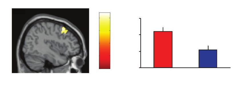
Straub et al.
to reach peak volumes earlier in life (around age 7 to 8 in and youth:9,24 in addition to the decreased hippocampal
girls and around age 10 in boys), grey-matter volume in fron- volumes found by Jaworska and colleagues,9 Hagan and
tal and parietal cortices peaks around age 10 to 12 and in the colleagues24 reported effects of depression on grey-matter
temporal cortex only around age 16, with girls showing volume, particularly in the thalamus and ACC.
earlier peaks than boys. Consequently, areas related to motor We formulated 2 main hypotheses. First, using whole-brain
or sensory functioning, such as somatosensory or visual corti- analysis, we expected to replicate an effect of age in the entire
ces, seem to mature earlier than higher-order association sample: that cortical volumes (particularly in phylogenetically
areas.11 Similarly, in terms of cortical thickness (as a measure newer areas of the brain such as the frontal, parietal and tem-
of another aspect of grey-matter structure) the dorsolateral poral cortices) decrease with age, in line with previous studies
prefrontal cortex (dlPFC) reaches adult levels particularly (hypothesis 1).24 In a second whole-brain analysis, we investi-
late.12 Differences in grey-matter volumes during adolescence gated the hypothesis that depressed adolescents would have
in depressed versus healthy young people may therefore relatively increased prefrontal cortex volumes compared to
reflect different maturational processes.13 Influences of age healthy controls, as has been found inconsistently in smaller
and sex on grey-matter volumes could explain different samples (hypothesis 2).15 We expected that our sample —
results for grey-matter volumes in prefrontal areas. There is which had a skewed distribution toward younger participants,
meta-analysis evidence for volume reductions in adults,14 and where maturational effects should be relatively large —
evidence of increased and decreased volumes in different would be well suited to detecting such differences.
groups of adolescents.15,16 However, for most brain regions, it As a secondary goal, we hypothesized that we would
has been suggested that lower grey-matter volumes may replicate the finding of decreased hippocampal volumes in
reflect greater maturity of the brain during adolescence.11 depressed adolescents relative to controls using a region-
Findings of greater prefrontal cortex volumes, particularly in of-interest (ROI) analysis, as consistently reported by Jaworska
younger depressed adolescents, may suggest that these indi- and colleagues9 and in several studies with smaller sample
viduals are more prone to delayed brain maturation as a risk sizes (hypothesis 3).20–23 We also investigated the effects of
factor for developing depression. Alternatively, increased illness on grey-matter volume in the ACC (ROI analysis) and
prefrontal volumes may also be linked to neuroplasticity pro- thalamus (whole-brain analysis) as previously reported by
cesses. In this vein, the rumination and compulsive symp- Hagan and colleagues,24 predicting decreased grey-matter
toms frequently seen in depression have been suggested to volumes in depressed participants (hypothesis 4).
relate to increased prefrontal volumes.17,18
For subcortical regions such as the hippocampus, the situ- Methods
ation is different: in healthy adolescents and young adults,
hippocampal volumes grow continuously.19 However, in de- Participants
pressed adolescents (as in adults), decreased volumes20–23
have been reported fairly consistently. A possible reason for We obtained participants’ morphological and clinical data as
this may be that maturation in this structure follows other part of 2 functional imaging projects on the effects of cogni-
principles, and a decrease in volume is not a sign of matura- tive behavioural therapy in depressed adolescents25 and on
tional processes but of already affected structural integrity as depression and nonsuicidal self-injury in adolescents and
a consequence of depression. A further explanation for lower young adults (part of the project published in Groschwitz
hippocampal volumes might be that the compulsive brood- and colleagues26). From these studies, we included data from
ing commonly seen in depression could interfere with the depressed patients (before cognitive behavioural therapy)
functioning of episodic memory, which in turn could ad- and healthy controls aged 12 to 27 years. After excluding
versely affect the volume of the hippocampus.17 1 patient and 1 control because of poor data quality, we had
In the current study, we investigated grey-matter volumes morphological data for 103 participants. All 60 participants in
in a reasonably large sample of 103 adolescents, all after the the depression group met DSM-IV criteria for current major
onset of puberty, and young adults (age range 12 to 27 years). depressive disorder at the time of the MRI. All were inpa-
We hypothesized that including both adolescents and young tients or outpatients of the Department of Child and Adoles-
adults would allow us to replicate earlier findings related to cent Psychiatry and Psychotherapy or of the Department of
maturational processes in the brain, irrespective of diagnosis. Psychiatry and Psychotherapy of Ulm University, or they
We expected to find significant effects of age in prefrontal were outpatients from a private child and adolescent psych
and temporal brain regions, where restructuring is assumed iatry practice in Ulm, Germany. Exclusion criteria were a cur-
to take place during this period of life (with greater grey- rent or previous diagnosis of bipolar disorder, schizophrenia
matter volumes in younger participants). We expected the or substance abuse; an intellectual disability; or a major so-
somatosensory and visual cortices, as well as phylogen matic or neurologic disorder. Of the patients with depression,
etically older brain areas such as the hippocampus and 20 were taking psychotropic medication at the time of the
amygdala, to show no such age effects irrespective of diagno- scan, usually antidepressants for their current episode. Of
sis, because maturational processes in these areas should be these, 9 had a history of current or previous long-term medi-
less evident after the onset of puberty.11 Furthermore, we ex- cation of more than 2 months. Four additional patients were
pected to replicate the findings of 2 previous studies on grey- currently unmedicated but had a history of previous long-
matter volumes in larger samples of depressed adolescents term medication. In the control group, we included only
2 J Psychiatry NeurosciAdolescent depression and brain development
articipants who had never been diagnosed with any psychi-
p participants, irrespective of diagnosis. We tested whole-brain
atric disorder and who were matched for age, education and group differences between healthy controls and patients
sex. None of the controls had a history of current or previous (hypothesis 2: whole brain analysis) for significance using the
psychotropic medication. All participants had reached pu- 2-sample t test module for unpaired samples in SPM 12. For
berty as indicated by Tanner stage 3 in males and menstrua- both analyses, we included age and sex as covariates, because
tion in females. All participants and caregivers (in the case of previous investigations showed a clear influence of these
minors under age 18 years) provided written informed con- variables on grey-matter volumes.11,24,31 Sex was a nuisance
sent. Procedures were carried out in accordance with the covariate in both analyses; age was the variable of interest for
Declaration of Helsinki (2013), and the studies were ap- hypothesis 1 and a nuisance covariate for hypothesis 2. We
proved by the Institutional Review Board of Ulm University. added long-term psychotropic medication (> 2 months
Diagnoses were assessed using the German version of the currently or in the past; n = 13) as another covariate of no
clinical interview Schedule for Affective Disorders and interest to the model. Thresholds for both analyses were set
Schizophrenia for School-Age-Children-Present and Lifetime at p < 0.001 at the voxel level, together with a family-wise
(K-SADS-PL) for DSM diagnoses27 in adolescents up to age error (FWE) correction for multiple comparisons at pFWE < 0.05
18 years, or the Structured Clinical Interview for DSM diag- at the cluster level. We extracted estimates of grey-matter
noses (SCID) in young adults.28 To assess current depressive volumes from regions with a significant statistical effect to
symptoms, we used the Beck Depression Inventory second visualize effects.
edition (BDI-II),29 German version.30
Replication of results from previous MRI studies
Structural MRI data acquisition To further investigate the validity of our data, we directed
additional analyses toward replication of the main results of
We used a 3.0 T Siemens MAGNETOM Allegra Scanner previous studies from other research groups in similar
(Siemens) equipped with a head coil to obtain MRI data. We samples, particularly those by Jaworska and colleagues9 and
acquired anatomic high-resolution T1-weighted images using a Hagan and colleagues.24 As in Jaworska and colleagues,9 we
magnetically prepared rapid acquisition gradient echo tested an a priori hypothesis (hypothesis 3, ROI analysis) for
sequence (MPRAGE: 1 × 1 × 1 mm voxels, band width 130 Hz/ differential effects in the hippocampus using an ROI
pixel, repetition time 2500 ms, inversion time 1.1 s, echo time approach and expecting smaller volumes in depressed youth.
4.57 ms, flip angle 12°, field of view 256 × 256, 192 sagittal Because the tracing procedures used by Jaworska and
slices) as part of a larger imaging protocol. colleagues9 to delineate the left and right hippocampus were
not available at our site, we used the hippocampus templates
Statistical analysis of behavioural data from the atlas for automated anatomic labelling accessible via
the WFU Pick Atlas for SPM (http://fmri.wfubmc.edu/
We performed statistical analyses using the Statistical software/PickAtlas). We thresholded the map at p < 0.01 at
Package for the Social Sciences 21 (SPSS Inc.). We com- the voxel level. To control for multiple comparisons, we used
puted between-group differences by means of 2-sample t a cluster-extent threshold of pFWE < 0.05 in combination with
tests and χ2 tests. For correlational analyses, we applied the the small-volume correction (SVC) in SPM, applying the
Pearson coefficient. All tests were performed with levels of hippocampus masks bilaterally.
significance established at p < 0.05 (2-tailed). To study the differential effects in the thalamus as one
main finding of Hagan and colleagues24 (hypothesis 4, whole-
Statistical analysis of structural MRI data brain analysis) we first performed an analysis at the whole-brain
level, expecting greater thalamic volumes, particularly in
Preprocessing younger depressed participants than in controls. The
We conducted image preprocessing using the Computational second main finding in that study was a differential effect in
Anatomy Toolbox for SPM 12 (CAT12, http://dbm.neuro. the ACC (hypothesis 4, ROI analysis). Like Hagan and
uni-jena.de/cat12/) with the following steps: normalization, colleagues, we used the atlas for automated anatomic
segmentation and quality check for sample homogeneity. labelling accessible via the WFU pickatlas for SPM and
Using standard settings of the toolbox, we normalized data combined templates for “Cingulum_Ant_R” and
into Montreal Neurological Institute (MNI) space and seg- “Cingulum_Ant_L” to delineate a bilateral ACC ROI. At the
mented them into grey matter, white matter and cerebrospinal voxel level, we used the same threshold for the ROI analysis
fluid using the SPM12 tissue probability maps for spatial regis- as Hagan and colleagues (p < 0.004), combined with an
tration and segmentation. We conducted spatial smoothing as FWE correction for multiple comparisons in small volumes
the final step of preprocessing with a Gaussian kernel of 6 mm (p FWE < 0.05). Because the sample studied by Hagan and
full width at half maximum using SPM 12 standard routines. colleagues included only participants younger than 18 years,
we investigated group differences between patients with
Whole-brain analyses depression and healthy controls in the whole group, but
We assessed the effect of age (hypothesis 1: whole brain calculated a separate analysis in participants up to age 18 years
analysis) on grey-matter volumes in the whole brain using a (n = 79). For hypotheses 3 and 4, we again included age, sex
simple regression analysis across the entire group of and medication as covariates of no interest in the analyses.
J Psychiatry Neurosci 3Straub et al.
Subgroup analyses: patients with psychiatric codiagnoses medial prefrontal cortex (MNI coordinates: x, y, z = −8, 59, 18;
excluded Z = 5.57; number of voxels = 28 181), very close to the region
To investigate whether psychiatric codiagnoses had an im- with the maximum effect of age on grey-matter volume
pact on our data, we recalculated each analysis. To exclude found by Hagan and colleagues (MNI coordinates: x, y, z =
effects of a history of anorexia nervosa (as found in 2 pa- −2, 52, 2).24 We observed further age effects in the ACC and
tients), we designed a model with 58 patients versus 43 con- medial prefrontal cortex, insula, lateral prefrontal cortex,
trols. To exclude effects of any psychiatric codiagnosis, we inferior and superior parietal regions, and precuneus. As
excluded the 18 patients with a history of psychiatric codiag- expected, we found no significant effects of age in the
nosis (including anorexia nervosa) from calculations, setting visual or somatosensory cortices, the amygdala or the
up a model with 42 patients versus 43 controls. hippocampus. We found no significant positive relationship
between larger grey-matter volume and increasing age.
Results
Group differences: hypothesis 2
Behavioural data The comparison of patients with depression and healthy con-
trols revealed a significant group difference in the dlPFC,
Both groups (patients and healthy controls) included more with greater grey-matter volumes in patients than in healthy
females than males, and the majority were right-handed. One controls (MNI coordinates: x, y, z = 44, 18, 50; Z = 4.19; num-
patient did not complete the BDI-II questionnaire. In the ber of voxels = 548; Fig. 2). We found no other significant
remaining group of 59 patients, the mean BDI-II score clusters for this comparison, and testing for the opposite
indicated a moderate degree of depressive symptoms (mean direction (smaller grey-matter volume in patients) provided
± standard deviation = 24.31 ± 10.37). Age, handedness and no significant results. An interaction analysis of group × age
sex did not differ between groups. Samples were roughly revealed no significant results (data not shown).
matched for education level. For more details, see Table 1.
Analyses according to a priori hypotheses from other studies
Whole-brain analyses
Differential effect in the hippocampus (ROI analysis):
Effect of age: hypothesis 1 hypothesis 3)
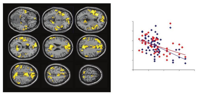
Regression analysis revealed a significant effect of age over a Comparing grey-matter volumes in the left and right
large array of cortical regions (Fig. 1): the older the hippocampus ROIs, we confirmed the finding of Jaworska
participant, the lower the grey-matter volumes. The global and colleagues.9 We found significantly lower grey-matter
maximum in this analysis was in the paracingulate and volumes in the posterior hippocampus in depressed youth
Table 1: Demographic and clinical characteristics of study participants
Characteristic Depressed patients (n = 60) Healthy controls (n = 43) Group differences
Sex, no. (%) 48 F (80) 38 F (88.4) NS*
BDI-II score, mean ± SD 24.31 ± 10.37 3.07 ± 3.44 t74 = 14.66, p < 0.001
Age, yr, mean ± SD (range) 17.30 ± 3.44 (13–27) 17.62 ± 3.85 (12–27) t101 = −0.44, p = 0.66
Secondary diagnosis, no.† Anorexia nervosa: 2 — —
Social phobia: 6
Attention deficit/hyperactivity disorder: 4
Socialized conduct disorder: 3
Specific phobia: 2
Borderline personality disorder: 1
Handedness, no. (%)‡ 58 right-handed (96.7) 40 right-handed (93) NS*
Intake of psychotropic Antidepressant (mirtazepine: 3; fluoxetine: 2; — —
medication currently or in the escitalopram: 2; sertraline: 3)
past > 2 months, no.§ Antipsychotic (quetiapine: 1)
Psychostimulant (methylphenidate: 4)
Education level, no.¶ Hauptschule: 10 Hauptschule: 5 NS*
Realschule: 23 Realschule: 13
Gymnasium: 20 Gymnasium: 22
Berufsschule: 5 Berufsschule: 3
Missing: 2
NS = not significant; SD = standard deviation.
*χ2 test.
†
Diagnosis according to DSM-IV; n = 18.
‡
Edinburgh Handedness Inventory; a independent sample t test.
§
n = 13.
¶
A Gymnasium is a type of school providing secondary education, comparable to English grammar schools and United States college preparatory high
schools. Hauptschule and Realschule are secondary schools. Berufsschule is a professional school that students attend along with an apprenticeship.
4 J Psychiatry NeurosciAdolescent depression and brain development
versus healthy controls (MNI coordinates: x, y, z = −18, −35, pFWE,SVC < 0.05, controlling for multiple comparisons. This
−2; Z = 2.54; number of voxels = 10). effect was not significant when analyzing data from
participants up to age 18 years. However, as described by
Group differences in the thalamus (whole-brain analysis) Hagan and colleagues24 in participants up to age 18 years and
and the ACC (ROI analysis): hypothesis 4 using the ROI approach, we found decreased grey-matter
We found a differential effect in the thalamus, with greater volumes in depressed patients compared with healthy
volumes in depressed participants than in healthy controls controls in the ACC, midcingular portion (MNI coordinates:
(MNI coordinates: x, y, z = −12, −17, 20; Z = 3.37; number of x, y, z = −6, 24, 20; Z = 3.21; number of voxels = 72). This
voxels = 6) at thresholds of p < 0.001 at the voxel level and effect was not significant for the whole group.
z = –15 z = –5 z=5
Grey-matter volume at global maximum
(MNI coordinates: x, y, z = –8, 59, 18)
0.8
Depressed participants
Healthy controls
0.7
z = 15 z = 25 z = 35
0.6
0.5
0.4
z = 45 z = 55 z = 65
0.3
10 15 20 25 30
Age, yr
Fig. 1: Left: Results of the whole-brain regression analysis over the entire group of participants (n = 103); p < 0.001 at the voxel level, pFWE <
0.05 at the cluster level. Right: Correlation of individual grey-matter volume (estimated relative concentration) and age at the global maxi-
mum of the age effect, with depressed participants and healthy controls in different colour coding. FWE = family-wise error; MNI, Montreal
Neurological Institute.
x = 42
4 0.55
Grey-matter volume
3
0.50
2
0.45
1
0 0.40
Depressed Healthy
participants controls
Fig. 2: Differential effect with greater grey-matter volumes (relative concentration) in the dorsolateral prefrontal cortex of depressed partici-
pants (n = 60) compared to matched healthy controls (n = 43). Whole-brain analysis, thresholded p < 0.001 at the voxel level, pFWE < 0.05 at
the cluster level. FWE = family-wise error.
J Psychiatry Neurosci 5Straub et al.
Subgroup analyses: patients with psychiatric codiagnoses lateral frontal cortex 32 and in frontal regions/prefrontal
excluded cortex.35 Our results point toward a similar situation in a
larger sample with respect to grey-matter volume, which is a
Exclusion of the 2 patients with a history of anorexia nervosa related, but not overlapping measure of grey-matter
revealed results that were not different from those of the structure.
whole sample. Exclusion of all 18 patients with any Despite these consistencies, it must be noted that our
psychiatric codiagnosis revealed unchanged results for findings were in contrast to those of a smaller study (n = 22
hypotheses 1 and 3, as well as for hypothesis 4 in the per group) by Shad and colleagues,16 which found lower
thalamus. For hypothesis 2, findings in the dlPFC survived grey-matter volumes in the dlPFC in depressed adolescents
correction for multiple comparisons, but only when applying compared with healthy controls. One explanation for this
a simple cluster-level correction at p < 0.05 (no FWE inconsistency, besides the smaller sample size, might be that
correction). The results of these analyses are reported in depressed participants in that study were younger (mean age
greater detail in Appendix 1, available at jpn.ca/170233. 15 years; range 12 to 20 years) than those in our study (mean
age 17.3 years; range 13 to 27 years). Furthermore, Hagan and
Discussion colleagues,24 investigating a similarly large sample, did not
report group differences in grey-matter volume between
Investigating grey-matter volumes in a comparatively large depressed and healthy adolescents. A reason for their finding
sample of peri- and postpubertal adolescents and young might be that participants were again younger (mean age
adults with depression, we found further support for the 15.65 years; range 11.83 to 17.96) than those in our study but
observation of relatively increased prefrontal volumes in this older than those in the study of Shad and colleagues.16 These
population; we also replicated findings from previous observations may guide conclusions that differences in brain
studies. Across all participants in the present study, results maturation reflected in different prefrontal grey-matter
revealed the expected age effect (hypothesis 1): the older the volumes may be more prominent in slightly older
participants, the lower their global grey-matter volumes, adolescents. Different findings might also have been due to
particularly in the paracingulate and the prefrontal cortex. A sex differences — Hagan and colleagues24 included more
comparison of both groups (hypothesis 2) revealed that males (27.8%) than we did (20%) — and the fact that
depressed patients had greater grey-matter volumes in the participants in that study were more often medicated
dlPFC than healthy controls. The interaction of group × age (approximately 33%) than participants in our study (13.3%).
(which could support assumptions of different maturation The dlPFC is an important structure when it comes to the
velocities) was not significant. Replicating the findings of processing of risk and fear, emotion regulation, cognitive
Jaworska and colleagues9 and Hagan and colleagues,24 our control, monitoring of performance, response inhibition and
results indicated lower grey-matter volumes in the behavioural adjustment.32,35,36 It has been suggested that
hippocampus in depressed patients (hypothesis 3) and in the maturational processes in the frontal lobe provide the
ACC in depressed patients under the age of 18 (hypothesis 4) neurophysiological basis for the acquisition of skills and
compared with healthy controls. knowledge related to higher cognitive functioning and social
Our observation of increased dlPFC grey-matter volumes behaviour.37 Significant increases in the development of
in depressed youth compared with healthy controls aligned attentional control are seen around age 15 years, 38 and
well with the results of previous studies and could indicate development of executive functions and problem-solving
that developmental processes in depressed adolescents abilities are found from this age into early adulthood.39
follow different trajectories in brain regions known to be Neurobiological models of depression, hand in hand with
involved in the regulation of emotions and stress. Because impaired frontal lobe functioning, conceptualize some of the
increased dlPFC brain volumes are not typically seen in key phenomena observed in depression. These include
adults,10 we think that a delayed onset of developmental biased attention to and increased processing of negative
processes is a plausible explanation for our finding in stimuli, as well as rumination that is related to impaired or
depressed adolescents. However, our sample did not include dysfunctional attentional and executive control. 40 In
preadolescent participants in each group, necessary to span particular, cognitive behavioural therapy explicitly targets
the entire range of brain maturation and monitor full-scale these aspects. Delayed maturational processes in the frontal
growth curves of grey-matter volumes. Therefore, this lobe may be a risk factor for adolescent depression. Immature
interpretation must remain tentative. Still, previous studies self-regulatory competence in combination with increased
on cortical thickness — a different aspect of grey-matter novelty and sensation-seeking (which have been suggested
structure in depression — have delivered results in a similar to drive commonly observed risky behaviours and emotional
direction to our findings. Studies investigating cortical imbalance in adolescents) may also facilitate depression.41
thickness in older adults (age > 60 years)32 and in younger Furthermore, because symptoms of depression such as
adults (age 24 to 35 years)33,34 have tended to support cortical rumination and compulsive behaviours have been associated
(in particular, prefrontal) thinning in patients with with increased prefrontal volumes,17,18 neuroplasticity effects
depression, but investigations in younger groups (males may stave off the normal age-related decline and keep dlPFC
average age 17 to 21.5 years) have shown thicker cortices in volumes larger. However, it remains unclear whether
depressed participants compared to healthy controls in the delayed cortical development predisposes a person to
6 J Psychiatry NeurosciAdolescent depression and brain development
depression or whether depression delays the brain (particularly in the subgenual portion) have been seen
maturation trajectories. consistently in adults,1 but not in a longitudinal study on
As well as demonstrating greater grey-matter volumes in adolescent depression.47 The ACC is associated with conflict
the prefrontal regions of depressed participants, where monitoring, social decision-making and determination of the
maturational processes peak during adolescence, we also source of social information.51,52 It also plays a key role in the
replicated previous findings of reduced grey-matter volumes processing, regulation and appraisal of emotions. 51
in regions such as the hippocampus.9,24 In these regions, brain Therefore, reduced grey-matter volume in the ACC might
maturation peaks earlier in development and is already relate to interpersonal and emotional impairments in
much more advanced in the age group we investigated.11 depressed patients.
More advanced maturational processes may be the reason Finally, we found tentative evidence for aberrant thalamic
why results from the hippocampus in adolescents are highly volume characteristics in depressed patients versus healthy
similar to those in adults.2 As in adults, prolonged exposure controls, similar to findings reported by Hagan and
to stress (which induces high glucocorticoid levels and colleagues.24 The thalamus undergoes a significant amount of
appears to harm the hippocampus in terms of neurogenesis reorganization from early childhood through adolescence to
and loss of dendritic spines42–44) has been suggested as a early adulthood. 53 However, structural changes in the
mechanism underlying these findings. In line with this idea, thalamus are not consistently found in adult depression,1
77% of depressed minors report that a stressful life preceded despite findings of functional changes.54,55
or triggered the onset of their first depressive episode.45
Furthermore Rao and colleagues46 found that early-life stress Limitations
was associated with both the onset of depression and smaller
hippocampal volumes. Besides that, younger age at onset The present study was limited because of a skewed sex
of major depressive disorder has been associated with distribution biased toward female participants, which limits
smaller hippocampi,9 and a longitudinal study noted that its generalizability to male patients. Furthermore, it was
attenuated hippocampal growth was associated with the limited owing to its cross-sectional design. Although even
onset of depression.47 These findings suggest that smaller short-term psychotropic medication might have affected
hippocampal volumes may predispose for depression, brain development and volume, we were able to control only
possibly because of impaired mnestic processes,42,48 impaired for the long term (> 2 months currently or in the past),
executive functioning and affect regulation.49 Indeed, the because the only available data for short-term medication in
meta-analysis from the ENIGMA group including data from the past were inconsistent. We did not systematically explore
1728 adult patients with major depression identified smaller experiences of triggering stressors such as childhood
hippocampal volume as the most robust marker of maltreatment/trauma in our study despite their well-known
depression, driven primarily by either earlier age of onset or influence on the limbic system. Normalization of images to a
recurrent depression, while recurrent depression did not standard template in voxel-based morphometry may result
moderate hippocampal volumes in early-onset patients.2 The in some deformation of the original brain structure and
authors concluded that early onset of depression, as in our possible errors in detecting small-volume differences. One
adolescent sample, may be independently associated with strength of the present study was the inclusion of relatively
lower hippocampal volumes. An alternative explanation for young participants, which reduced the likelihood that long-
lower hippocampal volumes could be a lack of effective term medication or chronicity of the disorder had already
epistemic foraging and information being consigned to induced marked changes in gross brain anatomy. Another
nonconscious episodic memory, with depressed patients strength was the comparably large sample size obtained from
getting stuck on compulsive, negative perspectives on the a single MRI scanner.
self, world and others. Such excessive top–down rumination
might limit the normal volumetric development of the Conclusion
hippocampus. Indeed, it has been shown that hippocampus
volume accounted for impaired memory function in We found further evidence to support different develop-
depressed versus healthy participants.50 Supporting this, mental trajectories in brain regions relevant for top–down
Wang and colleagues 17 found that depressed patients processing — particularly the dlPFC — in adolescents and
revealed a significant decrease of regional grey-matter young adults with depression. Other brain regions, such as
volume in the parahippocampal gyrus versus healthy the hippocampus, did not show signs in support of such a
controls and that rumination had a mediating effect on the rationale. Insufficient top–down control has been suggested
relationship between depression and regional grey-matter as an explanation for the increased incidence of substance
volume in the parahippocampal gyrus. abuse, risk behaviour and affective disorders during adoles-
In the present study, we also found reduced ACC grey- cence.56 However, it remains unclear whether these changes
matter volume in depressed patients versus healthy controls, are the cause of these disorders or an effect of already on
again in line with the findings of Jaworska and colleagues9 going disorders. In our study, we focused on depressed par-
and Hagan and colleagues24 in adolescents. Although the ticipants, although some might develop other affective disor-
maturation peak for the ACC is less clear, it seems to occur ders later on (for example, initial presentation with
earlier than in the dlPFC. 12 Smaller ACC volumes depression is common in bipolar disorder). Longitudinal
J Psychiatry Neurosci 7Straub et al.
studies in a large sample of younger adolescents are needed scans from 20 cohorts worldwide in the ENIGMA Major Depres-
sive Disorder Working Group. Mol Psychiatry 2016;22:900-9.
to shed further light on these processes and better under- 11. Lenroot RK, Giedd JN. Brain development in children and adoles-
stand natural brain maturation in healthy controls and pre- cents: insights from anatomical magnetic resonance imaging. Neurosci
dictors of depression and other disorders later in life. An- Biobehav Rev 2006;30:718-29.
12. Gogtay N, Giedd JN, Lusk L, et al. Dynamic mapping of human
other approach could be to calculate normative ranges of cortical development during childhood through early adulthood.
grey-matter volume in adolescents and young adults, taking Proc Natl Acad Sci U S A 2004;101:8174-9.
into account data from healthy populations in huge data 13. Paus T, Keshavan M, Giedd JN. Why do many psychiatric disor-
samples (e.g., ENIGMA, Human Connectome Project). In a ders emerge during adolescence? Nat Rev Neurosci 2008;9:947-57.
14. Koolschijn PC, van Haren NE, Lensvelt-Mulders GJ, et al. Brain
next-step deviation, scores of depressed participants versus volume abnormalities in major depressive disorder: a meta-analysis
normative data could be calculated by age range to learn of magnetic resonance imaging studies. Hum Brain Mapp 2009;
whether the brain age of depressed participants is lower than 30:3719-35.
15. Nolan CL, Moore GJ, Madden R, et al. Prefrontal cortical volume
their biological age. Besides analyses of grey-matter volume, in childhood-onset major depression: preliminary findings. Arch
it would be interesting to focus on other aspects of grey- Gen Psychiatry 2002;59:173-9.
matter structure in cortical thickness and surface analyses. 16. Shad MU, Muddasani S, Rao U. Gray matter differences between
healthy and depressed adolescents: a voxel-based morphometry
study. J Child Adolesc Psychopharmacol 2012;22:190-7.
Affiliations: From the Department of Child and Adolescent Psychia- 17. Wang K, Wei D, Yang J, et al. Individual differences in rumination
try and Psychotherapy, Ulm University Hospital, Ulm, Germany in healthy and depressive samples: association with brain struc-
(Straub, Brown, Bonenberger, Plener); the Department of Psychiatry ture, functional connectivity and depression. Psychol Med 2015;
and Psychotherapy III, Ulm University Hospital, Ulm, Germany 45:2999-3008.
(Malejko, Grön, Abler); and the Department of Child and Adolescent 18. Montigny C, Castellanos-Ryan N, Whelan R, et al. A phenotypic
Psychiatry and Psychotherapy, Medical University of Vienna, Vienna, structure and neural correlates of compulsive behaviors in adoles-
Austria (Plener). cents. PLoS One 2013;8: e80151.
19. Krogsrud SK, Tamnes CK, Fjell AM, et al. Development of hippo-
Competing interests: P. Plener declares grants from Servier and campal subfield volumes from 4 to 22 years. Hum Brain Mapp
Lundbeck for clinical studies outside the submitted work. No other 2014;35:5646-57.
competing interests declared. 20. Caetano SC, Fonseca M, Hatch JP, et al. Medial temporal lobe ab-
normalities in pediatric unipolar depression. Neurosci Lett 2007;
Contributors: J. Straub, P. Plener and B. Abler designed the study. 427:142-7.
J. Straub, R. Brown, K. Malejko and M. Bonenberger acquired the 21. MacMaster FP, Kusumakar V. Hippocampal volume in early onset
data, which J. Straub, G. Grön and B. Abler analyzed. J. Straub, depression. BMC Med 2004;2:2.
G. Grön and B. Abler wrote the article, which all authors reviewed. 22. MacMaster FP, Mirza Y, Szeszko PR, et al. Amygdala and hippo-
All authors approved the final version to be published and can certify campal volumes in familial early onset major depressive disorder.
that no other individuals not listed as authors have made substantial Biol Psychiatry 2008;63:385-90.
contributions to the paper. 23. MacMillan S, Szeszko PR, Moore GJ, et al. Increased amygdala:
hippocampal volume ratios associated with severity of anxiety in
pediatric major depression. J Child Adolesc Psychopharmacol
2003;13:65-73.
References 24. Hagan CC, Graham JM, Tait R, et al. Adolescents with current
major depressive disorder show dissimilar patterns of age-related
1. Bora E, Fornito A, Pantelis C, et al. Gray matter abnormalities in differences in ACC and thalamus. Neuroimage Clin 2015;7:391-9.
major depressive disorder: a meta-analysis of voxel based mor- 25. Straub J, Plener PL, Sproeber N, et al. Neural correlates of success-
phometry studies. J Affect Disord 2012;138:9-18. ful psychotherapy of depression in adolescents. J Affect Disord 2015;
2. Schmaal L, Veltman DJ, van Erp TG, et al. Subcortical brain altera- 183:239-46.
tions in major depressive disorder: findings from the ENIGMA 26. Groschwitz RC, Plener PL, Groen G, et al. Differential neural pro-
Major Depressive Disorder Working Group. Mol Psychiatry 2016; cessing of social exclusion in adolescents with non-suicidal self-
21:806-12. injury: an fMRI study. Psychiatry Res 2016;255:43-9.
3. Kendler KS, Thornton LM, Gardner CO. Stressful life events and 27. Delmo C, Weiffenbach O, Gabriel M, et al. Affective disorders and
previous episodes in the etiology of major depression in women: schizophrenia for school-age children – present and lifetime version
an evaluation of the “kindling” hypothesis. Am J Psychiatry 2000; (K-SADS-PL). Frankfurt: Klinik für Psychiatrie und Psychotherapie
157:1243-51. des Kindes- und Jugendalters; 2000.
4. Post RM. Transduction of psychosocial stress into the neurobiology 28. Ventura J, Liberman RP, Green MF, et al. Training and quality as-
of recurrent affective disorder. Am J Psychiatry 1992;149:999-1010. surance with the Structured Clinical Interview for DSM-IV
5. Garrison CZ, Addy CL, Jackson KL, et al. Major depressive disor- (SCID-I/P). Psychiatry Res 1998;79:163-73.
der and dysthymia in young adolescents. Am J Epidemiol 1992; 29. Beck AT, Steer RA, Brown GK. Beck depression inventory, manual.
135:792-802. 2nd ed. San Antonio (TX): The Psychological Corporation; 1996.
6. Whitaker A, Johnson J, Shaffer D, et al. Uncommon troubles in 30. Hautzinger M, Keller F, Kühner C. BDI-II Beck Depressions-Inventar
young people: prevalence estimates of selected psychiatric disor- Revision. Frankfurt am Main: Harcourt Test Services; 2006.
ders in a nonreferred adolescent population. Arch Gen Psychiatry 31. Bray S. Age-associated patterns in gray matter volume, cerebral
1990;47:487-96. perfusion and BOLD oscillations in children and adolescents. Hum
7. Lewinsohn PM, Rohde P, Seeley JR. Major depressive disorder in Brain Mapp 2017;38:2398-407.
older adolescents: prevalence, risk factors, and clinical implications. 32. Fonseka BA, Jaworska N, Courtright A, et al. Cortical thickness
Clin Psychol Rev 1998;18:765-94. and emotion processing in young adults with mild to moderate
8. Allen NB, Sheeber LB. Adolescent emotional development and the depression: a preliminary study. BMC Psychiatry 2016;16:1-9.
emergence of depressive disorders. Cambridge: Cambridge University 33. Salvadore G, Nugent AC, Lemaitre H, et al. Prefrontal cortical
Press; 2009. abnormalities in currently depressed versus currently remitted
9. Jaworska N, Yucel K, Courtright A, et al. Subgenual anterior cin- p atients with major depressive disorder. Neuroimage 2010;54:
gulate cortex and hippocampal volumes in depressed youth: the 2643-51.
role of comorbidity and age. J Affect Disord 2016;190:726-32. 34. Truong W, Minuzzi L, Soares CN, et al. Changes in cortical thick-
10. Schmaal L, Hibar DP, Samann PG, et al. Cortical abnormalities in ness across the lifespan in major depressive disorder. Psychiatry
adults and adolescents with major depression based on brain Res Neuroimaging 2013;214:204-11.
8 J Psychiatry NeurosciAdolescent depression and brain development
35. Reynolds S, Carrey N, Jaworska N, et al. Cortical thickness in 47. Whittle S, Lichter R, Dennison M, et al. Structural brain develop-
youth with major depressive disorder. BMC Psychiatry 2014;14:1-8. ment and depression onset during adolescence: a prospective lon-
36. Patel R, Spreng RN, Shin LM, et al. Neurocircuitry models of post- gitudinal study. Am J Psychiatry 2014;171:564-71.
traumatic stress disorder and beyond: a meta-analysis of func- 48. Peres JFP, Newberg AB, Mercante JP, et al. Cerebral blood flow
tional neuroimaging studies. Neurosci Biobehav Rev 2012;36:2130-42. changes during retrieval of traumatic memories before and after
37. Grattan LM, Eslinger PJ. Frontal lobe damage in children and psychotherapy: a SPECT study. Psychol Med 2007;37:1481-91.
adults: a comparative review. Dev Neuropsychol 1991;7:283-326. 49. Phillips ML, Drevets WC, Rauch SL, et al. Neurobiology of emo-
38. Anderson VA, Anderson P, Northam E, et al. Development of ex- tion perception I: the neural basis of normal emotion perception.
ecutive functions through late childhood and adolescence in an Biol Psychiatry 2003;54:504-14.
Australian sample. Dev Neuropsychol 2001;20:385-406. 50. Jayaweera HK, Hickie IB, Duffy SL, et al. Episodic memory in de-
39. De Luca CR, Wood SJ, Anderson V, et al. Normative data from the pression: the unique contribution of the anterior caudate and hip-
CANTAB: I. Development of executive function over the lifespan. pocampus. Psychol Med 2016;46:2189-99.
J Clin Exp Neuropsychol 2003;25:242-54. 51. Aupperle RL, Allard CB, Simmons AN, et al. Neural responses
40. Disner SG, Beevers CG, Haigh EAP, et al. Neural mechanisms of during emotional processing before and after cognitive trauma
the cognitive model of depression. Natl Rev 2011;12:467-77. therapy for battered women. Psychiatry Res Neuroimaging 2013;
41. Steinberg L. Cognitive and affective development in adolescence. 214:48-55.
Trends Cogn Sci 2005;9:69-74. 52. Behrens TE, Hunt LT, Woolrich MW, et al. Associative learning of
42. Fuchs E, Gould E. Mini-review: in vivo neurogenesis in the adult brain: social value. Nature 2008;456:245-9.
regulation and functional implications. Eur J Neurosci 2000;12:2211-4. 53. Lebel C, Walker L, Leemans A, et al. Microstructural maturation of
43. Kim EJ, Pellman B, Kim JJ. Stress effects on the hippocampus: a the human brain from chidhood to adulthood. Neuroimage
critical review. Learn Mem 2015;22:411-6. 2008;40:1044-55.
44. Conrad CD. Chronic stress-induced hippocampal vulnerability: 54. Graham J, Salimi-Khorshidi G, Hagan C, et al. Meta-analytic evi-
the glucocorticoid vulnerability hypothesis. Rev Neurosci dence for neuroimaging models of depression: state or trait? J Affect
2008;19:395-411. Disord 2013;151:423-31.
45. Essau CA, Conradt J, Petermann F. Frequency, comorbidity, and 55. Hamilton JP, Etkin A, Furman DJ, et al. Functional neuroimaging
psychosocial impairment of depressive disorders in adolescents. of major depressive disorder: a meta-analysis and new integration
J Adolesc Res 2000;15:470-81. of baseline activation and neural response data. Am J Psychiatry
46. Rao H, Betancourt L, Giannetta JM, et al. Early parental care is im- 2012;169:693-703.
portant for hippocampal maturation: evidence from brain mor- 56. Casey BJ, Jones RM, Hare TA. The adolescent brain. Ann N Y Acad
phology in humans. Neuroimage 2010;49:1144-50. Sci 2008;1124:111-26.
J Psychiatry Neurosci 9You can also read