Open Research Online - The ...
←
→
Page content transcription
If your browser does not render page correctly, please read the page content below
Open Research Online
The Open University’s repository of research publications
and other research outputs

Gold nanocarriers for transport of oligonucleotides
across brain endothelial cells
Journal Item
 How to cite:
 Fatima, Nayab; Gromnicova, Radka; Loughlin, Jane; Sharrack, Basil and Male, David (2020). Gold nanocarriers for
 transport of oligonucleotides across brain endothelial cells. PLOS ONE, 15(9), article no. e0236611.

For guidance on citations see FAQs.

c 2020 The Authors

Version: Version of Record

Link(s) to article on publisher’s website:
http://dx.doi.org/doi:10.1371/journal.pone.0236611

Copyright and Moral Rights for the articles on this site are retained by the individual authors and/or other copyright
owners. For more information on Open Research Online’s data policy on reuse of materials please consult the policies
page.

                                      oro.open.ac.uk
PLOS ONE

                                                       RESEARCH ARTICLE

                                                       Gold nanocarriers for transport of
                                                       oligonucleotides across brain endothelial cells
                                                       Nayab Fatima1, Radka Gromnicova1, Jane Loughlin1, Basil Sharrack2, David Male ID1*
                                                       1 Department of Life, Health and Chemical Sciences, The Open University, Milton Keynes, United Kingdom,
                                                       2 Academic Department of Neuroscience and Sheffield, NIHR Translational Neuroscience BRC, Sheffield
                                                       Teaching Hospitals, NHS Foundation Trust, University of Sheffield, Sheffield, United Kingdom

                                                       * David.Male@open.ac.uk
a1111111111
a1111111111
a1111111111                                            Abstract
a1111111111
a1111111111                                            Treatment of diseases that affect the CNS by gene therapy requires delivery of oligonucleo-
                                                       tides to target cells within the brain. As the blood brain barrier prevents movement of large
                                                       biomolecules, current approaches involve direct injection of the oligonucleotides, which is
                                                       invasive and may have only a localised effect. The aim of this study was to investigate the
    OPEN ACCESS                                        potential of 2 nm galactose-coated gold nanoparticles (NP-Gal) as a delivery system of oli-
Citation: Fatima N, Gromnicova R, Loughlin J,          gonucleotides across brain endothelium. DNA oligonucleotides of different types were
Sharrack B, Male D (2020) Gold nanocarriers for        attached to NP-Gal by the place exchange reaction and were characterised by EMSA (elec-
transport of oligonucleotides across brain             trophoretic mobility shift assay). Several nanoparticle formulations were created, with single-
endothelial cells. PLoS ONE 15(9): e0236611.
https://doi.org/10.1371/journal.pone.0236611
                                                       or double-stranded (20nt or 40nt) DNA oligonucleotides, or with different amounts of DNA
                                                       attached to the carriers. These nanocarriers were applied to transwell cultures of human
Editor: Yi Cao, Xiangtan University, CHINA
                                                       brain endothelium in vitro (hCMEC/D3 cell-line) or to a 3D-hydrogel model of the blood-brain
Received: July 7, 2020                                 barrier including astrocytes. Transfer rates were measured by quantitative electron micros-
Accepted: August 31, 2020                              copy for the nanoparticles and qPCR for DNA. Despite the increase in nanoparticle size
Published: September 17, 2020                          caused by attachment of oligonucleotides to the NP-Gal carrier, the rates of endocytosis
                                                       and transcytosis of nanoparticles were both considerably increased when they carried an oli-
Peer Review History: PLOS recognizes the
benefits of transparency in the peer review            gonucleotide cargo. Carriers with 40nt dsDNA were most efficient, accumulating in vesicles,
process; therefore, we enable the publication of       in the cytosol and beneath the basal membrane of the endothelium. The oligonucleotide
all of the content of peer review and author           cargo remained attached to the nanocarriers during transcytosis and the transport rate
responses alongside final, published articles. The
editorial history of this article is available here:
                                                       across the endothelial cells was increased at least 50fold compared with free DNA. The
https://doi.org/10.1371/journal.pone.0236611           nanoparticles entered the extracellular matrix and were taken up by the astrocytes in biologi-
Copyright: © 2020 Fatima et al. This is an open
                                                       cally functional amounts. Attachment of DNA confers a strong negative charge to the nano-
access article distributed under the terms of the      particles which may explain the enhanced binding to the endothelium and transcytosis by
Creative Commons Attribution License, which            both vesicular transport and the transmembrane/cytosol pathway. These gold nanoparticles
permits unrestricted use, distribution, and
                                                       have the potential to transport therapeutic amounts of nucleic acids into the CNS.
reproduction in any medium, provided the original
author and source are credited.

Data Availability Statement: All relevant data are
within the manuscript and its Supporting
Information files.
                                                       Introduction
Funding: NF was supported by a post-graduate
studentship from the Open University and funding       In recent years, there has been great interest in the potential for gene-therapy or gene editing
from Sheffield Hospital Trust. The funders had no      to treat conditions caused by single-gene defects. Many of these conditions affect the central

PLOS ONE | https://doi.org/10.1371/journal.pone.0236611 September 17, 2020                                                                                1 / 17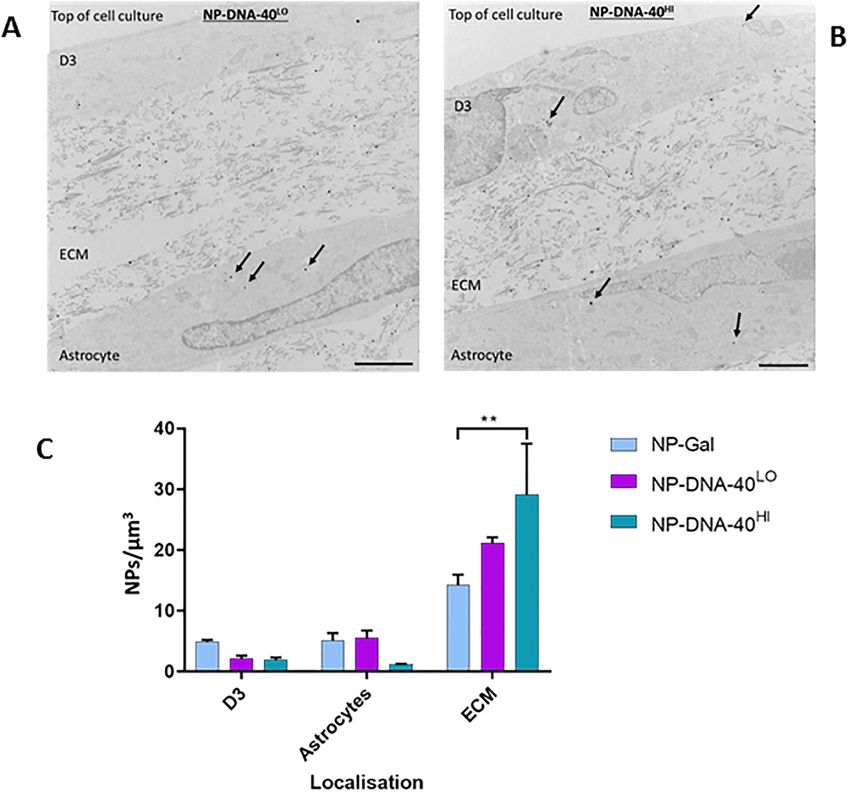
PLOS ONE                                                                         Gold nanocarriers for transport of oligonucleotides across brain endothelial cells

role in study design, data collection and analysis,   nervous system; examples include Huntington’s disease, hereditary forms of Alzheimer’s dis-
decision to publish or preparation of the             ease, lysosomal storage diseases and spinal muscular atrophy. One approach used to treat
manuscript.
                                                      Huntington’s disease is the use of microRNAs to suppress production of toxic proteins [1],
Competing interests: The authors declared that no     while another technique is to excise the defective gene segment with endonucleases [2]. Other
competing interests exist.                            neurological conditions have been treated with anti-sense oligonucleotides [3] or viral vectors
                                                      carrying a gene to replace the defective host gene [4].
                                                          A major obstacle in treating diseases of the CNS is the blood-brain barrier [5], which effec-
                                                      tively excludes all hydrophilic biomolecules greater than 1kDa. Large biomolecules including
                                                      nucleic acids and cytokines are unable to move through the tight junctions between brain
                                                      endothelial cells [6]. Consequently, it has been necessary to deliver gene- therapy by invasive
                                                      methods, such as direct intraventricular or intrathecal injection [7, 8]. With these approaches,
                                                      the distribution of the therapeutic agent is limited to the site of injection or areas close to the
                                                      ventricles. In contrast, transport of the agent across the brain endothelial cells of the blood
                                                      brain barrier following less invasive intravascular injection would potentially provide a
                                                      broader and more even distribution of the treatment. The overall aim of our research is to
                                                      develop methods for transporting large therapeutic biomolecules into the brain. The purpose
                                                      of this study was to determine the conditions that affect the transport of oligonucleotides
                                                      across the blood brain barrier by gold nanocarriers.
                                                          A variety of nanoparticles have been used to transport therapeutic agents across the blood-
                                                      brain barrier [9], including gold nanoparticles [10, 11]. Previously, we have shown that small
                                                      gold nanoparticles (2nm) are able to rapidly cross brain endothelium in vitro and in vivo and
                                                      enter astrocytes or other cells of the brain [12, 13]. The small size of these nanoparticles means
                                                      that they are able to cross brain endothelium by vesicular transcytosis or by direct movement
                                                      across the apical and basal plasma membranes via the cytosol [14]. Another advantage of these
                                                      nanoparticles is their relative safety—they have already been used in phase 1 and phase 2 clini-
                                                      cal trials for delivery of insulin. The FDA approval process for these trials included 24hr and
                                                      28day toxicity and pathology studies in rats and pigs as well as genotoxicity in bacteria. The
                                                      nanoparticles are synthesised with a covalently-bound coat of sugar molecules which main-
                                                      tains their solubility and limits removal by the mononuclear phagocyte system in vivo. The
                                                      sugars (glucose or galactose) are attached during synthesis of the nanoparticles by a disulphide
                                                      bond, which lends itself to an exchange reaction; the bound sugar can be exchanged subse-
                                                      quently with other biomolecules (cargo) that have a free—SH group. The conjugates are stable
                                                      for months when stored in an oxidising environment but will progressively release their cargo
                                                      in a reducing environment [15]. In other studies, gold-RNA or -DNA conjugates have been
                                                      used to modulate gene expression in cells outside the CNS [16], indicating that the oligonucle-
                                                      otides retain their biological activity after internalisation by target cells.
                                                          In the current study oligonucleotides have been attached to the gold nanoparticles, to inves-
                                                      tigate the potential for their transport across brain endothelium and uptake by astrocytes in
                                                      vitro. We have used the brain endothelial cell line hCMEC/D3 in transwell assays and a
                                                      3-dimensional hydrogel astrocyte co-culture model of the blood-brain barrier to investigate
                                                      the uptake and transport of gold nanoparticles with bound oligonucleotide cargos. Specifically,
                                                      we have investigated the transport of nanoparticles carrying varying amounts of single or dou-
                                                      ble-stranded oligonucleotides of 20 or 40 nucleotides in length.
                                                          DNA oligonucleotides of 20–40 nucleotides were chosen as cargo as DNA is less susceptible
                                                      to degradation in vitro than unmodified RNA and the length is in the order of that typically
                                                      employed for therapeutic nucleic acids. The intracellular route taken by nanoparticles has
                                                      been examined using electron microscopy and the distribution of the oligonucleotide cargo
                                                      has been assessed by qPCR.

PLOS ONE | https://doi.org/10.1371/journal.pone.0236611 September 17, 2020                                                                                   2 / 17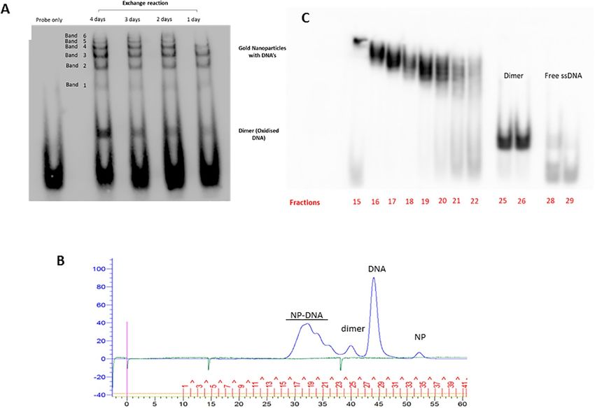
PLOS ONE                                                              Gold nanocarriers for transport of oligonucleotides across brain endothelial cells

                                              The results show that this class of gold nanoparticle can rapidly and effectively transport oli-
                                           gonucleotides across brain endothelial cells and into astrocytes, in quantities that could target
                                           gene-editing or modulate protein expression. Although this model system was designed to
                                           investigate oligonucleotide transport, rather than produce a treatment for any one disease, the
                                           system should be generally applicable for transport of oligonucleotides across the blood-brain
                                           barrier.

                                           Materials and methods
                                           Gold glyconanoparticles
                                           The gold nanoparticles (NP), obtained from Midatech Pharma Plc, were synthesised using a
                                           modified Brust-Schiffrin method [17] and characterised in water using DLS (dynamic light
                                           scattering) on a Nano ZSP Zetasizer (Malvern instruments). They were capped during synthe-
                                           sis with thiol-C2-galactose. The gold core of the nanoparticles was 2nm in diameter, as deter-
                                           mined by transmission electron microscopy, and the size of the glyconanoparticles was
                                           3.63 ± 1.09 nm (mean ± SD) measured by DLS. As determined by molecular mass of the gold
                                           core (~20,000) and physical size, these nanoparticles have approximately 100 gold atoms in the
                                           core and 40 covalently bound C2-galactose molecules on the outside, attached by disulphide
                                           bonds.
                                               Gold in the NP formulations was measured by a spectrophotometric method against gold
                                           standards (Sigma). The assay was performed in a 96 well plate, with a total volume of 200μl in
                                           each well. In each well, 10μl of sample, 10μl H2O and 30μl of 100% freshly prepared aqua regia
                                           (kept on ice) were added. The liquids were then mixed by gentle tapping and left to incubate
                                           for 1 min. Next, 150μl of 2M NaBr was added. The absorbance was read on a plate reader
                                           using OPTIMA FluoSTAR, at 390 nm. The concentrations (μg/ml) of NPs used in the biologi-
                                           cal assays refers to the gold concentration.

                                           Oligonucleotides and attachment to nanoparticles
                                           A 20nt, thiolated ssDNA oligonucleotide (5’ Thiol-C6- AAT ATC GCG GAC AGA
                                           AGA CG 3’) was obtained from Sigma. This sequence was derived from a plasmid of Neis-
                                           seria gonorrhoeae (pCmGFP) (GenBank: FJ172221.1) and was chosen on the basis that it has
                                           no homology in the human, rat or mouse genome and could therefore be readily detected by
                                           qPCR in human, rat or mouse systems without interference from genomic DNA. The oligonu-
                                           cleotide as supplied was substantially oxidised (at the 5’ thiol), and hence was dimerised.
                                           Before attachment to the glyconanoparticles, Tris(2-carboxyethyl)phosphine (TCEP, in 0.1M
                                           tris/HCl, pH 7.5) was used to reduce the DNA to yield free thiol monomers. This was achieved
                                           by incubating the oligonucleotide (7.5mM in 50μl) at room temperature for 4 hours with
                                           TCEP at a molar ratio of 1: 1.25 (DNA:TCEP).
                                               NP-DNA-20ss. After reduction, the oligonucleotide was attached to the NPs by an
                                           exchange reaction. NPs (2mg/ml equivalent of gold) were mixed with reduced 20nt ssDNA at
                                           a molar ratio of 1:14 in a 0.5ml Eppendorf tube, in a sealed container flushed with nitrogen gas
                                           to exclude oxygen. The reaction was allowed to continue for 48 hours at room temperature
                                           unless otherwise stated.
                                               NP-DNA-20ds. In order to make nanoparticles with 20nt double-stranded DNA attached,
                                           the NP-DNA-20 preparation was reacted at room temperature with an equimolar amount of
                                           complementary single-stranded DNA (5’-CGTCATCAGTCCGCGATATT -3’).
                                               NP-DNA-40. To synthesise nanoparticles with 40nt double-stranded DNA, the NP-
                                           DNA-20ss preparation was first hybridised with an equimolar amount of 40nt ssDNA, half
                                           complementary to the 20nt ssDNA bound to the NP (5’–

PLOS ONE | https://doi.org/10.1371/journal.pone.0236611 September 17, 2020                                                                        3 / 17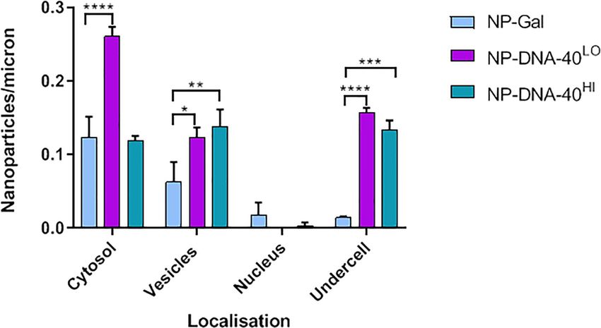
PLOS ONE                                                              Gold nanocarriers for transport of oligonucleotides across brain endothelial cells

                                           AAAAGCTCTGCCTTGGTTTCCGTCTTCTGTCCGCGATATT-3’) for 30 minutes. Then the
                                           extended single-strand of the 40nt oligonucleotide was filled in by hybridisation with an equi-
                                           molar amount of 20nt ssDNA (5’- GAAACCAAGGCAGAGCTTTT- 3’). A diagram showing
                                           the arrangement of the different oligonucleotides attached to the gold core is shown in S1 Fig
                                           in S1 File.
                                              FPLC of nanoparticles. In some experiments the nanoparticles were fractionated by
                                           FPLC (fast protein liquid chromatography) to remove any unbound oligonucleotides and to
                                           separate NPs with high or low levels of bound oligonucleotides. This technique is normally
                                           used to separate proteins, of 10–1000 kDa, but it is equally effective for gold glyconanoparticles
                                           [18]. The nanoparticles were fractionated on an ÄKTA pure FPLC system (GE Healthcare)
                                           using a Superdex 200 10/300 GL column in phosphate buffered saline (PBS). The elution vol-
                                           ume for all fractions was set at 0.5ml at a flow rate of 0.4 ml/min. Detection of oligonucleotides
                                           and nanoparticles was by UV absorption. The pooled fractions with different amounts of
                                           bound 40nt dsDNA are designated NP-DNA-40Hi and NP-DNA-40Lo.
                                              EMSA of nanoparticles. Nanoparticles were analysed by electrophoretic mobility shift
                                           assays (EMSA). This technique is normally used to determine whether proteins have bound to
                                           segments of DNA, but in this case, it was adapted to analyse oligonucleotide attachment to the
                                           nanoparticles. In these experiments the 20nt complementary DNA sequences were substituted
                                           with 5’-biotinylated versions, to allow detection of oligonucleotides on nanoparticles on the
                                           EMSA blots (Bio-CGTCATCAGTCCGCGATATT or Bio-GAAACCAAGGCAGAGCTTTT).
                                           Complementary biotinylated DNA was hybridised with NP-DNA-20ss to form NP-DNA-20ds.
                                           A molar ratio of 1nmol of NP-DNA-20ss to 1.15 nmol of 20nt biotinylated ssDNA (comple-
                                           mentary to thiol-C6-DNA) was used. The sample mixture was adjusted to pH ~ 8 by adding
                                           10X TBE buffer, leaving the final concentration of the TBE buffer 0.5X (pH 8.3) in the sample
                                           mixture. The mixture was incubated for 30 min at room temperature for the hybridization to
                                           take place. The EMSA technique is highly sensitive, so the samples were diluted x100 in pure
                                           water and then prepared with 1 μl NP sample, 6 μl 5X TBE, 13 μl water and 5 μl loading dye
                                           (6X, Fermentas).
                                              NP-DNA-20ds-biotin were separated on a 6% polyacrylamide gel and NP-DNA-40ds-biotin
                                           on a 5% gel. The gels were pre-run in 0.5x TBE at 85 V for 1hr. The samples (20μl in each well)
                                           were then loaded onto the gel and run at 90V for 70 mins for NP-DNA-20ds-biotin and 80
                                           mins for NP-DNA-40ds-biotin. The gels were transferred onto nylon membrane (Amersham
                                           Hybond N+: GE Healthcare) at 100 V for 1hr and 15 mins in 0.5x TBE at 4˚ C. DNA was then
                                           cross-linked onto the membrane with UV light (120mJ/cm2) and developed using a chemilu-
                                           minescent DNA detection kit, according to the manufacturer’s instructions (Thermofisher)
                                           and the blots imaged with a GelDoc.

                                           Tissue culture systems
                                           The human brain endothelial cell line hCMEC/D3 originally characterised in this laboratory
                                           [19] was used at passage 26–30, cultured in modified EBM-2 (Lonza)—containing 0.025%
                                           VEGF, IGF and EGF; 0.1% bFGF, 0.1% (v/v) rhFGF, 0.1% (v/v) gentamycin, 0.1% (v/v) ascor-
                                           bic acid, 0.04% (v/v) hydrocortisone. The amount of serum was reduced to 2.5% (v/v) of foetal
                                           bovine serum. Cytotoxicity of gold nanoparticles on these cells was measured by an Alamar
                                           blue assay [20].
                                              Primary human astrocytes (passage 2–7) obtained from ScienCell were cultured in astrocyte
                                           medium (ScienCell)–supplemented according to manufacturer’s instructions with FBS (2%),
                                           astrocyte growth supplement (1%), and penicillin/streptomycin solution (1%).

PLOS ONE | https://doi.org/10.1371/journal.pone.0236611 September 17, 2020                                                                        4 / 17
PLOS ONE                                                              Gold nanocarriers for transport of oligonucleotides across brain endothelial cells

                                               Transwell cultures for transport assays were produced by seeding hCMEC/D3 cells onto
                                           collagen-coated 0.4 μm polyester membrane transwell inserts (Corning Costar). The transwell
                                           inserts used were either 6.5 mm (seeding density 20,000 cells/insert) or 12 mm (seeding den-
                                           sity 70,000 cells/insert). The hCMEC/D3 cells were grown until confluent (2–3 days). At this
                                           point, nanocarriers (8 μg/ml gold) were added to the upper chamber in EBM-2 MV medium
                                           and incubated at 37˚C for 3 hours. After the incubation, both upper and lower chambers were
                                           washed 3x in 0.5ml Hank’s Balanced Salt Solution (HBSS). The cells were fixed for 1 hr at
                                           room temperature in 2.5% glutaraldehyde in 0.1M PB (Sörensons phosphate buffer). The
                                           fixative was removed and the chambers were washed 3x in PBS and stored in 0.1M PB at 4 ˚C.
                                           The inserts were then processed for electron microscopy to localise and quantitate the gold
                                           nanoparticles.
                                               For quantitation of DNA (qPCR), the medium was directly harvested from the upper and
                                           lower chambers, and the cells were lysed in 1% Triton X100. These samples were then immedi-
                                           ately frozen at -20˚C, until assayed.
                                               A 3-dimensional co-culture of human brain endothelial cells (hCMEC/D3) and human
                                           astrocytes (hA) was used for nanoparticle uptake and transport assays [21]. Astrocytes at 1.5
                                           x106/ml were seeded into a collagen gel solution, prepared using 80% (v/v) rat type I collagen
                                           (2 mg/ml), 10% 10x MEM and 10% human astrocyte cell suspension in astrocyte medium
                                           (500μl/well). The gelling of the collagen+MEM was initiated by careful, dropwise addition of
                                           5M NaOH, to neutral pH, before addition of the astrocytes. The gel was incubated for 20 min
                                           at 37 ˚C then RAFT™ absorber plungers (Lonza) were dropped carefully on top of the collagen
                                           gels to absorb ~90% of the liquid. The RAFT™ absorber plunger was removed after 20 mins.
                                           Astrocyte medium was added to the compressed gels, and the culture was incubated for 24hr.
                                           hCMEC/D3 cells were then seeded on top of the gel and grown over 2 days in modified EBM-
                                           2 medium until they formed a monolayer.
                                               For transport experiments, nanoparticles were added to the 3-dimensional co-culture gels
                                           at a final concentration of 8 μg/ml (gold) in modified EBM-2 media and incubated for 3 hrs at
                                           37˚C. After the incubation, the medium was removed, the cultures were washed 3x in 0.5ml
                                           HBSS and fixed in 2.5% glutaraldehyde for 1 hr. The compressed gels were washed 3x in PBS
                                           and gently detached from the bottom of the 24 well plates with a spatula. They were then
                                           stored in phosphate buffer at 4˚C and processed for electron microscopy.

                                           Localisation and quantitation of nanoparticles by transmission electron
                                           microscopy
                                           Cells were permeabilized in 0.01% Triton X100 for 15 minutes on a rocker and then washed 3x
                                           in 0.1M PB. Silver enhancement solution (Aurion) was prepared according to the manufactur-
                                           er’s instructions and applied to the fixed transwell inserts for 1 hr and 10 mins on a rocker and
                                           then washed 3x in distilled water. Cells were then osmicated in 1% (w/v) osmium tetroxide (in
                                           0.1M PB) for 30 min and then washed 3x in 0.1M PB. The insert was removed from the well,
                                           the membrane was cut out and strips (~3 mm wide) were prepared from the centre of the
                                           membrane. Finally, the membranes were dehydrated by immersion in increasing concentra-
                                           tions of ethanol as follows: 30% ethanol for 5 min, 50% ethanol for 5 min, 70% ethanol for 10
                                           min, 100% ethanol for 10 min (performed twice), 100% ethanol with molecular sieve for 10
                                           min.
                                               After dehydration, the membrane strips were incubated in a 50:50 mixture of 100% ethanol
                                           and Epon resin (Agar) and placed on a rocker overnight. The next day, the insert membrane
                                           pieces were penetrated with freshly prepared Epon resin and changed twice, each change was

PLOS ONE | https://doi.org/10.1371/journal.pone.0236611 September 17, 2020                                                                        5 / 17
PLOS ONE Gold nanocarriers for transport of oligonucleotides across brain endothelial cells
incubated for 2 hrs. The insert membrane pieces were embedded in Epon resin on a cushion
pad for 48 hrs at 60 ˚C.
Microsections of 80nm were collected onto pioloform film-covered copper grids. After at
least 2 hrs of air drying, they were counterstained with 3% aqueous uranyl acetate for 30 mins
followed by Reynolds lead citrate for 10 mins. Minor modifications of this protocol were
needed for preparation of the 3D cultures [21].
The sections were observed at a magnification of 20,000x on JEM 1010 (Jeol). The nanopar-
ticles were counted in cells and sorted into 4 cell compartment categories; vesicles, cytosol,
basal plasma membrane (under cell) and nucleus as previously described [12]. The length of
the insert which was assessed on TEM ranged usually between 300–1500 microns in length.
The number of nanoparticles was then calculated per micron of insert.
Quantification of DNA by qPCR
SYBR Green 2X master mix kit was from Qiagen. Stock forward and reverse primers, corre-
sponding to the sequence of the 40nt dsDNA (S1 Fig in S1 File) were made up at 10μM. The
20μl reaction mixture, assembled on ice, contained 10μl 2X SYBR green mastermix, 1μl of
each of the forward and reverse primers (0.5 μM final concentration), 7μl water and 1μl DNA
template. The template was diluted to give 1-10pg DNA in the reaction mix. Amplification
and detection conditions were 2 min at 50˚, 10 min at 95˚C, followed by 40 cycles of 15 s at
95˚C, and 1 min at 55˚C. The Ct values for each sample were normalised using a standard
curve that was established using the 40nt ssDNA strand used to prepare the NP-DNA-40.
Statistical methods
Data was analysed and graphs prepared using Graphpad Prism 8.0. Tests used, replication and
P values are reported in the figure legends.
Results
Synthesis and characterisation of nanoparticles with bound
oligonucleotides
It was necessary to first establish the conditions for production of gold glyconanoparticles
(NP) with attached oligonucleotides, by the place exchange reaction. Gold glyconanoparticles
with a 2nm core and coat of thiol-C2-galactose were mixed with reduced thiol-C6-DNA (20nt,
single stranded) for 1–4 days at a molar ratio of 1:14. In these conditions, the oligonucleotide
exchanges with theC2-galactose on the surface of the nanoparticles. The reaction products
examined by EMSA (Fig 1A) indicate the position of the oligonucleotide; the 6 bands at the
top of the gel, correspond to nanoparticles with different numbers of covalently bound oligo-
nucleotides. Previous work indicates that each band corresponds to one additional oligonucle-
otide bound to the nanoparticle [22], implying that these nanoparticles can carry up to 6
oligonucleotides. The proportion of larger bands increased up to 2 days but did not increase
further at day 3 (S2 Fig in S1 File). By day 4 there was a substantial increase in oxidised
(dimeric) oligonucleotide, which is unavailable for the exchange reaction. Based on these
observations, 2 days was identified as the optimum time for the exchange reaction in the stated
conditions and was used in subsequent preparations.
The reaction mixture was also examined by FPLC on a G200 column (Fig 1B). Fraction-
ation completely separated the unconjugated NPs and free oligonucleotides (monomers and
dimers) from the NP-DNA. The NP-DNA conjugates appeared as 3 overlapping peaks. Analy-
sis of these fractions by EMSA confirmed that these peaks contained the NP-DNA bands
PLOS ONE | https://doi.org/10.1371/journal.pone.0236611 September 17, 2020 6 / 17PLOS ONE Gold nanocarriers for transport of oligonucleotides across brain endothelial cells
Fig 1. Production and characterisation of nanoparticles with bound oligonucleotides. (A) EMSA of exchange
reaction (1–4 days) probed with a biotinylated oligonucleotide complementary to the ssDNA oligonucleotide attached
to the NPs. (B) FPLC trace of reaction products of exchange reaction with 20nt thiol-C6-ssDNA, at 48hrs. y-axis:
pressure and absorption at 280nm. x-axis: time (0–60 minutes) and fraction number (1–41). (C) EMSA of FPLC
fractions (15–29) of NP-DNA. The numbers 15–29 correspond to the fraction numbers shown in part Fig1B.
https://doi.org/10.1371/journal.pone.0236611.g001
previously identified (Fig 1C). However, FPLC was not able to fully resolve the different
NP-DNA conjugates. Therefore, in order to make NP-DNA preparations with different levels
of bound oligonucleotide, two pools were prepared: DNAHi (fractions 15–17); DNALo (frac-
tions 19–21). The DNAHi fraction corresponds to NPs in the upper bands and the DNALo frac-
tion with lower bands (Fig 1). These two preparations were subsequently used to prepare NPs
with high or low numbers of bound 40nt dsDNA oligonucleotides.
Nanoparticles with different numbers of bound oligonucleotides were examined by trans-
mission electron microscopy (S3 Fig in S1 File), and the size of the gold-cores measured.
There was no change in the core size of the NPs with different numbers of oligonucleotides
bound, indicating that the attachment of the DNA had not caused aggregation of the NPs.
Note that the TEM images identify the core rather than the hydrodynamic diameter of the
NPs. However, the FPLC trace does give data on the hydrodynamic diameter of the NP-DNA.
In the absence of nanoparticle standards, the G200 column was calibrated using protein stan-
dards (S4 Fig in S1 File) and values obtained for each of the FPLC fractions; by interpolation,
the mean hydrodynamic diameters of the NPs were: NP = 1.8nm, NP-DNA-20Lo = 3.7nm and
NP-DNA-20Hi = 4.9nm. The absolute values for the NPs are unlikely to be highly accurate, as
they were measured against protein standards and the nanoparticles with bound DNA are
asymmetric. Nevertheless, it indicates that attachment of the 20nt ssDNA oligonucleotides
increases the effective hydrodynamic diameter of the NP-conjugates by 2–3 fold.
To prepare NPs with 40nt dsDNA oligonucleotides, the NP-DNA-20Hi or NP-DNA-20Lo
preparations were hybridised with 40nt ssDNA, with its 3’ segment complementary to the oli-
gonucleotides attached to the nanoparticle. The remainder of the primary strand was hybri-
dised with a 20nt complementary ssDNA, to make 40nt dsDNA (S1 Fig in S1 File). Unreacted,
PLOS ONE | https://doi.org/10.1371/journal.pone.0236611 September 17, 2020 7 / 17PLOS ONE Gold nanocarriers for transport of oligonucleotides across brain endothelial cells
free oligonucleotides were removed by FPLC, and the products (NP-DNA-40Hi and NP-DNA-
40Lo) verified by EMSA (S5 Fig in S1 File). The hydrodynamic diameters of the NPs with 40nt
dsDNA attached were determined as before (S4 Fig in S1 File): NP-DNA-40Lo = 7.5nm and
NP-DNA-40Hi = 7.6nm.
Previous studies have shown that the base NPs were not cytotoxic for hCMEC/D3 cells up
to 32μg/ml [12]. The NP-DNA-40Hi and NP-DNA-40Lo preparations were also checked for
potential cytotoxicity on these endothelial cells with an Alamar blue assay (S6 Fig in S1 File).
There was no detectable cytotoxicity up to 32μg/ml (gold concentration) over 24 hours. In the
assays described below, NPs were used in cell cultures at 8 μg/ml for 3 hours.
Uptake and transport of DNA nanocarriers by brain endothelium
The uptake and transport of nanoparticles with 20nt ssDNA or dsDNA (unfractionated)
was compared, using TEM to quantitate nanoparticles in different cellular compartments of
hCMEC/D3 cells (Fig 2A). The data is represented as number of nanoparticles per micron of
insert membrane the cells grew on. The compartments assessed were: cytosol, vesicles, nucleus
and under the basal plasma membrane. Examples of electron micrographs with nanoparticles
in different subcellular compartments are shown in Fig 2B. Nanoparticles that have crossed
the basal plasma membrane indicating trans-endothelial transport are listed as ‘undercell’.
Note that the silver enhancement increases the size of the NPs for easier visualisation.
Despite the increase in size of the nanoparticles, the attachment of DNA did not reduce or
block their transport. In contrast, nanoparticles with double stranded DNA were present in
greater numbers in vesicles than NP-DNAss or control NP-Gal.
The transport rate of NPs with a 20nt dsDNA cargo or an equivalent 40nt dsDNA cargo
(unfractionated) was compared (Fig 3A). Examples of electron micrographs indicating the
localisation of nanoparticles are shown in Fig 3B. NP-DNA-40ds were taken up into cytosol
and vesicles more efficiently than NP-DNA-20ds. Moreover, the rate of transcytosis of
NP-DNA-40ds was much greater than NP-DNA-20ds or for NP without any bound DNA, as
indicated by the number of NPs that had crossed the basal membrane of the endothelium.
To investigate the effect that different numbers of bound oligonucleotides have on trans-
port, the rate of uptake of fractionated NP-DNA-40Hi and NP-DNA-40Lo were compared
(Fig 4). As noted previously, the number of NPs with attached oligonucleotides that had
crossed the basal membrane was significantly greater than that of the NPs without cargo,
however there was no consistent difference in transcytosis when comparing NPs with a high
density of DNA and those with a low density. In interpreting these experiments, it is impor-
tant to note that the results show a single time point (3hrs) in a dynamic process. Previous
work has shown that transcytosis of these NPs is detectable within 30 minutes of application
both in vitro and in vivo. The NPs accumulated beneath the basal membrane give a quantita-
tive measure of how many NPs have crossed the cells during the time-course (0-3hrs) but the
levels in the cellular compartments only detect what is present in those compartments at the
3hr time-point.
Transport of DNA across brain endothelium
The experiments described above detected the gold nanocarriers by TEM, but not the DNA.
It was therefore important to confirm that the DNA remains attached to the nanocarriers as
they cross the endothelium and to quantitate the effectiveness of the NPs as carriers. Initially,
NP-DNA bands were cut from the EMSA blots and 1mm2 pieces used as templates in the PCR
reaction. The products examined by agar gel electrophoresis demonstrated that DNA bound
to the gold NPs could still function as a template for PCR. Following optimisation of the
PLOS ONE | https://doi.org/10.1371/journal.pone.0236611 September 17, 2020 8 / 17PLOS ONE Gold nanocarriers for transport of oligonucleotides across brain endothelial cells
Fig 2. Endocytosis of gold nanocarriers with ssDNA or dsDNA by brain endothelium. (A) Nanoparticles in cytosol, vesicles and the nucleus of
hCMEC/D3 cells. NPs with 20nt ssDNA (NP-DNA-20ss) were compared with dsDNA(NP-DNA-20ds) and the base nanoparticles (NP-Gal). Three
experiments were performed, each individual experiment with three technical repeats. Tukey’s multiple comparisons test showed a significant
difference for NP-DNA-20ds compared to NP-Gal (�� P = 0.0034) and NP-DNA-20ss (� P = 0.0133) in vesicles. (B) Transmission electron
micrographs showing examples of silver enhanced gold NPs in vesicles (NP-DNA-20ds, A) or the cytosol (NP-Gal, B) of hCMEC/D3 cells after
application to the apical surface.
https://doi.org/10.1371/journal.pone.0236611.g002
reaction with different primers, the combination of a 20nt forward primer (AATATCGCGGA
CAGAAGACG) and 24nt reverse primer with a 4nt overhang (GAGCAAAAGCTCTGCCTTGGT
TTC) was adopted. This combination allowed the amplicon (44nt) to be distinguished from
any residual template (40nt) in the reaction products.
Having established conditions for PCR detection of NP-DNA, the transport of NP-DNA-
40Hi, NP-DNA-40Lo and free 40nt dsDNA were compared. Transport of the DNA was mea-
sured in hCMEC/D3 transwells collecting medium from the upper and lower chambers and
PLOS ONE | https://doi.org/10.1371/journal.pone.0236611 September 17, 2020 9 / 17PLOS ONE Gold nanocarriers for transport of oligonucleotides across brain endothelial cells
Fig 3. Endocytosis and transcytosis of nanocarriers with 20nt or 40nt dsDNA cargo. (A) Uptake of NP-DNA-20ds and NP-DNA-40ds into brain
endothelial cells. Nanoparticles were quantified in the cytosol, vesicles, nucleus, and at the basal membrane (undercell). NP-Gal was the nanoparticle
control, no DNA attached. Three experiments were performed, each experiment having three technical repeats. Tukey’s multiple comparisons test
showed significant difference for NP-DNA-40ds compared to NP-DNA-20ds in cytosol (��� P = 0.0003), vesicles (�� P = 0.0082) and under cell
(� P = 0.0108). (B) Electron micrographs of silver enhanced gold NPs in vesicles and at the basal membrane of hCMEC/D3 cells treated at the apical
membrane (top) with NP-DNA-40ds (A) or NP-DNA-20ds.
https://doi.org/10.1371/journal.pone.0236611.g003
from the endothelial layer following lysis of the cells. Amounts of transported DNA were
determined by qPCR and interpolated from a standard curve with known amounts of 40nt
ssDNA (Fig 5). The results show that free DNA was not taken up by the cells or transported
across the transwells, but significant amounts of DNA were transported to the lower chamber,
when attached to the gold nanocarriers. Moreover, NP-DNA-40Lo was more efficiently trans-
ported into the bottom chamber than NP-DNA-40Hi.
PLOS ONE | https://doi.org/10.1371/journal.pone.0236611 September 17, 2020 10 / 17PLOS ONE Gold nanocarriers for transport of oligonucleotides across brain endothelial cells
Fig 4. Endocytosis and transcytosis of nanocarriers with high or low levels of dsDNA cargo. Nanoparticles with high or low
levels of 40nt dsDNA located in cytosol, vesicles and nucleus of hCMEC/D3 cells and beneath the basal membrane (undercell).
Three experiments were performed, each individual experiment having three technical repeats. Tukey’s multiple comparisons test
showed significant difference for NP-DNA-40Lo in cytosol, vesicles and “undercell” compared to NP-Gal (���� PPLOS ONE Gold nanocarriers for transport of oligonucleotides across brain endothelial cells
Fig 6. Endocytosis of nanocarriers with dsDNA cargo in a blood-brain barrier model. (A and B) Representative TEM images from
3-independent experiments, showing sliver-enhanced hydrogel cocultures of hCMEC/D3 cells (D3) overlying collagen extracellular matrix (ECM)
with embedded astrocytes. Arrows indicate examples of nanoparticles. Scale bars = 2.0 μm. (C) Quantitation of nanoparticles in endothelium,
astrocytes and collagen ECM. The number of NP-DNA-40Hi in the matrix was significantly greater than for NP-Gal control, by Tukey’s multiple
comparison test (PPLOS ONE Gold nanocarriers for transport of oligonucleotides across brain endothelial cells
sections (80nm). The results show that nanoparticles do enter the astrocytes, although the den-
sity of nanoparticles is higher in the matrix than in either of the two cell types (Fig 6C). There
was no significant difference between levels of different nanoparticle types in the astrocytes,
although unlike endothelial cells in the transwell system, there was a higher proportion of
cytosolic uptake than vesicular internalisation in astrocytes (S7 Fig in S1 File). The results
demonstrate that these nanoparticles can move across the endothelial cells and be taken up by
astrocytes.
Discussion
The work of our group has shown that gold nanoparticles rapidly cross brain endothelium in
vitro and in vivo [13, 23]. Here, we have replaced some of the galactose ligand on the nanopar-
ticles with oligonucleotides to model a therapeutic cargo. The adaptation of FPLC to fraction-
ate gold nanoparticles with different stoichiometries and the development of EMSA for
characterisation of nanocarriers has allowed us to produce and characterise gold nanoparticles
with different types and amounts of attached oligonucleotides.
Taken together the results show that these gold-NP can transport dsDNA-oligonucleotides
across brain endothelial cells. Addition of oligonucleotides to the NP-Gal actually enhances
their internalisation. Moreover, NPs with longer oligonucleotides (40nt) are transported more
efficiently than those with shorter oligonucleotides (Fig 3). However, increasing the density of
the oligonucleotides on the nanoparticles had a limited effect on the transport rate (Fig 4).
It is known that both size and charge affect the rate of endocytosis of small (PLOS ONE Gold nanocarriers for transport of oligonucleotides across brain endothelial cells
route. Since the TEM data only provides a single-time point the images cannot show the rate
of transcytosis through different compartments, only the numbers of NPs present in each
compartment at the defined time-point. In addition, material that has been internalised may
be retained or diverted to other intracellular compartments [30, 31] or even be re-internalised
[32]. In the case of endothelial cells, transcytosis is a normal function and we have observed
NP-DNA being released at the basal plasma membrane from vesicles (Fig 3B). The simplest
explanation for these results is that an increase in the charge of the nanoparticles enhances
their binding to the apical cell surface and that this can increase movement across the plasma
membrane. However at the same time it also greatly increases the rate of NP endocytosis into
vesicles. Once inside the cell, NPs are transported within minutes to the basal membrane,
while NPs in the cytosol diffuse more slowly. Hence, it is likely that vesicular transport is the
major contributor for moving NP-DNA across brain endothelium, with an additional contri-
bution from trans-membrane/cytosolic diffusion.
An important finding was that a substantial amount of the DNA cargo was retained as the
NPs cross the endothelium (Fig 5). In oxidising conditions, the covalent linkage of the DNA to
the NP is stable for days or weeks. Potentially, release of cargo from the nanoparticles could
occur by exchange with glutathione in the cytosol. Experiments done with model nanoparti-
cles indicate that the rate of release depends on the molarity of glutathione and time; exchange
occurs over a number of hours at the glutathione concentrations typically found in the cytosol
[15]. Since vesicular, transport across the endothelium occurs within 30minutes of endocyto-
sis, the NPs are not exposed to conditions that would release large amounts of the DNA. More-
over, nanoparticles transported by vesicles are not exposed to the reducing conditions found
in the cytosol. Nanoparticles may be exposed to low pH in transport vesicles, however these
NPs with disulphide-bound ligands are stable in the acidic conditions typically found in vesi-
cles—down to pH 5.0. In support of the argument that the conjugates are stable during endo-
thelial transcytosis: the amount of DNA detected in the endothelium by qPCR was low by
comparison with the amount in the basal compartment (Fig 5). Moreover, the DNA detected
in the endothelium is likely to be bound to the NPs—i.e. most of the oligonucleotide DNA
detected in the endothelium is in transit across the cell.
The high rate of internalisation of the NP-DNA by brain endothelium was reflected in the
large numbers of nanoparticles released at the basal membrane. In the transwells, these nano-
particles were trapped between the cells and the filter, but in the hydrogel model they were
able to diffuse through the collagen matrix and be taken up by astrocytes (Fig 6). Interestingly,
in the astrocytes more nanoparticles were seen in the cytosol than vesicles (S7 Fig in S1 File).
This could be advantageous, since oligonucleotide release into cytosol would be required for
action of microRNAs, siRNA knockdown or targeting of CRISPR/Cas9 gene-editing.
An important question is whether gold nanocarriers can carry a sufficiently large amount
of nucleic acid across the blood-brain barrier in vivo, to be therapeutically useful? The data
(Fig 6) indicates up to 5 NP-DNA per μm3 in the astrocytes. If we assume that volume of astro-
cyte is ~3 X102 μm3 per cell [33] and if a single gold nanocarrrier can transport ~4 oligonucleo-
tides (Fig 1), then it would be possible to deliver up to 6000 oligonucleotides per cell. Hence
this would be an effective amount to target gene-editing which only requires one targeting oli-
gonucleotide and could be sufficient for short-term modulation of gene expression by Mirs or
siRNA interacting with individual mRNAs, which are typically present in the range 10–100
molecules per cell [34].
Another important consideration, is whether these nanocarriers could be used safely in
vivo? The size, formulation and capping of gold nanoparticles does affect cytotoxicity for
hCMEC/D3 cells in vitro [35] although the nanoparticles used in those studies were larger
than in this study and they did not have the C2-galactose cap as used on our nanocarriers.
PLOS ONE | https://doi.org/10.1371/journal.pone.0236611 September 17, 2020 14 / 17PLOS ONE Gold nanocarriers for transport of oligonucleotides across brain endothelial cells
Other cell types, particularly neurons, could be more sensitive to these 2nm gold NPs than the
endothelium, but other studies have shown low toxicity of 5nm negatively-charged gold NPs
(carboxylate groups) up to 250μg/ml [36]. Moreover, since the nanocarriers used in our study
are capped with C2-galactose and carry a DNA cargo, the gold core is completely shielded by
biological molecules. However, the most important argument for in vivo safety of these nano-
carriers is that they have been given FDA approval for use with a non-covalently bound insulin
cargo and phase 1 and 2 clinical trials have been carried out by Midatech Pharma plc.
This work demonstrates that small gold nanocarriers are effective at transporting 40nt
DNA oligonucleotides across brain endothelium and into astrocytes in vitro. It may be possi-
ble to further improve transport rates by adding targeting molecules to the nanocarriers, to
enhance receptor-mediated transcytosis. However, even the current formulations are capable
of transporting therapeutically useful amounts of oligonucleotides to target cells beyond the
endothelium.
Supporting information
S1 File.
(DOCX)
S1 Raw images.
(PDF)
Author Contributions
Conceptualization: Jane Loughlin, David Male.
Data curation: Nayab Fatima.
Formal analysis: Nayab Fatima.
Funding acquisition: Basil Sharrack, David Male.
Investigation: Nayab Fatima, Radka Gromnicova.
Methodology: Radka Gromnicova, David Male.
Project administration: David Male.
Supervision: Radka Gromnicova, Jane Loughlin, David Male.
Visualization: Nayab Fatima.
Writing – original draft: Nayab Fatima, David Male.
Writing – review & editing: Nayab Fatima, Radka Gromnicova, Jane Loughlin, Basil Sharrack,
David Male.
References
1. Dong X, Cong S. The emerging role of microRNAs in polyglutamine diseases. Front. Mol. Neurosci.
2019; 12: 156. https://doi.org/10.3389/fnmol.2019.00156 PMID: 31275113
2. Dabrowska M, Olejniczak M. Gene therapy for Huntington’s disease using targeted endonucleases.
Methods Mol. Biol. 20 2020; 56: 269–284.
3. Wurster CD, Ludolph AC. Antisense oligonucleotides in neurological disorders. In Therapeutic
Advances in Neurological Disorders, 2018; ( Vol. 11). SAGE Publications Ltd.
4. Deverman BE, Ravina BM, Bankiewicz KS, Paul SM, Sah DW. Gene therapy for neurological disorders:
progress and prospects. Nat. Rev. Drug Discov. 2018; 17: 641–659. https://doi.org/10.1038/nrd.2018.
110 PMID: 30093643
PLOS ONE | https://doi.org/10.1371/journal.pone.0236611 September 17, 2020 15 / 17PLOS ONE Gold nanocarriers for transport of oligonucleotides across brain endothelial cells
5. Crawford L, Rosch J, Putnam D. Concepts, technologies, and practices for drug delivery past the
blood–brain barrier to the central nervous system. Journal of Controlled Release, 2016; 240: 251–266.
https://doi.org/10.1016/j.jconrel.2015.12.041 PMID: 26724368
6. Juliano RL. The delivery of therapeutic oligonucleotides. Nucleic Acids Research, 2016; 44: 6518–
6548. https://doi.org/10.1093/nar/gkw236 PMID: 27084936
7. Gray SJ, Nagabhushan Kalburgi S, McCown TJ, Jude Samulski R. Global CNS gene delivery and eva-
sion of anti-AAV-neutralizing antibodies by intrathecal AAV administration in non-human primates.
Gene. Ther. 2013; 20: 450–459. https://doi.org/10.1038/gt.2012.101 PMID: 23303281
8. Evers MM, Toonen LJA, van Roon-Mom WMC. Antisense oligonucleotides in therapy for neurodegen-
erative disorders. In Advanced Drug Delivery Reviews, 2015; 87: 90–103. https://doi.org/10.1016/j.
addr.2015.03.008 PMID: 25797014
9. Teleanu DM, Negut I, Grumezescu V, Grumezescu AM, Teleanu RI. Nanomaterials for drug delivery to
the central nervous system. Nanomaterials 2019; 9: 371.
10. Shilo M, Motiei M, Hana P, Popovtzer R. Transport of nanoparticles through the blood-brain barrier for
imaging and therapeutic applications. Nanoscale 2014; 6: 2146–2152. https://doi.org/10.1039/
c3nr04878k PMID: 24362586
11. Sela H, Cohen H, Elia P, Zach R, Karpas Z, Zeiri Y. Spontaneous penetration of gold nanoparticles
through the blood brain barrier (BBB) J. Nanobiotechnol. 2015; 13: 71
12. Gromnicova R, Davies H, Sreekanthreddy P, Romero I A, Lund T, Roitt IM, et al. Glucose-coated gold
nanoparticles transfer across human brain endothelium and enter astrocytes in vitro. PLoS ONE, 2013;
8 (12): e81043. https://doi.org/10.1371/journal.pone.0081043 PMID: 24339894
13. Gromnicova R, Yilmaz CU, Orhan N, Kaya M, Davies H, Williams P, et al. Localisation and mobility of
glucose-coated gold nanoparticles within the brain. Nanomedicine (Lond). 2016; 6: 617–625.
14. Verma A, Uzun O, Hu Y, Han H-S, Watson N, Chen S, et al. Surface-structure-regulated cell-membrane
penetration by monolayer-protected nanoparticles. Nature Mater. 2008; 7: 588–595.
15. Male D, Gromnicova R, McQuaid C. Gold nanoparticles for imaging and drug transport to the CNS. Int.
Rev. Neurobiol. 2016; 130: 155–198. https://doi.org/10.1016/bs.irn.2016.05.003 PMID: 27678177
16. Ding Y, Jiang Z, Saha K, Kim CS, Kim ST, Landis RF, et al. Gold nanoparticles for nucleic acid delivery.
Molecular therapy 2014; 22: 1075–1083. https://doi.org/10.1038/mt.2014.30 PMID: 24599278
17. Brust M, Walker M, Bethell D, Schiffrin DJ, Whyman R. Synthesis of thiol-derivatised gold nanoparticles
in a two-phase liquid system. J. Chem. Soc., Chem. Commun. 1994; 7: 801–802.
18. Wei G-T, Liu FK. (1999). Separation of nanometer gold particles by size exclusion chromatography. J.
Chromatography A 1999; 836: 253–260.
19. Weksler BB, Subileau EA, Perrière N, Charneau P, Holloway K, Leveque M, et al. Blood-brain barriers
specific properties of a human adult brain endothelial cell line. FASEB J. 2005; 19: 1872–1874.
20. O’Brien J, Wilson I, Orton T, Pognan F. (2000). Investigation of the Alamar Blue (resazurin) fluorescent
dye for the assessment of mammalian cell cytotoxicity. In Eur. J. Biochem 2000; 267; 5421–5426.
21. Sreekanthreddy P, Gromnicova R, Davies H, Phillips J, Romero IA, Male D. A three-dimensional model of
the human blood-brain barrier to analyse the transport of nanoparticles and astrocyte/endothelial interac-
tions. F1000Research. 2015; 4: 1279. https://doi.org/10.12688/f1000research.7142.2 PMID: 26870320
22. Taton TA. Preparation of gold nanoparticle-DNA conjugates. Current protocols in nucleic acid chemistry
2002; 12.2: Supplement 9.
23. Gromnicova R, Kaya M, Romero IA, Williams P, Satchell S, Sharrack B, et al. Transport of gold nano-
particles by vascular endothelium from different human tissues. PLOS ONE 2016; 11(8): e0161610.
https://doi.org/10.1371/journal.pone.0161610 PMID: 27560685
24. Jiang Y, Huo S, Mizuhara T, Das R, Lee Y-W, Hou S, et al. The interplay of size and surface functional-
ity on the cellular uptake of sub-10 nm gold nanoparticles. ACS Nano. 2015; 10: 9986–9993.
25. Li Y, Gu N. Thermodynamics of charged nanoparticle adsorption on charge-neutral membranes: a sim-
ulation study. J. Phys. Chem. B 2010; 114: 2749–2754. https://doi.org/10.1021/jp904550b PMID:
20146444
26. Alkilany AM, Murphy CJ. Toxicity and cellular uptake of gold nanoparticles: What we have learned so
far? J. Nanoparticle Research 2010; 12: 2313–2333.
27. Weinbaum S, Tarbell JM, Damiano ER. The structure and function of the endothelial glycocalyx layer.
Annu Rev Biomed Eng. 2007; 9: 121–67. https://doi.org/10.1146/annurev.bioeng.9.060906.151959
PMID: 17373886
28. Chithrani B, Ghazani A, Chan W. Determining the size and shape dependence of gold nanoparticle
uptake into mammalian cells. Nano Letters 2006; 6: 662–668. https://doi.org/10.1021/nl052396o
PMID: 16608261
PLOS ONE | https://doi.org/10.1371/journal.pone.0236611 September 17, 2020 16 / 17PLOS ONE Gold nanocarriers for transport of oligonucleotides across brain endothelial cells
29. Van Lehn RC, Atukorale PU, Carney RP, Yang Y-S, Stellacci F, Irvine DJ, et al. Effect of particle diame-
ter and surface composition on the spontaneous fusion of monolayer-protected gold nanoparticles with
lipid bilayers. Nano Lett. 2013; 13: 4060–4067. https://doi.org/10.1021/nl401365n PMID: 23915118
30. Behzadi S, Serpooshan V, Tao W, Hamaly MA, Alkawareek MY, Dreaden EC, et al. Cellular uptake of
nanoparticles: Journey inside the cell. Chemical Society Reviews 2017; 46: 4218–4244. https://doi.org/
10.1039/c6cs00636a PMID: 28585944
31. Nativo P, Prior I, Brust M. Uptake and intracellular fate of surface-modified gold nanoparticles. ACS
Nano 2008; 2: 1639–1644. https://doi.org/10.1021/nn800330a PMID: 19206367
32. Bartczak D, Nitti S, Millar TM, Kanaras AG. Exocytosis of peptide functionalized gold nanoparticles in
endothelial cells. Nanoscale 2010; 4: 4470–4472.
33. Williams V, Grossman RG, Edmunds SM. Volume and surface area estimates of astrocytes in the sen-
sorimotor cortex of the cat. Neurosci. 1980; 5: 1151–1159.
34. Kempe H, Schwabe A, Crémazy F, Verschure P, Bruggeman FJ. The volumes and transcript counts of
single cells reveal concentration homeostasis and capture biological noise. Mol. Biol. Cell 2015; 26:
797–804. https://doi.org/10.1091/mbc.E14-08-1296 PMID: 25518937
35. Enea M, Peixoto de Almeida M, Eaton P, Dias da Silva D, Pereira E, Soares ME, et al. A multiparametric
study of gold nanoparticles cytotoxicity, internalization and permeability using an in vitro model of blood
brain barriers. Influence of size and capping agent. Nanotoxicology 2019; 13: 990–1004. https://doi.
org/10.1080/17435390.2019.1621398 PMID: 31106633
36. Vales G, Suhonen S, Siivola KM, Savolainen KM, Catalán J, Norppa H. Genotoxicity and cytotoxicity of
gold nanoparticles in vitro: role of surface functionalization and particle size. Nanomaterials 2020; 10:
271.
PLOS ONE | https://doi.org/10.1371/journal.pone.0236611 September 17, 2020 17 / 17You can also read