Radiation induces primary osteocyte senescence phenotype and affects osteoclastogenesis in vitro
←
→
Page content transcription
If your browser does not render page correctly, please read the page content below
INTERNATIONAL JOURNAL OF MOlecular medicine 47: 76, 2021

        Radiation induces primary osteocyte senescence phenotype
                   and affects osteoclastogenesis in vitro
  YUYANG WANG, LINSHAN XU, JIANPING WANG, JIANGTAO BAI, JIANGLONG ZHAI and GUOYING ZHU

      Department of Radiation Protection, Institute of Radiation Medicine, Fudan University, Shanghai 200032, P.R. China

                                    Received August 28, 2020; Accepted February 10, 2021

                                                 DOI: 10.3892/ijmm.2021.4909

Abstract. Irradiation‑induced bone remodeling imbalances          clinically for the management of cancer (1). However, the
arise as a consequence of the dysregulation of bone forma‑        efficacy of radiotherapy comes at the expense of damage
tion and resorption. Due to the abundance of osteocytes, their    to adjacent normal tissues, including tissues of the skeletal
long life and their dual‑regulatory effects on both osteoblast    system (2‑4). Irradiation initiates bone loss accompanied by
and osteoclast function, they serve as critical coordinators of   bone fractures or even increases the risk of bone metastasis
bone remolding. In the present study, femur and tibia‑derived     due to bone matrix degradation, which seriously impairs a
primary osteocytes were cultured and irradiated to observe        patient's quality of life and is a major dose‑limiting factor in
the functional changes and the cellular senescence phenotype      the use of radiotherapy (5‑7). Thus, further investigations to
in vitro. Irradiation directly reduced cell viability, affected   elucidate the mechanisms responsible for radiation‑induced
the crucial dendritic morphology and altered the expression       collateral damage of the skeleton should be performed.
of functional proteins, including upregulation of receptor        Osteocytes are the most abundant type of cells in the skel‑
activator of nuclear factor‑κ B ligand and sclerostin, and        eton, are embedded within the mineralized bone matrix, and
downregulation of osteoprotegerin. Irradiated osteocytes          have long been deemed to be passive, metabolically inactive
were shown to exhibit notable DNA damage, which resulted          cells (8). However, studies have shown that osteocytes are
in the initiation of a typical cellular senescence phenotype.     multifunctional cells that both compose bone and respond
Furthermore, it was found that irradiation‑induced prematurely    to mechanical loads for weight support, and also exhibit key
senescent osteocytes stimulate molecular secretion, referred to   roles in bone remodeling and mineral homeostasis through
as senescence‑associated secretory phenotype (SASP), which        secretory regulation of both osteoclasts and osteoblasts (9‑12).
may be involved in modulation of the bone microenvironment,       Targeting osteocytes and osteocyte‑mediated pathways may
including the promotion of osteoclastogenesis. Taken together,    hold promise in treating irradiation‑related bone damage,
the results showed that irradiation triggered osteocyte senes‑    therefore it is essential to investigate the effects of irradiation
cence and the acquisition of an associated secretory phenotype.   on osteocytes, and the underlying mechanisms.
This further resulted in an imbalance of bone remodeling              The characteristic dendrite‑like morphology of osteocytes
through senescent influence on proliferation, morphology and      is determined by the expression of genes associated with
marker protein production, but also indirectly via a paracrine    dendritic formation and branching, such as E11/gp38, and
pathway through SASP secretion. The results of the present        dendrites are involved in the ability of osteocytes to commu‑
study may highlight the potential of SASP‑targeted interven‑      nicate with other cells in the surrounding environment (13,14).
tions for the management of radiation‑induced bone loss.          Existing research has shown E11 is involved in early osteo‑
                                                                  cytogenesis and the formation of osteocyte dendrites (15).
Introduction                                                      Sclerostin (SOST), a marker of mature osteocytes and a
                                                                  negative modulator of bone formation, is specifically secreted
Radiotherapy, which has been used to treat malignant tumors       by osteocytes (16). Alternatively, osteocytes may generate a
for over a century, is still the most commonly used technique     variety of functional pro‑osteoclastogenic molecules, such as
                                                                  receptor activator of nuclear factor‑κ B ligand (RANKL) or
                                                                  inhibitors, such as osteoprotegerin (OPG), to regulate bone
                                                                  resorption. As a major source of RANKL, osteocyte was also
Correspondence to: Mrs. Guoying Zhu, Department of Radiation      reported to release RANKL to the cell surface for stimulation
Protection, Institute of Radiation Medicine, Fudan University,    with RANKL (17).
2094 Xietu Road, Shanghai 200032, P.R. China                          Radiation may result in notable DNA damage and place
E‑mail: zhugy@shmu.edu.cn                                         various stresses on cells, resulting in the initiation of typical
                                                                  cellular senescence, in which cells remain metabolically active,
Key    words: osteocyte, irradiation, cellular senescence,        yet undergo irreversible cell cycle arrest and develop distinct
senescence‑associated secretory phenotype, osteoclastogenesis     phenotypic alterations, including chromatin organization,
                                                                  upregulation of p16, p21 and acquisition of a secretory pheno‑
                                                                  type (5,18,19). Despite their maintained metabolic activity and
2                        WANG et al: RADIATION INDUCES PRIMARY OSTEOCYTE SENESCENCE PHENOTYPE

resistance to apoptosis, which serve roles in preserving cellular     EDTA solution (5 mM; pH, 7.4) was prepared in magnesium and
integrity and function, dysfunctional intra‑ and extra‑cellular       calcium‑free PBS with 1% calf serum (Gibco; Thermo Fisher
effects in cellular senescence cannot be ignored. The dysfunc‑        Scientific, Inc.). Trypsin (Gibco; Thermo Fisher Scientific,
tional secretion of pro‑inflammatory cytokines, chemokines            Inc.) was used directly, and all instruments were autoclaved
and matrix metalloproteinases, termed senescence‑associated           in advance. Cell suspensions as well as the bone pieces were
secretory phenotype (SASP), is a key feature of cellular              directly plated and cultured in α‑minimum essential medium
senescence, and is considered a modulatory mechanism for              (α‑MEM; Gibco; Thermo Fisher Scientific, Inc.) supplemented
surrounding cells (20). Previous research showed that radiation       with 5% FBS (Gibco; Thermo Fisher Scientific, Inc.), 5% calf
could generate senescent cells as well as the SASP in a chronic       serum and 1% penicillin/streptomycin (Sigma‑Aldrich; Merck
manner similar to aging, which influences fracture healing after      KGaA). Cells were cultured at 37˚C with 5% CO2 in a humidi‑
radiation treatment. In addition, several factors derived from        fied incubator for 7 days until cell confluency reached >90%.
the accumulation of senescent cells are implicated as causal
in age‑related osteoporosis and imbalance of bone metabo‑             Isolation of osteoblasts. Osteoblasts were isolated mechani‑
lism (21). Findings of previous studies of cellular senescence        cally from calvaria of newborn BALB/c mice, purchased from
have primarily focused on stem cells (22,23). By contrast, there      the Department of Experimental Animals at Fudan University
are a limited number of studies on post‑mitotic cells, such as        (Shanghai). All experimental procedures in mice were approved
the terminally differentiated osteocytes (24‑26). Therefore,          by the Committee for Ethical Use of Experimental Animals at
the biological functions and cellular senescence phenotypes           Fudan University (approval no. 2017‑03‑FYS‑ZGY‑01). After
regarding this type of bone cell should be explored to determine      carefully stripping off the periosteal layers and soft tissue on
its relevance. Osteocytes can survive for decades within the          bone surface in PBS, the calvaria were then cut into 1‑2 mm
bone matrix, making them one of the longest living cells in the       fragments. Subsequently, these bone pieces were digested with
body (9). However, the consequence of osteocyte senescence and        Trypsin solution for 20 min in an incubator, then pre‑digested
its role in the pathophysiology of irradiation‑related bone loss is   bone pieces were digested twice in collagenase type‑II solu‑
poorly understood. Despite the existence of several cell lines        tion for 1 h. Finally, the collagenase digestion was collected
that have been used to study osteocytes in vitro, the differences     and centrifuged at 200 x g for 10 min at room temperature and
between primary osteocytes and the immortalized cell lines            the sediment osteoblasts were resuspended in 4 ml DMEM
have not been studied, to the best of our knowledge. However,         (Gibco; Thermo Fisher Scientific, Inc.) supplemented with
in vitro experiments, albeit important in the preliminary stages,     10% fetal bovine serum (FBS; Gibco) and 1% penicillin/strep‑
cannot substitute for primary cells or in vivo models.                tomycin for further culture.
     Taken together, osteocytes act as important orchestrators
of bone remodeling, and may provide novel targets and strate‑         Identification of primary osteocytes. Alkaline phosphatase
gies for intervention and treatment of related bone diseases,         (ALP) staining was used to identify primary osteocytes.
including cancer radiotherapy‑induced bone loss. In the present       ALP staining is weak in osteocytes obtained from long
study, the effect of γ‑rays on the biological function of primary     bones (28,30). After removing the media, the cells were
osteocytes was assessed. Subsequently, irradiation‑induced            washed using PBS and then incubated with ALP staining
senescence and its secretory phenotype were further explored,         solution for 2 h at room temperature, which was provided in
particularly regarding regulation of osteoclastogenesis through       a staining kit (Beyotime Biotechnology). Images of random
the corresponding SASP paracrine pathway. The results of the          fields of view were taken at x100 magnification using a light
present study may provide a novel insight into the underlying         microscope (Leica Microsystems).
mechanism, and highlight the clinical potential to manage                 Expression levels of markers associated with osteocytes were
cancer treatment‑induced bone loss.                                   also analyzed. Primary osteocyte lysates were obtained using
                                                                      RIPA lysis buffer (Beyotime Institute of Biotechnology), loaded
Materials and methods                                                 on a 10% SDS‑gel, resolved using SDS‑PAGE and transferred
                                                                      to a PVDF membrane (EMD Millipore). After blocking with
Isolation of primary osteocytes. Primary osteocytes were              5% skimmed milk in TBS supplemented with 0.1% Tween 20
isolated from 7‑week‑old male BALB/c mice (weight,                    (TBST) for 1 h, the membrane was incubated with primary anti‑
~25 g) purchased from the Department of Experimental                  bodies specific to E11 (cat. no. ab11936, Abcam; 1:1,000), P16 (cat.
Animals at Fudan University (Shanghai). All experimental              no. ab51243, Abcam; 1:1,000), P21 (cat. no. ab188224, Abcam;
procedures were approved by the Committee for Ethical                 1:1,000), RANKL (cat. no. ab45039, Abcam; 1:1,000), OPG (cat.
Use of Experimental Animals at Fudan University (approval             no. ab183910, Abcam; 1:1,000), SOST (cat. no. ab63097, Abcam;
no. 2017‑03‑FYS‑ZGY‑01). A modified protocol of sequen‑               1:1,000) or β‑actin (cat. no. 4970S, Cell Signaling Technology,
tial collagenase/pancreatin digestion was utilized to isolate         Inc.; 1:10,000 at 4˚C overnight. Subsequently, the membrane
primary osteocytes (27‑29). The femur and tibia of BALB/c             was washed three times with TBST, each for 10 min, followed
male mice were aseptically dissected after sacrificing mice           by incubation with an anti‑mouse‑IgG‑HRP‑conjugated or
by cervical dislocation. The bones were placed in sterile PBS         anti‑rabbit‑IgG‑HRP‑conjugated secondary antibody (cat.
supplemented with 10% penicillin and streptomycin, followed           no. SA00001‑2, Proteintech; 1:1,000) at room temperature
by a series of digestion processes, as shown in Table I.              for 1 h. Signals were visualized using an ECL kit (Beyotime
Collagenase solution was prepared as 0.001 g/ml collagenase           Institute of Biotechnology) and an Omega Lum™ C Imaging
type‑I (Sigma‑Aldrich; Merck KGaA) and 0.001 g/ml collage‑            System (Gel Company). Densitometry analysis was performed
nase type‑II (Sigma‑Aldrich; Merck KGaA) dissolved in PBS.            using Image J Software (NIH).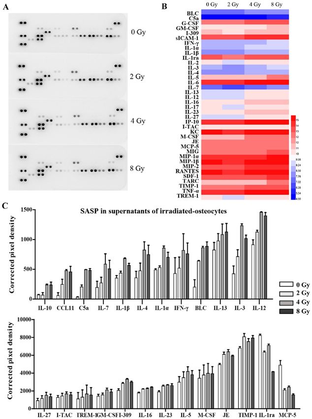
INTERNATIONAL JOURNAL OF MOlecular medicine 47: 76, 2021                                          3

Table Ⅰ. Stepwise procedure for isolating osteocytes from mouse long bones.

Step number                                                         Step description

 1             Soft tissue (adherent muscle and connective tissue) were cut off and the periosteum was scraped off using a
               surgical blade.
 2             Epiphyses were cut off and the marrow was rinsed with PBS using a syringe.
 3             After extensive washing, bones were cut into 1‑2 mm fragments in PBS.
 4             The remaining PBS was removed, and bone pieces were digested using trypsin solution for 20 min in an incubator.
 5             Bone pieces were digested with collagenase type‑II solution at 37˚C for 1 h in an incubator.
 6             Bone pieces were washed twice with PBS.
 7             Bone pieces were incubated with EDTA solution at 37˚C for 30 min in an incubator, and the EDTA solution was
               collected. The bone pieces were once again rinsed in PBS, and the PBS was subsequently added to the EDTA
               solution.
 8             The mixed EDTA solution obtained in the step 7 was centrifuged at room temperature at 200 x g for 10 min and
               the sedimented cells were resuspended in 2 ml culture medium (α Minimum Essential Medium supplemented
               with 10% FBS, calf serum and Pen/Strep).
 9             Bone pieces were incubated with collagenase type‑I solution at 37˚C for 30 min in an incubator, and the
               collagenase type‑I solution was collected. The bone pieces were once again rinsed in PBS, and the PBS was
               subsequently added to the collagenase solution.
10             The mixed collagenase solution obtained in step 9 was centrifuged at room temperature at 200 x g for 10 min and
               the sediment cells were resuspended in 2 ml culture medium.
11             Step 7 was repeated.
12             The mixed solution obtained in step 11 was centrifuged at room temperature at 200 x g for 10 min and the
               sedimented cells were resuspended in 2 ml culture medium.
13             Step 9 was repeated.
14             The mixed solution obtained in step 13 was centrifuged at 200 x g for 10 min at room temperature and the
               sedimented cells were resuspended in 2 ml culture medium.
15             Sedimented cells in steps 8, 10, 12 and 14 were mixed and cultured together with digested bone pieces at 37˚C
               with 5% CO2 in a humidified incubator for 7 days until cell confluency reached >90%.

Cell culture and irradiation treatment. Primary osteocytes            with DAPI (Dojindo Molecular Technologies, Inc.) for nuclei.
were cultured as described above. For ionizing radiation,             Each staining step was processed at room temperature and
1 day after cell plating, osteocytes were subjected to 2, 4, 6        incubated for 2 h in the dark. The number and dendritic length
or 8 Gy irradiation with 137Cs γ‑rays (Nordion). The dose rate        of osteocytes were quantitatively measured using SimplePCI
at the centre of the chamber was 66.7 cGy/min, which was              software (HCImage, SimplePCI 6.6). Six random fields were
annually calibrated by the Shanghai Institute of Measurement          chosen in three biological replicates, and representative
and Testing Technology. LiF (Mg, Cu, P) thermoluminescent             images were captured using a Leica fluorescent microscope
dosimeters were used to measure the actual radiation dose at          (Leica Microsystems, GmbH) with a magnification of x100.
different distances from the chamber centre (31,32). Untreated        The length of the dendrites was calculated by total length of
cells (0 Gy) were used as the control. Subsequent analysis was        dendrites/number of dendrites per cell.
performed 3 days after irradiation.
                                                                      Irradiation‑induced osteocyte DNA damage. A total of 7 h
Irradiation‑induced morphological and functional changes              after radiation treatment, osteocytes were fixed using 4%
in osteocytes. The cell viability of irradiated osteocytes was        formaldehyde at room temperature for 20 min. After washing
detected using a Cell Counting Kit‑8 assay (CCK8; Dojindo             with PBS, cells were permeabilized and blocked using 1%
Molecular Technologies, Inc.). Briefly, osteocytes seeded in          Triton X‑100, 1% BSA and 10% FBS in PBS buffer for 1 h
96‑well plates (3x10³ cells/well) were subsequently treated           at room temperature, and then incubated with primary anti‑
with irradiation (0, 2, 4 or 8 Gy), followed by incubation for 1,     bodies against γ‑H2AX (cat. no. ab81299, Abcam; 1:250)
3 or 5 days. CCK8 reagent was added at 10%, and incubated             overnight at 4˚C. Cells were washed with PBS twice and then
for 2 h at 37˚C. The absorbance at 450 nm was examined using          incubated in the dark with anti‑mouse‑IgG‑HRP‑conjugated
a microplate reader (Bioteck), and was considered to indicate         or anti‑rabbit‑IgG‑HRP‑conjugated secondary antibodies
cell viability.                                                       (cat. no. SA00001‑2, Proteintech; 1:1,000) and DAPI (1:500;
    The morphological changes including dendrite‑like synapse         Beyotime Institute of Biotechnology) for 60 min. The number
and cytoskeleton in the irradiated osteocytes were examined           of γ‑H2AX foci was quantified using a laser confocal micro‑
by staining with tetramethyl rhodamine‑phalloidin (Beijing            scope (Zeiss LSM 880; Carl Zeiss AG) with a magnification
Solarbio Science & Technology Co., Ltd.) for F‑actin and              of x60.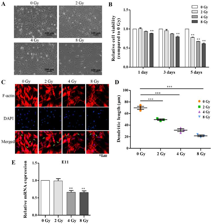
4                       WANG et al: RADIATION INDUCES PRIMARY OSTEOCYTE SENESCENCE PHENOTYPE

Irradiation‑induced osteocyte senescence and its secretory        Table Ⅱ. Primer sequences for reverse transcription‑quantita‑
phenotype. Senescence‑associated β‑galactosidase (SA‑β‑gal)       tive PCR.
expression was visualized using an SA‑ β ‑gal staining kit
(Beyotime Institute of Biotechnology) according to the manu‑      Genes                              Sequence (5'‑3')
facturer's protocol. Cells were washed with PBS twice and
fixed using a fixative solution at room temperature for 15 min,   E11                   F: ACCGTGCCAGTGTTGTTCTG
and then stained with X‑gal solution for 24 h at 37˚C (without                          R: AGCACCTGTGGTTGTTATTTTGT
CO2). Cells were observed using a light microscope (Leica         P16                   F: CGCAGGTTCTTGGTCACTGT
Microsystems, GmbH) with a magnification of x100, and the                               R: TGTTCACGAAAGCCAGAGCG
percentage of SA‑β‑gal‑positive cells in 10 random fields was     P21                   F: CCTGGTGATGTCCGACCTG
calculated.                                                                             R: CCATGAGCGCATCGCAATC
    The levels of 40 different cytokines and chemokines in        NF‑κB                 F: TGCGATTCCGCTATAAATGCG
700 µl supernatant of cultured osteocytes 3 days after irradia‑                         R: ACAAGTTCATGTGGATGAGGC
tion were simultaneously detected using R&D Systems Mouse         TNF‑α                 F: TCAGAATGAGGCTGGATAAG
Cytokine Array, Panel A (cat. no. ARY006; R&D Systems,
                                                                                        R: GGAGGCAACAAGGTAGAG
Inc.), according to the manufacturer's protocol. Signals were
                                                                  MMP13                 F: CCTTGATGCCATTACCAGTCTC
detected using chemiluminescence (Chemi Scope 6300) and
subsequently quantitated with HLImage++ computer vision                                 R: TCCACATGGTTGGGAAGTTCT
systems (1997, Western Vision Software).                          IL‑1α                 F: CTGAAGAAGAGACGGCTGAGT
                                                                                        R: CTGGTAGGTGTAAGGTGCTGAT
Irra diation‑induced senescent osteocyte‑ mediated                IL‑6                  F: ATGAACAACGATGATGCACTTG‑
osteoclastogenesis. Primary osteocytes were inoculated                                  R: GGTACTCCAGAAGACCAGAGG
in a 24‑well plate (4x10³ cells/well), cultured for 24 h and      GAPDH                 F: GGAGTCTACTGGTGTCTTC
subsequently irradiated (0, 2, 4, and 8 Gy). RAW264.7 cells                             R: TCATCATACTTGGCAGGTT
(2x10³ cells/well) were plated on top of the irradiated osteo‑
cytes and co‑cultured in α‑MEM supplemented with 10%              MMP, matrix metalloproteinase; IL, interleukin; TNF‑α, tumor
FBS and 25 ng/ml RANKL (PeproTech, Inc.). The induction           necrosis factor‑α.
medium was changed every 2 days. After 7 days, cells were
fixed in 2.5% glutaraldehyde for 10 min at room temperature
and stained for tartrate‑resistant acid phosphatase (TRAP)
activity using a TRAP staining kit according to the manufac‑      Results
turer's protocol (Sigma‑Aldrich, Merck KGaA). TRAP‑positive
multinucleated cells with >3 nuclei were counted as osteoclasts   Identification of primary osteocytes. Osteocytes, tradition‑
and the area was analyzed using ImageJ.                           ally viewed as a descendant of osteoblasts, exhibit distinct
                                                                  characteristics from osteoblasts (30,34). In order to determine
Reverse transcription‑quantitative PCR (RT‑qPCR). Gene            whether the isolated cells could reflect osteocyte metabolism
expression analysis was performed using RT‑qPCR. Three            traits in vitro, cellular dendrite‑like synapse morphology,
days after irradiation, cells were washed with cold PBS           expression of ALP and expression levels of marker proteins
three times and lysed using TRIzol® reagent (Invitrogen;          were observed. Three days after isolation, digested primary
Thermo Fisher Scientific, Inc.) to obtain total RNA.              osteocytes were present and were suspended in the medium,
Reverse transcription was performed using a Quantscript           whereas on the 5th day, primary osteocytes emerged from the
RT kit (Tiangen Biotech) at 45˚C for 15 min and 95˚C for          bone pieces, and cell populations obtained by direct diges‑
3 min, and PCR was performed in an ABI QuantStudio 3              tion were observed. In the following culture process, it was
(Applied Biosystems; Thermo Fisher Scientific, Inc.) using        noted that primary osteocytes proliferated continuously and
PowerUp SYBR‑Green MasterMix (Invitrogen; Thermo                  presented a characteristic stellate shape with the formation
Fisher Scientific, Inc.) to a final volume of 10 µl. The          of specific dendritic processes, characteristic to osteocytes,
thermocycling conditions used were: 40 cycles of 95˚C for         which are essential for osteocyte function (Fig. 1A).
15 sec followed by 55˚C for 15 sec and 72˚C for 1 min. The            In addition to the above morphological characteristics,
primer sequences are listed in Table II. The relative mRNA        osteocytes showed negative or only weak positive staining for
expression levels of the indicated genes were quantified          ALP expression, whereas osteoblasts exhibited strong staining
using the 2 ‑ΔΔCq method (33). GAPDH was used as the              (Fig. 1B). Furthermore, primary osteocytes were confirmed
loading control.                                                  by the expression of several marker proteins, including OPG,
                                                                  E11, RANKL, and SOST, which were produced almost exclu‑
Statistical analysis. Data were analyzed by one‑way ANOVA         sively in osteocytes (Fig. 1C). Overall, the process of isolation
using SPSS 16.0 (SPSS, Inc.) and GraphPad Prism 5.0               detailed in Table I was effectively used to obtain and identify
(GraphPad Software, Inc.). Following each one‑way ANOVA,          primary osteocytes isolated from femur and tibia.
Tukey's post hoc test was performed to compare all treatments
against the control. Data are presented as the mean ± standard    Irradiation reduces cell viability and directly disrupts funda‑
deviation. P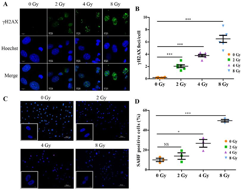
INTERNATIONAL JOURNAL OF MOlecular medicine 47: 76, 2021                                                                5

Figure 1. Proliferative primary osteocytes exhibit a typical morphology and characteristic marker protein expression. (A) Proliferation and characteristic
morphology of primary osteocytes was observed using bright‑field microscopy, 3, 5 and 7 days after isolation. Bone pieces are indicated by the arrows. Scale
bar, 100 µm. Magnification, x100. (B) ALP staining of primary osteocytes and osteoblasts, respectively. Scale bar, 100 µm. Magnification, x100. (C) Analysis
of expression of the osteocyte markers, E11, SOST, RANKL and OPG in primary osteocytes using western blot analysis. ALP, alkaline phosphatase; SOST,
sclerostin; RANKL, receptor activator of nuclear factor‑κ B ligand; OPG, osteoprotegerin.

Figure 2. Irradiation disrupts cell viability and dendrite morphology. (A) Morphology and number of primary osteocyte was altered at 5 days after exposure
to γ‑rays. Scale bar, 100 µm. (B) Cell viability was quantitatively evaluated using a Cell Counting Kit‑8 assay (n=6). (C) F‑actin and nuclei were stained for
osteocyte cytoskeleton and typical dendrite‑like synapse using phalloidin and DAPI fluorescence staining, respectively, 3 days after irradiation, and the typical
dendrites are indicated by the green arrows. Scale bar, 100 µm. (D) Quantitative analysis of the change in dendritic length (n=6). (E) Relative mRNA expres‑
sion levels of E11. Gene expression was normalized to β‑actin and the control (n=3). Expression was analyzed 3 days after irradiation. Data are presented as
the mean ± standard deviation. **P
6                            WANG et al: RADIATION INDUCES PRIMARY OSTEOCYTE SENESCENCE PHENOTYPE

Figure 3. Irradiation affects expression of marker proteins in primary osteocytes. (A‑C) Changes in expression of E11, SOST, RANKL, OPG and RANKL/OPG
ratio in osteocytes 3 days after irradiation exposure were assessed by western blot analysis (n=3). Data are presented as the mean ± standard deviation. *P
INTERNATIONAL JOURNAL OF MOlecular medicine 47: 76, 2021                                                               7

Figure 4. Irradiation results in DNA damage and the development of SAHF in primary osteocytes. (A) Immunofluorescence staining of γH2AX in osteocytes
7 h post irradiation. Scale bar, 10 µm. (B) Quantitative analysis of γ‑H2AX foci per cell. n=6. (C) SAHF formation was observed using confocal microscopy
3 days after irradiation, and was characterized by punctate DNA foci in irradiated‑osteocyte nuclei. Scale bar, 10 µm. (D) Quantification of SAHF posi‑
tive cells from three random fields of view. Data are presented as the mean ± standard deviation. NS, not significant, *P
8 WANG et al: RADIATION INDUCES PRIMARY OSTEOCYTE SENESCENCE PHENOTYPE
Figure 6. Prematurely senescent osteocytes induced by irradiation secrete multiple SASP components in a dose‑depended manner. (A) Levels of soluble SASP
components in the CS of irradiated osteocytes at third day after exposure were detected using Mouse Cytokine Array membrane analysis (n=2). (B) Heat map
representation of secreted cytokines. (C) Expression levels of the indicated secretory cytokines and chemokines were quantified and presented as the corrected
pixel density (n=2). Bar graphs show the mean intensity of the developed spots ± standard deviation. Results are presented as mean ± standard deviation. SASP,
senescence‑associated secretory phenotype; CS, culture supernatants.
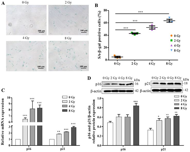
aging‑pathway proteins, p16 and p21, were assayed by western Thus, in the present study the SASP factors from osteocyte
blotting and RT‑qPCR analysis, and the results were consistent culture supernatants were investigated using a Mouse Cytokine
with cellular senescence (Fig. 5C and D). Taken together, the Array panel (Fig. 6A). The analysis showed that the release
results suggested that irradiation induced DNA damage and of 23 cytokines associated with SASP were significantly
may thus stimulate cellular senescence in primary osteocytes. increased in supernatants from radiation‑induced senescent
osteocytes, including pro‑inflammatory interleukin family
Irradiation induces prematurely senescent osteocytes and members (IL‑4, IL‑7 and IL‑3), chemokines (CCL11 and
regulates osteoclastogenesis via a paracrine pathway. I‑309), interferon‑γ, and various other cytokines. Interestingly,
Accumulating evidence has suggested that SASP production certain cytokines, such as IL‑1ra and MCP‑5, were down‑
is typically a consequence of activated downstream path‑ regulated following irradiation (Fig. 6B and C). Expression of
ways of senescence, and it serves a pivotal role in specific several chemokines were not significantly altered and are thus
pathologies of aging diseases (36,37). However, the underlying not listed. Additionally, RT‑qPCR analysis further confirmed
mechanism of the senescent phenotype in the premature aging that irradiation‑induced prematurely senescent osteocytes
of irradiation‑induced cells in the bone microenvironment, could activate multiple SASP components, including NF‑κ B,
particularly for osteocytes, is still incompletely understood. TNF‑α, MMP13, IL‑1α and IL‑6 (Fig. 7A), all of which areINTERNATIONAL JOURNAL OF MOlecular medicine 47: 76, 2021 9 Figure 7. SASP from irradiation‑induced prematurely senescent osteocytes participate in unbalancing the homeostasis of osteoclastogenesis. (A) mRNA expression levels of several SASP components from radiation‑induced senescent osteocytes (n=3). (B) TRAP staining of RAW264.7 cells co‑cultured with OCY for 7 days with RANKL stimulation. Scale bar, 100 µm. Magnification, x100. (C) TRAP positive area was calculated in OCY co‑cultured with RAW264.7 cells (n=3). Results are presented as the mean ± standard deviation. *P
10                      WANG et al: RADIATION INDUCES PRIMARY OSTEOCYTE SENESCENCE PHENOTYPE

addition, during the isolation of osteocytes, a small number        paracrine pathways (54), and MCP‑1 has been indicated to
of osteoblasts are still inevitably mixed into the osteocytes       play an important role in bone remodelling and bone‑related
population. Considering these factual evidences, the presence       cancers (55). Additionally, drug intervention targeting the
of ALP in Fig. 1B is caused by either contaminating osteo‑          SASP profile may be a hopeful approach that can potentially
blasts or weakly‑expressed osteocytes. However, compared            improve bone integrity and function (19,50,56). For example,
with the strong expression of osteoblasts in Fig. 1B, identifica‑   it was confirmed that inhibition of C5a/C5aR axis could
tion of osteocytes and effectiveness for isolation remain to be     alleviate osteoclast degradation (57). In the current study,
elucidated. Overall, a detailed methodology was adapted to          we indicated that besides the direct dysfunctional damage,
enable the isolation and identification of primary osteocytes       osteocytes undergoing irradiation‑induced senescence may
from mice skeletal tissue. This novel primary osteocyte model       significantly increase the expression and secretion of SASP
possesses several advantages over cell lines and may assist in      in the surrounding environment. Following co‑culture,
improving our understanding of the roles of mature osteocytes       irradiated osteocytes promoted the differentiation of nearby
in skeletal homeostasis and remodeling.                             RAW264.7 cells, highlighting the paracrine effects of
    Concerning the direct damage of irradiation to biological       senescent osteocytes in co‑ordinating other cell activities
function in osteocytes, irradiation can markedly inhibit the        in the bone microenvironment. These results suggest that
viability and proliferation of osteocytes, which ultimately         senescent osteocytes modulate bone remodeling via a para‑
leads to a decreased number of osteocytes in a dose‑depen‑          crine pathway targeting the associated bone‑metabolizing
dent manner. Although a high dose of radiation tends to             cells. A recent study demonstrated that targeted reduction
induce more apoptosis and necrosis, previous research               of senescent cells could alleviate focal radiotherapy‑related
had demonstrated that the initial apoptotic cells tend to die       bone loss (27), consistent with the results of the present study,
rapidly, and the majority of surviving cells maintain senes‑        and this may partially be explained by the possible cellular
cence (46‑48). Given these evidences, at 5 days after radiation     senescence mechanism. Taken together, these results suggest
a small number of apoptosis or necrosis osteocytes were             that the accumulation of senescent osteocytes and increased
inevitably mixed into exiting populations, while the majority       SASP secretion induced by radiation are likely one of the
of surviving cells maintain senescence. Additionally, it was        primary initiators of bone damage, or even increased risk of
shown that irradiated primary osteocytes presented obvious          bone metastasis in post‑radiotherapy cancer patients.
morphological changes, including sparsely distributed cells,            Collectively, these results suggested that irradiation
shortened or disappeared dendritic branches and disordered          decreased cell viability and disrupted fundamental biological
cytoskeleton, which were consistent with reduced mRNA and           functions of osteocytes, as well as altered the crucial dendritic
protein expression levels of E11, leading to the dysfunction        morphology and the regulatory function via changes in
of direct communication between embedded osteocytes and             the expression levels of E11, RANKL, OPG and SOST.
the surrounding cells. Accordingly, this study also demon‑          Additionally, it was shown that irradiation resulted in the accu‑
strated that irradiation upregulated the expression of SOST         mulation of DNA damage and thereby initiated senescence
and RANKL, whereas OPG expression was decreased. In                 in osteocytes. Furthermore, irradiation induced prematurely
conclusion, the above results reflect the functional damage of      senescent osteocytes to obtain a SASP, which in turn, indi‑
osteocytes by irradiation in the modulatory process of bone         rectly participated in osteoclastogenesis imbalance. The
homeostasis.                                                        paracrine modulatory process of osteocytes was also involved
    Furthermore, increasing in vitro and in vivo evidence has       in a series of molecular mechanisms. However, it remains
shown that bone loss is a normal consequence associated             to be determined which specific upstream and downstream
with aging (49,50). More recently, several molecular mecha‑         responses SASP and senescent osteocytes modulate to exhibit
nisms, including oxidative stress and DNA damage have been          their effects in radiation‑induced bone loss.
observed to be increased in senescence and irradiation (21).            In conclusion, the present study provides insights into
Interestingly, the present study demonstrated that irradiated       the mechanisms underlying irradiation‑induced bone loss
osteocytes showed several markers associated with DNA               by showing that irradiated osteocytes prematurely senescent
damage and cellular senescence, including the elevated              damage and SASP secretion serve as crucial regulators of
accumulation of γH2AX, positive SA‑ β ‑gal staining, and            bone metabolism.
upregulated expression of p16 and p21 at both the mRNA and
protein level, as well as a novel SAHF indicator. These results     Acknowledgements
suggested that irradiation, as an alternative promoter of
oxidative stress and DNA damage, may also initiate cellular         Not applicable.
senescence of osteocytes in vitro. Moreover, the methods
used in the present study may improve the identification of         Funding
senescence, and allow for easier differentiation between
stimulator‑induced premature senescence compared with               This study was funded by Shanghai Municipal Health
age‑related natural aging.                                          Commission under contract number GWV‑10.1‑XK10.
    It was previously reported that SASP triggers senes‑
cence of the surrounding normal cells (51,52). Many studies         Availability of data and materials
have focused on these secreted factors and their proposed
mechanism in bone metabolism (12,53). IL‑17A was found              The datasets generated and/or analyzed in the present study
to mediate the promotion of osteoclastogenesis by osteocyte         are all included in this published article.
INTERNATIONAL JOURNAL OF MOlecular medicine 47: 76, 2021 11
Authors' contributions 16. Bonewald LF: The amazing osteocyte. J Bone Miner Res 26:
229‑238, 2011.
17. Honma M, Ikebuchi Y, Kariya Y, Hayashi M, Hayashi N, Aoki S
GZ designed the study and revised the article. YW and JW and Suzuki H: RANKL subcellular trafficking and regulatory
performed the experiments. JB, LX and JZ analyzed the data. mechanisms in osteocytes. J Bone Miner Res 28: 1936‑1949, 2013.
18. Mas‑Bargues C, Viña‑Almunia J, Inglés M, Sanz‑Ros J,
YW wrote the manuscript. All authors have read and approved Gambini J, Ibáñez‑Cabellos JS, García‑Giménez JL, Viña J and
the final manuscript. Borrás C: Role of p16INK4a and BMI‑1 in oxidative stress‑induced
premature senescence in human dental pulp stem cells. Redox
Biol 12: 690‑698, 2017.
Ethics approval and consent to participate 19. Pignolo RJ, Samsonraj RM, Law SF, Wang H and Chandra A:
Targeting cell senescence for the treatment of age‑related bone
All the animal experimental procedures were approved by the loss. Curr Osteoporos Rep 17: 70‑85, 2019.
20. Dou Z, Ghosh K, Vizioli MG, Zhu J, Sen P, Wangensteen KJ,
Committee for Ethical Use of Experimental Animal at Fudan Simithy J, Lan Y, Lin Y, Zhou Z, et al: Cytoplasmic chromatin
University (approval no. 201703FYSZGY01). triggers inflammation in senescence and cancer. Nature 550:
402‑406, 2017.
21. Chandra A, Park SS and Pignolo RJ: Potential role of senescence
Patient consent for publication in radiation‑induced damage of the aged skeleton. Bone 120:
423‑431, 2019.
Not applicable. 22. Cmielova J, Havelek R, Soukup T, Jiroutová A, Visek B,
Suchánek J, Vavrova J, Mokry J, Muthna D, Bruckova L, et al:
Gamma radiation induces senescence in human adult mesen‑
Competing interests chymal stem cells from bone marrow and periodontal ligaments.
Int J Radiat Biol 88: 393‑404, 2012.
23. Bai J, Wang Y, Wang J, Zhai J, He F and Zhu G: Irradiation‑induced
The authors declare that they have no competing interests. senescence of bone marrow mesenchymal stem cells aggravates
osteogenic differentiation dysfunction via paracrine signaling.
References Am J Physiol Cell Physiol 318: C1005‑C1017, 2020.
24. Sapieha P and Mallette FA: Cellular senescence in postmitotic
cells: Beyond growth arrest. Trends Cell Biol 28: 595‑607, 2018.
1. Allen C, Her S and Jaffray DA: Radiotherapy for cancer: Present 25. Anderson R, Lagnado A, Maggiorani D, Walaszczyk A, Dookun E,
and future. Adv Drug Deliv Rev 109: 1‑2, 2017. Chapman J, Birch J, Salmonowicz H, Ogrodnik M, Jurk D, et al:
2. Mendes EM, Irie MS, Rabelo GD, Borges JS, Dechichi P, Diniz RS Length‑independent telomere damage drives post‑mitotic cardio‑
and Soares PBF: Effects of ionizing radiation on woven bone: myocyte senescence. EMBO J 38: e100492, 2019.
Influence on the osteocyte lacunar network, collagen maturation, 26. Riessland M, Kolisnyk B, Kim TW, Cheng J, Ni J, Pearson JA,
and microarchitecture. Clin Oral Investig 24: 2763‑2771, 2020. Park EJ, Dam K, Acehan D, Ramos‑Espiritu LS, et al: Loss of
3. Schmeler KM, Jhingran A, Iyer RB, Sun CC, Eifel PJ, Soliman PT, SATB1 induces p21‑dependent cellular senescence in post‑mitotic
Ramirez PT, Frumovitz M, Bodurka DC and Sood AK: Pelvic dopaminergic neurons. Cell Stem Cell 25: 514‑530.e8, 2019.
fractures after radiotherapy for cervical cancer: Implications for 27. Gu G, Nars M, Hentunen TA, Metsikkö K and Väänänen HK:
survivors. Cancer 116: 625‑630, 2010. Isolated primary osteocytes express functional gap junctions
4. Zhang J, Qiu X, Xi K, Hu W, Pei H, Nie J, Wang Z, Ding J, Shang P, in vitro. Cell Tissue Res 323: 263‑271, 2006.
Li B and Zhou G: Therapeutic ionizing radiation induced bone 28. Stern AR, Stern MM, Van Dyke ME, Jähn K, Prideaux M
loss: A review of in vivo and in vitro findings. Connect Tissue and Bonewald LF: Isolation and culture of primary osteocytes
Res 59: 509‑522, 2018. from the long bones of skeletally mature and aged mice.
5. Saintigny Y, Cruet‑Hennequart S, Hamdi DH, Chevalier F and Biotechniques 52: 361‑373, 2012.
Lefaix JL: Impact of therapeutic irradiation on healthy articular 29. Stern AR and Bonewald LF: Isolation of osteocytes from mature
cartilage. Radiat Res 183: 135‑146, 2015. and aged murine bone. Methods Mol Biol 1226: 3‑10, 2015.
6. Alwood JS, Shahnazari M, Chicana B, Schreurs AS, Kumar A, 30. Franz‑Odendaal TA, Hall BK and Witten PE: Buried alive: How
Bartolini A, Shirazi‑Fard Y and Globus RK: Ionizing radiation osteoblasts become osteocytes. Dev Dyn 235: 176‑190, 2006.
stimulates expression of pro‑osteoclastogenic genes in marrow 31. Fartaria MJ, Reis C, Pereira J, Pereira MF, Cardoso JV, Santos LM,
and skeletal tissue. J Interferon Cytokine Res 35: 480‑487, 2015. Oliveira C, Holovey V, Pascoal A and Alves JG: Assessment
7. Jia D, Gaddy D, Suva LJ and Corry PM: Rapid loss of bone mass of the mean glandular dose using LiF:Mg,Ti, LiF:Mg,Cu,P,
and strength in mice after abdominal irradiation. Radiat Res 176: Li2B4O7:Mn and Li2B4O7:Cu TL detectors in mammography
624‑635, 2011. radiation fields. Phys Med Biol 61: 6384‑6399, 2016.
8. Bonewald LF: The role of the osteocyte in bone and nonbone 32. Lucas PA, Aubineau‑Lanièce I, Lourenço V, Vermesse D and
disease. Endocrinol Metab Clin North Am 46: 1‑18, 2017. Cutarella D: Using LiF:Mg,Cu,P TLDs to estimate the absorbed
9. Dallas SL, Prideaux M and Bonewald LF: The osteocyte: An dose to water in liquid water around an 192Ir brachytherapy
endocrine cell ... and more. Endocr Rev 34: 658‑690, 2013. source. Med Phys 41: 011711, 2014.
10. Klein‑Nulend J, Bakker AD, Bacabac RG, Vatsa A and 33. Livak KJ and Schmittgen TD: Analysis of relative gene expres‑
Weinbaum S: Mechanosensation and transduction in osteocytes. sion data using real‑time quantitative PCR and the 2(‑Delta Delta
Bone 54: 182‑190, 2013. C(T)) method. Methods 25: 402‑408, 2001.
11. Robling AG and Bonewald LF: The osteocyte: New insights. 34. Manolagas SC: Birth and death of bone cells: Basic regulatory
Annu Rev Physiol 82: 485‑506, 2020. mechanisms and implications for the pathogenesis and treatment
12. Kitaura H, Marahleh A, Ohori F, Noguchi T, Shen WR, Qi J, of osteoporosis. Endocr Rev 21: 115‑137, 2000.
Nara Y, Pramusita A, Kinjo R and Mizoguchi I: Osteocyte‑related 35. Ikpegbu E, Basta L, Clements DN, Fleming R, Vincent TL,
cytokines regulate osteoclast formation and bone resorption. Int Buttle DJ, Pitsillides AA, Staines KA and Farquharson C: FGF‑2
J Mol Sci 21: 5169, 2020. promotes osteocyte differentiation through increased E11/podo‑
13. Staines KA, Javaheri B, Hohenstein P, Fleming R, Ikpegbu E, planin expression. J Cell Physiol 233: 5334‑5347, 2018.
Unger E, Hopkinson M, Buttle DJ, Pitsillides AA and 36. Childs BG, Durik M, Baker DJ and van Deursen JM: Cellular
Farquharson C: Hypomor phic conditional deletion of senescence in aging and age‑related disease: From mechanisms
E11/Podoplanin reveals a role in osteocyte dendrite elongation. to therapy. Nat Med 21: 1424‑1435, 2015.
J Cell Physiol 232: 3006‑3019, 2017. 37. Childs BG, Gluscevic M, Baker DJ, Laberge RM, Marquess D,
14. Bellido T: Osteocyte‑driven bone remodeling. Calcif Tissue Dananberg J and van Deursen JM: Senescent cells: An emerging
Int 94: 25‑34, 2014. target for diseases of ageing. Nat Rev Drug Discov 16: 718‑735,
15. Staines KA, Hopkinson M, Dillon S, Stephen LA, Fleming R, 2017.
Sophocleous A, Buttle DJ, Pitsillides AA and Farquharson C: 38. Zhang Y, Huang H, Zhao G, Yokoyama T, Vega H, Huang Y,
Conditional deletion of E11/Podoplanin in bone protects against Sood R, Bishop K, Maduro V, Accardi J, et al: ATP6V1H defi‑
ovariectomy‑induced increases in osteoclast formation and ciency impairs bone development through activation of MMP9
activity. Biosci Rep 40: BSR20190329, 2020. and MMP13. PLoS Genet 13: e1006481, 2017.12 WANG et al: RADIATION INDUCES PRIMARY OSTEOCYTE SENESCENCE PHENOTYPE
39. Hameister R, Lohmann CH, Dheen ST, Singh G and Kaur C: The 50. Farr JN, Xu M, Weivoda MM, Monroe DG, Fraser DG, Onken JL,
effect of TNF‑α on osteoblasts in metal wear‑induced peripros‑ Negley BA, Sfeir JG, Ogrodnik MB, Hachfeld CM, et al:
thetic bone loss. Bone Joint Res 9: 827‑839, 2020. Targeting cellular senescence prevents age‑related bone loss in
40. Dinarello CA: The IL‑1 family of cytokines and receptors in mice. Nat Med 23: 1072‑1079, 2017.
rheumatic diseases. Nat Rev Rheumatol 15: 612‑632, 2019. 51. Hitomi K, Okada R, Loo TM, Miyata K, Nakamura AJ and
41. Kim MH, Lee H, Ha IJ and Yang WM: Zanthoxylum Takahashi A: DNA damage regulates senescence‑associated
piperitum alleviates the bone loss in osteoporosis via inhibi‑ extracellular vesicle release via the ceramide pathway to prevent
tion of RANKL‑induced c‑fos/NFATc1/NF‑ κ B pathway. excessive inflammatory responses. Int J Mol Sci 21: 3720, 2020.
Phytomedicine 80: 153397, 2021. 52. Faget DV, Ren Q and Stewart SA: Unmasking senescence:
42. Maré A, D'Haese PC and Verhulst A: The role of sclerostin in Context‑dependent effects of SASP in cancer. Nat Rev Cancer 19:
bone and ectopic calcification. Int J Mol Sci 21: 3199, 2020. 439‑453, 2019.
43. Zhang J, Wang Z, Wu A, Nie J, Pei H, Hu W, Wang B, Shang P, 53. Farr JN, Fraser DG, Wang H, Jaehn K, Ogrodnik MB,
Li B and Zhou G: Differences in responses to X‑ray exposure Weivoda MM, Drake MT, Tchkonia T, LeBrasseur NK,
between osteoclast and osteoblast cells. J Radiat Res 58: 791‑802, Kirkland JL, et al: Identification of senescent cells in the bone
2017. microenvironment. J Bone Miner Res 31: 1920‑1929, 2016.
44. Metzger CE and Narayanan SA: The role of osteocytes in inflam‑ 54. Liao C, Cheng T, Wang S, Zhang C, Jin L and Yang Y: Shear
matory bone loss. Front Endocrinol (Lausanne) 10: 285, 2019. stress inhibits IL‑17A‑mediated induction of osteoclastogenesis
45. Bonewald LF: Establishment and characterization of an osteo‑ via osteocyte pathways. Bone 101: 10‑20, 2017.
cyte‑like cell line, MLO‑Y4. J Bone Miner Metab 17: 61‑65, 1999. 55. Mulholland BS, Forwood MR and Morrison NA: Monocyte
46. He F, Bai J, Wang J, Zhai J, Tong L and Zhu G: Irradiation‑induced chemoattractant protein‑1 (MCP‑1/CCL2) drives activation
osteocyte damage promotes HMGB1‑mediated osteoclastogen‑ of bone remodelling and skeletal metastasis. Curr Osteoporos
esis in vitro. J Cell Physiol 234: 17314‑17325, 2019. Rep 17: 538‑547, 2019.
47. Alessio N, Esposito G, Galano G, De Rosa R, Anello P, Peluso G, 56. Chandra A, Lagnado AB, Farr JN, Monroe DG, Park S,
Tabocchini MA and Galderisi U: Irradiation of mesenchymal Hachfeld C, Tchkonia T, Kirkland JL, Khosla S, Passos JF and
stromal cells with low and high doses of alpha particles induces Pignolo RJ: Targeted reduction of senescent cell burden allevi‑
senescence and/or apoptosis. J Cell Biochem 118: 2993‑3002, ates focal radiotherapy‑related bone loss. J Bone Miner Res 35:
2017. 1119‑1131, 2020.
48. Alessio N, Capasso S, Di Bernardo G, Cappabianca S, Casale F, 57. D'Angelo R, Mangini M, Fonderico J, Fulle S, Mayo E, Aramini A
Calarco A, Cipollaro M, Peluso G and Galderisi U: Mesenchymal and Mariggiò S: Inhibition of osteoclast activity by complement
stromal cells having inactivated RB1 survive following low regulation with DF3016A, a novel small‑molecular‑weight C5aR
irradiation and accumulate damaged DNA: Hints for side effects inhibitor. Biomed Pharmacother 123: 109764, 2020.
following radiotherapy. Cell Cycle 16: 251‑258, 2017.
49. Tiede‑Lewis LM, Xie Y, Hulbert MA, Campos R, Dallas MR, This work is licensed under a Creative Commons
Dusevich V, Bonewald LF and Dallas SL: Degeneration of the Attribution-NonCommercial-NoDerivatives 4.0
osteocyte network in the C57BL/6 mouse model of aging. Aging International (CC BY-NC-ND 4.0) License.
(Albany NY) 9: 2190‑2208, 2017.You can also read