The Roles of Phospholipase A2 in Phagocytes - Frontiers
←
→
Page content transcription
If your browser does not render page correctly, please read the page content below
REVIEW
                                                                                                                                          published: 10 June 2021
                                                                                                                                   doi: 10.3389/fcell.2021.673502

                                            The Roles of Phospholipase A2 in
                                            Phagocytes
                                            Deepti Dabral* and Geert van den Bogaart*
                                            Department of Molecular Immunology and Microbiology, Groningen Biomolecular Sciences and Biotechnology Institute,
                                            University of Groningen, Groningen, Netherlands

                                            Phagocytic cells, such as macrophages, neutrophils, and dendritic cells, ingest particles
                                            larger than about 0.5 µM and thereby clear microbial pathogens and malignant
                                            cells from the body. These phagocytic cargoes are proteolytically degraded within
                                            the lumen of phagosomes, and peptides derived from them are presented on Major
                                            Histocompatibility Complexes (MHC) for the activation of T cells. Mammalian PLA2
                                            isozymes belong to a large family of enzymes that cleave phospholipids at the second
                                            position of the glycerol backbone, releasing a free fatty acid and a lysolipid moiety. In
                                            human macrophages, at least 15 different PLA2 forms are expressed, and expression of
                                            many of these is dependent on pathogenic stimulation. Intriguing questions are why
                          Edited by:        so many PLA2 forms are expressed in macrophages, and what are the functional
                    James T. Murray,        consequences of their altered gene expression after encountering pathogenic stimuli. In
  Swansea University Medical School,
                     United Kingdom
                                            this review, we discuss the evidence of the differential roles of different forms of PLA2 in
                      Reviewed by:
                                            phagocytic immune cells. These roles include: lipid signaling for immune cell activation,
                    Scott Kobayashi,        initial phagocytic particle uptake, microbial action for the killing and degradation of
Rocky Mountain Laboratories (NIAID),        ingested microbes, and the repair of membranes induced by oxygen radicals. We also
                       United States
                     Anetta Härtlova,       discuss the roles of PLA2 in the subsequent digestion of ingested phagocytic cargoes
  University of Gothenburg, Sweden          for antigen presentation to T cells.
                 *Correspondence:
                                            Keywords: phagocytosis, PLA2 , pathogens, macrophages, trafficking
                        Deepti Dabral
                      d.dabral@rug.nl
               Geert van den Bogaart
            g.van.den.bogaart@rug.nl       INTRODUCTION
                    Specialty section:     A role for phospholipase A2 (PLA2 ) in phagocytic immune cells has been suggested since the 1980s
         This article was submitted to     (Waite et al., 1979; Scott et al., 1980). These phagocytic cells, such as macrophages, neutrophils,
                              Signaling,   and dendritic cells, ingest particles larger than about 0.5 µM and thereby clear microbial
                a section of the journal   pathogens and malignant cells from the body (Uribe-Querol and Rosales, 2020). Particularly
  Frontiers in Cell and Developmental
                                           macrophages and dendritic cells also present peptides derived from these phagocytosed antigens
                                Biology
                                           on major histocompatibility complex (MHC) types I and II to activate cytolytic and helper T cells,
         Received: 27 February 2021
                                           respectively, while lipids can be presented on CD1 (Burgdorf and Kurts, 2008). The process of
            Accepted: 11 May 2021
                                           phagocytosis and the subsequent processing of the ingested antigen are essential processes that
           Published: 10 June 2021
                                           contribute to both innate and adaptive immunity. As we will discuss in this review, evidence
                               Citation:
                                           suggests that PLA2 is involved in all stages of pathogen encounter, from the initial signaling, to
     Dabral D and van den Bogaart G
(2021) The Roles of Phospholipase A2
                                           the uptake, degradation and presentation of the antigen.
                        in Phagocytes.        The nomenclature of PLA2 is very complex and disordered as it follows a chronology reflecting
       Front. Cell Dev. Biol. 9:673502.    their time-line of discovery (Dennis et al., 2011; Murakami et al., 2015; Ramanadham et al., 2015).
      doi: 10.3389/fcell.2021.673502       Mammalian PLA2 isozymes belong to a large family, which have been assigned into groups I to

Frontiers in Cell and Developmental Biology | www.frontiersin.org                1                                           June 2021 | Volume 9 | Article 673502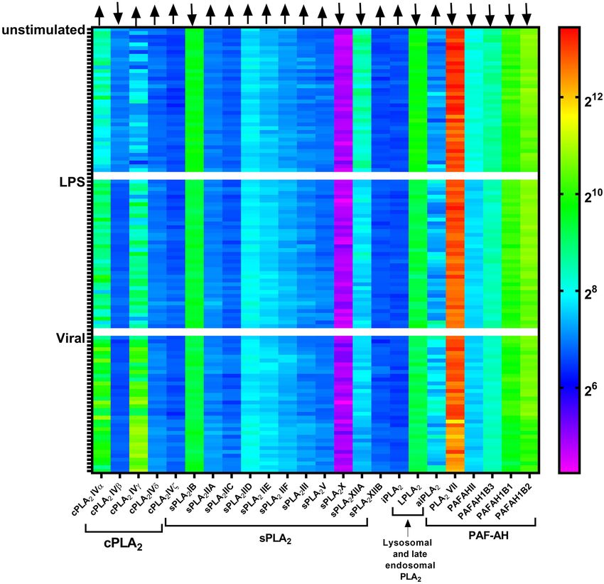
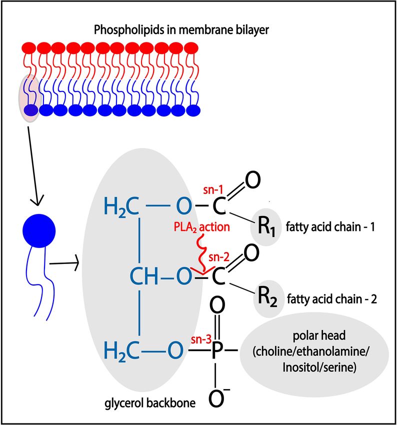
Dabral and van den Bogaart                                                                                            PLA2 in Phagocytic Immune Cells

XVI based on their primary amino acid sequence (Dennis et al.,                signaling by lipids carried out by cPLA2 α, (ii) the focal exocytosis
2011; Murakami et al., 2015; Ramanadham et al., 2015). Based on               to support phagosome formation by iPLA2 β, (iii) the killing of
their subcellular localization and Ca2+ requirement, these groups             the microbe by sPLA2 -V, and LPLA2 forms, and (iv) the repair
can be further categorized into six types called sPLA2 , iPLA2 ,              of damaged phagosomal membranes inflicted by reactive oxygen
cPLA2 , LPLA2 , aiPLA2 , and PAF-AH, and all these types are                  species by LPLA2 and aiPLA2 . In this review, we aim is to provide
further categorized into subtypes (Table 1) (i) sPLA2 are Ca2+ -              an overview of these functions of PLA2 forms.
dependent secretory PLA2 forms found in secretions, such as a
tears, plasma and pancreatic juice (Dennis et al., 2011; Murakami
et al., 2015; Ramanadham et al., 2015). These enzymes belong                  LIPID SIGNALING BY cPLA2 α
to groups I, II, III, V, IX, X, XI, XII, XIII, and XIV (Dennis
et al., 2011; Murakami et al., 2015; Ramanadham et al., 2015).                Phospholipase A2 enzymes hydrolyze membrane phospholipids
Within the sPLA2 subgroup, a capital English letter indicates                 that are composed of a glycerol backbone esterified to two
the subtype of enzymes. For example, sPLA2 -IIA is present in                 hydrophobic fatty acids tails at the sn- (stereospecifically
synovial fluid, while sPLA2 -IID is present in pancreas and spleen            numbered) 1 and 2 positions, and a hydrophilic head group
(Dennis et al., 2011). (ii) cPLA2 are Ca2+ -dependent cytosolic               at the sn-3 position (Figure 2). Phospholipids with the head
enzymes which belong to group IV, and its subtypes are cPLA2 α,               groups choline, ethanolamine, serine, and inositol form the main
-β, -γ, -δ, -ε, and -ζ (Dennis et al., 2011; Murakami et al., 2015;           classes of phospholipids and are called phosphatidylcholine
Ramanadham et al., 2015). (iii) iPLA2 are Ca2+ -independent                   (PC), phosphatidylethanolamine (PE), phosphatidylserine (PS),
cytosolic forms which belong to group VI, and its subtypes are                and phosphatidylinositol (PI), respectively. The catalytic action
iPLA2 -β, -γ, -δ, -ε, -ζ, and -η (Dennis et al., 2011; Murakami et al.,       of PLA2 releases the free fatty acid from the sn-2 position,
2015; Ramanadham et al., 2015). Within the cPLA2 and iPLA2                    such as arachidonic acid (ARA; 20:4), docosahexaenoic acid
types, the different subtypes are mostly indicated by Greek letters           (C22:6), oleic acid (C18:1), while lysophospholipids, such as lyso-
(Dennis et al., 2011; Murakami et al., 2015; Ramanadham et al.,               phosphatidyl-choline/-ethanolamine/-inositol (LPC/LPE/LPI),
2015). However, this nomenclature is not always consistently                  remain esterified in the membrane.
used and for example cPLA2 -IVα and iPLA2 -VIβ are also known                    Both the lyso-phospholipids and the free fatty acids produced
as cPLA2 -IVA and iPLA2 -VIA. (iv) LPLA2 is a lysosomal PLA2                  by PLA2 can be bioactive molecules and/or form precursors
which belongs to group XV (Shayman et al., 2011; Shayman                      for the generation of bioactive lipid hormones (Brash, 2001;
and Tesmer, 2019). (v) aiPLA2 are acidic Ca2+ -independent                    Carneiro et al., 2013). PLA2 activity is especially important for
PLA2 forms which belong to group XVI, and are better known                    the generation of a class of lipid hormones called eicosanoids.
as peroxisomal PLA2 (Sorokina et al., 2009; Fisher, 2018). (vi)               Eicosanoids are a broad family of oxygenated lipid compounds
platelet-activating factor hydrolases (PAFAH) belong to groups                that include prostaglandins, thromboxanes, and leukotrienes.
VIIA (also known as lipoprotein-associated PLA2; Lp-PLA2 ),                   Eicosanoids are generated via non-enzymatic oxidation of ARA
VIIB and VIII (Stafforini, 2009; Dennis et al., 2011).                        or by the action of enzymes, such as cyclooxygenase (COX),
   In human macrophages, 15 PLA2 forms are found at the                       lipooxygenase (LOX), and cytochrome P450 (CYP) (Dennis
transcript level, including sPLA2 (sPLA2 -IID, -V, and -XIIA),                and Norris, 2015; Gil-de-Gómez et al., 2020). Eicosanoids exert
cPLA2 (cPLA2 -IV α, β, and γ), iPLA2 (iPLA2 -VI β, γ, δ, ε, ζ, and            immunomodulatory functions and, depending on the species
η), LPLA2 , LpPLA2 , and aiPLA2 (Elstad et al., 1989; Rubio et al.,           of eicosanoids, can have pro- or anti-inflammatory effects
2015). Of these forms, LPLA2 and LpPLA2 carry a signal peptide                (Dennis and Norris, 2015). Upon encountering a pathogen,
required for cotranslational insertion into the ER and subsequent             immune phagocytes produce elevated levels of eicosanoids and
transport through the Golgi where they undergo N-glycosylation,               an increased amount of eicosanoids in circulation is considered
while others are either cytosolic or membrane bound proteins                  a hallmark of inflammation (Dennis and Norris, 2015). Non-
(Dennis et al., 2011; Murakami et al., 2015; Ramanadham et al.,               steroidal anti-inflammatory drugs (NSAIDs), such as ibuprofen,
2015). Analysis of published gene expression data of blood                    indomethacin, and asprin, target COX and PLA2 to alleviate pain,
monocyte-derived dendritic cells from 38 healthy individuals                  redness, and swelling associated with inflammation (Singh et al.,
(Lee et al., 2014) revealed that the expression of some of these              2005; Dennis and Norris, 2015).
PLA2 forms is dependent on pathogenic stimulation (Figure 1).                    Mammalian cells are rich in ARA, but this is mostly
For example, the expression of cPLA2 -IVα and -IVγ are                        incorporated in phospholipids by esterification to the glycerol
upregulated, while sPLA2 -XIIA and PAFAHII are downregulated                  backbone at the sn-2 position. For example, human cultured
upon stimulation of dendritic cells with lipopolysaccharide (LPS)             platelets contain ∼30 µg esterified arachidonate per 109 cells,
or influenza virus. This information is important, because it                 which approximately corresponds to 5 mM, while free ARA is
shows that many forms of PLA2 undergo substantial up- or                      only present at ∼3 pmol per 106 cells, corresponding to about
down-regulation upon pathogenic stimulation, suggesting that                  0.5–1 µM (Brash, 2001). However, the concentration of free ARA
they have roles in the immune function of the phagocytes.                     significantly increases upon pathogenic stimulation. For example,
   Indeed, although the roles of most PLA2 forms in                           plasma concentrations in mice increase from ∼1 to ∼1.5 mM
macrophages and dendritic cells are largely unknown, literature               upon infection with Salmonella pneumonia (Eijkelkamp et al.,
shows that various PLA2 forms function during all the events                  2018). Similarly, concentrations of ARA in the whole blood of
that occur following the encounter of a pathogen: (i) the immune              sepsis patients increase significantly from ∼200 to ∼250 ng/ml

Frontiers in Cell and Developmental Biology | www.frontiersin.org         2                                       June 2021 | Volume 9 | Article 673502
Dabral and van den Bogaart                                                                                                           PLA2 in Phagocytic Immune Cells

TABLE 1 | Phospholipase A2 (PLA2 ) family and role of specific forms discussed in this review.

Type        Group           Sub types       Other name          Molecular            Catalytic residue    References                   PLA2 forms discussed
                                                                weight (kDa)

cPLA2       IV              α (A)                               60–114               Ser/asp              Dennis et al., 2011;         cPLA2 -IVα in lipid signaling
                            β (B)                                                                         Leslie, 2015
                            γ (C)
                            δ (D)
                            ε (E)
                            ζ (F)
iPLA2       VI              β (A)           PNPLA9              27–146               Ser/Asp              Dennis et al., 2011;         iPLA2 -VIβ in supporting focal
                            γ (B)           PNPLA8                                                        Ramanadham et al.,           exocytosis at the phagocytic
                            δ (C)           PNPLA6                                                        2015                         cup
                            ε (D)           PNPLA3
                            ζ (E)           PNPLA2
                            η (F)           PNPLA4
sPLA2       I               A, B                                10–19                His/Asp              Dennis et al., 2011;         sPLA2 -IIA, V, X, XII as
            II              A, B,                                                                         Murakami et al., 2015;       antimicrobial forms at the
            III             C, D,                                                                         Murakami, 2017               phagocytic cup and within
            V               E, F                                                                                                       closed phagosomes
            IX              A, B
            X               A, B
            XI
            XII
            XIII
            XIV
LPLA2       XV                                                  45                   Ser/His/Asp          Dennis et al., 2011;         LPLA2 having bacteriocidal
                                                                                                          Fisher, 2018                 activity at the phagocytic cup
                                                                                                                                       and within phagolysosomes
                                                                                                                                       Also, its membrane repair
                                                                                                                                       mechanism
aiPLA2      XVI                             Peroxiredoxin 6     25                   Ser/His/Asp          Fisher, 2018                 aiPLA2 regulating NOX2
                                                                                                                                       assembly, and its membrane
                                                                                                                                       repair mechanism
PAF-AH      VIIA                            Lp-PLA2 ,           26–45                Ser/His/Asp          Dennis et al., 2011;         Not discussed as relevant
            VIIB                            PLA2 VII                                                      Karasawa and Inoue,          information was not found
            VIII                            PAF-AH II                                                     2015
                                            PAF-AH I
                                            (PAFAH1B1
                                            PAFAH1B2
                                            PAFAH1B3)

(Bruegel et al., 2012). Phagocytosis also triggers ARA release                          domain for interacting with membranes (Leslie, 2015). cPLA2
in vitro, as shown for macrophages labeled with radioisotope                            forms require mM concentrations of free Ca2+ for calcium-
labeled ARA (Waite et al., 1979; Gil-de-Gómez et al., 2020).                            dependent membrane binding and activation (Dennis et al.,
This has been observed for a wide range of phagocytic cargoes,                          2011; Leslie, 2015). Members of this family include cPLA2 α,
such as serum-opsonized zymosan, native zymosan, and live                               -β, -γ, -δ, -ε, and -ζ, which vary in their molecular weight,
pathogenic bacteria (Adolph et al., 2012; Gil-de-Gómez et al.,                          tissue expression, and subcellular localization (Dennis et al.,
2020). These studies indicate that pathogenic encounter results                         2011; Leslie, 2015). These members have about 30% sequence
in an increased PLA2 activity resulting in the production of                            homology and although they have overlapping activities, their
free ARA. The elevated ARA levels, in turn, promote the                                 functions are largely non-redundant. cPLA2 α is the only PLA2
production of eicosanoids for inflammatory signaling. As will be                        form that contains mitogen-activated protein kinase (MAPK)
discussed in more detail below, the elevated ARA levels might                           phosphorylation sites (S505 and S727 ) (Murakami, 2017). Thereby,
also facilitate the phagocytic process because supplementation                          infection-induced activation of the Ras and MAPK pathways
of ARA to macrophage-like cell lines RAW264.7 and THP-1                                 results in the phosphorylation of cPLA2 α (Zhou et al., 2003; Su
accelerated phagocytosis, and potentiated their anti-microbial                          et al., 2004). This phosphorylation results in increased activation
ability against intracellular microbes (Adolph et al., 2012;                            of cPLA2 α, which in turn leads to increased production of ARA
Eijkelkamp et al., 2018).                                                               (Dieter et al., 2002). Infection also induces cPLA2 α transcription
   The most important PLA2 form involved in eicosanoid                                  via transcriptional factors, such as nuclear factor κB (NF-κB),
signaling is cPLA2 α. cPLA2 isozymes are characterized by an                            Krüppel-like factor, hypoxia-inducible factor (Hif), specificity
N-terminal CalB domain for Ca2+ binding, an active site Ser-Asp                         protein 1 (Sp1), and c-Jun, that are well known to regulate
dyad for lipid hydrolysis, and a C-terminal phospholipid-binding                        immune cell activation (Su et al., 2004; Dennis and Norris, 2015;

Frontiers in Cell and Developmental Biology | www.frontiersin.org                   3                                            June 2021 | Volume 9 | Article 673502
Dabral and van den Bogaart PLA2 in Phagocytic Immune Cells
FIGURE 1 | Gene microarray heatmap of phospholipase A2 (PLA2 ) gene expression in monocyte-derived dendritic cells from a published microarray study. Each
column shows a PLA2 gene. Each row shows dendritic cells derived from an individual donor, either unstimulated or stimulated with LPS or the influenza virus (Viral).
Microarray data is from the reference (Lee et al., 2014).
Lane et al., 2019). In line with this, transcriptomics analysis of GFP–cPLA2 α also translocates to the phagocytic cup (Casas
human blood monocyte-derived dendritic cells revealed that both et al., 2010) and to zymosan containing phagosomes (Casas et al.,
LPS and viral stimulation increase cPLA2 α expression (Figure 1). 2009). This suggests that, next to lipid signaling, cPLA2 α has
cPLA2 α localizes in the cytosol in unstimulated RAW264.7 additional roles in the phagocytic process. These additional roles
macrophage-like cells (Casas et al., 2006) and in mouse seem to be independent of its catalytic activity, because mutated
peritoneal primary macrophages (Gijón and Leslie, 1999). It cPLA2 α versions with an inactive catalytic domain showed no
translocates to a perinuclear membrane-rich area (likely the phagocytic defect in RAW264.7 macrophage-like cells, whereas
Golgi network) in response to chemically induced increases mutation of the C2 domain, which is needed for membrane
in PI (4,5)-bisphosphate [PI(4,5)P2 ] (Bechler et al., 2012) and attachment, resulted in significantly less phagocytic ability (Zizza
Ca2+ concentrations at this location (Gijón and Leslie, 1999). et al., 2012).
A similar translocation has been observed in LPS-stimulated
P388D1 macrophage-like cells (Balboa et al., 2003b; Shirai et al.,
2005). In mouse peritoneal macrophages, cPLA2 α and COX2 are iPLA2 β SUPPORTS FOCAL EXOCYTOSIS
both located close to the perinuclear Golgi network, which likely FOR PATHOGEN UPTAKE
facilitates their functional coupling (Gijón and Leslie, 1999; Su
et al., 2004). Thus, the increased expression and MAPK-mediated Unlike cPLA2 forms, group-VI iPLA2 s do not require Ca2+ for
phosphorylation of cPLA2 α upon encountering a pathogen, lead their activity and membrane association (Dennis et al., 2011;
to its translocation to the Golgi, resulting in increased production Ramanadham et al., 2015). These are intracellular membrane-
of ARA (Gijón and Leslie, 1999). This mechanism underlies the associated and cytoplasmic isozymes with a molecular weight
increased production of eicosanoids upon infection. ranging from 27 to 146 kDa (Dennis et al., 2011; Ramanadham
However, in human monocyte-derived macrophages, et al., 2015). These isozymes are characterized by lipase (GXSXG)
immunofluorescence microscopy experiments revealed that and nucleotide-binding (GXGXXG) consensus sequences
Frontiers in Cell and Developmental Biology | www.frontiersin.org 4 June 2021 | Volume 9 | Article 673502Dabral and van den Bogaart PLA2 in Phagocytic Immune Cells
a resting cell, and iPLA2 -β is a critical enzyme for this
pathway (Kennedy and Weiss, 1956; Lands, 1958). In the Lands
pathway, iPLA2 -β generates free ARA, but most of it rapidly
gets re-incorporated into phospholipids by first linking to Co-
Enzyme A (CoA) by long-chain fatty acyl CoA synthetases.
CoA-linked ARA then gets incorporated into PC by a CoA-
dependent acyltransferase (Balsinde et al., 1997). Thus, iPLA2 β-
produced ARA is an intermediate of connected de-acylation
and re-acylation reactions of membrane phospholipids. In
unstimulated P388D1 macrophages, re-acylation dominates over
de-acylation, which limits the presence of free ARA (Chilton
et al., 1996; Balsinde et al., 1997), thereby also limiting eicosanoid
production. The Kennedy pathway is a low-affinity mechanism
that incorporates free ARA in membranes as triacylglycerol
(TAG) and diacylglycerol (DAG) species (Kennedy and Weiss,
1956; Astudillo et al., 2012; Gil-de-Gómez et al., 2020). Thus, the
incorporation of free ARA into membranes depends upon the
concentration of free ARA: at low concentrations most ARA will
be incorporated in phospholipids via the Lands pathway, while at
higher concentrations it will be incorporated in TAG and DAG
species via the Kennedy pathway. Since pathogenic stimulation
results in increased activities of cPLA2 as described above, the
rate of ARA hydrolysis exceeds that of its reincorporation into
FIGURE 2 | Catalytic action of PLA2 . Membrane phospholipids such as membrane phospholipids, leading to activation of the Kennedy
phosphatidylcholine (PC)/phosphatidylethanolamine (PE)/phosphatidylinositol pathway to esterify bulk ARA back in the membrane.
(PI), and phosphatidylserine (PS), are cleaved at the sn-2 position of the
However, evidence suggests that iPLA2 -β has a second role in
glycerol backbone by the action of PLA2 , thereby releasing free fatty acid such
as arachidonic acid. supporting the phagocytic uptake. During phagocytosis, iPLA2 -β
translocates to the plasma membrane within 5 mins after particle
engagement (Tay and Melendez, 2004). The catalytic activity of
iPLA2 -β is essential for particle uptake, as its inhibition using
(Dennis et al., 2011; Ramanadham et al., 2015). They also carry bromoenolactone (BEL)–a selective iPLA2 -β and -γ inhibitor
ankyrin repeats that mediate protein-protein interactions and (Ramanadham et al., 2015)–leads to the polarized accumulation
this enables them to form homo-oligomers which is essential of electronlucent structures that appear as intracellular vesicles
for their activity (Dennis et al., 2011; Ramanadham et al., 2015). in the cytosol polarized toward the target attachment site in
Members of this family include iPLA2 -β, -γ, -δ, -ε, -ζ, and -η. primary human monocytes (Lennartz et al., 1997). Moreover,
Notably, transcripts of iPLA2 -β, -γ, -δ, -ε, -ζ, and -η have been enrichment of BEL-sensitive iPLA2 to F-actin rich pseudopods
detected in human monocyte derived macrophages (Rubio et al., occurs in murine monocytes that are stimulated with monocyte
2015), iPLA2 -β in human primary blood monocytes (Mishra chemoattractant protein-1 (MCP-1) (Mishra et al., 2008), a
et al., 2008) and the human monocyte cell line U937 (Tay chemokine known to induce increased ARA release in human
and Melendez, 2004). Of these, only iPLA2 -β (a cytoplasmic monocytes (Gschwandtner et al., 2019). Together, these findings
form), and -γ (membrane-associated form) isozymes are widely indicate that vesicles carrying BEL-sensitive iPLA2 translocate
characterized (Dennis et al., 2011; Ramanadham et al., 2015). to the plasma membrane at the site of the nascent phagosome,
iPLA2 -β localizes to the ER–Golgi intermediate compartment presumably to support membrane extension by promoting the
(ERGIC), likely by associating to the cytosolic leaflet of the local fusion of these vesicles with the plasma membrane (Bajno
ERGIC membrane (Ben-Tekaya et al., 2010; Bechler et al., et al., 2000). These iPLA2 carrying vesicles might be of endosomal
2012). Studies in mammalian cell lines, such as HeLa, showed nature, as iPLA2 has been reported on endosomes (Karli et al.,
that iPLA2 -β mediates the formation of membrane tubules that 1990; Mayorga et al., 1993, 1994). Alternatively or additionally,
bridge between separate ERGIC clusters and thereby regulate these might be secretory lysosomes, as BEL-sensitive iPLA2
intra-ERGIC trafficking (Ben-Tekaya et al., 2010; Bechler et al., is required for lysozyme secretion from these compartments
2012). However, the widely described role of iPLA2 -β is a (Balboa et al., 2003a).
housekeeping function of remodeling phospholipids (Winstead
et al., 2000; Ramanadham et al., 2015) that occurs in nearly
all cellular membranes via two pathways: the Lands and the ANTI-MICROBIAL ROLES OF sPLA2
Kennedy (or de novo) pathways (Pasternak and Bergeron, 1970;
Dabral and Coorssen, 2017). The Lands pathway maintains Bactericidal Roles of sPLA2 Forms
cellular homeostasis and operates in the presence of low Members of the sPLA2 group were first discovered in snake
concentrations of free ARA, which typically is the case in and bee venom, and in bovine pancreatic juice (Dennis et al.,
Frontiers in Cell and Developmental Biology | www.frontiersin.org 5 June 2021 | Volume 9 | Article 673502Dabral and van den Bogaart PLA2 in Phagocytic Immune Cells 2011). Humans express 10 catalytically active sPLA2 forms (IB, and rat liver macrophages (Dieter et al., 2002). In line with IIA, IIC, IID, IIE, IIF, III, V, X, and XIIA), and one inactive this, transcriptomics analysis (Lee et al., 2014) also showed an form (XIIB) (Murakami et al., 2015). Several of these forms upregulation in the expression of sPLA2 -V in LPS and viral are expressed in phagocytic immune cells. For instance, human stimulated human monocyte-derived dendritic cells (Figure 1). primary macrophages express sPLA2 -IIA, -IID, -V, -XIIA, and - However, sPLA2 -XIIA seems down regulated upon pathogenic XIIB (Anthonsen et al., 2000; Rubio et al., 2015), the macrophage- stimulation of monocyte-derived dendritic cells (Figure 1), but like cell line U937 expresses sPLA2 -IID, -V, and -XIIA (Dennis the functional significance of this is unknown. et al., 2011), human monocyte-derived dendritic cells express The release of sPLA2 -V might occur in a polarized fashion sPLA2 -IIA, -IIC, -IID, -IIE, -IIF, -III, -V, -X, -XIIA, and -XIIB at the phagocytic cup, as it translocates from the Golgi and (Anthonsen et al., 2000; Lee et al., 2014), and human monocytes recycling endosomes to the forming phagosome in zymosan- express sPLA2 -IIA, and -V (Anthonsen et al., 2000). sPLA2 forms stimulated mouse peritoneal macrophages (Balestrieri et al., are Ca2+ -dependent and low molecular weight proteins (
Dabral and van den Bogaart PLA2 in Phagocytic Immune Cells
Second, LPLA2 might mediate pathogen killing similar to the phagosomal membrane, particularly because polyunsaturated
sPLA2 described above, because LPLA2 is also released in fatty acids, such as ARA, are highly susceptible to lipid
the extracellular space of zymosan-stimulated mouse peritoneal peroxidation (Yin et al., 2011). LPLA2 might thus potentially limit
and alveolar macrophages (Wightman et al., 1981; Abe et al., or repair the damaging effects of ROS, by removing oxidized
2008). Also similar to sPLA2 , LPLA2 translocates to forming ARA from phospholipids and transfer it to ceramides. Supporting
phagosomes in RAW264.7 macrophage-like cells within 4 mins this transfer, ceramides containing long acyl chains such as
after particle engagement, and the fusion of lysosomes with the ARA are high on mature phagosomes while ceramide synthase
plasma membrane results in the exocytosis of LPLA2 (Sun et al., 2 expression and activity are high on early phagosomes in
2020). Because the V-ATPase at the plasma membrane pumps H+ RAW264.7 macrophage-like cells (Pathak et al., 2018).
ions across the membrane to the extracellular side, potentially
lowering the local extracellular pH at the phagocytic cup, this
might provide the required acidic microenvironment for secreted Regulation of Oxidative Damage by
LPLA2 to attack the microbial cargo. aiPLA2
Third, LPLA2 might play a role in antigen presentation of Acidic Ca2+ -independent PLA2 (aiPLA2 ) has a molecular weight
lipids to T cells in CD1, because LPLA2 knock-out mice displayed of ∼25 kDa and is better known as peroxiredoxin 6 (Fisher, 2011,
lower T cell activation and recruitment to infected lungs 2018). It is expressed in mouse alveolar macrophages (Chatterjee
(Shayman and Tesmer, 2019). Furthermore, Mycobacterium et al., 2011) and mainly localizes to the cytosol where it has
infected LPLA2 knock-out mice show an enhanced bacterial only low PLA2 activity due to its acidic optimum of pH 4.0
burden and a low recruitment of CD1-expressing cells to (Fisher, 2018). However, a fraction of aiPLA2 is also targeted
the infection site (Schneider et al., 2014). This role in lipid to the lumen of lysosomes and late endosomes, in a manner
presentation is supported by the finding that LPLA2 can depending on direct interactions with the chaperone 14-3-3ε
cleave bacterial cardiolipin, and the resulting lipid species are (Sorokina et al., 2009; Fisher, 2018). Besides possessing PLA2
incorporated into the membranes of phagosomes and other activity, aiPLA2 is a multifunctional enzyme that also possesses
organelles in Mycobacterium bovis infected murine macrophages lyso-phosphatidyl acyltransferase and glutathione peroxidase
(Fischer et al., 2001). Similarly, LPLA2 can cleave mycobacterial activities (Fisher, 2011).
tetra-acylated glycolipid antigens (phosphatidyl-myo-inositol Because aiPLA2 binds to liposomes carrying oxidized lipids,
mannosides) into diacylated forms in THP-1 monocyte-like cells and translocates to the plasma membrane in A549 cells following
(Balboa et al., 2003a). Because cardiolipin carries four fatty acid treatment with an oxidization agent (Manevich et al., 2009),
chains, just as mycobacterial tetra-acylated glycolipids, LPLA2 membrane oxidation might promote association of aiPLA2
appears to preferentially cleave tetra-acylated lipids (Fischer et al., with the membrane. Hence, oxidized sn-2 fatty acids might
2001; Gilleron et al., 2016). Thus, current evidence suggests be replaced by the lyso-phosphatidyl acyltransferase activity of
that LPLA2 processes microbial tetra-acylated lipids within aiPLA2 (Fisher, 2018; Fisher et al., 2018). Additionally, aiPLA2
phagolysosomes, and the resulting diacylated forms might be can reduce peroxidized phospholipids to their corresponding
presented on CD1 molecules to stimulate T cells. alcohol due to its glutathione peroxidase activity (Chatterjee
Fourth, LPLA2 could play a role in membrane repair, as et al., 2011; Fisher, 2018; Fisher et al., 2018). Therefore, similar
explained in the next section. to LPLA2 , aiPLA2 can protect against oxidative damage due to
its ability to (i) hydrolyze the sn-2 position peroxidized fatty
acids to generate lyso-phospholipids, (ii) reacylate these lyso-
phospholipids to form a new phospholipid, and (iii) convert
MEMBRANE REPAIR BY LPLA2 AND
oxidized phospholipids to their corresponding alcohol (Fisher
aiPLA2 et al., 2018). The physiological roles of aiPLA2 therefore include
the repair of oxidized membranes (Chatterjee et al., 2011).
Transacylase Activity of LPLA2 for Repair However, aiPLA2 also promotes ROS production by
of Membrane Damage promoting the assembly of the NOX2 complex in alveolar
In addition to its antimicrobial roles described in the previous macrophages (Chatterjee et al., 2011). As mentioned above,
section, LPLA2 also has a potential role in membrane repair. NOX2 generates ROS within the lumen of phagolysosomes,
It can remove oxidized fatty acids at the sn-2 position of and also at the plasma membrane, in order to kill and degrade
phospholipids and transfer these to ceramides in the transacylase microbial pathogens (Burgdorf and Kurts, 2008; Uribe-Querol
reaction (Shayman and Tesmer, 2019). This ability of LPLA2 and Rosales, 2020). The assembly and activation of NOX2 occurs
might be important, because in neutrophils, macrophages, when MAPK phosphorylates cytosolic aiPLA2 at T177 which
and dendritic cells, the killing of ingested pathogens is a results in the translocation of aiPLA2 to the plasma membrane
radical-mediated mechanism where the NADPH oxidase NOX2 (Fisher et al., 2018). At the plasma membrane, aiPLA2 generates
generates large amounts of reactive oxygen species (ROS) in lyso-PC which in turn converts to lyso-phospatidic acid (LPA)
the lumen of phagolysosomes (Vulcano et al., 2004). These by lysophospholipase D (Vázquez-Medina et al., 2016; Fisher,
radicals not only damage the ingested pathogen, but also oxidize 2018). Binding of LPA to the LPA receptor-1 activates the small-
membranes of the host cell (Dingjan et al., 2016). Therefore, GTPase Rac, which is a component required for the activation
the host cell could need a mechanism to repair damage to of the NOX2 complex (Fisher, 2018). In stark contrast, another
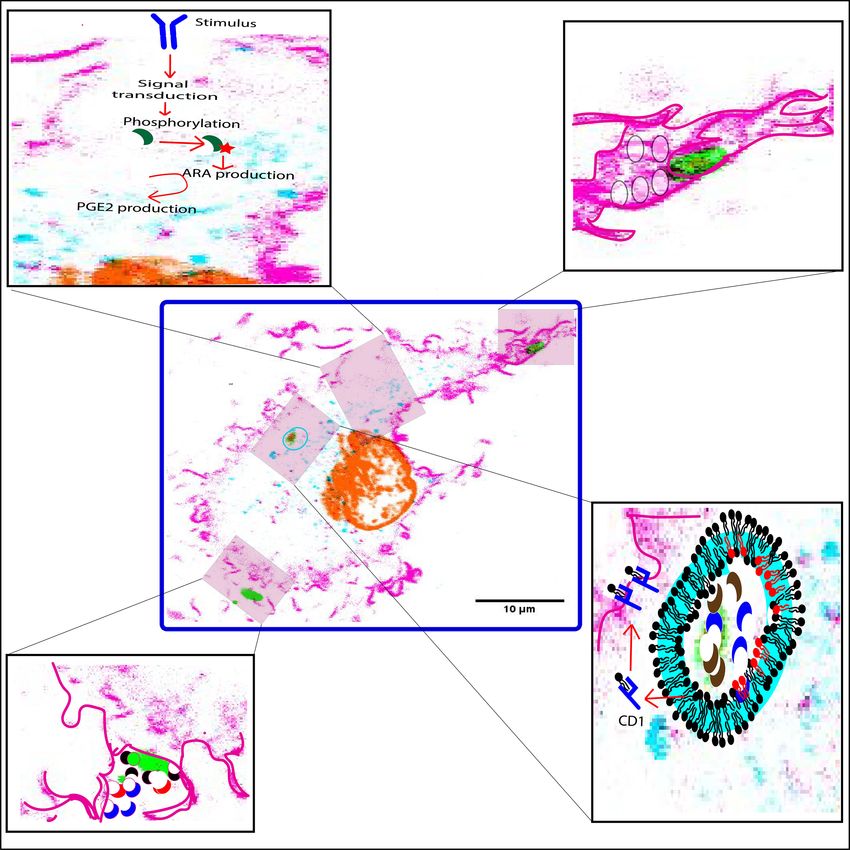
Frontiers in Cell and Developmental Biology | www.frontiersin.org 7 June 2021 | Volume 9 | Article 673502Dabral and van den Bogaart PLA2 in Phagocytic Immune Cells FIGURE 3 | Functions of PLA2 forms at different stages of phagocytosis. (A) cPLA2 α (green crescent) is phosphorylated in response to an extracellular pathogenic stimulus. This leads to translocation of cPLA2 α to the perinuclear area, where it hydrolyzes membrane phospholipids to generate arachidonic acid (ARA) which in-turn gets metabolized to eicosanoids by the action of the COX-2 enzyme. (B) iPLA2 (not shown) translocates to the phagocytic cup, and facilitates fusion of secretory vesicles (translucent circles) to provide additional membrane required for the extension of the pseudopodia. (C) sPLA2 -II/V (red and black crescent) and LPLA2 (blue crescent) are released at the phagocytic cup to degrade invading microbes (green). (D) LPLA2 (blue crescent) and aiPLA2 (brown crescent) work together to degrade and process microbes within the phagolysosome. The processed lipids are then loaded onto a CD1 molecule (blue structure) to be presented on the cell surface to activate T cells. Both LPLA2 and aiPLA2 repair peroxidized lipids of the phagosomal membrane that might occur due to increased NOX2 activity. The repaired phospholipids are shown in red. F-actin is pink, DNA is orange, LAMP1 is turquoise and LPS is green. Highlighted pink boundary in insets (B–D) shows pseudopodia, phagocytic cup, and plasma membrane, respectively. Highlighted turquoise boundary at the phagosome membrane is to show recruited LAMP1. component of the NOX2, p67phox , can bind to phosphorylated aiPLA2 might also mediate the repair of membranes form of aiPLA2 , and this inhibits its PLA2 activity (Fisher, 2018). damaged by intracellular pathogens. The expression of Thus, aiPLA2 has a dual function: it both repairs oxidation- aiPLA2 shows a biphasic response in Brucella suis infected induced membrane damage and promotes NOX2-mediated ROS RAW267.4 macrophages: aiPLA2 expression initially decreases formation. Perhaps at initial stages of pathogen recognition, the until 10 h post-infection, after which it increases until membrane damage by NOX2-produced ROS is still low. At this 50 h post-infection (Wang et al., 2019). During the first stage, unassembled p67phox (i.e., not in the NOX2 complex) might phase, B. suis is non-replicative within phagosomes (Celli, negatively regulate aiPLA2 activity, as the need to repair damaged 2019), whereas it becomes replicative in the second phase membranes is low. However, as more NOX2 is assembled and and this eventually leads to rupture of the phagosomal ROS production is increased, most p67phox might be assembled membrane (Celli, 2019; Wang et al., 2019). Therefore, the in the NOX2 complex and no longer be available to inhibit expression of aiPLA2 might initially be low because there aiPLA2 . At this stage, aiPLA2 would be free to repair peroxidized is limited need for repair of membrane damage, whereas phospholipids in the membrane. later its expression increases to perhaps repair the damaged Frontiers in Cell and Developmental Biology | www.frontiersin.org 8 June 2021 | Volume 9 | Article 673502
Dabral and van den Bogaart                                                                                      PLA2 in Phagocytic Immune Cells

phagosomal membrane. This mechanism is speculative and                   the administration of LPC and oleic acid to the cell culture
needs experimental support.                                              medium, which increases their concentrations in the outer
                                                                         leaflet of the plasma membrane, accelerated and deaccelerated
                                                                         nurotransmitter release, respectively (Dhara et al., 2020). In
DISCUSSION AND CONCLUDING                                                contrast, the intracellular administration of LPC and oleic acid
REMARKS                                                                  by microinjection, which increased their concentration in the
                                                                         inner leaflet of the plasma membrane, blocked and promoted
As discussed above, PLA2 forms have different effects on                 membrane fusion, respectively (Dhara et al., 2020). Similar results
phagocytosis: aiPLA2 , LPLA2 , and sPLA2 forms mainly act on the         were also obtained in homotypic fusion of cortical vesicles
luminal and/or extracellular leaflet of the (nascent) phagosomal         isolated from eggs of the sea urchin, in which increasing the LPC
membrane and play roles in the killing of the pathogen and               concentrations in the outer leaflets of these vesicles by external
the repair of the membrane from oxidative damage. In contrast,           LPC blocked fusion (Dabral and Coorssen, 2019). However, the
iPLA2 and cPLA2 forms mainly act on the cytosolic leaflets of            PLA2 products might not only affect the membrane fusion, but
the plasma membrane, phagosomes and other organelles, and                also upstream events as the external application of ARA also
play roles in eicosanoid signaling and regulation of organellar          affected the SNARE dependent docking or priming in homotypic
trafficking. These roles are summarized in Figure 3. In addition,        fusion of cortical vesicles (Dabral and Coorssen, 2019). Because
since the products from PLA2 hydrolysis directly alter the               many SNARE proteins are both functionally and structurally
physicochemical properties of the membrane, PLA2 s might                 homologous, and since many SNARE proteins are involved in
also directly affect the phagocytic process. ARA and LPC have            endosomal trafficking and phagocytosis (Dingjan et al., 2018),
negative and positive spontaneous curvatures, respectively, and          similar interactions with PLA2 metabolites could potentially
thereby can directly stabilize or destabilize membrane assemblies        inhibit or promote membrane fusion in phagocytosis as well.
(Chernomordik et al., 1995). Increased levels of ARA in the                 Whereas the roles of the different PLA2 types in phagocytes
cis leaflets of merging bilayers (i.e., the cytoplasmic leaflet of       are fairly well established, as discussed in this review, a main
the plasma membrane and the outer leaflet of the organellar              open question is why immune cells express so many different
membrane) promote fusion, while more LPC blocks fusion                   members of each PLA2 group. For example, human monocyte
(Chernomordik et al., 1995). In contrast, more ARA in the trans          derived macrophages express three forms of sPLA2 (sPLA2 -
leaflets (i.e., outer leaflet of the plasma membrane and luminal         IID, -V, and -XIIA), three forms of cPLA2 (cPLA2 IV-α, -β,
leaflet of the organellar membrane) of merging bilayers blocks           and -γ) and six forms of iPLA2 (iPLA2 VI-β, -γ, -δ, -ε, -ζ,
fusion, while more LPC promotes fusion (Chernomordik et al.,             and -η) (Rubio et al., 2015). While these different types have
1995). Therefore, membrane fusion might be both promoted                 non-overlapping functions in eicosanoid signaling, membrane
and inhibited by these PLA2 metabolites depending on the                 repair, organellar trafficking and pathogen killing, it is largely
site of PLA2 action, the PLA2 substrates, and downstream                 unknown why multiple members of each different type are
metabolism of the PLA2 products. Moreover, the fusion of                 expressed. PLA2 members from each type perhaps are essential
intracellular membrane compartments at the phagocytic cup                for carrying out the same critical processes. Hence, multiple
results in the delivery of more PLA2 s to the nascent phagosome,         genes coding for these essential enzymes might ensure the
including cPLA2 , sPLA2 , LPLA2 , aiPLA2 , and iPLA2 forms. This         functional redundency in the absence of one of the forms,
local delivery of more PLA2 at the nascent phagosome might               thereby increasing the fidelity of the immune response. It
thus modulate the phagocytic process by either facilitating or           might also be that these different members have different
inhibiting the membrane reshaping required for the membrane              specificities for headgroups and acyl chains, and/or they might
wrapping and internalization of the phagocytic cargo.                    have different subcellular localizations. Thereby, the various
    In addition to these spontaneous effects on membrane                 PLA2 subgroup members might have non-overlapping roles, and
fusion by the physicochemical properties of PLA2 products,               could for instance act during different stages of the phagocytic
PLA2 might potentially also affect phagocytosis by affecting             process: antigen recognition, formation of the phagocytic cup,
soluble N-ethylmaleimide-sensitive factor attachment protein             particle internalization, and maturation of the phagosome into
receptors (SNARE)-mediated membrane fusion. In animal cells,             a phagolysosome. Supporting such non-overlapping roles, is the
all organellar membrane fusion (except mitochondrial fusion)             finding that expression of different subtype members of cPLA2
is mediated by members of the SNARE protein family (Hong,                is differently regulated upon pathogenic stimulation: transcript
2005). Cognate SNARE proteins in both the vesicular and                  levels of cPLA2 -IVα and -IVγ are upregulated, whereas cPLA2 -
target membranes, called v- and t-SNAREs, engage and “zipper”            IVβ is downregulated (Figure 1) (Lee et al., 2014).
from their N-terminal toward their C-terminal termini, thereby              Another open question is whether microbial PLA2 forms
forming a tight alpha-helical coiled-coil bundle that overcomes          also modulate phagocytic process. Several bacterial pathogens
the energy barrier of membrane fusion (Dingjan et al., 2018).            express PLA2 enzymes as virulence factors. For example, PLA2
In mouse chromaffin cells, the transmembrane domain of the               activity of Helicobacter pylori is responsible for the degradation
v-SNAREs VAMP2 is affected by the PLA2 metabolites LPC and               of the gut mucosal barrier (Baj et al., 2020). The putative PLA2
oleic acid in a leaflet specific manner that either promotes or          protein RV3091 of Mycobacterium tuberculosis is involved in the
inhibits fusion pore formation and expansion, hence affecting            phagosomal escape of this pathogen (Cui et al., 2020) and a
neurotransmitter release (Dhara et al., 2020). In line with this,        secreted form of PLA2 by Toxoplama gondii contributes to its

Frontiers in Cell and Developmental Biology | www.frontiersin.org    9                                      June 2021 | Volume 9 | Article 673502
Dabral and van den Bogaart PLA2 in Phagocytic Immune Cells
replicative cycle (Cassaing et al., 2000). These bacterial PLA2 s shaping the downstream immune response, (ii) stimulates or
are structurally similar to mammalian sPLA2 forms and thus inhibits the phagocytic process, (iii) directly supports the killing
are evolutionary conserved (Murakami et al., 2015). Hence, we of pathogenic microbes, and (iv) repairs oxidation induced
presume that within the lumen of a phagosome or at the nascent phagosomal membrane damage. Therefore, the understanding
phagocytic cup, both host and microbial PLA2 likely engage in a of the expression, membrane association, substrate specificity
contest to cleave the lipids of the bacterial and host membranes, and the associated immunomodulatory actions of PLA2 forms
respectively, but the functional impact of such engagement in in regulating the immune response in phagocytic cells is critical,
phagocytic cells is yet unexplored. as this might also lead to new therapeutic approaches to combat
Experimentally, it is difficult to discriminate the actions of microbial infections.
the different PLA2 forms, both from host and microbial origin,
because many PLA2 inhibitors are not entirely specific for one
specific species of PLA2 . Moreover, all PLA2 forms act on AUTHOR CONTRIBUTIONS
membrane phospholipids, and lipidomics approaches therefore
do not readily allow the assignment of the results to a single DD conceptualize and drafted the original draft. GB reviewed and
species of PLA2 . The potential functional redundancy of PLA2 s provided critical feedback. Both authors equally contributed to
also make them difficult to study, as knockout or knockdown the final preparation of the manuscript.
might not always result in a clear phenotype. Nevertheless, we
expect that the side-by-side comparison of mammalian cell lines
with specific knockouts of one or more PLA2 forms, for instance FUNDING
using CRISPR technology, coupled with MS based organellar
lipidomics and high resolution microscopy, will enable to address GB is funded by a Young Investigator Grant from the Human
this functional redundancy and better delineate the unique and Frontier Science Program (HFSP; RGY0080/2018). GB has
overlapping roles of the different PLA2 forms. received funding from the European Research Council (ERC)
As summarized in Figure 3, PLA2 is important for the under the European Union’s Horizon 2020 Research and
clearance of pathogens, because it (i) triggers eicosanoid signaling Innovation Program (Grant Agreement No. 862137).
REFERENCES maturation and regulates the innate immune response against Candida
albicans. J. immunol. 182, 4891–4898. doi: 10.4049/jimmunol.0803776
Abe, A., Kelly, R., Kollmeyer, J., Hiraoka, M., Lu, Y., and Shayman, J. A. (2008). The Balsinde, J., Balboa, M. A., and Dennis, E. A. (1997). Antisense inhibition of
secretion and uptake of lysosomal phospholipase A2 by alveolar macrophages. group VI Ca2+-independent phospholipase A2 blocks phospholipid fatty acid
J. Immunol. 181, 7873–7881. doi: 10.4049/jimmunol.181.11.7873 remodeling in murine P388D1 macrophages. J. Biol. Chem. 272, 29317–29321.
Adolph, S., Fuhrmann, H., and Schumann, J. (2012). Unsaturated fatty acids doi: 10.1074/jbc.272.46.29317
promote the phagocytosis of P. aeruginosa and R. equi by RAW264.7 Bechler, M. E., de Figueiredo, P., and Brown, W. J. (2012). A PLA1-2 punch
macrophages. Curr. Microbiol. 65, 649–655. doi: 10.1007/s00284-012-0207-3 regulates the Golgi complex. Trends Cell Biol. 22, 116–124. doi: 10.1016/j.tcb.
Anthonsen, M. W., Stengel, D., Hourton, D., Ninio, E., and Johansen, B. (2000). 2011.10.003
Mildly oxidized LDL induces expression of group IIa secretory phospholipase Ben-Tekaya, H., Kahn, R. A., and Hauri, H.-P. (2010). ADP ribosylation
A2 in human monocyte derived macrophages. Arterioscler. Thromb. Vasc. Biol. factors 1 and 4 and group VIA phospholipase A2 regulate morphology
20, 1276–1282. doi: 10.1161/01.atv.20.5.1276 and intraorganellar traffic in the endoplasmic reticulum-Golgi intermediate
Astudillo, A. M., Balgoma, D., Balboa, M. A., and Balsinde, J. (2012). Dynamics compartment. Mol. Biol. Cell 21, 4130–4140. doi: 10.1091/mbc.e10-01-0022
of arachidonic acid mobilization by inflammatory cells. Biochim. Biophys. Acta. Brash, A. R. (2001). Arachidonic acid as a bioactive molecule. J. Clin. Investig. 107,
1821, 249–256. doi: 10.1016/j.bbalip.2011.11.006 1339–1345. doi: 10.1172/jci13210
Baj, J., Forma, A., Sitarz, M., Portincasa, P., Garruti, G., Krasowska, D., et al. (2020). Bruegel, M., Ludwig, U., Kleinhempel, A., Petros, S., Kortz, L., Ceglarek, U., et al.
Helicobacter pylori virulence factors-mechanisms of bacterial pathogenicity (2012). Sepsis-associated changes of the arachidonic acid metabolism and their
in the gastric microenvironment. Cells 10:27. doi: 10.3390/cells100 diagnostic potential in septic patients. Crit. Care. Med. 40, 1478–1486. doi:
10027 10.1097/ccm.0b013e3182416f05
Bajno, L., Peng, X. R., Schreiber, A. D., Moore, H. P., Trimble, W. S., and Grinstein, Burgdorf, S., and Kurts, C. (2008). Endocytosis mechanisms and the cell biology of
S. (2000). Focal exocytosis of VAMP3-containing vesicles at sites of phagosome antigen presentation. Curr. Opin. Immunol. 20, 89–95. doi: 10.1016/j.coi.2007.
formation. J. Cell Biol. 149, 697–706. doi: 10.1083/jcb.149.3.697 12.002
Balboa, M. A., Sáez, Y., and Balsinde, J. (2003a). Calcium-independent Carneiro, A. B., Iaciura, B. M. F., Nohara, L. L., Lopes, C. D., Veas, E. M. C.,
phospholipase A2 is required for lysozyme secretion in U937 promonocytes. Mariano, V. S., et al. (2013). Lysophosphatidylcholine triggers TLR2- and TLR4-
J. Immunol. 170, 5276–5280. doi: 10.4049/jimmunol.170.10.5276 mediated signaling pathways but counteracts LPS-induced NO synthesis in
Balboa, M. A., Shirai, Y., Gaietta, G., Ellisman, M. H., Balsinde, J., and Dennis, peritoneal macrophages by inhibiting NF-κB translocation and MAPK/ERK
E. A. (2003b). Localization of group V phospholipase A2 in caveolin-enriched phosphorylation. PLoS One 8:e76233. doi: 10.1371/journal.pone.0076233
granules in activated P388D1 macrophage-like cells. J. Biol. Chem. 278, 48059– Casas, J., Gijón, M. A., Vigo, A. G., Crespo, M. S., Balsinde, J., and Balboa,
48065. doi: 10.1074/jbc.m305904200 M. A. (2006). Phosphatidylinositol 4,5-bisphosphate anchors cytosolic group
Balestrieri, B., Hsu, V. W., Gilbert, H., Leslie, C. C., Han, W. K., Bonventre, IVA phospholipase A2 to perinuclear membranes and decreases its calcium
J. V., et al. (2006). Group V secretory phospholipase A2 translocates to requirement for translocation in live cells. Mol. Biol. Cell 17, 155–162. doi:
the phagosome after zymosan stimulation of mouse peritoneal macrophages 10.1091/mbc.e05-06-0545
and regulates phagocytosis. J. Biol. Chem. 281, 6691–6698. doi: 10.1074/jbc. Casas, J., Meana, C., Esquinas, E., Valdearcos, M., Pindado, J., Balsinde, J.,
m508314200 et al. (2009). Requirement of JNK-mediated phosphorylation for translocation
Balestrieri, B., Maekawa, A., Xing, W., Gelb, M. H., Katz, H. R., and Arm, of group IVA phospholipase A2 to phagosomes in human macrophages.
J. P. (2009). Group V secretory phospholipase A2 modulates phagosome J. Immunol. 183, 2767–2774. doi: 10.4049/jimmunol.0901530
Frontiers in Cell and Developmental Biology | www.frontiersin.org 10 June 2021 | Volume 9 | Article 673502Dabral and van den Bogaart PLA2 in Phagocytic Immune Cells Casas, J., Valdearcos, M., Pindado, J., Balsinde, J., and Balboa, M. A. Fisher, A. B. (2011). Peroxiredoxin 6: a bifunctional enzyme with glutathione (2010). The cationic cluster of group IVA phospholipase A2 peroxidase and phospholipase A2 activities. Antioxid. Redox Signal 15, 831–844. (Lys488/Lys541/Lys543/Lys544) is involved in translocation of the doi: 10.1089/ars.2010.3412 enzyme to phagosomes in human macrophages. J. Lipid Res. 51, 388–399. Fisher, A. B. (2018). The phospholipase A(2) activity of peroxiredoxin 6. J. Lipid doi: 10.1194/jlr.m001461 Res. 59, 1132–1147. doi: 10.1194/jlr.r082578 Cassaing, S., Fauvel, J., Bessières, M.-H., Guy, S., Séguéla, J.-P., and Chap, H. (2000). Fisher, A. B., Vasquez-Medina, J. P., Dodia, C., Sorokina, E. M., Tao, J. Q., and Toxoplasma gondii secretes a calcium-independent phospholipase A2. Int. J. Feinstein, S. I. (2018). Peroxiredoxin 6 phospholipid hydroperoxidase activity Parasitol. 30, 1137–1142. doi: 10.1016/s0020-7519(00)00101-6 in the repair of peroxidized cell membranes. Redox Biol. 14, 41–46. doi: 10. Celli, J. (2019). The intracellular life cycle of Brucella spp. Microbiol. Spectr. 1016/j.redox.2017.08.008 7:10.1128/microbiolsec.BAI-0006-2019. Gijón, M. A., and Leslie, C. C. (1999). Regulation of arachidonic acid release Chatterjee, S., Feinstein, S. I., Dodia, C., Sorokina, E., Lien, Y. C., Nguyen, S., and cytosolic phospholipase A2 activation. J. Leukoc. Biol. 65, 330–336. doi: et al. (2011). Peroxiredoxin 6 phosphorylation and subsequent phospholipase 10.1002/jlb.65.3.330 A2 activity are required for agonist-mediated activation of NADPH oxidase Gil-de-Gómez, L., Monge, P., Rodríguez, J. P., Astudillo, A. M., Balboa, M. A., in mouse pulmonary microvascular endothelium and alveolar macrophages. and Balsinde, J. (2020). Phospholipid arachidonic acid remodeling during J. Biol. Chem. 286, 11696–11706. doi: 10.1074/jbc.m110.206623 phagocytosis in mouse peritoneal macrophages. Biomedicines 8:274. doi: 10. Chernomordik, L., Chanturiya, A., Green, J., and Zimmerberg, J. (1995). The 3390/biomedicines8080274 hemifusion intermediate and its conversion to complete fusion: regulation by Gilleron, M., Lepore, M., Layre, E., Cala-De Paepe, D., Mebarek, N., Shayman, J. A., membrane composition. Biophys. J. 69, 922–929. doi: 10.1016/s0006-3495(95) et al. (2016). Lysosomal lipases PLRP2 and LPLA2 process mycobacterial multi- 79966-0 acylated lipids and generate T cell stimulatory antigens. Cell Chem. Biol. 23, Chilton, F. H., Fonteh, A. N., Surette, M. E., Triggiani, M., and Winkler, J. D. (1996). 1147–1156. doi: 10.1016/j.chembiol.2016.07.021 Control of arachidonate levels within inflammatory cells. Biochim. Biophys. Grönroos, J. O., Laine, V. J. O., and Nevalainen, T. J. (2002). Bactericidal group IIA Acta. 1299, 1–15. doi: 10.1016/0005-2760(95)00169-7 phospholipase A2 in serum of patients with bacterial infections. J. Infect. Dis. Cui, Z., Dang, G., Song, N., Cui, Y., Li, Z., Zang, X., et al. (2020). Rv3091, 185, 1767–1772. doi: 10.1086/340821 an extracellular patatin-like phospholipase in Mycobacterium tuberculosis, Gschwandtner, M., Derler, R., and Midwood, K. S. (2019). More than just attractive: prolongs intracellular survival of recombinant Mycolicibacterium smegmatis by how CCL2 influences myeloid cell behavior beyond chemotaxis. Front. Immun mediating phagosomal escape. Front. Microbiol. 11:2204. 10:2759. Dabral, D., and Coorssen, J. R. (2017). Phospholipase A2: potential roles in native Hong, W. (2005). SNAREs and traffic. Biochim. Biophys. Acta, Mol. Cell Res. 1744, membrane fusion. Int. J. Biochem. Cell Biol. 85, 1–5. doi: 10.1016/j.biocel.2017. 120–144. 01.011 Karasawa, K., and Inoue, K. (2015). Overview of PAF-degrading enzymes. Enzymes Dabral, D., and Coorssen, J. R. (2019). Arachidonic acid and 38, 1–22. doi: 10.1016/bs.enz.2015.09.006 lysophosphatidylcholine inhibit multiple late steps of regulated exocytosis. Karli, U. O., Schäfer, T., and Burger, M. M. (1990). Fusion of neurotransmitter Biochem. Biophys. Res. Commun. 515, 261–267. doi: 10.1016/j.bbrc.2019. vesicles with target membrane is calcium independent in a cell-free system. 05.106 Proc. Natl. Acad. Sci. U.S.A. 87, 5912–5915. doi: 10.1073/pnas.87.15.5912 Dennis, E. A., and Norris, P. C. (2015). Eicosanoid storm in infection and Kennedy, E. P., and Weiss, S. B. (1956). The function of cytidine coenzymes in inflammation. Nat. Rev. Immunol. 15, 511–523. doi: 10.1038/nri3859 the biosynthesis of phospholipides. J. Biol. Chem. 222, 193–214. doi: 10.1016/ Dennis, E. A., Cao, J., Hsu, Y.-H., Magrioti, V., and Kokotos, G. (2011). s0021-9258(19)50785-2 Phospholipase A2 enzymes: physical structure, biological function, disease Koduri, R. S., Grönroos, J. O., Laine, V. J., Calvez, C. Le, Lambeau, G., Nevalainen, implication, chemical inhibition, and therapeutic intervention. Chem. Rev. 111, T. J., et al. (2002). Bactericidal properties of human and murine groups I, II, 6130–6185. doi: 10.1021/cr200085w V, X, and XII secreted phospholipases A(2). J. Biol. Chem. 277, 5849–5857. Dhara, M., Mantero Martinez, M., Makke, M., Schwarz, Y., Mohrmann, R., and doi: 10.1074/jbc.m109699200 Bruns, D. (2020). Synergistic actions of v-SNARE transmembrane domains Lands, W. E. (1958). Metabolism of glycerolipides; a comparison of lecithin and and membrane-curvature modifying lipids in neurotransmitter release. eLife triglyceride synthesis. J. Biol. Chem. 231, 883–888. doi: 10.1016/s0021-9258(18) 9:e55152. 70453-5 Dieter, P., Kolada, A., Kamionka, S., Schadow, A., and Kaszkin, M. (2002). Lane, K., Andres-Terre, M., Kudo, T., Monack, D. M., and Covert, M. W. (2019). Lipopolysaccharide-induced release of arachidonic acid and prostaglandins in Escalating threat levels of bacterial infection can be discriminated by distinct liver macrophages: regulation by Group IV cytosolic phospholipase A2 , but not MAPK and NFkB signaling dynamics in single host cells. Cell Syst. 8, 183– by Group V and Group IIA secretory phospholipase A2. Cell Signal 14, 199–204. 196.e4. doi: 10.1016/s0898-6568(01)00243-1 Lee, M. N., Ye, C., Villani, A. C., Raj, T., Li, W., Eisenhaure, T. M., et al. (2014). Dingjan, I., Linders, P. T. A., Verboogen, D. R. J., Revelo, N. H., Beest, M. Ter, and Common genetic variants modulate pathogen-sensing responses in human van den Bogaart, G. (2018). Endosomal and phagosomal SNAREs. Physiol. Rev. dendritic cells. Science 343:1246980. doi: 10.1126/science.1246980 98, 1465–1492. doi: 10.1152/physrev.00037.2017 Lennartz, M. R., Yuen, A. F., Masi, S. M., Russell, D. G., Buttle, K. F., and Smith, Dingjan, I., Verboogen, D. R., Paardekooper, L. M., Revelo, N. H., Sittig, S. P., J. J. (1997). Phospholipase A2 inhibition results in sequestration of plasma Visser, L. J., et al. (2016). Lipid peroxidation causes endosomal antigen release membrane into electronlucent vesicles during IgG-mediated phagocytosis. for cross-presentation. Sci. Rep. 6:22064. J. Cell Sci. 110(Pt 17), 2041–2052. doi: 10.1242/jcs.110.17.2041 Eijkelkamp, B. A., Begg, S. L., Pederick, V. G., Trapetti, C., Gregory, M. K., Whittall, Leslie, C. C. (2015). Cytosolic phospholipase A2: physiological function and role in J. J., et al. (2018). Arachidonic acid stress impacts pneumococcal fatty acid disease. J. Lipid Res. 56, 1386–1402. doi: 10.1194/jlr.r057588 homeostasis. Front. Microbiol 9:813. Manevich, Y., Shuvaeva, T., Dodia, C., Kazi, A., Feinstein, S. I., and Fisher, Elstad, M. R., Stafforini, D. M., McIntyre, T. M., Prescott, S. M., and Zimmerman, A. B. (2009). Binding of peroxiredoxin 6 to substrate determines differential G. A. (1989). Platelet-activating factor acetylhydrolase increases during phospholipid hydroperoxide peroxidase and phospholipase A2 activities. Arch. macrophage differentiation. A novel mechanism that regulates accumulation Biochem. Biophys. 485, 139–149. doi: 10.1016/j.abb.2009.02.008 of platelet-activating factor. J. Biol. Chem. 264, 8467–8470. doi: 10.1016/s0021- Mayorga, L. S., Berón, W., Sarrouf, M. N., Colombo, M. I., Creutz, C., and Stahl, 9258(18)81811-7 P. D. (1994). Calcium-dependent fusion among endosomes. J. Biol. Chem. 269, Fischer, K., Chatterjee, D., Torrelles, J., Brennan, P. J., Kaufmann, S. H. E., 30927–30934. doi: 10.1016/s0021-9258(18)47370-x and Schaible, U. E. (2001). Mycobacterial lysocardiolipin is exported from Mayorga, L. S., Colombo, M. I., Lennartz, M., Brown, E. J., Rahman, K. H., Weiss, phagosomes upon cleavage of cardiolipin by a macrophage-derived lysosomal R., et al. (1993). Inhibition of endosome fusion by phospholipase A2 (PLA2) phospholipase A2. J. Immunol. 167, 2187–2192. doi: 10.4049/jimmunol.167.4. inhibitors points to a role for PLA2 in endocytosis. Proc. Natl. Acad. Sci. U.S.A. 2187 90, 10255–10259. doi: 10.1073/pnas.90.21.10255 Frontiers in Cell and Developmental Biology | www.frontiersin.org 11 June 2021 | Volume 9 | Article 673502
You can also read