Coffee intake (Coffea arabica L.) reduces advanced glycation end product (AGEs) formation and platelet aggregation in diabetic rats
←
→
Page content transcription
If your browser does not render page correctly, please read the page content below
RESEARCH ARTICLE
Coffee intake (Coffea arabica L.) reduces advanced
glycation end product (AGEs) formation and
platelet aggregation in diabetic rats
Alessandra dos Santos Danziger Silvério1, Rosemary Gualberto Fonseca Alvarenga Pereira2,
Stella Maris da Silveira Duarte3, Sônia Aparecida Figueiredo3, Fernanda Borges de Araújo Paula3,
Tomaz Henrique Araújo4, Cláudia de Souza Ferreira3, Bruno César Corrêa Salles3,
André Luiz Machado Viana3, Eric Batista Ferreira5, Maria Rita Rodrigues3*
1
Faculdade de Medicina, Universidade José do Rosário Vellano, Unifenas, Alfenas, MG, Brasil
2
Departamento de Ciência dos Alimentos, Universidade Federal de Lavras, Lavras, MG, Brasil
3
Departamento de Análises Clínicas e Toxicológicas, Faculdade de Ciências Farmacêuticas, Universidade Federal de Alfenas,
Alfenas, MG, Brasil
4
Instituto de Ciências Biomédicas, Universidade Federal de Alfenas, Alfenas, MG, Brasil
5
Instituto de Ciências Exatas, Universidade Federal de Alfenas, Alfenas, MG, Brasil
*Corresponding author: rodrigues.mrita@gmail.com
Abstract
Objectives: The study aimed to determine the effect of coffee intake on AGEs formation and
platelet aggregation in diabetic Wistar rats. Methods: Coffee powder samples were used to
prepare a 10% beverage. Diabetes mellitus was induced in the animals by administering 2%
alloxan. All animal experiments were approved by the ethics committee for animal experiments
under N°. 420/2012 and 536/2013. Diabetic and non-diabetic rats were divided into 6 groups
treated and untreated with coffee (7.2 mL/Kg body weight) and aminoguanidine (AGE inhibiting
agent) (100 mg/Kg body weight) for 50 days. After 50 days, the animals were fasted for 12 h and
anesthetized (40 mg/Kg sodium pentobarbital) intraperitoneally. Blood samples were collected
from the abdominal artery puncture. Hematological parameters (red cells, hemoglobin,
hematocrit and leukocyte) and glycemic and HbA1c levels were measured. AGEs quantification
(spectrofluorometric method) and the platelet aggregation test (aggregation of cuvettes in a four-
channel platelet aggregometer) were also conducted. The rats’ renal function was evaluated by
measuring serum urea and creatinine. Results: Data showed that coffee intake had no effect on
the hematological parameters. Fasting glucose and HbA1c dosage were significantly higher in
diabetic animals compared to non-diabetic animals (confirmed the effectiveness of inducing and
maintaining diabetic status). Results showed that coffee reduced AGE formation and platelet
aggregation in our animal model, not altering the animals’ renal function. Conclusions: These
results suggest beneficial effects on vasculopathy, a common complication in diabetic patients.
Keywords: Coffea arabica L. Diabetes Mellitus. End Products of Advanced Glycation.
Aminoguanidine. Platelet Aggregation
How to cite
Silvério ASD, Pereira RGFA, Duarte SMS, Figueiredo SA, Paula FBA, Araújo TH, Ferreira CS, Salles
BCC, Viana ALM, Ferreira EB, Rodrigues MR. Coffee intake (Coffea arabica L.) reduces advanced
glycation end product (AGEs) formation and platelet aggregation in diabetic rats. Rev Ciênc Farm
Básica Apl. 2021;42:e711. https://doi.org/10.4322/2179-443X.0711
Funding source: Nothing to declare.
Conflict of interests: The authors declare that there are no conflicts of interest regarding the publication of this article.
The study was carried out at Federal University of Alfenas (UNIFAL-MG), Alfenas, MG, Brazil.
Received on October 26, 2020. Accepted on December 09, 2020.
Copyright © Silvério et al. This is an Open Access article distributed under the terms of the Creative Commons Attribution License, which permits
unrestricted use, distribution, and reproduction in any medium, provided the original work is properly cited.
Rev Ciênc Farm Básica Apl. 2021;42:e711 | https://doi.org/10.4322/2179-443X.0711 1/14
Coffee intake (Coffea arabica L.) reduces advanced glycation end product (AGEs) formation and platelet aggregation in diabetic rats

1. INTRODUCTION
   Diabetes mellitus (DM) is a chronic metabolic disease characterized by hyperglycemia that
affects 415 million people worldwide, with an estimate for the coming decades of 642 million
new cases1,2. One of its causes may be the resistance of the insulin present in the body or the
reduction in the amount of insulin produced (type 2 DM). Another manifestation of this
disease occurs when the insulin-producing cells (pancreatic β) are destroyed, hindering
hormone secretion (type 1 DM)2,3.
   As the illness progresses, it may trigger disorders such as nephropathy, neuropathy,
retinopathy (microvascular complications), coronary heart disease, peripheral vascular
disease and heart failure (macrovascular complications)1,3,4.
   Complications resulting from chronic hyperglycemia during DM are caused by oxidative
stress triggered in different pathways, such as advanced glycation end products (AGEs)
formation via polyol, protein kinase C (PKC) and hexosamine biosynthesis. These oxidative
changes alter genetic expressions that damage pancreatic β cells and induce insulin
resistance5-8. Thus, AEGs formation act as one of the key factors that explain the ability of
these compounds to alter the chemical and functional properties of various biological
structures9-11. Chronic hyperglycemia contributes significantly to endothelial injury by
formation of free radicals, interactions with cellular receptors, or via cross-linking with
proteins. The irreversible glycation of subendothelial collagen and other structural proteins
changes vase structure and promotes vascular permeability and synthesis of pro-
inflammatory cytokines, leading to diabetic complications such as macrovascular
disorders10,12,13. Platelets, in turn, adhere to subendothelial components and are activated by
cellular signaling, resulting in alteration, secretion and aggregation of the cellular form.
Hyperglycemia has been reported to increase the propensity for platelets to aggregate and
degranulate, leading to hypercoagulability14-16.
   Currently, studies consider this chronic metabolic disorder as one of the main causes of
increased morbidity and mortality in the world, constituting the main public health issue17-19.
   Demand for diabetes treatment and prevention measures is imperative, and natural
products with bioactive substances are currently a new research trend20. Phenolic compounds
present in foods and beverages have been associated with a decreased risk of developing
several chronic diseases1,21,22. Coffee, which contains chlorogenic acid as a main component,
is the main source of phenolic compounds in the human diet23-25 and one of the most
appreciated beverages worldwide20,22,26. Due to its potential protective effects on human
health, coffee has gained prominence and has garnered the interest of researchers in the
search for a treatment to chronic diseases18,19,21,27.
   The chemical composition of the coffee bean is quite complex and depends on the variety,
farming, processing type, roasting degree and grinding, and the brewing method20,28,29.
Factors and dietary habits of coffee consumers, such as frequency of coffee intake, lifestyle,
and genetic predisposition to developing certain diseases, can equally affect the possible
effects of coffee on health30. Coffee consumption can affect key factors for blood sugar
regulation, such as mechanisms of glucose capture in the intestine, hepatic gluconeogenesis
and oxidative process in the liver20.
   Containing a combination of bioactive components, especially phenols, coffee appears as
one of the several therapeutic candidates studied in the search for compounds that can
prevent complications caused by diabetes. This study aims to determine the effects of coffee
on AGEs formation and platelet aggregation in diabetic rats.

2. METHODS
   All in vivo experiments followed the ethical principles outlined for animal experimentation
adopted by the guidelines of the International Council for Laboratory Animal Science (ICLAS)
and were approved by the Ethical Committee for Animal Experimentation of UNIFAL-MG doc.
n° 420/2012 and 536/2013.

Rev Ciênc Farm Básica Apl. 2021;42:e711                                                                                               2/14
Coffee intake (Coffea arabica L.) reduces advanced glycation end product (AGEs) formation and platelet aggregation in diabetic rats

2.1. Coffee preparation and characterization
   The coffee (Coffea arabica L.) samples used in this study came from Fazenda Conquista
(865 m altitude), belonging to Ipanema Agricola S.A., located in Alfenas, MG. The natural coffee
beans were dried in the sun and resulted in 12% of the unit (gravimetric method -
Farmacopeia Brasileira31. They were roasted in a Probat roaster and then ground in a Raiar
electric grinder (model RA21) in fine granulometry, vacuum-packed and stored at -20°C, until
use.
   For study, an aliquot powdered coffee was placed in a paper filter funnel (Whatman Nº3,
General Electric Company), and water at 90 °C added over32, resulting in a beverage with a
10% concentration. These beverage samples were lyophilized for the chemical analyses28. For
the bioassays, drinks were always prepared at the time of administration.

2.2. Coffee administration, diabetes induction and treatment with an AGE inhibitor
(aminoguanidine)
   All in vivo experiments followed the ethical principles outlined for animal experiments
adopted by the International Council for Laboratory Animal Science (ICLAS) guidelines and
were approved by the Ethics Committee for Animal Experiments of the Federal University of
Alfenas, Alfenas, MG, Brazil (UNIFAL-MG) under n° 420/2012 and 536/2013.
   Male Wistar rats (22 weeks old, 320 ± 20 g, n = 60) were obtained from the animal house of
the UNIFAL-MG and kept under controlled temperature of 23°C, 12 h-light/dark conditions
with food and water access ad libitum.
   Filtered coffee beverages were administered daily to the animals by gavage corresponding
to 7.2 mL/kg body weight for a period of 50 days, which is equivalent to the daily human
consumption of eight 50-mL cups of coffee. The control group received the same dosage of
water.
   Diabetes was induced in animals kept fasting for 12 hours by intraperitoneal administration
of alloxan (2% solution) at a dose of 150 mg/kg body weight33,34. We considered as diabetic the
rats with glycemia above 250 mg/dL verified by the Trinder reaction, seven days after
induction35. Blood glucose levels were measured weekly by collecting blood from the rats’ tails
to supervise the diabetic process.
   The aminoguanidine (AMG) (Sigma Chemical Co, St. Louis-USA), AGE inhibiting agent,
treatment was conducted by administering 100 mg/kg body weight by gavage for 50 days,
starting seven days after alloxan administration and confirmed diabetic status36,37.

2.3. Experiment design
   After a week of acclimatization, the rats were divided into 6 groups (n = 10 rats, group):
non-diabetic rats treated with distilled water (ND), non-diabetic rats treated with
aminoguanidine (ND + AMG), non-diabetic rats treated with full Arabica coffee (ND + CF),
diabetic rats treated with distilled water (D), diabetic rats treated with aminoguanidine
(D + AMG), and diabetic rats treated with full Arabica coffee (D + CF).
   On day 51, after treatment, the animals underwent fasting for 12 hours, and then
anesthetized and euthanized via deep anesthesia37 after intraperitoneal administration of 40
mg/kg of sodium pentobarbital., Blood samples for biological examinations were collected by
abdominal artery puncture with a 19G needle and distributed in: siliconized test tubes without
additives, siliconized tubes with ethylenediaminetetraacetic acid (EDTA), and tubes with
sodium citrate.

Rev Ciênc Farm Básica Apl. 2021;42:e711                                                                                               3/14
Coffee intake (Coffea arabica L.) reduces advanced glycation end product (AGEs) formation and platelet aggregation in diabetic rats

2.4. Biological sample preparation and collection

2.4.1. Obtaining serum
  A blood aliquot was distributed in siliconized test tubes without additives containing gel
separator. After blood clotting, the samples were centrifuged at 2000 g for 10 minutes. Serum
was separated and used for evaluating blood glucose, renal profile and AGEs quantification38.

2.4.2. Obtaining whole blood
  A blood aliquot was distributed in siliconized test tubes containing EDTA. Whole blood was
used for determining glycated hemoglobin (HbA1c) and hematological parameters.

2.4.3. Obtaining platelet-rich plasma (PRP)
   Blood was collected by abdominal artery puncture and distributed in Falcon type centrifuge
tubes containing anticoagulant citric acid 3% w/v, trisodium citrate 4% w/v, glucose 2% w/v,
sodium citrate (ADC-C), at a 1:9 (v/v) ratio and homogenized. Then, the whole blood samples
were centrifuged at 600 g for 12 minutes at room temperature within four hours after blood
collection, to obtain platelet rich plasma (PRP)39.

2.4.4. Obtaining washed platelet
   A wash buffer (140 mM NaCl, 0.5 mM KCl, 12 mM trisodium citrate, 10 mM glucose and
12.5 mM sucrose, pH 6) was added to the PRP at a 7:5 (buffer/plasma) ratio and then
centrifuged for 13 minutes at 822 g. The platelet pellet was resuspended in Krebs-Ringer
solution devoid of calcium with pH precisely adjusted to track 7.2 - 7.4 CO2 39.

2.5. Hematological parameters
  Hematological blood samples, collected in tubes containing EDTA, were processed in an
Analyzer Counter 19 (Wiener Laboratories) to determine hematological parameters (red cells,
hemoglobin, hematocrit, and leukocyte).

2.6. Glycemic and HbA1c levels
  Blood glucose was determined by enzymatic method [39]. HbA1c was assessed by ion
exchange chromatography (HPLC), using D-10 Hemoglobin A1c Program and Dual Program
BIO-RAD® Kit37.

2.7. AGEs spectrofluorometric measure
   To quantify AGE-peptides we measured fluorescence according to Zilin et al.38. Serum
samples (70 μL) were treated with trichloroacetic acid (TCA) and chloroform, under vigorous
agitation to precipitate proteins and extract lipids in the organic phase and were then
centrifuged. Measurements were performed with a spectrofluorometer adjusted at
wavelengths of 350 nm (excitation) and 440 nm (emission) with a gap width of 5 nm. The
results were expressed as arbitrary unit divided by the concentration of total proteins
(method of Biuret – Layne)40 in the serum (AU/ mg protein)37,41.

2.8. Hemostatic parameters
   Platelet count in the PRP followed the impedance method proposed by Wallace Counter
automated apparatus (Wiener Lab. Counter 19).
   To conduct the platelet aggregation test using the aggregation curve we initially performed
a platelet count in the PRP, which were adjusted to 250.000 platelets/mm3. After adjustment,
the PRP was incubated with 1 mM calcium chloride (CaCl2), for 5 minutes at 37°C, then added

Rev Ciênc Farm Básica Apl. 2021;42:e711                                                                                               4/14
Coffee intake (Coffea arabica L.) reduces advanced glycation end product (AGEs) formation and platelet aggregation in diabetic rats

1 μM agent adenosine agonist 5'-diphosphate (ADP), and the variation of light transmission
was monitored by standardized turbidimetric method for 5 minutes at 37°C. The platelet
aggregation measurements were carried out in bucket aggregations in 4-channel platelet
aggregometer (Qualiterm) with ultimate reaction volume of 410 μL 39.

2.9. Renal function biomarkers
   Urea and creatinine were measured by a commercial kit according to the manufacturers’
instructions and using an automatic analyzer (HumanStar, InVitro Diagnóstica®)37.

2.10. Statistical analysis
   Three experiments were conducted in Completely Randomized Design (CRD) and analyzed
under double factorial scheme (2 levels of diabetes and 3 levels of inhibitors). Tukey test was
performed for qualitative factors, when necessary (5% of significance level).
   Eventual missing data were estimated by analysis of variance in a linear model, resulting in
a balanced scores table.
   All analyses were performed in R (R CORE TEAM42 using ExpDes package43.

3. RESULTS
   We evaluated the animals’ hematological parameters (red cells, hemoglobin, hematocrit,
and leukocyte measurements) to verify whether the diabetes induction or treatments with
coffee and AMG modified the erythrogram and the number of leukocytes in these animals.
   For the mean erythrocyte count, we observed no differences between the diabetic and non-
diabetic groups (Table 1). AMG treatment and coffee intake effected no change on the rats’
erythrocyte profile and leukocyte counts. The obtained data suggest that coffee consumption
does not alter hemoglobin levels or the number of circulating red blood cells and leukocytes
when animals have a balanced diet.

Table 1. Determination of the rats’ hematological parameters in the different treatment groups.
    Parameters               Hem (x 1012 L)              Hb (g/dL)                Ht (%)                Leuk (mm3)
    Non-diabetic                8.7 (±0.4)               16.7 (±0.7)            46.0 (±1.5)            5785.0 (±348.4)
       Diabetic                 8.8 (±0.4)               17.2 (±1.1)            48.7 (±2.9)            5522.0 (±243.8)
     ND + AMG                   8.6 (±0.6)               17.0 (±0.4)            46.6 (±2.0)            5825.0 (±436.7)
      D + AMG                   8.6 (±0.5)               17.1 (±1.1)            47.9 (±2.8)            5514.0 (±402.9)
       ND + CF                  8.8 (±0.5)               17.9 (±0.3)            47.7 (±1.9)            5542.0 (±395.2)
        D + CF                  8.7 (±0.5)               17.0 (±1.0)            47.8 (±2.9)            5275.0 (±377.0)
The results represent the mean ± standard deviation (SD) for five measurements per treatment.

Non-diabetic: non-diabetic animals, Diabetic: diabetic animals, ND + AMG: non-diabetics animals treated with
aminoguanidine, ND + CF: non-diabetics animals treated with coffee, D + AMG: diabetic animals treated with
aminoguanidine, D + CF: diabetic animals treated with coffee, Hem: red blood cells, Hb: hemoglobin, Ht: hematocrit,
Leuk: leucocytes. The data statistical analysis showed no statistically significant difference between the groups and
treatments performed.

   The fasting glucose (kept above 250 mg/dL throughout the experiment) and HbA1c levels
were significantly higher in diabetic animals (D) compared with non-diabetic animals (ND)
(Figure 1), showing the effectiveness of alloxan in inducing and maintaining diabetic
conditions throughout the experiment. Here, coffee intake had no effect on fasting blood
glucose and HbA1c levels for either group (Figure 1). As expected, the HbA1c levels decreased
in diabetic animals treated with AMG, a known glycation inhibitor (Figure 1).

Rev Ciênc Farm Básica Apl. 2021;42:e711                                                                                               5/14
Coffee intake (Coffea arabica L.) reduces advanced glycation end product (AGEs) formation and platelet aggregation in diabetic rats
Figure 1. Measurement of glycated hemoglobin (HbA1c) percentage in rats. Values are presented as
mean ± SD of the three experiments performed in triplicate to measure the HbA1c percentage in the
non-diabetic and diabetic groups. Non-diabetic and diabetic rats were treated with water,
aminoguanidine (AMG) or coffee. SD: standard deviation. ***p < 0.001 indicates statistical difference.
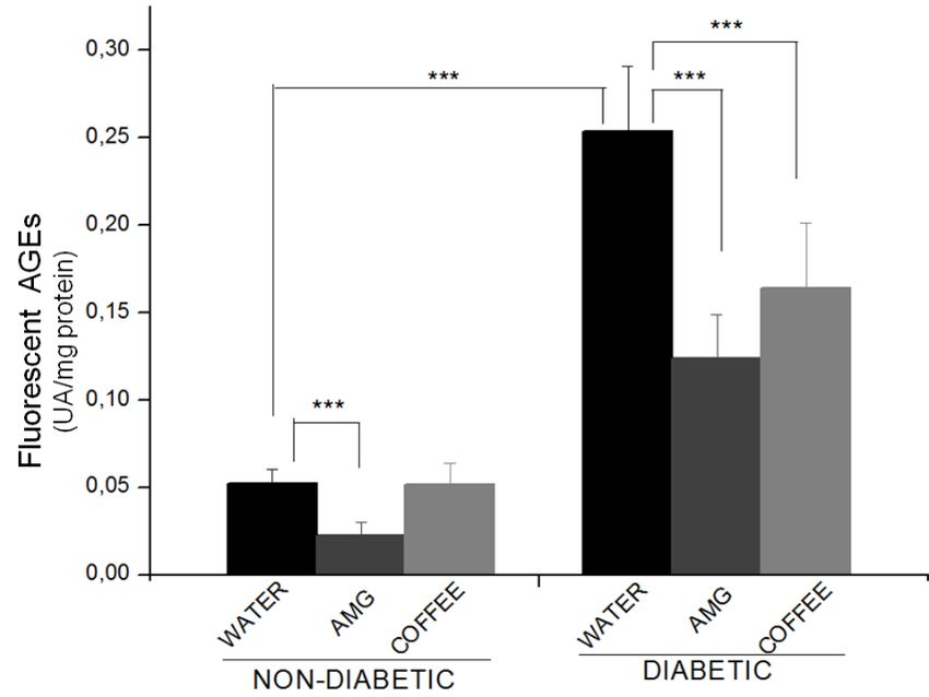
Circulating levels of fluorescent AGEs were significantly higher in diabetic animals
compared with non-diabetic animals (Figure 2). AGEs serum concentration decreased in both
groups treated with AMG (Figure 2). As expected, our results show an efficacy of AMG
treatment in preventing AGEs formation in diabetic animals. Coffee was as efficient as AMG
in reducing AGEs formation in diabetic animals (Figure 2).
Figure 2. Determination of fluorescent advanced glycation end products (AGEs) in rat’s serum. Values
are presented as mean ± SD of the three experiments performed in triplicate to measure serum
fluorescent AGEs in the non-diabetic and diabetic groups. Non-diabetic and diabetic rats were treated
with water, aminoguanidine (AMG) or coffee. SD: standard deviation. ***p < 0.001 indicates statistical
difference.
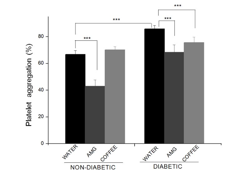
To assess the effects of coffee on primary hemostasis in diabetic rats, we measured the
platelet aggregation against the ADP agonist26. Platelet aggregation significantly increased in
the diabetic animals (Figure 3), suggesting an excess of glucose-induced osmotic effect and
reactive oxygen species (ROS) in the serum.
Rev Ciênc Farm Básica Apl. 2021;42:e711 6/14Coffee intake (Coffea arabica L.) reduces advanced glycation end product (AGEs) formation and platelet aggregation in diabetic rats
Figure 3. Evaluation of platelet aggregation percentage in rats. Values are presented as mean ± SD of
the three experiments performed in triplicate to measure platelet aggregation in the non-diabetic and
diabetic groups. Non-diabetic and diabetic rats were treated with water, aminoguanidine (AMG) or
coffee. SD: standard deviation. ***p < 0.001 indicates statistical difference.
We observed that AMG significantly reduced platelet aggregation in non-diabetic and
diabetic animals (Figure 3). Our results show that coffee reduced platelet aggregation in
diabetic animals to levels comparable to those of the non-diabetic group (Figure 3).
Finally, we evaluated the renal parameters in both animal groups by measuring urea and
creatinine levels. Diabetic rats had significantly higher levels of urea in the serum (Figure 4A).
Hyperglycemia did not alter the serum creatinine levels, that is, we found no kidney damage
due to hyperglycemia in the evaluated period (Figure 4B). The AMG treatment or coffee intake
had no effect on these animals’ renal profile.
Figure 4. Determination of the rats’ renal profile with serum urea (mg/mL) (A) and creatinine (mg/mL)
(B) measurements. Values are presented as mean ± SD of the three experiments performed in triplicate
to measure serum urea and creatinine in the non-diabetic and diabetic groups. Non-diabetic and
diabetic rats were treated with water, aminoguanidine (AMG) or coffee. SD: standard deviation.
***p < 0.001 indicates statistical difference.
4. DISCUSSION
The search for therapeutic plants for treating diabetes mellitus has been stimulated by the
World Health Organization (WHO) due to low side effects from their prolonged and traditional
use, in addition to being a low cost and significantly effective therapeutic alternative44.
Rev Ciênc Farm Básica Apl. 2021;42:e711 7/14Coffee intake (Coffea arabica L.) reduces advanced glycation end product (AGEs) formation and platelet aggregation in diabetic rats
Coffee is a fruit rich in bioactive compounds, such as caffeine, trigonelline, chlorogenic
acids, phenolic compounds, diterpenes and melanoidins. Its long-term consumption has
shown beneficial effects in chronic diseases such as diabetes45. The decreased risk of
developing chronic diseases has been related to the presence of phenolic compounds1,21,22.
Untreated chronic hyperglycemia leads to macro and microvascular diabetic complications.
The progression of these complications triggers multiple renal, cardiac and hepatic disorders
and, consequently, derangements in the biochemical, hematological and histological
parameters that contribute to increase free radicals, associated with the pathophysiology of
diabetes44.
In the present study, the diabetic status did not alter the number of leukocytes (Table 1);
but this change can occur and is typical of the damage caused by induced diabetes, as
reported by46. The other parameters (Hem and Hb) also showed no statistical change.
Hyperglycemia can modify red blood cell count and cause oxidative stress, hemoglobin
glycation and other hematological disorders46. Studies have shown the ability of phenolic
compounds to form complexes with Fe (II), present in large quantities in the hemoglobin
molecule, which could interfere with its absorption in the gastrointestinal tract and
consequently lead to iron deficiency47-49. The doses of coffee consumption used in this study,
however, were unable to change the hematological parameters in rats with a balanced diet.
Regarding the glycemic profile, diabetes induction and maintenance was confirmed in
animals by the increase in fasting glucose and glycated hemoglobin levels (Figure 1). Among
the bioactive substances present in coffee, caffeine has controversial effects on blood glucose
levels. While short-term metabolic studies performed in humans have shown that caffeine at
acute doses reduces insulin sensitivity due to its rapid metabolism27,50,51, others have
observed increased sensitivity in insulin receptors upon caffeine treatment52. Long-term
coffee consumption can induce glucose tolerance and insulin sensitivity, helping to prevent
diabetes symptoms and improving insulin sensitivity53.
Several mechanisms have been proposed to explain the beneficial effect of coffee on
glucose, insulin homeostasis and diabetes progression. This protective effect can be
attributed to other coffee components such as chlorogenic acid and their derivatives – lignans,
trigonelline, N-methylpyridinium, minerals and vitamins, proteins and lipids (diterpenes)19,20.
These compounds can act on harmful free radicals, participate in cell signaling pathways
performing, among other functions, anti-glycation activity54. Chlorogenic acids and their
derivatives represent the most prevalent phenolic compounds in coffee20,55.
The HbA1c level is an important biomolecular marker. It is a chromatographic fraction of
hemoglobin that carries an Amadori product in its β chain and that reflects the occurrence of
hyperglycemia in the three months prior to its measurement and, indirectly, advanced
glycation56. After 50 days of treatment, measuring this marker also confirmed the diabetic
state induced in the animals.
AMG, an agent commonly used to prevent AGEs formation in animal models, is effective in
reducing AGEs and lowering the severity of diabetes-associated structural and functional
alterations37,57. The use of a treatment that blocks AGEs or reduces oxidative and nitrosative
stress can be effective in controlling diabetic complications.
The contribution of AGEs in the development and progression of diabetic complications is
well established1,10,37,58. AGEs may act by modifying intracellular functional proteins involved
in gene regulation in the nearby extracellular matrix molecules, by interfering in the signaling
between the matrix and the cell causing dysfunction in proteins such as albumin. AGE
accumulation results in endothelial dysfunction, fibrosis, chronic inflammation and,
consequently, in generalized cellular dysfunction1,10,59.
As AGEs are one of the main factors that could potentially explain how chronic
hyperglycemia leads to the cell and tissue damage observed in diabetes, we measured the
serum concentration of the well characterized and widely studied fluorescent AGEs
pentosidine and argipiridine in the animals60. Circulating levels of fluorescent AGE-peptides
were significantly higher in diabetic animals compared with the non-diabetic animals (Figure
2). These results were similar to those found by Ferreira et al.37. Although it has already been
Rev Ciênc Farm Básica Apl. 2021;42:e711 8/14Coffee intake (Coffea arabica L.) reduces advanced glycation end product (AGEs) formation and platelet aggregation in diabetic rats
shown that AGE production increases significantly under hyperglycemia or oxidative stress,
several of its mechanical details remain unclear1,10,61,62. As mentioned above, AGEs serum
concentration significantly decreased in aminoguanidine-treated diabetic and non-diabetic
rats (Figure 2). Our results show the effectiveness of aminoguanidine treatment in preventing
AGE formation in diabetic animals. These data are consistent with the results obtained
previously by Ferreira et al.37. Similarly, coffee efficiently reduced AGE formation in diabetic
animals (Figure 2).
There has been significant interest in identifying dietary components that combat or
attenuate diabetic complications by reducing AGEs accumulation or inhibiting its effects on
the body63,64. Although no food component has been specified as being anti-AGE in vivo,
certain coffee components, such as phenolic compounds, have been intensively studied and
shown to have the ability to inhibit AGE formation63.
Being highly damaging to the integrity and function of blood vessels, AGEs result in the
main cause of diabetes complications related to vasculopathy64,65. The interaction of AGEs
with components of the vessel wall increases vascular permeability, expression of
procoagulant activity and ROS generation, resulting in increased leukocyte adhesion
molecules in the endothelial layer65. Additionally, platelet-monocyte aggregates and platelet
hyperactivity are important factors commonly associated with chronic diabetes
complications26,66.
Diabetic patients generally present symptoms of hypercoagulability and hypofibrinolysis,
but the degree of hemostatic abnormality in these patients is still poorly understood. To
evaluate the effects of coffee on primary hemostasis in diabetic rats, we measured the platelet
aggregation against the ADP agonist26. Platelet aggregation significantly increased in the
diabetic animals (Figure 3), suggesting an excess of glucose-induced osmotic effect and ROS
in the serum, leading to platelet aggregation and degranulation and, consequently, decreased
endothelial functions26,67. Increased platelet activity and thrombus formation has been
suggested to play a role in diabetes, atherosclerosis, cardiac disease, and hypertension26,68.
Several studies have shown increased platelet reactivity in diabetic patients by increased
levels of specific markers, such as beta-thromboglobulin and platelet factor 4 in platelet
granules in plasma69, and increased expression of platelet membrane glycoproteins GPIb and
GPIIb/IIIa. Reduced membrane fluidity has also been correlated with glycation of platelet
membrane proteins70,71.
We observed that aminoguanidine significantly reduced platelet aggregation in non-
diabetic and diabetic animals (Figure 3). This result may be explained by its antioxidant
activity72, which blocks protein glycation and can reduce reactivity changes in platelets, thus
helping ameliorate the endothelial dysfunction that is intrinsic to diabetes.
AGE-induced pathogenesis of diabetic micro- and macroangiopathy is well established in
the literature10,73,74. As kidneys are the major site for AGE clearance, they constitute the main
target of deleterious AGE-mediated changes75. The severity of vascular damage in renal tissue
may result from a decline in soluble RAGE (sRAGE) and the AGEs/sRAGE ratio; associated with
urinary albumin/serum creatinine levels, it can measure reno-vascular complications in
diabetic patients10. Thus, we evaluated the renal function in both animal groups by measuring
urea and creatinine levels. Diabetic rats showed significantly higher levels of urea in the
serum, and aminoguanidine treatment or coffee intake effected no change in these animals’
renal profile (Figure 4A). Urea, the main product of protein catabolism in the liver, is
synthesized from CO2 and ammonia and circulates in the blood before being filtered in the
kidneys, where most of it is excreted in urine. Urea levels are sensitive to changes, appear
faster in primary renal functions, and is an important marker in conditions that affect primary
renal function alterations, like diabetes37,76. DM2 promotes progressive cachexia, which
causes skeletal muscle atrophy77 – result of the imbalance on protein synthesis in favor of its
proteolysis. Excessive proteolysis results in an increase in plasma urea, as observed in the
study. The atrophy stems from reduced insulin-like growth factor 1 (IGF-1) and hyperglycemic
conditions78. But as urea concentration is influenced by hydration levels and dietary habits, it
is less specific than creatinine for assessing renal function.
Rev Ciênc Farm Básica Apl. 2021;42:e711 9/14Coffee intake (Coffea arabica L.) reduces advanced glycation end product (AGEs) formation and platelet aggregation in diabetic rats
In this study, coffee intake or aminoguanidine treatment had no effect on serum creatinine
levels in the diabetic animals (Figure 4B). Creatinine, which is a breakdown product of creatine
phosphate in muscle, is usually produced at a constant rate and its levels are directly
proportional to muscle mass. Interestingly, creatinine levels also reflect the glomerular
filtration rate, for its concentration in the blood increases as the renal filtration rate declines79.
According to Fagbohun et al.44, plasma creatinine acts as a better indicator of stage 1 kidney
toxicity than urea.
Our results showed that aminoguanidine did not alter renal biomarkers. Although not
evaluated in this work, recent studies have shown that this AGE-inhibitor can alter liver
markers such as alanine aminotransferase (ALT), aspartate aminotransferase (AST) and
alkaline phosphatase (ALP)80.
It is important to highlight that the coffee variety, roasting process, and method of brewing,
among other variables, changes the amount of bioactive components, such as chlorogenic
acid, caffeine, and melanoidins, in the beverage. Such components are the main responsible
for the antioxidative effects of coffee and can alter the effects described in other studies.
Thus, it is possible to conclude that the study results showed that coffee intake reduced
AGEs formation and platelet aggregation in our experimental model. The protective role of
coffee may result from the action of its bioactive components, such as chlorogenic acid.
Aminoguanidine was effective in inhibiting AGE formation and platelet aggregation. We also
observed that coffee had no effect on the animals’ renal function. These results are especially
interesting because diabetes is an important risk factor for thrombosis, characterized by
hypercoagulability and hypofibrinolysis, and coffee is a widely consumed beverage in the
contemporary world.
REFERENCES
1. Jud P, Sourij H. Theraoeutic options to reduce advanced glycation end products in patients with
diabetes ellitus: A review. Diabetes Res Clin Pract. 2019;148:54-63.
http://dx.doi.org/10.1016/j.diabres.2018.11.016. PMid:30500546.
2. Pang M, Li Y, Gu W, Sun Z, Wang Z, Li L. Recent Advances in Epigenetics of Macrovascular
Complications in Diabetes mellitus. Heart Lung Circ. 2020;30(2):186-196.
3. Mirza S, Khan AA, Al-Kheraif AA, Khan SZ, Shafqat SS. Efficacy of adjunctive photodynamic therapy
on the clinical periodontal, HbA1c and advanced glycation end product level among mild to
moderate chronic periodontal disease patients with type 2 diabetes mellitus: A randomized
controlled clinical trial., Photodiagn Photodyn Ther. 2019;28:177-82.
http://dx.doi.org/10.1016/j.pdpdt.2019.08.003. PMid:31394300.
4. Triches C, Schaan B, Gross JL, Azevedo MJ. Complicações macrovasculares do diabetes melito:
peculiaridades clínicas, de diagnóstico e manejo. Arq Bras Endocrinol Metabol. 2009;53(6):698-708.
http://dx.doi.org/10.1590/S0004-27302009000600002. PMid:19893911.
5. Reis JS, Veloso CA, Mattos RT, Purish S, Nogueira-Machado JA. Estresse oxidativo: revisão de
sinalização metabólica no diabetes tipo 1. Arq Bras Endocrinol Metabol. 2008;52(7):1096-105.
http://dx.doi.org/10.1590/S0004-27302008000700005. PMid:19082297.
6. Sheweita SA, ElHady SA, Hammoda HM. Trigonella stellate reduced the deleterious effects of
diabetes mellitus through alleviation of oxidative stress, antioxidant- and drug-metabolizing
enzymes activities. J Ethnopharmacol. 2020;256:112821.
http://dx.doi.org/10.1016/j.jep.2020.112821. PMid:32251758.
7. Wu CH, Huang HW, Lin JA, Huang SM, Yen GC. The proglycation effect caffeic acid leads to the
elevation of oxidative stress and inflammation in monocytes, macrophages and vascular
endothelial cells. J Nutr Biochem. 2011;22(6):585-95.
http://dx.doi.org/10.1016/j.jnutbio.2010.05.002. PMid:20951562.
8. Zeng C, Li Y, Ma J, Niu L, Tay FR. Clinical/Translational aspects of advanced glycation end-products.
Trends Endocrinol Metab. 2019;30(12):959-73. http://dx.doi.org/10.1016/j.tem.2019.08.005.
PMid:31597608.
9. Brownlee M. Biochemistry and molecular cell biology of diabetic complications. Nature.
2001;414(6865):813-20. http://dx.doi.org/10.1038/414813a. PMid:11742414.
Rev Ciênc Farm Básica Apl. 2021;42:e711 10/14Coffee intake (Coffea arabica L.) reduces advanced glycation end product (AGEs) formation and platelet aggregation in diabetic rats
10. Farhan SS, Hussain SA. Advanced glycation end products (AGEs) and their soluble receptors
(sRAGE) as early predictors of reno-vascular complications in patients with uncontrolled type 2
diabetes mellitus. Diabetes Metab Syndr. 2019;13(4):2457-61.
http://dx.doi.org/10.1016/j.dsx.2019.06.019. PMid:31405660.
11. Peppa M, Uribarri J, Vlassara H. Glucose, advanced glycation end products, and diabetes
complications: what is new and what works. Clin Diabetes. 2003;21(4):186-7.
http://dx.doi.org/10.2337/diaclin.21.4.186.
12. Borissoff JI, Spronk HMH, ten Cate H. The hemostatic system as a modulator of atherosclerosis. N
Engl J Med. 2011;364(18):1746-60. http://dx.doi.org/10.1056/NEJMra1011670. PMid:21542745.
13. Reasner CA. Reducing cardiovascular complications of type 2 diabetes by targeting multiple risk
factors. J Cardiovasc Pharmacol. 2008;52(2):136-44.
http://dx.doi.org/10.1097/FJC.0b013e31817ffe5a. PMid:18670366.
14. Barg A, Ossig R, Goerge T, Schneider MF, Schillers H, Oberleithner H, Schneider SW. Soluble plasma-
deived Von Willebrand factor assembles to a haemostatically active filamentous network. J Thromb
Haemost. 2007;97(4):514-26. http://dx.doi.org/10.1160/TH06-05-0274. PMid:17393012.
15. Gray E, Mulloy B, Barrowcliffe TW. Heparin and low-molecular-weight heparin. J Thromb Haemost.
2008;99(5):807-18. PMid:18449410.
16. Michelson AD. Antiplatelet therapies for the treatment of cardiovascular disease. Nat Rev Drug
Discov. 2010;9(2):154-69. http://dx.doi.org/10.1038/nrd2957. PMid:20118963.
17. American Diabetes Association. Position statement: standards of medical care in diabetes.
Diabetes Care. 2013;36:11-66.
18. Fangel MV, Nielsen PB, Kristensen JK, Larsen TB, Overvad TF, Lip GYH, Jensen MB. Albuminuria and
risk of cardiovascular events and mortality in a general population of patients with Type 2 diabetes
without cardiovascular disease: a danish cohort study. Am J Med. 2020;133(6):e269-79.
http://dx.doi.org/10.1016/j.amjmed.2019.10.042. PMid:32205071.
19. Mirmiran P, Carlström M, Bahadoran Z, Azizi F. Long-term effects of coffee and caffeine intake on
the risk of pre-diabetes and type 2 diabetes: findings from a population with low coffee
consumption. Nutr Metab Cardiovasc Dis. 2018;28(12):1261-6.
http://dx.doi.org/10.1016/j.numecd.2018.09.001. PMid:30352712.
20. Costabile A, Sarnsamak K, Hauge-Evans AC. Coffee, type 2 diabetes and pancreatic islet function – A
mini-review. J Funct Foods. 2018;45:409-16. http://dx.doi.org/10.1016/j.jff.2018.04.011.
21. Muñoz E, Hernández SS, Tolosa AR, Burillo SP, Olalla-Herrera M. Evaluation of differences in the
antioxidant capacity and phenolic compounds of green and roasted coffee and their relationship
with sensory properties. Lebensm Wiss Technol. 2020;128:109457.
http://dx.doi.org/10.1016/j.lwt.2020.109457.
22. Vithana MDK, Singh Z, Johnson SK. Harvest maturity stage affects the concentrations of health-
promoting compounds: Lupeol, mangiferin and phenolic acids in the pulp and peel of ripe
‘Kensington Pride’ mango fruit. Sci Hortic (Amsterdam). 2019;243:125-30.
http://dx.doi.org/10.1016/j.scienta.2018.08.019.
23. Gómez-Ruiz JA, Leake DS, Ames JM. In vitro antioxidant activity of coffee compounds and their
metabolites. J Agric Food Chem. 2007;5(17):6962-9. http://dx.doi.org/10.1021/jf0710985.
PMid:17655324.
24. Lafay S, Morand C, Manach C, Besson C, Scalbert A. Absorption and metabolism of caffeic acid and
chlorogenic acid in the small intestine of rats. Br J Nutr. 2006;96(1):39-46.
http://dx.doi.org/10.1079/BJN20061714. PMid:16869989.
25. Natella F, Scaccini C. Role of coffee in modulation of diabetes risk. Nutr Rev. 2012;70(4):207-17.
http://dx.doi.org/10.1111/j.1753-4887.2012.00470.x. PMid:22458694.
26. Olas B, Bryś M. Effects of coffee, energy drinks and their components on hemostasis: the
hypothetical mechanisms of their action. Food Chem Toxicol. 2019;127:31-41.
http://dx.doi.org/10.1016/j.fct.2019.02.039. PMid:30844438.
27. Reis CEG, Dórea JG, da Costa THM. Effects of coffee consumption on glucose metabolism: A
systematic review of clinical trials. J Tradit Complement Med. 2019;9(3):184-91.
http://dx.doi.org/10.1016/j.jtcme.2018.01.001. PMid:31193893.
Rev Ciênc Farm Básica Apl. 2021;42:e711 11/14Coffee intake (Coffea arabica L.) reduces advanced glycation end product (AGEs) formation and platelet aggregation in diabetic rats
28. Lima AR, Pereira RGFA, Abrahão SA, Duarte SMS, Paula FBA. Compostos bioativos do café: atividade
antioxidante in vitro do café verde e torrado antes e após a descafeinação. Quim Nova.
2010;33(1):20-4. http://dx.doi.org/10.1590/S0100-40422010000100004.
29. Tripetch P, Borompichaichartkul C. Effect of packaging materials and storage time on changes of
colour, phenolic content, chlorogenic acid and antioxidant activity in Arabica green coffee beans
(Coffea Arabica L. cv. Catimor). J Stored Prod Res. 2019;84:101510.
http://dx.doi.org/10.1016/j.jspr.2019.101510.
30. Higdon JV, Frei B. Coffee and health: a review of recent human research. Crit Rev Food Sci Nutr.
2006;46(2):101-23. http://dx.doi.org/10.1080/10408390500400009. PMid:16507475.
31. Farmacopeia Brasileira. Métodos de Farmacognosia. Métodos de análises de drogas vegetais.
Determinação de água em drogas vegetais. 5. ed. Brasília: ANVISA; 2017. p. 54.
32. Duarte SMS, Abreu CMP, Menezes HC, Santos MR, Gouvêa CMCP. Effect of processing and roasting
on the antioxidant activity of coffee brews. Ciência Tecnol Alime. 2005;25(2):387-93.
http://dx.doi.org/10.1590/S0101-20612005000200035.
33. Ferreira C, Araújo TH, Ângelo ML, Pennacchi PC, Okada SS, Paula FBA, Migliorini S, Rodrigues MR.
Neutrophil dysfunction induced by hyperglycemia: modulation of myeloperoxidase activity. Cell
Biochem Funct. 2012;30(7):604-10. http://dx.doi.org/10.1002/cbf.2840. PMid:22610543.
34. Szkudelski T. The mechanism of alloxan and streptozotocin action in B cells of the rat pancreas.
Physiol Res. 2001;50(6):537-46. PMid:11829314.
35. Stoppa GR, Cesquini M, Roman EA, Ogo SH, Torsoni MA. Aminoguanidine prevented impairment of
blood antioxidant system in insulin-dependent diabetic rats. Life Sci. 2006;78(12):1352-61.
http://dx.doi.org/10.1016/j.lfs.2005.07.031. PMid:16229859.
36. Edelstein D, Brownlee M. Aminoguanidine ameliorates albuminuria in diabetic hypertensive rats.
Diabetologia. 1992;35(1):96-7. http://dx.doi.org/10.1007/BF00400859. PMid:1541387.
37. Ferreira CS, Pennacchi PC, Araújo TH, Taniwaki NN, Paula FBA, Duarte SMS, Rodrigues MR.
Aminoguanidine treatment increased NOX2 response in diabetic rats: improved phagocytosis and
killing of Candida albicans by neutrophils. Eur J Pharmacol. 2016;772:83-91.
http://dx.doi.org/10.1016/j.ejphar.2015.12.044. PMid:26724393.
38. Zilin S, Naifeng L, Bicheng L, Jiping W. The determination of AGE-peptides by flow injection assay, a
practical marker of diabetic nephropathy. Clin Chim Acta. 2001;313(1-2):69-75.
http://dx.doi.org/10.1016/S0009-8981(01)00651-9. PMid:11694241.
39. Silvério ASD, Pereira RGFA, Lima AR, Paula FBA, Rodrigues MR, Baldissera L Jr, Duarte SM. The
effects of the decaffeination of coffee samples on platelet aggregation in hyperlipidemic rats. Plant
Foods Hum Nutr. 2013;68(3):268-73. http://dx.doi.org/10.1007/s11130-013-0365-x. PMid:23780748.
40. Layne E. Spectrophotometric and Turbidimetric Methods for Measuring Proteins. Methods
Enzymol. 1957;10:447-55. http://dx.doi.org/10.1016/S0076-6879(57)03413-8.
41. Alhamdani MS, Al-Azzawie HF, Abbas FK. Decreased formation of advanced glycation end-products
in peritoneal fluid by carnosine and related peptides. J Int Society Perit Dial., 2007;27(1):86-9.
http://dx.doi.org/10.1177/089686080702700118. PMid:17179517.
42. R Core Team (2020). R: A language and environment for statistical computing. R Foundation for
Statistical Computing, Vienna, Austria. https://www.R-project.org/
43. Ferreira EB, Cavalcanti PP, Nogueira DA. ExpDes: Experimental Designs package. R package version
1.1.2; 2013.
44. Fagbohun OF, Awoniran PO, Babalola OO, Agboola FK, Msagati TAM. Changes in the biochemical,
hematological and histopathological parameters in STZ-Induced diabetic rats and the ameliorative
effect of Kigelia africana fruit extract. Heliyon. 2020;6(5):e03989.
http://dx.doi.org/10.1016/j.heliyon.2020.e03989. PMid:32462092.
45. Wang X, Meng Q, Peng X, Hu G, Qiu M. Identification of new diterpene esters from greens Arabica
coffee beans, and their platelet aggregation accelerating activities. Food Chem. 2018;263:251-7.
http://dx.doi.org/10.1016/j.foodchem.2018.04.081. PMid:29784314.
46. Hajam YA, Rai S, Ghosh H, Basheer M. Combined administration of exogenous melatonin and
insulin ameliorates streptozotocin induced toxic alteration on hematological parameters in diabetic
male Wistar rats. Toxicol Rep. 2020;7:353-9. http://dx.doi.org/10.1016/j.toxrep.2020.01.020.
PMid:32095432.
Rev Ciênc Farm Básica Apl. 2021;42:e711 12/14Coffee intake (Coffea arabica L.) reduces advanced glycation end product (AGEs) formation and platelet aggregation in diabetic rats
47. Szajdek A, Borowska EJ. Bioactive Compounds and Health-Promoting Properties of Berry Fruits: A
Review. Plant Foods Hum Nutr. 2008;63(4):147-56. http://dx.doi.org/10.1007/s11130-008-0097-5.
PMid:18931913.
48. Elhabiri M, Carrër C, Marmolle F, Traboulsi H. Complexation of iron (III) by catecholate-type
polyphenols. Inorg Chim Acta. 2007;360(1):353-9. http://dx.doi.org/10.1016/j.ica.2006.07.110.
49. Mennen LI, Walker R, Bennetau-Pelissero C, Scalbert A. Risks and safety of polyphenol
consumption. Am J Clin Nutr. 2005;81(1, Suppl):326S-9S. http://dx.doi.org/10.1093/ajcn/81.1.326S.
PMid:15640498.
50. Keijzers GB, de Galan BE, Tack CJ, Smits P. Caffeine can decrease insulin sensitivity in humans.
Diabetes Care. 2002;25(2):364-9. http://dx.doi.org/10.2337/diacare.25.2.364. PMid:11815511.
51. Thong FS, Derave W, Kiens B, Graham TE, Urso B, Wojtaszewski JF, Hansen BF, Richter EA. Caffeine-
induced impairment of insulin action but not insulin signaling in human skeletal muscle is reduced
by exercise. Diabetes. 2002;51(3):583-90. http://dx.doi.org/10.2337/diabetes.51.3.583.
PMid:11872654.
52. van Dam RM, Willett WC, Manson JE, Hu FB. Coffee, caffeine, and risk of type 2 diabetes: a
prospective cohort study in younger and middle-aged U.S. women. Diabetes Care. 2006;29(2):398-
403. http://dx.doi.org/10.2337/diacare.29.02.06.dc05-1512. PMid:16443894.
53. Akash MSH, Rehman K, Chen S. Effects of coffee on type 2 diabetes mellitus. Nutrition. 2014;30(7-
8):755-63. http://dx.doi.org/10.1016/j.nut.2013.11.020. PMid:24984989.
54. Khan M, Liu H, Wang J, Sun B. Inhibitory effect of phenolic compounds and plant extracts on the
formation of advance glycation end products: A comprehensive review. Food Res Int.
2020;130:108933. http://dx.doi.org/10.1016/j.foodres.2019.108933. PMid:32156381.
55. Clifford MN. Chlorogenic Acids. Coffee. 1985;1:153-202. http://dx.doi.org/10.1007/978-94-009-4948-5_5.
56. Mustafa IO, Tanko Y, Yusuf R, Musa SA. The use of glycated haemoglobin (HbA1c) in determining
glycemic control (and relevance of BMI) in diabetic patients in Ahmadu Bello University Teaching
Hospital Zaria, Nigeria. Diabetes Metab Syndr. 2019;13(5):2967-292.
http://dx.doi.org/10.1016/j.dsx.2019.07.057. PMid:31437811.
57. Huebschmann AG, Regensteiner JG, Vlassara H, Reusch JE. Diabetes and glicoxidation end
products. Diabetes Care. 2006;29(6):1420-32. http://dx.doi.org/10.2337/dc05-2096. PMid:16732039.
58. Jakus V, Rietbrock N. Advanced glycation end-products and the progress of diabetic vascular
complications. Physiol Res. 2004;53(2):131-42. PMid:15046548.
59. Goldin A, Beckman JA, Schmidt A, Creager MA. Review. Advanced glycation end products: sparking
the development of diabetic vascular injury. Circulation. 2006;114(6):597-605.
http://dx.doi.org/10.1161/CIRCULATIONAHA.106.621854. PMid:16894049.
60. Ahmed N. Advanced glycaton endproducts-role in pathology of diabetic complications. Diabetes
Res Clin Pract. 2005;67(1):3-21. http://dx.doi.org/10.1016/j.diabres.2004.09.004. PMid:15620429.
61. Jay D, Hitomi H, Griendling KK. Oxidative stress and diabetic cardiovascular complications. Free
Radic Biol Med. 2006;40(2):183-92. http://dx.doi.org/10.1016/j.freeradbiomed.2005.06.018.
PMid:16413400.
62. Lapolla A, Fedele D, Traldi P. Glyco-oxidation in diabetes and related diseases. Clin Chim Acta.
2005;357(2):236-50. http://dx.doi.org/10.1016/j.cccn.2005.03.032. PMid:15939413.
63. Babu PV, Sabitha KE, Shyamaladevi CS. Green tea impedes dyslipidemia, lipid peroxidation, protein
glycation and ameliorates Ca2+-ATPase activity in the heart of streptozotocin-diabetic rats. Chem
Biol Interact. 2006;162(2):157-64. http://dx.doi.org/10.1016/j.cbi.2006.05.020. PMid:16846594.
64. Joglekar MM, Bavkar LN, Sistla S, Arvindekar AU. Effective inhibition of protein glycation by
combinatorial usage of limonene and aminoguanidine through differential and synergistic
mechanisms. Int J Biol Macromol. 2017;99:563-8. http://dx.doi.org/10.1016/j.ijbiomac.2017.02.104.
PMid:28274869.
65. Bierhaus A, Hofmann MA, Ziegler R, Nawroth PP. AGEs and their interaction with AGE-receptors in
vascular disease and diabetes mellitus I. The AGE concept Nawroth. Cardiovasc Res. 1998;37(3):586-
600. http://dx.doi.org/10.1016/S0008-6363(97)00233-2. PMid:9659442.
Rev Ciênc Farm Básica Apl. 2021;42:e711 13/14Coffee intake (Coffea arabica L.) reduces advanced glycation end product (AGEs) formation and platelet aggregation in diabetic rats
66. Ollas B, Wachowiez B, Stochhmal A, Olessezk W. Inhibition of blood platelet adhesion and aecretion
by different phenolics from Yucca achidigera Roezl Bark. Nutr. 2005;21(2):199-206.
http://dx.doi.org/10.1016/j.nut.2004.03.024.
67. Keating FK, Sobel BE, Schneider DJ. Effects of increased concentrations of glucose on platelet
reactivity in healthy subjects and in patients with and without diabetes mellitus. Am J Cardiol.
2003;92(11):1326-1325. http://dx.doi.org/10.1016/j.amjcard.2003.08.033. PMid:14636925.
68. El Haouari M, Rosado JA. Platelet signaling abnormalities in patients with type 2 diabetes mellitus: A
review. Blood Cells Mol Dis. 2008;41(1):119-23. http://dx.doi.org/10.1016/j.bcmd.2008.02.010.
PMid:18387322.
69. Grant PJ. Diabetes melito as prothrombotic condition. J Intern Med. 2007;262(2):157-72.
http://dx.doi.org/10.1111/j.1365-2796.2007.01824.x. PMid:17645584.
70. Kuhne T, Hornstein A, Semple J, Chang W, Blanchette V, Freedman J. Flow cytometric evaluation of
platelet activation in blood collected into EDTA vs. Diatube-H a sodium citate solution
supplemented with theophylline, adenosine, and dipyridamole. Am J Hematol. 1995;50(1):40-5.
http://dx.doi.org/10.1002/ajh.2830500108. PMid:7668222.
71. Winocour PD. Platelets, vascular disease, and diabetes mellitus [Review]. Can J Physiol Pharmacol.
1994;72(3):295-303. http://dx.doi.org/10.1139/y94-045. PMid:8069776.
72. Freedman JE. Oxidative stress and Platelets. Arterioscler Thromb Vasc Biol. 2008;28(3):11-6.
http://dx.doi.org/10.1161/ATVBAHA.107.159178. PMid:18174453.
73. Coughlan MT, Mibus AL, Forbes JM. Oxidative stress and advanced glycation in diabetic
nephropathy. Ann N Y Acad Sci. 2008;1126(1):190-3. http://dx.doi.org/10.1196/annals.1433.018.
PMid:18448815.
74. Noordzij MJ, Lefrandt JD, Smit AJ. Advanced glycation end products in renal failure: an overview. J
Ren Care. 2008;34(4):207-12. http://dx.doi.org/10.1111/j.1755-6686.2008.00038.x. PMid:19090900.
75. Miyata T, Ueda Y, Horie K, Nangaku M, Tanaka S, Van Ypersele de Strihou C, Kurokawa K. Renal
catabolism of advanced glycation end products: the fate of pentosidine. Kidney Int. 1998;53(2):416-
22. http://dx.doi.org/10.1046/j.1523-1755.1998.00756.x. PMid:9461101.
76. Nosadini R. Hypertension and renal complications in type diabetes. Semin Vasc Med. 2002;2(1):109-
19. http://dx.doi.org/10.1055/s-2002-23101. PMid:16222601.
77. Yadav A, Singh A, Phogat J, Dahuja A, Dabur R. Magnoflorine prevent the skeletal muscle atrophy
via Akt/mTOR/FoxO signal pathway and increase slow-MyHC production in streptozotocin-induced
diabetic rats. J Ethnopharmacol. 2020;267:113510. http://dx.doi.org/10.1016/j.jep.2020.113510.
PMid:33141056.
78. Kordi M, Khoramshahi S, Eshghi S, Gaeeni A, Moosakhani A. The effect of high intensity interval
training on some atrophic and anti-atrophic gene expression in rat skeletal muscle with diabetes.
Sci Sports. 2020;35(3):e75-81. http://dx.doi.org/10.1016/j.scispo.2019.04.003.
79. Gómez-Marcos MA, Martínez-Salgado C, Grandes G, Recio-Rodríguez JI, Castaño Sánchez Y,
Rodríguez SE, García-Ortiz L. Subclinical and established kidney disease in recently diagnosed
hypertensive patients. Med Clin (Barc). 2010;134(6):246-53. PMid:20045531.
80. Lima TFO, Costa MC, Figueiredo ID, Inácio MD, Rodrigues MR, Assis RP, Baviera AM, Brunetti IL.
Curcumin, alone or in combination with aminoguanidine, increases antioxidant defenses and
glycation product detoxification in streptozotocin-diabetic rats: a therapeutic strategy to mitigate
glycoxidative stress. Oxid Med Cell Longev. 2020;2020:1036360.
http://dx.doi.org/10.1155/2020/1036360. PMid:32566072.
Authors' contributions
ASDS and CSF performed the experiments, collected and analyzed the data; RGFAP, SMSD and
MRR designed the study and performed the data evaluation; BCCS and ALMV performed the
experiments; FBAP and THA performed the data evaluation; EBF statistical analysis; ASDS and MRR
wrote the paper; SAF analysis and evaluation of data and update of article writing.
Rev Ciênc Farm Básica Apl. 2021;42:e711 14/14You can also read