Common and Specific Alterations of Amygdala Subregions in Major Depressive Disorder With and Without Anxiety: A Combined Structural and ...
←
→
Page content transcription
If your browser does not render page correctly, please read the page content below
ORIGINAL RESEARCH
                                                                                                                                            published: 15 February 2021
                                                                                                                                      doi: 10.3389/fnhum.2021.634113

                                            Common and Specific Alterations of
                                            Amygdala Subregions in Major
                                            Depressive Disorder With and
                                            Without Anxiety: A Combined
                                            Structural and Resting-State
                                            Functional MRI Study
                                            Yao Yao Li 1† , Xiao kang Ni 2† , Ya feng You 2† , Yan hua Qing 2 , Pei rong Wang 3 , Jia shu Yao 2 ,
                                            Ke ming Ren 2 , Lei Zhang 2 , Zhi wei Liu 4 , Tie jun Song 4 , Jinhui Wang 5 , Yu-Feng Zang 6 ,
                                            Yue di Shen 7* and Wei Chen 2,8,9*
                            Edited by:      1
                                             Department of Psychiatry, Hangzhou Seventh People’s Hospital, Hangzhou, China, 2 Department of Psychiatry, Sir Run Run
                              Wei Liao,     Shaw Hospital, Zhejiang University School of Medicine, Hangzhou, China, 3 Zhejiang Academy of Traditional Chinese
  University of Electronic Science and      Medicine, Hangzhou, China, 4 Clinical Laboratory, Sir Run Run Shaw Hospital, Zhejiang University School of Medicine,
           Technology of China, China       Hangzhou, China, 5 Guangdong Key Laboratory of Mental Health and Cognitive Science, Center for Studies of Psychological
                       Reviewed by:         Application, Institute for Brain Research and Rehabilitation, South China Normal University, Guangzhou, China, 6 Zhejiang Key
                         Yifeng Wang,       Laboratory for Research in Assessment of Cognitive Impairments, Hangzhou, China, 7 Department of Diagnostics, School of
     Sichuan Normal University, China       Medicine, Hangzhou Normal University, Hangzhou, China, 8 Department of Psychology and Behavioral Sciences, Zhejiang
                             Lixia Tian,    University, Hangzhou, China, 9 Key Laboratory of Medical Neurobiology of Zhejiang Province, Hangzhou, China
     Beijing Jiaotong University, China

                   *Correspondence:
                                            Anxious major depressive disorder is a common subtype of major depressive disorder;
                           Yue di Shen
                    shenyd5@126.com         however, its unique neural mechanism is not well-understood currently. Using multimodal
                             Wei Chen       MRI data, this study examined common and specific alterations of amygdala subregions
                      srrcw@zju.edu.cn
                                            between patients with and without anxiety. No alterations were observed in the gray
     † These   authors have contributed     matter volume or intra-region functional integration in either patient group. Compared
                    equally to this work
                                            with the controls, both patient groups showed decreased functional connectivity between
                    Specialty section:      the left superficial amygdala and the left putamen, and between the right superficial
          This article was submitted to     amygdala and the bilateral anterior cingulate cortex and medial orbitofrontal cortex,
        Brain Imaging and Stimulation,
                                            while only patients with anxiety exhibited decreased activity in the bilateral laterobasal
                 a section of the journal
     Frontiers in Human Neuroscience        and superficial amygdala. Moreover, the decreased activity correlated negatively with the
       Received: 27 November 2020           Hamilton depression scale scores in the patients with anxiety. These findings provided
        Accepted: 22 January 2021           insights into the pathophysiologic processes of anxious major depressive disorder and
       Published: 15 February 2021
                                            may help to develop new and effective treatment programs.
                               Citation:
Li YY, Ni Xk, You Yf, Qing Yh, Wang Pr,     Keywords: major depressive disorder, multimodal MRI, amygdala subregion, anxiety, functional connectivity
     Yao Js, Ren Km, Zhang L, Liu Zw,
  Song Tj, Wang J, Zang Y-F, Shen Yd
     and Chen W (2021) Common and           INTRODUCTION
       Specific Alterations of Amygdala
       Subregions in Major Depressive
                                            Major depressive disorder (MDD) is a common mental disorder that affects more than 300 million
 Disorder With and Without Anxiety: A
               Combined Structural and
                                            people globally (Gaspersz et al., 2017a). MDD is becoming the leading cause of disability worldwide
   Resting-State Functional MRI Study.      and contributes significantly to the global disease burden. MDD is a clinically heterogeneous
    Front. Hum. Neurosci. 15:634113.        disease with multiple subtypes, among which anxious MDD (AMDD) is one of the most
     doi: 10.3389/fnhum.2021.634113         common, with a prevalence of 40–60% (Gaspersz et al., 2017a). Compared with non-anxious MDD

Frontiers in Human Neuroscience | www.frontiersin.org                              1                                         February 2021 | Volume 15 | Article 634113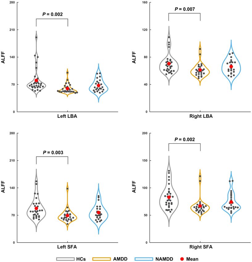
Li et al.                                                                                                         Altered Amygdala Subregions in MDD

(nAMDD), patients with AMDD often suffer from more severe                     baseline brain architecture and clinical outcomes of patients with
depressive illness (Wiethoff et al., 2010; Goldberg et al., 2014),            MDD after antidepressant treatment using multimodal MRI data.
greater functional impairment (Rao and Zisook, 2009; Goldberg                 According to the aim of the current study, 49 patients with MDD
and Fawcett, 2012), reduced response to antidepressant treatment              (26 AMDD and 23 nAMDD) were selected. MDD was diagnosed
(Fava et al., 2008; Ionescu et al., 2014), and a higher risk of suicide       according to the Diagnostic and Statistical Manual of Mental
(Seo et al., 2011; Gaspersz et al., 2017b). Thus, it is clinically            Disorders, 4th Edition, Text Revision (DSM-IV-TR) criteria,
significant to explore the unique neural mechanism underlying                 using the Structured Clinical Interview for DSM-V (SCID)-I.
this specific MDD subtype, which could help to develop new and                Exclusion criteria for MDD included (1) patients who could
effective treatment programs for AMDD.                                        not undergo an MRI examination because of claustrophobia,
    The amygdala, which is located in the medial part of the                  metallic implants, or other contraindication for MRI; (2) patients
anterior temporal lobe, is one of the most important structures               with severe suicidal tendency; (3) pregnant or lactating women;
in the limbic system (Ongur and Price, 2000). It plays a vital role           (4) any severe physical diseases as assessed by personal history;
in emotion processing and regulation (Phillips and Swartz, 2014).             (5) a history of organic brain disorders, neurological disorders,
Mounting evidence from multimodal MRI studies has indicated                   other psychiatric disorders, cardiovascular diseases, head trauma,
that the amygdala is widely implicated in the pathophysiology                 or loss of consciousness; and (6) a history of substance abuse,
of depression, as characterized by structural and functional                  including tobacco, alcohol, or other psychoactive substances.
alterations in patients (Savitz and Drevets, 2009; Licznerski and             The patients were recruited from outpatients and inpatients
Duman, 2013; Fonseka et al., 2018). With respect to AMDD, a                   of the Sir Run Run Shaw Hospital, School of Medicine,
previous study used diffusion and structural MRI and showed                   Zhejiang University, Hangzhou, China. All patients were free of
no significant differences in volume or white matter integrity                psychotropic medications for at least 4 weeks before the MRI scan
of the amygdala between patients with AMDD and nAMDD                          and had a Hamilton depression rating (HAMD) score ≥ 14. We
(Delaparte et al., 2017). Analogously, analyses of functional                 chose a HAMD ≥ 14 instead of 18 as in our previous studies
MRI (fMRI) revealed that patients with AMDD and those with                    (Shen et al., 2015; Sheng et al., 2018) to maximize the sample size
nAMDD exhibited common patterns of abnormal activation and                    of the current study. Out of the 49 patients, 26 were categorized
connectivity of the amygdala compared with controls (Etkin and                as having AMDD in terms a HAMD anxiety/somatization factor
Schatzberg, 2011; van Tol et al., 2012). However, it is worth                 score ≥ 7 (Fava et al., 2008). In addition, a cohort of 30 HCs
noting that all the above studies treated the amygdala as a unified           was enrolled using community recruitment via an advertisement.
structure. Recent evidence indicates anatomical and functional                All HCs had no lifetime history of psychiatric or neurological
heterogeneity of the human amygdala, which can be divided                     illness and no lifetime history of substance abuse. All participants
into three major subdivisions: The laterobasal amygdala (LBA),                were right-handed Han Chinese individuals, aged 18–60 years
the superficial amygdala (SFA), and the centromedial amygdala                 old, and had a mood Disorder Questionnaire (MDQ) score < 7.
(CMA) (Amunts et al., 2005; Ball et al., 2007). These subregions              Demographic and clinical characteristics of all the participants
exhibit different connectivity patterns in healthy individuals (Li            are summarized in Table 1. This study was approved by the
et al., 2012) and show unique alterations in MDD (Wang et al.,                Ethics Committee of the Sir Run Run Shaw Hospital, School
2017) and anxiety disorder (Qin et al., 2014). However, whether               of Medicine, Zhejiang University, and the Affiliated Hospital
and how the amygdala subregions are differentially involved in                of Hangzhou Normal University. All participants gave written
AMDD are largely unknown.                                                     informed consent.
    In the present study, we investigated common and specific
structural and functional brain alterations of amygdala
subregions between patients with AMDD and those with                          MRI Data Acquisition
nAMDD by combining structural MRI and resting-state fMRI                      All MRI data were acquired on a 3.0 T MR scanner (GE Discovery
(R-fMRI). Specifically, for each subregion of the amygdala, we                MR750, GE Medical Systems, Milwaukee, WI, USA) equipped
compared 26 patients with AMDD and 23 patients with nAMDD                     with an eight-channel head coil array in the Center for Cognition
vs. 30 healthy controls (HCs) in terms of regional gray matter                and Brain Disorders, Hangzhou Normal University, China.
volume (GMV) using structural MRI, and amplitude of low                       During the scan, all participants were instructed to lie quietly
frequency fluctuation (ALFF), cross-correlation coefficients                  in the scanner with their eyes closed, to keep their head still,
of spontaneous low-frequency (COSLOF), and seed-based                         but awake, and to try not to think of anything systematically.
functional connectivity (sFC) using R-fMRI. We hypothesized                   High-resolution T1-weighted images were acquired with a
that compared with the HCs, patients with AMDD and nAMDD                      three-dimensional spoiled gradient-recalled sequence with the
had common and specific alterations in the amygdala that were                 following parameters: 176 axial slices; repetition time (TR) =
dependent on the subregions.                                                  8.1 ms; echo time (TE) = 3.1 ms; ip angle (FA) = 8◦ ; field of
                                                                              view (FOV) = 250 × 250 mm2 ; matrix = 256 × 256; slice
MATERIALS AND METHODS                                                         thickness = 1.0 mm; and no gap. The R-fMRI images were
                                                                              acquired axially using a single-shot, gradient-recalled echo planar
Participants                                                                  imaging sequence parallel to the line of the anterior–posterior
All patients included in this study were screened from an ongoing             commissure: 37 slices; TR = 2,000 ms; TE = 30 ms; FA = 90◦ ;
follow-up project that aims to explore the relationships between              FOV = 220 × 220 mm2 ; matrix = 128 × 128; slice thickness

Frontiers in Human Neuroscience | www.frontiersin.org                     2                                  February 2021 | Volume 15 | Article 634113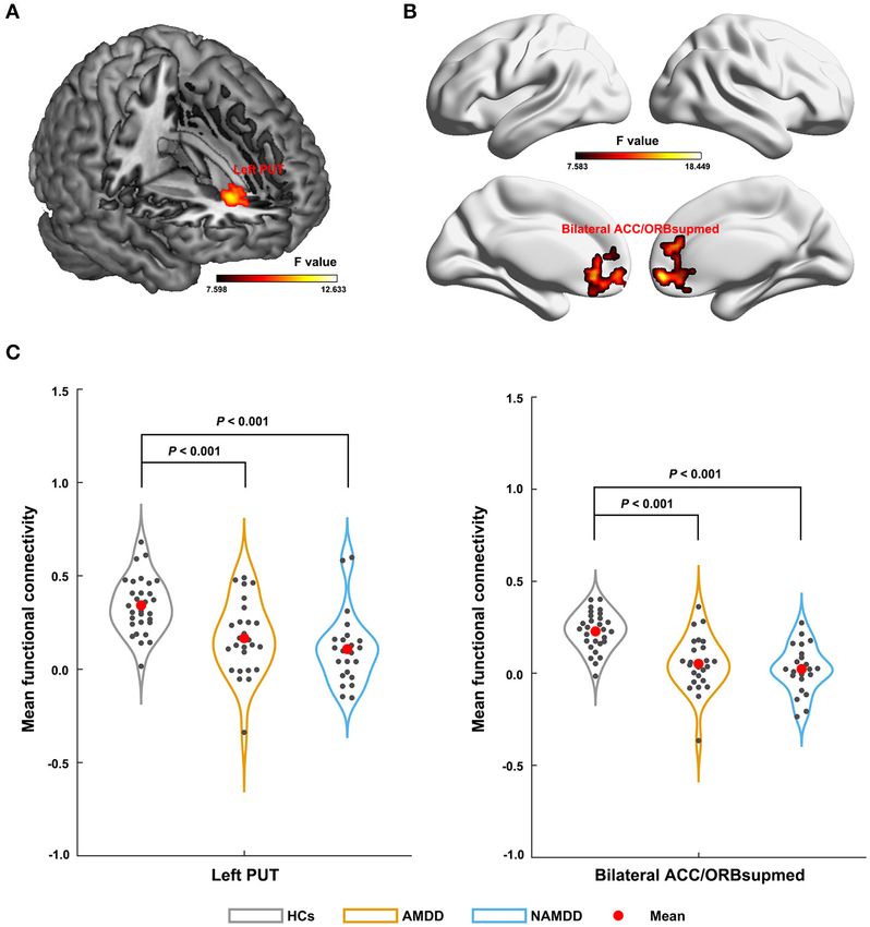
Li et al.                                                                                                                             Altered Amygdala Subregions in MDD

TABLE 1 | Demographics and clinical measures of all participants.

                                                       HCs (n = 30)                        AMDD (n = 26)                       NAMDD (n = 23)                       P-value

Age (years)                                           39.233 ± 12.484                      41.308 ± 10.884                        36 ± 10.135                         0.264
Gender (M/F)                                               12/18                                11/15                                 15/8                            0.147
HAMD                                                                                       22.808 ± 4.578                       18.044 ± 2.931                        2.5◦ in rotation                         analysis was restricted in a gray matter mask that was obtained
in any direction (the maximum head motion was 2.275 mm                                    by thresholding the gray matter tissue probability map in the
in translation and 1.617◦ in rotation over all participants).                             SPM12 toolbox (thres = 0.4). The relatively loose threshold was
To ensure functional specificity of the amygdala subregions,                              chosen to ensure sufficient search space for gray matter.
all regions of interest (ROIs) were masked out before spatial
smoothing. The smoothed images subsequently underwent                                     Statistical Analysis
removal of linear trends and band-pass filtering (0.01–0.08 Hz).                          For the demographic data, one-way analysis of variance
Finally, the white matter signals, cerebrospinal fluid signals,                           (ANOVA) and chi-square tests were used to compare age

Frontiers in Human Neuroscience | www.frontiersin.org                                 3                                         February 2021 | Volume 15 | Article 634113
Li et al. Altered Amygdala Subregions in MDD
and gender data among the three groups, respectively. For Functional sFC of Amygdala Subregions
the clinical data, two-sample t-tests were used to compare Significant main effects (P < 0.05, FWE corrected) were observed
HAMD anxiety/somatization factor scores, HAMD without for the left SFA-based sFC in a cluster that primarily embraced
anxiety/somatization factor scores, age of onset, and duration of the left putamen (Figure 2A) and for the right SFA-based sFC
illness between the two patient groups. For multimodal MRI- in a cluster that was predominantly involved in the bilateral
based GMV, ALFF, and COSLOF of each amygdala subregion, anterior cingulate cortex (ACC) and medial orbital parts of the
one-way ANOVA was used to infer differences among the bilateral superior frontal gyri (ORBsupmed) (Figure 2B). Further
three groups, followed by a Bonferroni method to correct post-hoc comparisons revealed that both patient groups showed
for multiple comparisons across subregions. For the whole- significantly decreased FC compared with those of the HCs,
brain sFC maps, a voxel-wise F-test was performed to infer but with no significant differences between them (Figure 2C).
differences among the three groups. A cluster-level family-wise For the bilateral CMA and LBA, no regions were found to
error rate procedure was used to determine significant clusters show significant main effects among the three groups (P > 0.05,
by combining a voxel-level P < 0.001 and an extend-level P FWE corrected).
< 0.05. Post-hoc pair-wise comparisons were further performed
with two-sample t-tests when significant main effects were Relationships Between Functional
observed. For the sFC, the post-hoc comparisons were conducted Characteristics of Amygdala Subregions
on the intra-cluster mean FC strength. Finally, non-parametric
Spearman correlations were used to test the relationships
and Clinical Variables
between multimodal MRI-based amygdala alterations and Significant negative correlations were observed for the ALFF of
clinical variables (HAMD anxiety/somatization factor scores, the left LBA and SFA with HAMD without anxiety/somatization
HAMD without anxiety/somatization factor scores, duration of factor scores in the patients with AMDD (r = −0.645 and
illness, and age of onset) in each patient group. Again, the −0.622, respectively, P < 0.05, Bonferroni corrected) rather than
Bonferroni method was used to correct for multiple comparisons. in those with nAMDD (P > 0.05) (Figure 3). No significant
correlations were found between ALFF and other clinical
variables or between sFC and any clinical variables.
RESULTS
Demographic and Clinical Characteristics DISCUSSION
As shown in Table 1, there were no significant differences in
age or gender among the three groups (P > 0.05). Both patient This study investigated the structural and functional alterations
groups did not differ significantly in their HAMD without of amygdala subregions in patients with AMDD and nAMDD
anxiety/somatization factor scores, age of onset, or duration vs. HCs. Our results showed common and specific amygdala
of illness (P > 0.05). As expected, the HAMD scores were alterations between patients with AMDD and nAMDD that
significantly higher in the AMDD group than the nAMDD group were dependent on different amygdala subdivisions. These
(P < 0.001) because of their higher anxiety/somatization factor findings provide new insights into the unique neural mechanism
scores (P < 0.001). underlying AMDD.
Numerous studies have examined the amygdala volume in
Structural GMV of Amygdala Subregions MDD; however, inconsistent even opposing results were obtained
(Munn et al., 2007; Tang et al., 2007). Many factors may
No significant differences were observed in the GMV for
collectively account for the discrepancy, in which the clinical
any amygdala subregion among the three groups (P > 0.05,
heterogeneity of the patients and the functional heterogeneity
Bonferroni corrected).
of the amygdala might be two key ones. Focusing on individual
amygdala subregions in MDD patients with and without anxiety,
Functional ALFF of Amygdala Subregions we showed that there were no significant GMV differences in
Significant main effects (P < 0.05, Bonferroni corrected) were the patients compared with those in the HCs for any amygdala
found for the ALFF of the bilateral LBA (left: P = 0.002; right: subregion. This is consistent with previous studies showing
P = 0.007) and SFA (left: P = 0.003; right: P = 0.002), but not the an unchanged amygdala volume in MDD (Bremner et al.,
bilateral CMA (P > 0.05), among the three groups (Figure 1). 2000; Frodl et al., 2004; Munn et al., 2007; Lorenzetti et al.,
Post-hoc comparisons revealed a common pattern of HCs > 2010). Moreover, no significant GMV differences were observed
nAMDD > AMDD that were shared by the four subregions, between the two patient groups for any amygdala subregion. This
while only the differences between the patients with AMDD and is in line with a previous study showing no significant volume
HCs were significant (P < 0.05, Bonferroni corrected). differences of the whole amygdala between patients with anxious
unipolar major depressive disorder and those with non-anxious
Functional COSLOF of Amygdala unipolar major depressive disorder (Delaparte et al., 2017). These
Subregions findings collectively suggest that gray matter volume of the
No significant differences were observed in the COSLOF for amygdala is not a suitable sign to differentiate anxious depressed
any amygdala subregion among the three groups (P > 0.05, from non-anxious depressed individuals. Notably, there are a
Bonferroni corrected). number of other variables that may confound results for the
Frontiers in Human Neuroscience | www.frontiersin.org 4 February 2021 | Volume 15 | Article 634113Li et al. Altered Amygdala Subregions in MDD
FIGURE 1 | Differences in the ALFF of amygdala subregions among the groups. Significant ALFF decreases in the bilateral LBA and SFA were observed in patients
with AMDD but not in those with nAMDD compared with the HCs. ALFF, amplitude of low frequency fluctuation; LBA, laterobasal amygdala; SFA, superficial
amygdala; AMDD, anxious major depressive disorder; nAMDD, non-anxious major depressive disorder; HCs, healthy controls.
amygdala volume, including depression duration or number of The SFA and LBA are closely related to emotional processing
episodes (Frodl et al., 2003; MacMaster et al., 2008; Kronenberg and regulation: The SFA is mainly involved in olfactory function
et al., 2009), antidepressant medication (Hamilton et al., 2008), (Heimer and Van Hoesen, 2006; Moreno and Gonzalez, 2007)
suicidal tendency (Monkul et al., 2007; Spoletini et al., 2011), and affective processes (Roy et al., 2009), and the LBA plays
genetic variations (Zetzsche et al., 2008; Savitz and Drevets, a central role in modulating the fear response and evaluating
2009), and the state of MDD (van Eijndhoven et al., 2009; sensory information (Jovanovic and Ressler, 2010). In contrast,
Malykhin et al., 2012; Zavorotnyy et al., 2018). Further studies are the CMA is mainly associated with the generation of behavioral
needed to examine the reproducibility of our findings by taking responses through projections to the brainstem and cortical and
these factors into consideration to gain a more comprehensive striatal regions (Roy et al., 2009). Accordingly, our results suggest
picture of amygdala volume abnormalities in MDD. In addition, that AMDD selectively affects local spontaneous brain activity of
postmortem neuroanatomical studies may further clarify the emotion-related amygdala subregions. However, no significant
issue by examining, for example, neuron or glia numbers or ALFF differences were found between patients with nAMDD
densities at the cellular level (Rubinow et al., 2016). and HCs for any amygdala subregion. Therefore, the observed
In addition to structural GMV, we characterized the functional ALFF alterations in SFA and LBA appear to be specific to
organization of the amygdala subregions from the perspectives of AMDD. This implies that the SFA and the LBA may be involved
local fluctuation amplitude, intra-region functional integration, in the pathophysiological processes of AMDD and may serve
and inter-regional functional communication. First, we found as potential biomarkers to differentiate AMDD from nAMDD.
that patients with AMDD showed significantly decreased ALFF However, no significant differences were found in the ALFF of
in the bilateral SFA and the LBA compared with those in the HCs. the bilateral SFA and the LBA between the two patient groups.
Frontiers in Human Neuroscience | www.frontiersin.org 5 February 2021 | Volume 15 | Article 634113Li et al. Altered Amygdala Subregions in MDD FIGURE 2 | Differences in sFC of amygdala subregions among groups. The left SFA-based sFC (A); the right SFA-based sFC (B); Significant sFC decreases were observed in both the patient groups compared with that in the HCs between the left SFA and the left putamen, and between the right SFA and the bilateral anterior cingulate cortex and medial orbital parts of the bilateral superior frontal gyri (C). PUT, putamen; ACC, anterior cingulate cortex; ORBsupmed, medial orbital part of the superior frontal gyrus; AMDD, anxious major depressive disorder; nAMDD, non-anxious major depressive disorder; HCs, healthy controls. These findings imply that there may exist a trend effect in the on this point by examining the associations between alterations observed ALFF alterations from nAMDD to AMDD. Notably, of amygdala subregions and each factor in a larger sample size. using a whole-brain voxel-wise analysis, two previous studies Second, no significant difference was found in the COSLOF found increased ALFF in the bilateral amygdala in patients with among the three groups for each amygdala subregion. This MDD compared with that in HCs (Du et al., 2016; Chen et al., suggests intact functional integration within each amygdala 2017). The discrepancy might be caused by different scales at subregion in patients with MDD, regardless of the status which the analyses were performed (i.e., voxel-level vs. region- of anxiety. level) or because of the differentiation of AMDD and nAMDD Finally, compared with the HCs, both patients with AMDD in the current study. Moreover, we found that decreased ALFF and those with nAMDD exhibited decreased functional in the left LBA and SFA were negatively correlated with the connectivity of the bilateral SFA, with no significant differences HAMD without anxiety/somatization factor scores in the AMDD between the two patient groups. This suggests that functional patients. The HAMD without anxiety/somatization factor scores connectivity reductions of the SFA might serve as intrinsic include several factors, such as cognitive disturbance, retardation, features for MDD. Mounting evidence from both animal sleep disturbance, and weight loss. These factors represent and human studies indicates that the SFA typically shows different symptom clusters that are closely related to the connectivity with the limbic system to form a neural circuit characteristics and medication of the disease. Thus, the ALFF that is closely associated with affective processes (Moreno and decreases may underlie the specific symptoms in patients with Gonzalez, 2007; Goossens et al., 2009; Roy et al., 2009; Gabard- AMDD. Further studies are needed to deepen our understanding Durnam et al., 2014). Since affective disturbances are commonly Frontiers in Human Neuroscience | www.frontiersin.org 6 February 2021 | Volume 15 | Article 634113
Li et al.                                                                                                                      Altered Amygdala Subregions in MDD

  FIGURE 3 | Scatter plots of relationships between altered ALFF of amygdala subregions and clinical variables in patients. Significant negative correlations were
  observed for the ALFF of the left LBA and SFA with HAMD without anxiety/somatization factor scores in the AMDD group, but not in the nAMDD group. ALFF,
  amplitude of low frequency fluctuation; LBA, laterobasal amygdala; SFA, superficial amygdala; AMDD, anxious major depressive disorder; nAMDD, non-anxious major
  depressive disorder; HAMD, Hamilton Rating Scale for Depression.

observed in MDD, regardless of subtype, it is plausible to observe                   due to the limitation of cross-species homologies. This issue can
disrupted functional connectivity of the SFA in both AMDD                            be partially addressed in the future by establishing cross-species
and nAMDD. However, a previous task-fMRI study showed                                correspondence in brain structures via network approaches
common deficits in both activation and connectivity of the                           (Goulas et al., 2015).
amygdala among patients with MDD, patients with anxiety, and                             Several limitations of this study should be mentioned. First,
comorbid subjects, suggesting a shared origin between anxiety                        the sample size was relatively small, which may limit the
and depression (Etkin and Schatzberg, 2011). Specifically, the                       generalizability of our findings. Second, about half of the patients
right SFA showed decreased functional connectivity with the                          were treated with psychotropic medication before the MRI scan,
bilateral ACC and ORBsupmed in both MDD groups. Both the                             which may have significantly modulated their functional brain
ACC and ORBsupmed are parts of the prefrontal cortex and                             organization (Wang et al., 2015c; Abdallah et al., 2017; Sheng
are related to emotional processing and regulation (Mayberg                          et al., 2018). Although all patients were free of psychotropic
et al., 2002; Milad and Rauch, 2007). Numerous previous studies                      medications for at least 4 weeks before the MRI scan, we
demonstrated the existence of functional connectivity between                        cannot fully exclude the possible long-term effects of medication.
the amygdala and different parts of the prefrontal regions (e.g.,                    Studies on non-medicated patients are expected in the future to
the ACC, and the ventral prefrontal and orbitofrontal cortex) in                     examine to what extent the current findings are contaminated
healthy individuals (Zald and Pardo, 1997; Hariri et al., 2003;                      by this factor. Third, similar to previous studies, we employed
Ochsner et al., 2004) and disruptions of these connectivities                        dimensional diagnosis to define AMDD, based on the severity
in MDD (Almeida et al., 2009; Versace et al., 2010; Etkin and                        of anxiety symptoms in MDD, as measured by the HAMD
Schatzberg, 2011). The disruptions imply impaired prefrontal                         anxiety/somatization factor. However, the anxiety/somatization
modulation of the amygdala in MDD, which may contribute to                           factor only contains seven items representing anxiety symptoms
emotional disturbances in patients suffering from the disease.                       and thus may suffer from the risk of misclassification to
Here, our findings further suggested that the disruptions are                        some extent. Nevertheless, it should be noted that a previous
selectively targeted to the SFA in MDD. As for the left SFA,                         study in Level 1 of the Sequenced Treatment Alternatives
decreased functional connectivity with the left putamen was                          to Relieve Depression indicated that the association between
found in both MDD groups, as compared with that in the HCs.                          anxious unipolar major depressive disorder and poorer treatment
The putamen belongs to the dorsal striatum and is thought                            outcome was independent of how we defined anxious unipolar
to play a crucial role in the development of mood disorders.                         major depressive disorder (Fava et al., 2008). Future studies are
For instance, one animal study found that the putamen was                            required to replicate the current findings by integrating multiple
significantly different between anhedonic-like and resilient                         dimension diagnosis to minimize the classification error. Fourth,
animals, indicating important implications of the putamen in                         although spatial smoothing was performed after masking out
anhedonia (Delgado y Palacios et al., 2014), which is one of the                     all amygdala subregions to ensure functional specificity, the
core symptoms of depression. Thus, the disrupted interaction                         signals in the amygdala subregions might still be polluted
between the left SFA and the putamen may be involved in the                          by those from regions adjacent to them. Future studies are
pathological mechanism of depression, in particular the origin                       required to address this issue by using more sophisticated
of anhedonia. It should be noted that to what extent the findings                    methods, such as orthogonalization. Finally, several previous
from animal studies apply to humans remain to be determined                          studies have shown that MDD-related alterations in both local

Frontiers in Human Neuroscience | www.frontiersin.org                            7                                       February 2021 | Volume 15 | Article 634113
Li et al.                                                                                                                                          Altered Amygdala Subregions in MDD

fluctuation amplitude and interregional functional connectivity                                    Shaw Hospital, Zhejiang University School of Medicine. The
are frequency-dependent (Yue et al., 2015; He et al., 2016; Wang                                   patients/participants provided their written informed consent to
et al., 2016). It would be interesting to investigate whether                                      participate in this study.
the observed amygdala alterations are modulated by choices of
different frequency intervals.                                                                     AUTHOR CONTRIBUTIONS
CONCLUSION                                                                                         YL, XN, and YY conducted the statistical analysis and drafted the
                                                                                                   initial manuscript. YQ, PW, JY, KR, LZ, ZL, and TS contributed
In summary, we demonstrated common and specific functional                                         to the conduct of the study. JW conducted the data analysis.
alterations of the amygdala between patients with AMDD and                                         Y-FZ and JW were responsible for the interpretation of MRI data.
those with nAMDD. Moreover, the alterations were dependent                                         WC and JW reviewed and revised the manuscript. WC and YS
on the functional features chosen and different amygdala                                           designed and supervised the study. All authors contributed to the
subregions. These findings deepen our understanding neural                                         article and approved the submitted version.
mechanisms underlying anxiety co-morbid with MDD and
have implications for future development of new and effective                                      FUNDING
treatment programs.
                                                                                                   This work was supported by the National Key Research
DATA AVAILABILITY STATEMENT                                                                        & Development Program of China (2017YFC1310502
                                                                                                   and 2016YFC1307200), the NSFC Projects of International
The original contributions presented in the study are included                                     Cooperation and Exchanges (8161101010), the National Natural
in the article/supplementary material, further inquiries can be                                    Science Foundation of China (81671764 and 81671350), the
directed to the corresponding authors.                                                             Key Project of the Department of Science and Technology of
                                                                                                   Zhejiang Province (2018C03023), the Department of Science and
ETHICS STATEMENT                                                                                   Technology of Zhejiang Province (2016F50046), the Science and
                                                                                                   Technology Program of Hangzhou Municipality (20190101A10),
The studies involving human participants were reviewed                                             and the Project of Chinese Traditional Medicine of Zhejiang
and approved by The ethics committee of Sir Run Run                                                Province (2020ZX012).

REFERENCES                                                                                         Du, L., Wang, J., Meng, B., Yong, N., Yang, X., Huang, Q., et al. (2016). Early
                                                                                                      life stress affects limited regional brain activity in depression. Sci. Rep. 6:25338.
Abdallah, C. G., Averill, L. A., Collins, K. A., Geha, P., Schwartz, J., Averill, C., et al.          doi: 10.1038/srep25338
   (2017). Ketamine treatment and global brain connectivity in major depression.                   Etkin, A., and Schatzberg, A. F. (2011). Common abnormalities and disorder-
   Neuropsychopharmacology 42, 1210–1219. doi: 10.1038/npp.2016.186                                   specific compensation during implicit regulation of emotional processing in
Almeida, J. R., Versace, A., Mechelli, A., Hassel, S., Quevedo, K., Kupfer, D. J.,                    generalized anxiety and major depressive disorders. Am. J. Psychiatry 168,
   et al. (2009). Abnormal amygdala-prefrontal effective connectivity to happy                        968–978. doi: 10.1176/appi.ajp.2011.10091290
   faces differentiates bipolar from major depression. Biol. Psychiatry 66, 451–459.               Fava, M., Rush, A. J., Alpert, J. E., Balasubramani, G. K., Wisniewski, S. R.,
   doi: 10.1016/j.biopsych.2009.03.024                                                                Carmin, C. N., et al. (2008). Difference in treatment outcome in outpatients
Amunts, K., Kedo, O., Kindler, M., Pieperhoff, P., Mohlberg, H., Shah, N. J.,                         with anxious versus nonanxious depression: a STAR∗ D report. Am. J. Psychiatry
   et al. (2005). Cytoarchitectonic mapping of the human amygdala, hippocampal                        165, 342–351. doi: 10.1176/appi.ajp.2007.06111868
   region, and entorhinal cortex: intersubject variability and probability maps.                   Fonseka, T. M., MacQueen, G. M., and Kennedy, S. H. (2018). Neuroimaging
   Anat. Embryol. (Berl.) 210, 343–352. doi: 10.1007/s00429-005-0025-5                                biomarkers as predictors of treatment outcome in Major Depressive
Ball, T., Rahm, B., Eickhoff, S. B., Schulze-Bonhage, A., Speck, O., and Mutschler,                   Disorder. J. Affect. Disord. 233, 21–35. doi: 10.1016/j.jad.2017.
   I. (2007). Response properties of human amygdala subregions: evidence based                        10.049
   on functional MRI combined with probabilistic anatomical maps. PLoS ONE                         Friston, K. J., Williams, S., Howard, R., Frackowiak, R. S., and Turner, R. (1996).
   2:e307. doi: 10.1371/journal.pone.0000307                                                          Movement-related effects in fMRI time-series. Magn. Reson. Med. 35, 346–355.
Bremner, J. D., Narayan, M., Anderson, E. R., Staib, L. H., Miller, H. L., and                        doi: 10.1002/mrm.1910350312
   Charney, D. S. (2000). Hippocampal volume reduction in major depression.                        Frodl, T., Meisenzahl, E. M., Zetzsche, T., Born, C., Jager, M., Groll, C., et al. (2003).
   Am. J. Psychiatry 157, 115–118. doi: 10.1176/ajp.157.1.115                                         Larger amygdala volumes in first depressive episode as compared to recurrent
Chen, V. C., Shen, C. Y., Liang, S. H., Li, Z. H., Hsieh, M. H., Tyan, Y. S.,                         major depression and healthy control subjects. Biol. Psychiatry 53, 338–344.
   et al. (2017). Assessment of brain functional connectome alternations and                          doi: 10.1016/S0006-3223(02)01474-9
   correlation with depression and anxiety in major depressive disorders. PeerJ.                   Frodl, T., Meisenzahl, E. M., Zetzsche, T., Höhne, T., Banac, S., Schorr, C., et al.
   5:e3147. doi: 10.7717/peerj.3147                                                                   (2004). Hippocampal and amygdala changes in patients with major depressive
Delaparte, L., Yeh, F. C., Adams, P., Malchow, A., Trivedi, M. H., Oquendo,                           disorder and healthy controls during a 1-year follow-up. J. Clin. Psychiatry 65,
   M. A., et al. (2017). A comparison of structural connectivity in anxious                           492–499. doi: 10.4088/JCP.v65n0407
   depression versus non-anxious depression. J. Psychiatr. Res. 89, 38–47.                         Gabard-Durnam, L. J., Flannery, J., Goff, B., Gee, D. G., Humphreys, K. L., Telzer,
   doi: 10.1016/j.jpsychires.2017.01.012                                                              E., et al. (2014). The development of human amygdala functional connectivity
Delgado y Palacios, R., Verhoye, M., Henningsen, K., Wiborg, O., and Van                              at rest from 4 to 23 years: a cross-sectional study. Neuroimage 95, 193–207.
   der Linden, A. (2014). Diffusion kurtosis imaging and high-resolution MRI                          doi: 10.1016/j.neuroimage.2014.03.038
   demonstrate structural aberrations of caudate putamen and amygdala after                        Gaspersz, R., Lamers, F., Kent, J. M., Beekman, A. T., Smit, J. H., van Hemert,
   chronic mild stress. PLoS ONE 9:e95077. doi: 10.1371/journal.pone.0095077                          A. M., et al. (2017a). Longitudinal predictive validity of the DSM-5 anxious

Frontiers in Human Neuroscience | www.frontiersin.org                                          8                                             February 2021 | Volume 15 | Article 634113
Li et al. Altered Amygdala Subregions in MDD
distress specifier for clinical outcomes in a large cohort of patients with major Mayberg, H. S., Silva, J. A., Brannan, S. K., Tekell, J. L., Mahurin, R. K., McGinnis,
depressive disorder. J. Clin. Psychiatry 78, 207–213. doi: 10.4088/JCP.15m10221 S., et al. (2002). The functional neuroanatomy of the placebo effect. Am. J.
Gaspersz, R., Lamers, F., Kent, J. M., Beekman, A. T. F., Smit, J. H., van Hemert, A. Psychiatry 159, 728–737. doi: 10.1176/appi.ajp.159.5.728
M., et al. (2017b). Anxious distress predicts subsequent treatment outcome and Milad, M. R., and Rauch, S. L. (2007). The role of the orbitofrontal cortex in anxiety
side effects in depressed patients starting antidepressant treatment. J. Psychiatr. disorders. Ann. N. Y. Acad. Sci. 1121, 546–561. doi: 10.1196/annals.1401.006
Res. 84, 41–48. doi: 10.1016/j.jpsychires.2016.09.018 Monkul, E. S., Hatch, J. P., Nicoletti, M. A., Spence, S., Brambilla, P., Lacerda, A.
Goldberg, D., and Fawcett, J. (2012). The importance of anxiety in both L., et al. (2007). Fronto-limbic brain structures in suicidal and non-suicidal
major depression and bipolar disorder. Depress Anxiety 29, 471–478. female patients with major depressive disorder. Mol. Psychiatry 12, 360–366.
doi: 10.1002/da.21939 doi: 10.1038/sj.mp.4001919
Goldberg, D. P., Wittchen, H. U., Zimmermann, P., Pfister, H., and Moreno, N., and Gonzalez, A. (2007). Evolution of the amygdaloid complex in
Beesdobaum, K. (2014). Anxious and non-anxious forms of major depression: vertebrates, with special reference to the anamnio-amniotic transition. J. Anat.
familial, personality, and symptom characteristics. Psychol. Med. 44:1223. 211, 151–163. doi: 10.1111/j.1469-7580.2007.00780.x
doi: 10.1017/S0033291713001827 Munn, M. A., Alexopoulos, J., Nishino, T., Babb, C. M., Flake, L. A., Singer, T., et al.
Goossens, L., Kukolja, J., Onur, O. A., Fink, G. R., Maier, W., Griez, E., et al. (2009). (2007). Amygdala volume analysis in female twins with major depression. Biol.
Selective processing of social stimuli in the superficial amygdala. Hum. Brain Psychiatry 62, 415–422. doi: 10.1016/j.biopsych.2006.11.031
Mapp. 30, 3332–3338. doi: 10.1002/hbm.20755 Ochsner, K. N., Ray, R. D., Cooper, J. C., Robertson, E. R., Chopra, S., Gabrieli,
Goulas, A., Schaefer, A., and Margulies, D. S. (2015). The strength of weak J. D., et al. (2004). For better or for worse: neural systems supporting the
connections in the macaque cortico-cortical network. Brain Struct. Funct. 220, cognitive down- and up-regulation of negative emotion. Neuroimage 23,
2939–2951. doi: 10.1007/s00429-014-0836-3 483–499. doi: 10.1016/j.neuroimage.2004.06.030
Hallquist, M. N., Hwang, K., Luna, B. The nuisance of nuisance regression: spectral Ongur, D., and Price, J. L. (2000). The organization of networks within the orbital
misspecification in a common approach to resting-state fMRI preprocessing and medial prefrontal cortex of rats, monkeys and humans. Cereb. Cortex 10,
reintroduces noise and obscures functional connectivity. Neuroimage. (2013) 206–219. doi: 10.1093/cercor/10.3.206
82, 208–25. doi: 10.1016/j.neuroimage.2013.05.116 Phillips, M. L., and Swartz, H. A. (2014). A critical appraisal of neuroimaging
Hamilton, J. P., Siemer, M., and Gotlib, I. H. (2008). Amygdala volume in major studies of bipolar disorder: toward a new conceptualization of underlying
depressive disorder: a meta-analysis of magnetic resonance imaging studies. neural circuitry and a road map for future research. Am. J. Psychiatry. 171,
Mol. Psychiatry 13, 993–1000. doi: 10.1038/mp.2008.57 829–843. doi: 10.1176/appi.ajp.2014.13081008
Hariri, A. R., Mattay, V. S., Tessitore, A., Fera, F., and Weinberger, D. R. (2003). Qin, S., Young, C. B., Duan, X., Chen, T., Supekar, K., and Menon, V. (2014).
Neocortical modulation of the amygdala response to fearful stimuli. Biol. Amygdala subregional structure and intrinsic functional connectivity predicts
Psychiatry 53, 494–501. doi: 10.1016/S0006-3223(02)01786-9 individual differences in anxiety during early childhood. Biol. Psychiatry 75,
He, Z., Cui, Q., Zheng, J., Duan, X., Pang, Y., Gao, Q., et al. (2016). 892–900. doi: 10.1016/j.biopsych.2013.10.006
Frequency-specific alterations in functional connectivity in treatment-resistant Rao, S., and Zisook, S. (2009). Anxious depression: clinical features and treatment.
and -sensitive major depressive disorder. J. Psychiatr. Res. 82, 30–39. Curr. Psychiatry Rep. 11, 429–436. doi: 10.1007/s11920-009-0065-2
doi: 10.1016/j.jpsychires.2016.07.011 Roy, A. K., Shehzad, Z., Margulies, D. S., Kelly, A. M., Uddin, L. Q., Gotimer, K.,
Heimer, L., and Van Hoesen, G. W. (2006). The limbic lobe and its output et al. (2009). Functional connectivity of the human amygdala using resting state
channels: implications for emotional functions and adaptive behavior. fMRI. Neuroimage 45, 614–626. doi: 10.1016/j.neuroimage.2008.11.030
Neurosci. Biobehav. Rev. 30, 126–147. doi: 10.1016/j.neubiorev.2005.06.006 Rubinow, M. J., Mahajan, G., May, W., Overholser, J. C., Jurjus, G. J., Dieter, L.,
Ionescu, D. F., Niciu, M. J., Richards, E. M., and Zarate, C. A. Jr. (2014). et al. (2016). Basolateral amygdala volume and cell numbers in major depressive
Pharmacologic treatment of dimensional anxious depression: a review. Prim. disorder: a postmortem stereological study. Brain Struct. Funct. 221, 171–184.
Care Companion CNS Disord. 16:PCC.13r01621. doi: 10.4088/PCC.13r01621 doi: 10.1007/s00429-014-0900-z
Jovanovic, T., and Ressler, K. J. (2010). How the neurocircuitry and genetics of Savitz, J. B., and Drevets, W. C. (2009). Imaging phenotypes of major
fear inhibition may inform our understanding of PTSD. Am. J. Psychiatry 167, depressive disorder: genetic correlates. Neuroscience 164, 300–330.
648–662. doi: 10.1176/appi.ajp.2009.09071074 doi: 10.1016/j.neuroscience.2009.03.082
Kronenberg, G., Tebartz van Elst, L., Regen, F., Deuschle, M., Heuser, I., and Seo, H. J., Jung, Y. E., Kim, T. S., Kim, J. B., Lee, M. S., Kim, J. M.,
Colla, M. (2009). Reduced amygdala volume in newly admitted psychiatric et al. (2011). Distinctive clinical characteristics and suicidal tendencies
in-patients with unipolar major depression. J. Psychiatr. Res. 43, 1112–1117. of patients with anxious depression. J. Nerv. Ment. Dis. 199, 42–48.
doi: 10.1016/j.jpsychires.2009.03.007 doi: 10.1097/NMD.0b013e3182043b60
Li, S. J., Li, Z., Wu, G., Zhang, M. J., Franczak, M., and Antuono, P. Shen, Y., Yao, J., Jiang, X., Zhang, L., Xu, L., Feng, R., et al. (2015). Sub-hubs of
G. (2002). Alzheimer disease: evaluation of a functional MR imaging baseline functional brain networks are related to early improvement following
index as a marker. Radiology 225, 253–259. doi: 10.1148/radiol.22510 two-week pharmacological therapy for major depressive disorder. Hum. Brain
11301 Mapp. 36, 2915–2927. doi: 10.1002/hbm.22817
Li, Y., Qin, W., Jiang, T., Zhang, Y., and Yu, C. (2012). Sex-dependent correlations Sheng, J., Shen, Y., Qin, Y., Zhang, L., Jiang, B., Li, Y., et al. (2018).
between the personality dimension of harm avoidance and the resting- Spatiotemporal, metabolic, and therapeutic characterization of altered
state functional connectivity of amygdala subregions. PLoS ONE 7:e35925. functional connectivity in major depressive disorder. Hum. Brain Mapp. 39,
doi: 10.1371/journal.pone.0035925 1957–1971. doi: 10.1002/hbm.23976
Licznerski, P., and Duman, R. S. (2013). Remodeling of axo-spinous synapses in Spoletini, I., Piras, F., Fagioli, S., Rubino, I. A., Martinotti, G., Siracusano, A.,
the pathophysiology and treatment of depression. Neuroscience 251, 33–50. et al. (2011). Suicidal attempts and increased right amygdala volume in
doi: 10.1016/j.neuroscience.2012.09.057 schizophrenia. Schizophr. Res. 125, 30–40. doi: 10.1016/j.schres.2010.08.023
Lorenzetti, V., Allen, N. B., Whittle, S., and Yücel, M. (2010). Amygdala Tang, Y., Wang, F., Xie, G., Liu, J., Li, L., Su, L., et al. (2007). Reduced
volumes in a sample of current depressed and remitted depressed patients ventral anterior cingulate and amygdala volumes in medication-naive
and healthy controls. J. Affect. Disord. 120, 112–119. doi: 10.1016/j.jad.2009. females with major depressive disorder: a voxel-based morphometric
04.021 magnetic resonance imaging study. Psychiatry Res. 156, 83–86.
MacMaster, F. P., Mirza, Y., Szeszko, P. R., Kmiecik, L. E., Easter, P. C., doi: 10.1016/j.pscychresns.2007.03.005
Taormina, S. P., et al. (2008). Amygdala and hippocampal volumes in van Eijndhoven, P., van Wingen, G., van Oijen, K., Rijpkema, M., Goraj,
familial early onset major depressive disorder. Biol. Psychiatry 63, 385–390. B., Jan Verkes, R., et al. (2009). Amygdala volume marks the acute
doi: 10.1016/j.biopsych.2007.05.005 state in the early course of depression. Biol. Psychiatry 65, 812–818.
Malykhin, N. V., Carter, R., Hegadoren, K. M., Seres, P., and Coupland, N. doi: 10.1016/j.biopsych.2008.10.027
J. (2012). Fronto-limbic volumetric changes in major depressive disorder. J. van Tol, M. J., Demenescu, L. R., van der Wee, N. J., Kortekaas, R., Marjan, M. A.
Affect. Disord. 136, 1104–1113. doi: 10.1016/j.jad.2011.10.038 N., Boer, J. A., et al. (2012). Functional magnetic resonance imaging correlates
Frontiers in Human Neuroscience | www.frontiersin.org 9 February 2021 | Volume 15 | Article 634113Li et al. Altered Amygdala Subregions in MDD of emotional word encoding and recognition in depression and anxiety Yue, Y., Jia, X., Hou, Z., Zang, Y., and Yuan, Y. (2015). Frequency-dependent disorders. Biol. Psychiatry 71, 593–602. doi: 10.1016/j.biopsych.2011.11.016 amplitude alterations of resting-state spontaneous fluctuations in late-onset Versace, A., Thompson, W. K., Zhou, D., Almeida, J. R., Hassel, S., Klein, C. depression. Biomed. Res. Int. 2015:505479. doi: 10.1155/2015/505479 R., et al. (2010). Abnormal left and right amygdala-orbitofrontal cortical Zald, D. H., and Pardo, J. V. (1997). Emotion, olfaction, and the human amygdala: functional connectivity to emotional faces: state versus trait vulnerability amygdala activation during aversive olfactory stimulation. Proc. Natl. Acad. Sci. markers of depression in bipolar disorder. Biol. Psychiatry 67, 422–431. U.S.A. 94, 4119–4124. doi: 10.1073/pnas.94.8.4119 doi: 10.1016/j.biopsych.2009.11.025 Zang, Y. F., He, Y., Zhu, C. Z., Cao, Q. J., Sui, M. Q., Liang, M., et al. (2007). Wang, J., Wang, X., He, Y., Yu, X., Wang, H., and He, Y. (2015a). Apolipoprotein E Altered baseline brain activity in children with ADHD revealed by resting-state ε4 modulates functional brain connectome in Alzheimer’s disease. Hum. Brain functional MRI. Brain Dev. 29, 83–91. doi: 10.1016/j.braindev.2006.07.002 Mapp. 36, 1828–1846. doi: 10.1002/hbm.22740 Zavorotnyy, M., Zollner, R., Schulte-Gustenberg, L. R., Wulff, L., Schoning, S., Wang, J., Wang, X., Xia, M., Liao, X., Evans, A., and He, Y. (2015b). GRETNA: Dannlowski, U., et al. (2018). Low left amygdala volume is associated with a graph theoretical network analysis toolbox for imaging connectomics. Front. a longer duration of unipolar depression. J. Neural. Transm. (Vienna) 125, Hum. Neurosci. 9:386. doi: 10.3389/fnhum.2015.00386 229–238. doi: 10.1007/s00702-017-1811-y Wang, J., Wei, Q., Bai, T., Zhou, X., Sun, H., Becker, B., et al. (2017). Zetzsche, T., Preuss, U. W., Bondy, B., Frodl, T., Zill, P., Schmitt, G., et al. Electroconvulsive therapy selectively enhanced feedforward connectivity from (2008). 5-HT1A receptor gene C−1019 G polymorphism and amygdala fusiform face area to amygdala in major depressive disorder. Soc. Cogn. Affect. volume in borderline personality disorder. Genes Brain Behav. 7, 306–313. Neurosci. 12, 1983–1992. doi: 10.1093/scan/nsx100 doi: 10.1111/j.1601-183X.2007.00353.x Wang, L., Kong, Q., Li, K., Su, Y., Zeng, Y., Zhang, Q., et al. (2016). Frequency-dependent changes in amplitude of low-frequency oscillations Conflict of Interest: The authors declare that the research was conducted in the in depression: a resting-state fMRI study. Neurosci. Lett. 614, 105–111. absence of any commercial or financial relationships that could be construed as a doi: 10.1016/j.neulet.2016.01.012 potential conflict of interest. Wang, L., Xia, M., Li, K., Zeng, Y., Su, Y., Dai, W., et al. (2015c). The effects of antidepressant treatment on resting-state functional brain networks in Copyright © 2021 Li, Ni, You, Qing, Wang, Yao, Ren, Zhang, Liu, Song, Wang, patients with major depressive disorder. Hum. Brain Mapp. 36, 768–778. Zang, Shen and Chen. This is an open-access article distributed under the terms doi: 10.1002/hbm.22663 of the Creative Commons Attribution License (CC BY). The use, distribution or Wiethoff, K., Bauer, M., Baghai, T. C., Möller, H. J., Fisher, R., Hollinde, D., reproduction in other forums is permitted, provided the original author(s) and the et al. (2010). Prevalence and treatment outcome in anxious versus nonanxious copyright owner(s) are credited and that the original publication in this journal depression: results from the German Algorithm Project. J. Clin. Psychiatry 71, is cited, in accordance with accepted academic practice. No use, distribution or 1047–1054. doi: 10.4088/JCP.09m05650blu reproduction is permitted which does not comply with these terms. Frontiers in Human Neuroscience | www.frontiersin.org 10 February 2021 | Volume 15 | Article 634113
You can also read