Photosynthesis and Bio-Optical Properties of Fluorescent Mesophotic Corals
←
→
Page content transcription
If your browser does not render page correctly, please read the page content below
ORIGINAL RESEARCH
                                                                                                                                                    published: 15 April 2021
                                                                                                                                           doi: 10.3389/fmars.2021.651601

                                            Photosynthesis and Bio-Optical
                                            Properties of Fluorescent
                                            Mesophotic Corals
                                            Or Ben-Zvi 1,2* , Daniel Wangpraseurt 3,4 , Omri Bronstein 1,5 , Gal Eyal 1,2,6,7 and Yossi Loya 1
                                            1
                                             School of Zoology, Faculty of Life Sciences, Tel Aviv University, Tel Aviv, Israel, 2 The Interuniversity Institute for Marine
                                            Sciences in Eilat, Eilat, Israel, 3 Department of Nanoengineering, University of California, San Diego, San Diego, CA,
                                            United States, 4 Department of Chemistry, University of Cambridge, Cambridge, United Kingdom, 5 The Steinhardt Museum
                                            of Natural History, Tel Aviv University, Tel Aviv, Israel, 6 ARC Centre of Excellence for Coral Reef Studies, School of Biological
                                            Sciences, The University of Queensland, Brisbane, QLD, Australia, 7 The Mina and Everard Goodman Faculty of Life
                                            Sciences, Bar-Ilan University, Ramat Gan, Israel

                                            Mesophotic coral ecosystems (MCEs) are light-dependent coral-associated
                                            communities found at 30–150 m depth. Corals inhabiting these deeper reefs are often
                                            acclimatized to a limited and blue-shifted light environment, enabling them to maintain
                                            the relationship with their photosynthetic algal symbionts (family Symbiodiniaceae)
                                            despite the seemingly suboptimal light conditions. Among others, fluorescent proteins
                                            produced by the coral host may play a role in the modulation of the quality and spectral
                           Edited by:
                                            distribution of irradiance within the coral tissue through wavelength transformation.
                       Noga Stambler,       Here we examined the bio-optical properties and photosynthetic performances
              Bar-Ilan University, Israel   of different fluorescence morphs of two mesophotic coral species Goniopora
                       Reviewed by:         minor and Alveopora ocellata, in order to test the photosynthesis enhancement
                    Michael P. Lesser,
        University of New Hampshire,        hypothesis proposed for coral fluorescence. The green morph of G. minor and the low
                        United States       fluorescence morph of A. ocellata exhibit, in their natural habitats, higher abundance.
                       Charles Mazel,
           NIGHTSEA, United States
                                            The morphs also presented different spectral reflectance and light attenuation
                  *Correspondence:
                                            within the tissue. Nevertheless, chlorophyll a fluorescence-based, and O2 evolution
                           Or Ben-Zvi       measurements, revealed only minor differences between the photosynthetic abilities
                    orbzvi@gmail.com        of three fluorescence morphs of the coral G. minor and two fluorescence morphs of
                   Specialty section:
                                            A. ocellata. The fluorescence morphs did not differ in their algal densities or chlorophyll
         This article was submitted to      concentrations and all corals harbored Symbiodiniaceae from the genus Cladocopium.
                 Coral Reef Research,
                                            Thus, despite the change in the internal light quantity and quality that corals and their
               a section of the journal
          Frontiers in Marine Science       symbionts experience, we found no evidence for the facilitation or enhancement of
         Received: 15 January 2021          photosynthesis by wavelength transformation.
          Accepted: 24 March 2021
                                            Keywords: photosynthesis, mesophotic coral ecosystems (MCEs), Symbiodiniaceae, fluorescence, microsensors
           Published: 15 April 2021
                             Citation:
          Ben-Zvi O, Wangpraseurt D,
      Bronstein O, Eyal G and Loya Y
                                            INTRODUCTION
               (2021) Photosynthesis
           and Bio-Optical Properties
                                            Stony corals are sessile organisms and are considered to be highly dependent on the photosynthates
   of Fluorescent Mesophotic Corals.        derived from their algal symbionts (family Symbiodiniaceae; LaJeunesse et al., 2018) as their
           Front. Mar. Sci. 8:651601.       main energy source (Muscatine, 1990). Sustaining high rates of photosynthesis in the underwater
    doi: 10.3389/fmars.2021.651601          light environment is challenging as (1) light attenuates rapidly with depth (Kirk, 2011a),

Frontiers in Marine Science | www.frontiersin.org                                    1                                                 April 2021 | Volume 8 | Article 651601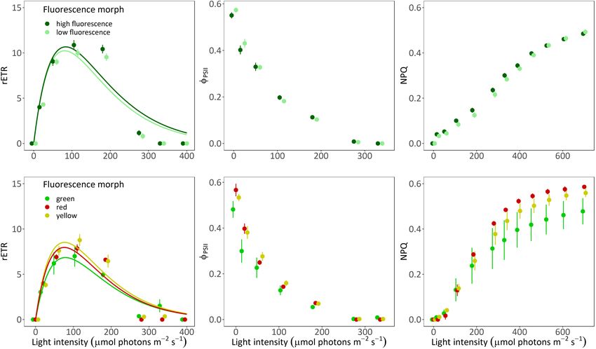
Ben-Zvi et al.                                                                                      Mesophotic Corals Photosynthesis and Bio-Optics

(2) wave movement creates a flickering effect that causes                 habitats, usually results in green wavelengths that are the least
rapid and extreme localized changes in light intensity (Walsh             efficient for photosynthesis in Symbiodiniaceae (Scott and Jitts,
and Legendre, 1983), (3) the macro-structure of the reef                  1977; Kühl et al., 1995). Another mechanism for photosynthesis
may shade coral colonies (Stimson, 1985; Kaniewska et al.,                enhancement was suggested to rely on electron transport through
2008), and (4) certain substrates may be highly reflective                fluorescence resonance energy transfer (FRET) which depends
consequently contributing to downwelling irradiance (Kirk,                on a close proximity between the donor of electrons and the
2011b). Scleractinian corals in mesophotic coral ecosystems               recipient (Förster, 1955). Despite some evidences indicating that
(MCEs; 30–150 m) experience light intensities that are up to              FRET is occurring between FPs (Cox et al., 2007), there is
99% lower than those experienced by their shallow counterparts            currently a lack of sufficient support for electron transfer between
(0–30 m; Kahng and Kelley, 2007; Lesser et al., 2009; Tamir               FPs and the photosynthetic apparatus (Gilmore et al., 2003; Cox
et al., 2019). Additionally, MCEs are also exposed to a restricted        and Salih, 2005). Nevertheless, recent studies suggest that FPs
light spectrum centered around the blue region of the spectrum            may alter (by wavelength conversion) and disperse (by scattering)
(Jerlov, 1968; Kahng et al., 2019). Coral photoacclimatization            the available light more efficiently through the tissue, enabling it
to MCEs is manifested at the coral-host level as changes in               to reach the Symbiodiniaceae residing in the deeper tissue layers
colony morphology (Kaniewska et al., 2008; Nir et al., 2011),             of the coral host (Smith et al., 2017), and that symbiotic algae
skeletal features that modify the internal light environment              are attracted to green light (Hollingsworth et al., 2005) and green
(Enríquez et al., 2005; Kahng et al., 2012), changes in the               fluorescence (Aihara et al., 2019). The enhanced reflectance and
populations of endosymbiotic Symbiodiniaceae expressed as a               scattering of light are also supported by direct measurements
shift in their genetic identity (Cooper et al., 2011; Bongaerts           and is correlated with higher FP fluorescence (Salih et al., 2000;
et al., 2013; Einbinder et al., 2016) and modifications in the            Lyndby et al., 2016).
composition of photosynthetic pigments and structure of the                   Thus far, hypotheses regarding the role of coral fluorescence
photosynthetic complex (Einbinder et al., 2016). These changes,           have mostly been tested on shallow corals (DeSalvo et al., 2008;
among others, potentially assist corals in maintaining a successful       Dove et al., 2008; D’Angelo et al., 2012) and among coral morphs
symbiosis with Symbiodiniaceae. Accordingly, deeper corals will           that display different expression levels of the same FP but not
usually present a higher maximal quantum yield of photosystem             among morphs that differ in the FPs they contain (Gittins
II (Einbinder et al., 2016; Ben-Zvi et al., 2020), a lower                et al., 2015; Roth et al., 2015). In MCEs, the phenomenon of
Symbiodiniaceae density accompanied by higher chlorophyll                 fluorescence polymorphism is common and multiple species can
concentration within algal cells (Mass et al., 2007), and a reduced       exhibit several color morphs occurring side by side at the same
capacity to manage excess light (Einbinder et al., 2016; Ben-Zvi          site and depth (Eyal et al., 2015; Ben-Zvi et al., 2019).
et al., 2020). Additionally, mesophotic corals may rely more on               Here we sought to investigate the photosynthetic and bio-
heterotrophy rather than autotrophy as their main strategy for            optical properties of different fluorescence morphs of two
acquiring energy (Mass et al., 2007; Lesser et al., 2010).                mesophotic coral species which differ either in the levels of
    Corals are recognized for being extremely colorful under              expression (i.e., Alveopora ocellata) or by the emission peak of
short-wavelength lighting conditions, which is attributed to the          their FPs (i.e., Goniopora minor), in order to determine the
phenomenon of fluorescence (Kawaguti, 1944; Matz et al., 1999).           potential links between the coral host’s fluorescent pigments and
Fluorescence refers to the conversion of light wavelength, usually        coral photosynthesis, in the unique mesophotic environment.
from short wavelengths into longer ones. In corals, fluorescence
results from proteins belonging to the green fluorescent protein
(GFP)-like family, that are produced by the coral host. The               MATERIALS AND METHODS
fluorescent proteins (FPs) may exhibit a diversity of excitation
and emission peaks (Alieva et al., 2008), and an individual coral         Coral Collection, Sampling, and
may possess a single or multiple FPs (Dove et al., 2001; Ben-Zvi          Maintenance
et al., 2014, 2019; Eyal et al., 2015). Fluorescence polymorphism         Ten colonies of the mesophotic scleractinian coral A. ocellata
within the same coral species has been previously described as            were collected from the reef in front of the Interuniversity
resulting either from a difference in the expression levels of a          Institute for Marine Sciences in Eilat (IUI; 29◦ 300 1600 N,
single protein (Gittins et al., 2015; Takahashi-Kariyazono et al.,        34◦ 550 700 E) and seven colonies of G. minor were collected
2018), or the presence of different FPs (Eyal et al., 2015; Ben-Zvi       from the Dekel Beach site (29◦ 320 1700 N, 34◦ 560 5600 E), Gulf of
et al., 2019).                                                            Eilat/Aqaba (GoE/A), northern Red Sea. All corals were collected
    One of the suggested functional roles for coral fluorescence          at 45 m depth using open-circuit technical diving. Corals were
is that of photosynthesis enhancement. Earlier studies have               transferred to a running seawater system at the IUI in dark
contended that fluorescence may enhance photosynthesis where              containers and were subsampled and preserved by dipping the
light is limited, as in deeper habitats, by conversion of short           fragments in liquid nitrogen and storing them at −80◦ C until
wavelengths into longer wavelengths, capable of absorption by             analyses (for Symbiodiniaceae density and genetic identification,
the photosynthetic pigments of the algal symbiont using host              and chlorophyll concentration analyses). The remaining corals
associated cyan FPs (Schlichter et al., 1986, 1994; Schlichter            (used for the scalar irradiance, chlorophyll fluorescence, and O2
and Fricke, 1990). However, this notion is not fully supported            evolution measurements) were kept for further analyses under a
since a conversion of blue light, which is abundant in deeper             lighting filter (“Lagoon blue,” Lee Filters, United States) providing

Frontiers in Marine Science | www.frontiersin.org                     2                                        April 2021 | Volume 8 | Article 651601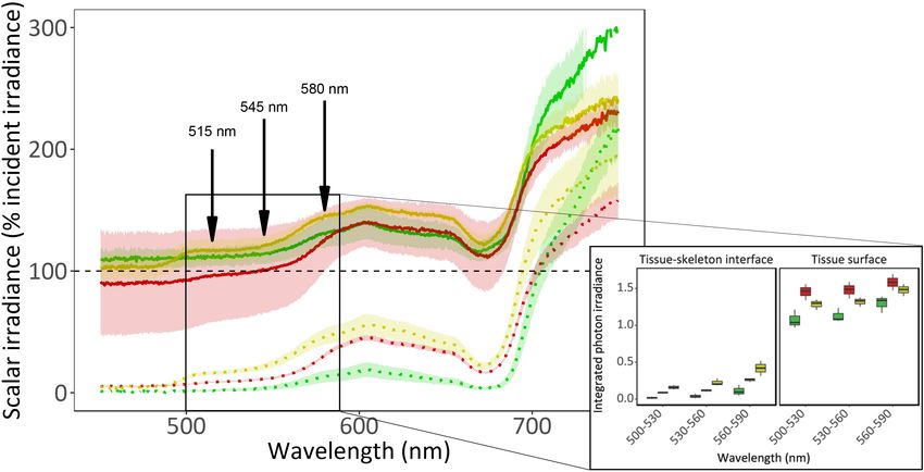
Ben-Zvi et al.                                                                                      Mesophotic Corals Photosynthesis and Bio-Optics

a light regime similar to that of the natural mesophotic reefs at          Chlorophyll Fluorescence Measurements
Eilat (Dishon et al., 2012; Ben-Zvi et al., 2021).                         We used an imaging-pulse amplitude modulation fluorometer
                                                                           (imaging-PAM; blue maxi-version Walz GmbH, Germany) to
Field Survey                                                               perform rapid light curves (RLCs) and measure photosystem
Eight line transects (each 10 m long) were deployed at 45 m depth          II (PSII) chlorophyll fluorescence on the intact corals. The
at each collection site (i.e., IUI and Dekel Beach; total of 160 m).       photosynthetic quantum yield of PSII (8PSII ), relative electron
Each colony crossed by the transect was classified as either a             transport rate (rETR) and non-photochemical quenching (NPQ)
high or low fluorescence morph (for A. ocellata), or as green,             were calculated following Kramer et al. (2004) as:
yellow, or red morph (for G. minor), based on their appearance
                                                                                                8PSII = Fm0 − F /F 0

under ambient light. A given morph’s abundance was calculated                                                         m                  (2)
by dividing the number of colonies from each morph by the total                     rETR = 0.5 × 8PSII × PAR × 0.84                              (3)
number of colonies from the specific species it belongs to, that                                      h                  i
were found along the transect.                                                    NPQ = 1 − 8PSII − 1/ NPQ1 + qL FFm0 − 1                        (4)

Host Fluorescence and Spectral Analysis                                    Where F m 0 is the steady-state maximal fluorescence yield,
Representative fragments from each morph were imaged with                  PAR is the photosynthetically active radiation, F 0 is the
a SONY RX100 camera under white illumination (for the non-                 fluorescence yield, F m is the maximal fluorescence followed by
fluorescent images) and under a blue excitation light source               a saturating pulse, and qL is the fraction of open PSII centers.
(BlueStar, NightSea, United States) and a commercial yellow                It should be noted that rETR calculation is based on the PAR
barrier filter (Y12, Tiffen, United States) mounted on the camera          absorbance (0.84) and photosystems ratio (1:1) of terrestrial
(for the fluorescent images). Host fluorescence was excited                plants (Björkman and Demmig, 1987), as it has not yet been
by a light source peaking at 450 nm (BlueStar, NightSea,                   determined for the selected coral species in this study.
United States) positioned horizontally to the coral colony and                Following a 30 min dark incubation, measurements were
emission spectra were recorded with a flat cut 600 µm core UV-             taken using a saturation pulse intensity of 2,700 µmol photons
Visible fiber (QP600-2-UV-VIS, Ocean Optics, United States)                m−2 s−1 for 800 ms after 2.5 min incubation at each light
equipped with a long pass barrier filter (cut-off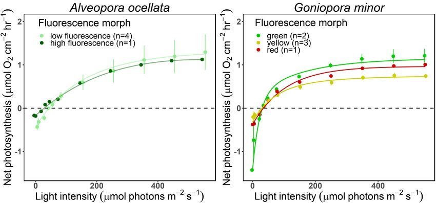
Ben-Zvi et al. Mesophotic Corals Photosynthesis and Bio-Optics
were normalized to coral surface area that was determined RESULTS
from top-view images and to the volume of water in the
chambers. Net and gross photosynthesis were fitted using a Fluorescence Polymorphism and
double exponential decay following Platt et al. (1980), and α,
Abundance
maximal photosynthesis rate (Pmax ), compensation irradiance
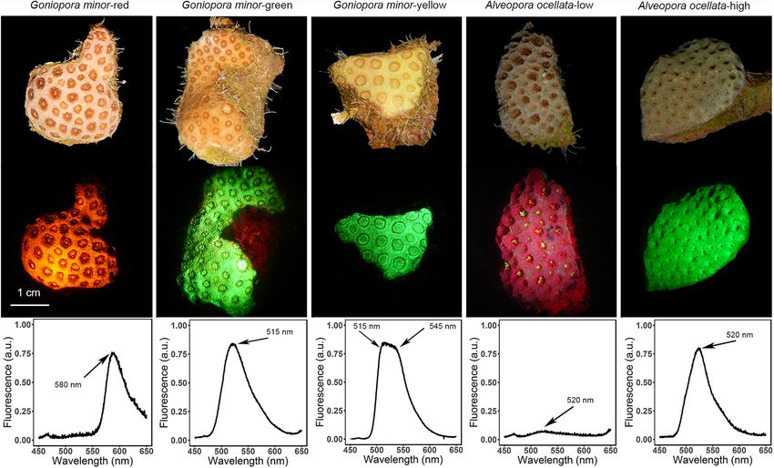
Fluorescence morphs of A. ocellata and G. minor visually differ
(Ec ), Ek , and dark respiration (Rd ) were extracted from
from one another under white illumination, under blue light
the fitted models.
illumination, and in their fluorescence emission peak (λem ;
Figure 1). Three distinct fluorescence morphs are described
Symbiodiniaceae Density and for G. minor; a red morph (Figure 1A; λem = 580 nm),
a green morph (Figure 1B; λem = 515 nm), and a yellow
Chlorophyll Concentration morph (Figure 1C; λem = 515 and 545 nm). Two fluorescence
Frozen fragments of G. minor and A. ocellata were thawed and morphs are described for A. ocellata: a low fluorescence morph
tissue was removed in the presence of 0.22 µ filtered seawater (Figure 1D; λem = 520 nm) and a high fluorescence morph
using an artist’s air brush into 50 ml tubes. Surface area was (Figure 1E; λem = 520 nm), both of which present the same green
determined using the single dip wax method for normalization emission peak, with the distinction that the low fluorescence
(Veal et al., 2010). Coral tissue was mechanically broken using a morph appears red under both illuminations due to chlorophyll
motorized homogenizer and centrifuged at 5,000 rpm for 5 min fluorescence of its symbionts (λem = 680 nm). The green
to separate the host (i.e., coral) supernatant from the algal pellet. morph of G. minor presents the typical, brownish color of
The host fraction was discarded, and the algal pellet was used corals under white light (Figure 1B, top image), commonly
for the quantification of chlorophyll and Symbiodiniaceae cell attributed to the symbiotic algae, yet exhibits a green fluorescence
density. A small subsample from each algal pellet was taken for under the excitation light (Figure 1B, middle image); while the
Symbiodiniaceae genetic identification. Algal cells were counted yellow morph displays a yellowish appearance under white light
in triplicates using a hemocytometer under a light microscope. illumination (Figure 1C, top image) and exhibits a bright green
The remaining algal pellet was used for chlorophyll analysis. glow under blue illumination (Figure 1C, middle image).
Chlorophyll was extracted in the presence of 100% cold acetone For G. minor, out of 63 surveyed colonies, the green
for 15 h at 4◦ C and chlorophyll a and c2 concentrations (pg morph was more prevalent compared to the yellow morph
chlorophyll cell−1 ) were determined spectrometrically as in (74 ± 14.7% and 26 ± 14.7%, respectively). The red morph
Jeffrey and Humphrey (1975). of G. minor was extremely rare and was not crossed in our
field surveys. For A. ocellata, we found that out of 40 colonies,
84 ± 17.4% (mean ± SD) presented low fluorescence and only
Symbiodiniaceae Genetic Identification 15.15 ± 17.43% of them displayed high fluorescence.
DNA was extracted using the DNeasy Blood and Tissue kit
(Qiagen, Germany) from the Symbiodiniaceae sub-samples
following the manufacturer’s protocol. A ∼700 bp sequence In vivo Light Microenvironment
fragment of the internal transcribed spacer 2 (ITS2) was Scalar irradiance (E0 ) measurements revealed a strong light
PCR amplified using the primers SYM_VAR_FWD and gradient within the coral tissue (Figure 2 and Supplementary
SYM_VAR_REV following the procedure of Hume et al. Figure 1). At the tissue surface, irradiance is 1.5-fold higher than
(2013). PCR products were purified by ExoSAP-IT (Thermo the incident irradiance for visible light (PAR; 400–700 nm) and
Fisher Scientific, United States) and bi-directionally sequenced. at the tissue-skeleton interface, available light is greatly reduced
Individual sequences were aligned using Geneious, and a to 50% of the incident light for PAR. While this surface light
consensus sequence was constructed for comparison against the enhancement is prominent between 400 and 700 nm for the
GeoSymbio database (Franklin et al., 2012). yellow and green morphs, it only occurs above 580 nm for
the red morph (the λem of this morph). Light absorbance by
photosynthetic pigments (i.e., chlorophyll a) can be observed as a
Statistical Analyses drop around 680 nm in all fluorescence morphs, and in the yellow
All statistical analyses were performed using R software (R morph the contribution of the host fluorescent proteins can be
Core Team, 2013). Data were tested for normality using the observed as a shoulder between 500 and 600 nm. The scattering of
Shapiro–Wilk test and homogeneity of variance with Levene’s light at wavelengths above 700 nm was found to be 11–14% higher
test. G. minor data was tested using PERMANOVA (for repeated in the green morph compared to the other morphs. Comparing
measure ANOVA in the light curves analyses), ANOVA (for the integrated photon irradiance (Figure 2B) revealed significant
normally distributed data), or permutational ANOVA when differences in all morphs between the two areas in which the
data did not follow a normal distribution. A. ocellata data was measurement was taken (i.e., tissue surface and tissue-skeleton
tested with PERMANOVA (for repeated measure ANOVA in the interface; permutational ANOVA, p < 0.0001). When examining
light curves analyses), t-tests (for normally distributed data) or the differences among morphs within each location we found
Wilcoxon signed-rank test (for non-normal data). Results were that the yellow morph differed from the green and red morphs
considered significant if p < 0.05. Where appropriate, data were in the surface measurements (permutational ANOVA, p < 0.01),
further examined using a Tukey’s post hoc test. while the green and red morphs did not (permutational ANOVA,
Frontiers in Marine Science | www.frontiersin.org 4 April 2021 | Volume 8 | Article 651601Ben-Zvi et al. Mesophotic Corals Photosynthesis and Bio-Optics
FIGURE 1 | Fluorescence diversity of mesophotic corals. Representative non-fluorescent images (top image in each panel); fluorescent images (middle image in
each panel); and fluorescence emission spectra of Goniopora minor (A–C) and Alveopora ocellata (D,E) fluorescence morphs. Fluorescence emission peaks are
indicated by arrows.
p = 0.71). Near the skeleton the green morph differed form the while the minimum saturating irradiance (Ek ) was slightly higher
two other morphs (permutational ANOVA, p < 0.05) while the in the high fluorescence morph (t-test, t = −2.17, p = 0.06). In
red and yellow did not (permutational ANOVA, p = 0.89). G. minor, the fluorescence morph had no significant effect on
any of the parameters (Supplementary Table 1; permutational
ANOVA, p < 0.05).
Chlorophyll a Fluorimetry
Relative electron transport rate values did not differ significantly
between morphs of A. ocellata (Figures 3A,C; PERMANOVA, O2 Evolution
F = 0.85, p = 0.37) or G. minor (PERMANOVA, F = 0.41, O2 evolution differed when measured under white or blue
p = 0.7). In A. ocellata, we found a significant effect of illuminations (Supplementary Figure 3, Table 1, and
the fluorescence morph on 8PSII (Figure 3B; PERMANOVA, Supplementary Table 2; PERMANOVA, p = 0.03). Since
F = 5.68, p = 0.04), being higher in the low fluorescence morph, P-E curves performed under blue illumination resulted in
while in G. minor it was found to be lowest in the green morph fully extended polyps, smoother curves (i.e., higher R2 ), and
(Figure 3E; PERMANOVA, F = 58.13, p = 0.01). The maximum had been previously suggested to be more appropriate for
quantum yield of PSII (F v /F m ; or 8PSII measured after a dark mesophotic corals (Mass et al., 2010), we present in Figure 4
incubation) was found to be similar among the fluorescence and Supplementary Figure 4 measurements, and in Table 1
morph of A. ocellata (Figure 3B and Supplementary Figure 3A; parameters derived from the curves performed under blue light
t-test, t = −1.37, p = 0.18) and among morphs of G. minor (parameters derived from the white illuminated curves can be
(Figure 3E and Supplementary Figure 2B; permutational found in Supplementary Table 2).
ANOVA, p = 0.18). We did not find differences in the P-E derived parameters
Non-photochemical quenching values were also similar between species (permutational ANOVA, p > 0.05) or between
between morphs of A. ocellata (Figure 3C; PERMANOVA, A. ocellata and G. minor morphs (t-test for A. ocellata or
F = 0.1, p = 0.74), and G. minor (Figure 3F; PERMANOVA, permutational ANOVA for G. minor, p > 0.05).
F = 0.06, p = 0.09).
The initial slope (α), and relative maximal electron transport Symbiodiniaceae Density and
rate (rETRmax ) calculated from the RLC were found to be Chlorophyll Concentration
similar among A. ocellata morphs (Supplementary Table 1; t- Symbiodiniaceae density and chlorophyll (a and c2 )
test, t = −0.05, p = 0.96 and t = −0.38, p = 0.71, respectively), concentrations did not significantly differ between species
Frontiers in Marine Science | www.frontiersin.org 5 April 2021 | Volume 8 | Article 651601Ben-Zvi et al. Mesophotic Corals Photosynthesis and Bio-Optics
FIGURE 2 | In vivo spectral scalar irradiance of three fluorescence morphs of the mesophotic coral Goniopora minor. (A) Scalar irradiance (E 0 ) was measured at the
coral tissue surface (solid lines) and at the tissue-skeleton interface (dotted lines) of red (colored red), green (colored green), and yellow (colored yellow) fluorescence
morphs of the mesophotic coral G. minor. Dashed black line indicates 100% of the incident irradiance (E d ). Colored lines represent the mean relative scalar
irradiance (n = 3) of each morph and confidence intervals are represented by transparent corresponding areas. Fluorescence emission peaks are indicated by
arrows. The solid box indicates an area of interest corresponding to (B) the integrated photon irradiance (n = 3) for specific wave bands (500–530 nm, 530–560 nm,
and 560–590 nm) for the three fluorescence morphs. Boxes represent the upper and lower quartile, center lines represent medians, and whiskers extend to data
measurements that are less than 1.5∗ IQR (interquartile range) away from first/third quartile.
(Figure 5; permutational ANOVA, p < 0.05). The mean (±SD) A. ocellata presented one fluorescence emission peak (at 520 nm),
Symbiodiniaceae density was 2.6 × 10⁷ ± 1.4 × 10⁷ cells G. minor presented three (515, 545, and 580 nm). In the GoE/A,
cm−2 . Moreover, we did not find a significant effect of the A. ocellata mostly inhibits mesophotic depths (Eyal-Shaham et al.,
fluorescence morph, of both species, on Symbiodiniaceae density 2016), whereas G. minor is a depth generalist and can be found
(Figure 5A, t-test, t = −0.54, p = 0.6, and permutational ANOVA, also in the shallower parts of the reefs (Tamir et al., 2019).
p = 0.88 for A. ocellata and G. minor, respectively), chlorophyll The differences in the zonation characteristics of these species
a concentration (Figure 5B; Wilcoxon test, W = 7, p = 0.35 may explain why G. minor possesses a range of FPs. A broader
and ANOVA, F = 0.53, p = 0.61 for A. ocellata and G. minor, FPs arsenal covers a broader spectrum of emission peaks which
respectively), and chlorophyll c2 (Figure 5C; Wilcoxon test, potentially correspond to a wider range of excitations that may
W = 7, p = 0.35 and ANOVA, F = 0.86, p = 0.47 for A. ocellata mediate excess light (at shallow depths) or provide wavelengths
that are absent (at mesophotic depths). A. ocellata, presents
and G. minor, respectively).
only one fluorescence emission which will have a narrower
Genetic Analysis excitation range. Field surveys revealed that for A. ocellata the low
fluorescence morph was the dominant one, while for G. minor the
Genetic identification based on the ITS2 region revealed that
dominant morph was the green one. Despite the reported higher
all our coral samples harbored Symbiodiniaceae from the genus
abundance of red FPs in deeper habits compared to shallower
Cladocopium (formerly known as clade C; LaJeunesse et al.,
ones, and their suggested advantage in the dispersal of light
2018), regardless of species or fluorescence morph. Based on the
deeper into the coral tissue (Smith et al., 2017), the red morph
ITS2 sequences we found that A. ocellata harbored Cladocopium
of G. minor is extremely rare.
types C3.10 (n = 7), C101 (n = 1), C3 (n = 1), and C66b (n = 1),
Host fluorescence is known to play a critical role in the
and G. minor harbored types C1 (n = 4), C78 (n = 1), C40 (n = 1),
modification of irradiance intensity and the spectral tuning
and C3 (n = 1). We found no effect of the fluorescence morph on
of in-hospite irradiance environment (Salih et al., 2000; Mazel
Symbiodiniaceae types.
and Fuchs, 2003; Wangpraseurt et al., 2012; Lyndby et al.,
2016; Smith et al., 2017; Quick et al., 2018; Wangpraseurt
DISCUSSION et al., 2019; Bollati et al., 2020). Our light microsensors
measurements indicate that the optical environment within
The spectral analyses revealed a range of fluorescence emission the coral tissue is influenced by the presence of different FPs
peaks for two mesophotic coral species (Figure 1). While, (Figure 2 and Supplementary Figure 1). Despite the bright
Frontiers in Marine Science | www.frontiersin.org 6 April 2021 | Volume 8 | Article 651601Ben-Zvi et al. Mesophotic Corals Photosynthesis and Bio-Optics
FIGURE 3 | Rapid light curves of two mesophotic coral species. Fitted (solid lines) and mean (circles) ± SE (error bras) of rETR values (A,D), effective quantum yield
[8PSII ; (B,E)], and non-photochemical quenching [NPQ; (C,F)] of high fluorescence (dark green; n = 5) and low fluorescence (light green; n = 5) morphs of Alveopora
ocellata (A–C), and green (green; n = 2), red (red; n = 1), and yellow (yellow; n = 2) fluorescence morphs of Goniopora minor (D–F).
TABLE 1 | Photosynthetic parameters derived from photosynthesis-irradiance (P-E) curves under blue illumination.
Species Morph n Slope Pmax Ec Ek Rd
(α) (µmol O2 µmol (µmol O2 cm−2 hr−1 ) (µmol photons (µmol photons (µmol O2 cm−2 hr−1 )
photons−1 m−2 s−1 ) m−2 s−1 ) m−2 s−1 )
A. ocellata HF 1 0.006 1.12 26 176 0.17
A. ocellata LF 4 0.01 ± 2e-3 1.27 ± 0.87 37 ± 8 110 ± 77 0.43 ± 0.15
G. minor Green 2 0.08 ± 0.09 1.14 ± 0.4 23 ± 8 67 ± 87 1.42 ± 1.57
G. minor Red 1 0.01 0.96 29 75 0.37
G. minor Yellow 3 0.01 ± 8e-3 0.74 ± 0.13 22 ± 7 86 ± 76 0.31 ± 0.23
Mean (±SD) value of the initial slope (α), maximal photosynthetic rate (Pmax ), minimum saturating irradiance (Ek ), compensation irradiance (Ec ), and dark respiration (Rd )
of high fluorescence (HF) and low fluorescence (LF) morphs of Alveopora ocellata and green, red, and yellow fluorescence morphs of Goniopora minor.
appearance of G. minor under white light (in the red and (Lesser et al., 2004), which correspond to the emission
yellow morph; Figures 1A,C) and under blue excitation light peaks of several FPs and may confound the interpretation
(all morphs), the contribution of the FPs to the spectral of fluorescence spectra. Nonetheless, the in-hospite irradiance
signature of the morphs was not as strong as expected and differed among the morphs (Figure 2). The yellow morph
previously documented (Mazel and Fuchs, 2003; Wangpraseurt presented the greatest scalar irradiance enhancement at the
et al., 2012). This may be explained by the light source used shorter wavelengths (i.e., 400–700 nm), and the green morph
for these measurements which is poor in blue, FP-exciting showed greater light enhancement at longer wavelengths (700–
photons, or by the absorbance of light by the photosynthetic 800 nm). The dominance of certain morphs over others and
pigments sharing these emission peaks of host fluorescence. the differences found in the internal light environment within
For example, the yellow morph of G. minor has a fluorescence the coral tissue, led us to hypothesize that certain FPs may
emission peak at 545 nm (Figure 1C), while peridinin has be advantageous for photosynthesis within the mesophotic
an absorbance peak at 540 nm (Niedzwiedzki et al., 2014). light environment.
Additionally, coral-associated cyanobacteria may also contain Chlorophyll fluorescence-based measurements revealed that
phycoerythrin with absorption bands around 505 and 571 nm A. ocellata and G. minor fluorescence morphs mostly did
Frontiers in Marine Science | www.frontiersin.org 7 April 2021 | Volume 8 | Article 651601Ben-Zvi et al. Mesophotic Corals Photosynthesis and Bio-Optics FIGURE 4 | Net photosynthesis of two mesophotic corals fluorescence morphs. Fitted (solid lines) and mean (circles) ± SE (error bars) values of O2 evolution (µmol O2 cm−2 hr−1 ) measured under blue light at increasing intensities (0, 5, 23, 36, 50, 78, 150, 250, 350, 450, and 550 µmol photons m−2 s−1 ) of Alveopora ocellata [(A), n = 1 and 4 for high and low fluorescence morphs, respectively] and Goniopora minor [(B), n = 2, 1, and 3 for green, red, and yellow morphs, respectively]. not differ in the quantum yield of PSII (Figure 3A,B for Symbiodiniaceae has already been established as being more and Supplementary Figure 2). Likewise, Roth et al. (2015), efficient at shorter wavelengths than at longer ones (Szabó et al., did not find differences in 8PSII between high and low 2014). Furthermore, as corals FPs are largely excited by blue fluorescence morphs of Leptoseris spp. at mesophotic depths. light, the FPs will in turn affect the internal light environment. Although it has been shown that changes in the in-hospite We therefore compared measurements taken under white and irradiance environment can affect absolute electron transport blue light (Supplementary Figure 3 and Supplementary Table 2) rates in corals (Wangpraseurt et al., 2014), we found no and indeed found differences in coral response. Under blue light differences in rETR values among the examined morphs corals expanded their tentacles, while under white light they (Figures 3A,D) despite an enhanced irradiance measured contracted them. Conducting the metabolic measurements under within the tissue of G. minor’s yellow morph (Figure 2). blue light resulted in smoother P-E curves (Supplementary When the photoprotective role of FPs was examined in Figure 3), which may represent a more natural performance of mesophotic Euphyllia paradivisa, no differences in the amount mesophotic corals. Most of the previously reported values for of UVR-induced DNA damage were found between fluorescence P-E derived parameters that we were able to compare to our morphs (Ben-Zvi et al., 2019). However, this does not exclude measurement aligned with the current results (Cooper et al., the putative photoprotective role of FPs as NPQ values 2011; Nir et al., 2014; Eyal et al., 2019). The results indicate that were slightly (but not significantly) higher for the red and mesophotic corals usually present relatively low compensation yellow morphs of G. minor compared to the green morph irradiance ranging between 15 and 96 µmol photons m−2 s−1 (Figure 3F), indicating their potentially greater capability as well as low saturating irradiances, ranging between 28 and in mediating excess light and preventing damage to the 80 µmol photons m−2 s−1 leaving a narrow window of light photosynthetic apparatus. intensities which enable photosynthesis. Respiration and photosynthesis data from mesophotic corals Klueter et al. (2006) found that a highly fluorescent morph are very limited and there is currently no agreed protocol of Montipora digitata had higher Symbiodiniaceae densities and for performing these measurements. Although Mass et al. chlorophyll a concentration compared to a low fluorescent (2010) provided evidence for the chromatic dependence of morph. We examined the Symbiodiniaceae densities as well as photosynthetic performance on the light provided during chlorophyll a and c2 concentrations in all our studied morphs measurements, ex-situ measurements on mesophotic corals but found no differences among our samples (Figure 5). Our are still commonly performed under white light (Cooper measured values of Symbiodiniaceae density are higher than et al., 2011; Ben-Zvi et al., 2020). Since corals are known to previously reported values in mesophotic corals (Bongaerts photoacclimatize to their natural light conditions, mesophotic et al., 2011; Cooper et al., 2011), however this parameter corals are most likely acclimatize to blue light, as this is the can greatly vary between species, depth, and light availability prominent wavelength at depths of 30–150 m (Kahng et al., 2019). [reviewed by Roth (2014)]. Chlorophyll concentration values Moreover, the wavelength-dependent absorption cross-section determined in this study, align with previously reported values Frontiers in Marine Science | www.frontiersin.org 8 April 2021 | Volume 8 | Article 651601
Ben-Zvi et al.                                                                                                         Mesophotic Corals Photosynthesis and Bio-Optics

  FIGURE 5 | Photobiology of fluorescence morphs of the mesophotic corals Alveopora ocellata and Goniopora minor. Areal Symbiodiniaceae density (A), cellular
  chlorophyll a (B), and cellular chlorophyll c2 (C) of low fluorescence (LF) and high fluorescence (HF) morphs of A. ocellata and green, yellow, and red fluorescence
  morphs of G. minor. Boxes represent the upper and lower quartile, center lines represent medians, and whiskers extend to data measurements that are less than
  1.5*IQR away from first/third quartile. Outliers are represented by dots. Sample size (n) is indicated below each box.

(Lesser et al., 2010; Cooper et al., 2011; Eyal et al., 2019). The                         In this study, we investigated the bio-optical properties and
latter result also indicates that the brighter color of the yellow                      photosynthetic performances of mesophotic corals exhibiting
morph of G. minor is probably the result of a higher expression                         different host fluorescence emissions resulting from differential
of FPs rather than a low algal density or low chlorophyll                               expression of the same FP (A. ocellata) or multiple FPs
concentration. Similarly, in A. ocellata, the red/brown appearance                      (G. minor). Our results, showing only negligible differences in
of the low fluorescence morph compared to the green appearance                          the photobiology of the different coral fluorescence morphs,
of the high fluorescence morph may also be a result of a higher                         do not support any of the prevailing mechanisms that
concentrations of the host FP and not of a change in the                                have been suggested to enhance photosynthesis in coral by
algal symbionts.                                                                        means of FPs in deeper habitats. Nevertheless, the bio-optical
    Aihara et al. (2019) demonstrated that coral fluorescence may                       properties revealed changes among morphs, indicating that
serve as an attractive signal for symbiotic algae, and specific                         fluorescence may mediate the internal light environment of
Symbiodiniaceae genera/species were found to be correlated                              corals, potentially affecting other aspects of coral physiology
with “redder” juveniles of Acropora millepora (Quigley et al.,                          through cellular mechanisms that are light-regulated such as
2018). We therefore sought to determine whether a specific                              circadian clocks entrainment (Levy et al., 2007), spawning
fluorescent signal would indeed attract symbionts that differ                           (Sweeney et al., 2011), or growth (Roth et al., 1982). Future
genetically. The genetic identity of Symbiodiniaceae revealed                           research should focus on depicting the pathways, such as
no significant effect of the fluorescence morph, and all corals                         gene regulation and expression, by which the effect of the
harbored Symbiodiniaceae from the genus Cladocopium, as                                 diverse internal light regimes found among morphs may play a
previously described in other coral species at the mesophotic                           significant role.
reefs of the GoE/A (Nir et al., 2011; Einbinder et al.,
2016; Eyal et al., 2019), as well as at other mesophotic
reefs worldwide (Ziegler et al., 2015; Goulet et al., 2019).                            DATA AVAILABILITY STATEMENT
We therefore conclude that despite the possibly of serving
as general Symbiodiniaceae attractant, specific fluorescence                            The raw data supporting the conclusions of this article will be
emissions do not attract specific Symbiodiniaceae genotypes.                            made available by the authors, without undue reservation.
Since all the species and morphs we examined share the
same habitat and are found in close proximity to each
other, and hence experience similar environmental conditions,                           AUTHOR CONTRIBUTIONS
there may not be an apparent reason to attract different
symbionts, which may or may not have an advantage in their                              OB-Z and YL conceived the study. GE and OB-Z collected
photosynthetic performances or in their tolerance to stressors,                         the coral samples and conducted the field surveys.
such as temperature.                                                                    DW performed the bio-optical measurements. OB-Z

Frontiers in Marine Science | www.frontiersin.org                                   9                                               April 2021 | Volume 8 | Article 651601
Ben-Zvi et al. Mesophotic Corals Photosynthesis and Bio-Optics
performed all other analyses, visualized all data, and wrote ACKNOWLEDGMENTS
the first draft of the manuscript. OB-Z, DW, and OB
analyzed the data. All authors reviewed, commented, and We would like to thank O. Levy for the use of the Imaging-PAM
approved the manuscript. and oxygen evolution measurement system, T. Treibitz for the
use of the JAZ system, and N. Paz for editing the manuscript. We
would also like to thank the Interuniversity Institute for Marine
FUNDING Sciences in Eilat and its staff for making their facilities available
for us and for all the YL group for their ongoing support.
This research was funded by the Israel Science Foundation
(ISF) grant agreement No. 1191/16 to YL, Ministry of Science,
Technology and Space doctoral fellowship grant agreement No. SUPPLEMENTARY MATERIAL
3-18487 to OB-Z, Assemble Plus Consortium grant to DW,
and European Union’s Horizon 2020 Research and Innovation The Supplementary Material for this article can be found
Program under the Marie Skłodowska-Curie grant agreement online at: https://www.frontiersin.org/articles/10.3389/fmars.
No. 796025 to GE. 2021.651601/full#supplementary-material
REFERENCES D’Angelo, C., Smith, E., Oswald, F., Burt, J., Tchernov, D., and Wiedenmann,
J. (2012). Locally accelerated growth is part of the innate immune response
Aihara, Y., Maruyama, S., Baird, A. H., Iguchi, A., Takahashi, S., and Minagawa, J. and repair mechanisms in reef-building corals as detected by green fluorescent
(2019). Green fluorescence from cnidarian hosts attracts symbiotic algae. Proc. protein (GFP)-like pigments. Coral Reefs 31, 1045–1056. doi: 10.1007/s00338-
Nat. Acad. Sci. U.S.A. 116, 2118–2123. doi: 10.1073/pnas.1812257116 012-0926-8
Alieva, N. O., Konzen, K. A., Field, S. F., Meleshkevitch, E. A., Hunt, M. E., Beltran- DeSalvo, M., Voolstra, C., Sunagawa, S., Schwarz, J., Stillman, J., Coffroth, M.,
Ramirez, V., et al. (2008). Diversity and evolution of coral fluorescent proteins. et al. (2008). Differential gene expression during thermal stress and bleaching
PLoS One 3:e2680. doi: 10.1371/journal.pone.0002680 in the Caribbean coral Montastraea faveolata. Mol. Ecol. 17, 3952–3971. doi:
Ben-Zvi, O., Eyal, G., and Loya, Y. (2014). Light-dependent fluorescence in the 10.1111/j.1365-294X.2008.03879.x
coral Galaxea fascicularis. Hydrobiologia 759, 15–26. doi: 10.1007/s10750-014- Dishon, G., Dubinsky, Z., Fine, M., and Iluz, D. (2012). Underwater light field
2063-6 patterns in subtropical coastal waters: a case study from the Gulf of Eilat
Ben-Zvi, O., Eyal, G., and Loya, Y. (2019). Response of fluorescence morphs of the (Aqaba). Isr. J. Plant Sci. 60, 265–275. doi: 10.1560/IJPS.60.1-2.265
mesophotic coral Euphyllia paradivisa to ultra-violet radiation. Sci. Rep. 9:5245. Dove, S., Hoegh-Guldberg, O., and Ranganathan, S. (2001). Major colour patterns
doi: 10.1038/s41598-019-41710-3 of reef-building corals are due to a family of GFP-like proteins. Coral Reefs 19,
Ben-Zvi, O., Ofer, E., Eyal, G., and Loya, Y. (2021). Experimental evidence of 197–204. doi: 10.1007/PL00006956
temperature-induced bleaching in two fluorescence morphs of a Red Sea Dove, S. G., Lovell, C., Fine, M., Deckenback, J., Hoegh-Guldberg, O., Iglesias-
mesophotic coral. Coral Reefs 40, 187–199. doi: 10.1007/s00338-020-02027-0 Prieto, R., et al. (2008). Host pigments: potential facilitators of photosynthesis
Ben-Zvi, O., Tamir, R., Keren, N., Tchernov, D., Berman-Frank, I., Kolodny, in coral symbioses. Plant Cell Environ. 31, 1523–1533. doi: 10.1111/j.1365-3040.
Y., et al. (2020). Photophysiology of a mesophotic coral 3 years after 2008.01852.x
transplantation to a shallow environment. Coral Reefs 39, 903–913. doi: 10. Einbinder, S., Gruber, D. F., Salomon, E., Liran, O., Keren, N., and Tchernov, D.
1007/s00338-020-01910-0 (2016). Novel adaptive photosynthetic characteristics of mesophotic symbiotic
Björkman, O., and Demmig, B. (1987). Photon yield of O2 evolution and microalgae within the reef-building coral, Stylophora pistillata. Front. Mar. Sci.
chlorophyll fluorescence characteristics at 77K among vascular plants of diverse 3:195. doi: 10.3389/fmars.2016.00195
origins. Planta 170, 489–504. doi: 10.1007/BF00402983 Enríquez, S., Méndez, E. R., and Prieto, R. I. (2005). Multiple scattering on coral
Bollati, E., D’angelo, C., Alderdice, R., Pratchett, M., Ziegler, M., and Wiedenmann, skeletons enhances light absorption by symbiotic algae. Limnol. Oceanogr. 50,
J. (2020). Optical feedback loop involving dinoflagellate symbiont and 1025–1032. doi: 10.4319/lo.2005.50.4.1025
scleractinian host drives colorful coral bleaching. Curr. Biol. 30, 2433–2445.e3. Eyal, G., Cohen, I., Eyal-Shaham, L., Ben-Zvi, O., Tikochinski, Y., and Loya, Y.
doi: 10.1016/j.cub.2020.04.055 (2019). Photoacclimation and induction of light-enhanced calcification in the
Bongaerts, P., Riginos, C., Hay, K. B., Van Oppen, M. J., Hoegh-Guldberg, O., mesophotic coral Euphyllia paradivisa. R. Soc. Open Sci. 6:180527. doi: 10.1098/
and Dove, S. (2011). Adaptive divergence in a scleractinian coral: physiological rsos.180527
adaptation of Seriatopora hystrix to shallow and deep reef habitats. BMC Evol. Eyal, G., Wiedenmann, J., Grinblat, M., D’angelo, C., Kramarsky-Winter, E.,
Biol. 11:303. doi: 10.1186/1471-2148-54011-303 Treibitz, T., et al. (2015). Spectral diversity and regulation of coral fluorescence
Bongaerts, P., Frade, P. R., Ogier, J. J., Hay, K. B., Van Bleijswijk, J., Englebert, in a mesophotic reef habitat in the Red Sea. PLoS One 10:e0128697. doi: 10.
N., et al. (2013). Sharing the slope: depth partitioning of agariciid corals and 1371/journal.pone.0128697
associated Symbiodinium across shallow and mesophotic habitats (2-60 m) on a Eyal-Shaham, L., Eyal, G., Tamir, R., and Loya, Y. (2016). Reproduction, abundance
Caribbean reef. BMC Evol. Biol. 13:205. doi: 10.1186/1471-2148-13-205 and survivorship of two Alveopora spp. in the mesophotic reefs of Eilat. Red Sea
Cooper, T. F., Ulstrup, K. E., Dandan, S. S., Heyward, A. J., Kühl, M., Muirhead, Sci. Rep. 6:20964. doi: 10.1038/srep20964
A., et al. (2011). Niche specialization of reef-building corals in the mesophotic Förster, T. (1955). Intermolecular Energy Transfer and Fluorescence. Ottawa, ON:
zone: metabolic trade-offs between divergent Symbiodinium types. Proc. Royal National Research Council of Canada.
Soc. B 278, 1840–1850. doi: 10.1098/rspb.2010.2321 Franklin, E. C., Stat, M., Pochon, X., Putnam, H. M., and Gates, R. D.
Cox, G., Matz, M., and Salih, A. (2007). Fluorescence lifetime imaging of (2012). GeoSymbio: a hybrid, cloud-based web application of global geospatial
coral fluorescent proteins. Microsc. Res. Tec. 70, 243–251. doi: 10.1002/jemt. bioinformatics and ecoinformatics for Symbiodinium–host symbioses. Mol.
20410 Ecol. Resour. 12, 369–373. doi: 10.1111/j.1755-0998.2011.03081.x
Cox, G., and Salih, A. (2005). “Fluorescence lifetime imaging of symbionts and Gilmore, A. M., Larkum, A. W. D., Salih, A., Itoh, S., Shibata, Y., Bena, C., et al.
fluorescent proteins in reef corals,” in Biomedical Optics 2005: International (2003). Simultaneous time resolution of the emission spectra of fluorescent
Society for Optics and Photonics, (San Jose, CA: SPIE), 162–170. doi: 10.1117/ proteins and zooxanthellar chlorophyll in reef-building corals. Photochem.
12.600708 Photobiol. 77, 515–523. doi: 10.1562/0031-865520030770515STROTE2.0.CO2
Frontiers in Marine Science | www.frontiersin.org 10 April 2021 | Volume 8 | Article 651601Ben-Zvi et al. Mesophotic Corals Photosynthesis and Bio-Optics
Gittins, J. R., D’angelo, C., Oswald, F., Edwards, R. J., and Wiedenmann, J. zone: light, food, and genetics. Ecology 91, 990–1003. doi: 10.1890/09-
(2015). Fluorescent protein-mediated colour polymorphism in reef corals: 0313.1
multicopy genes extend the adaptation/acclimatization potential to variable Levy, O., Appelbaum, L., Leggat, W., Gothlif, Y., Hayward, D. C., Miller, D. J., et al.
light environments. Mol. Ecol. 24, 453–465. doi: 10.1111/mec.13041 (2007). Light-responsive cryptochromes from a simple multicellular animal, the
Goulet, T. L., Lucas, M. Q., and Schizas, N. V. (2019). “Symbiodiniaceae coral Acropora millepora. Science 318, 467–470. doi: 10.1126/science.1145432
genetic diversity and symbioses with hosts from shallow to mesophotic coral Lyndby, N. H., Kühl, M., and Wangpraseurt, D. (2016). Heat generation and light
ecosystems,” in Mesophotic Coral Ecosystems, eds Y. Loya, K. A. Puglise, and scattering of green fluorescent protein-like pigments in coral tissue. Sci. Rep.
T. C. L. Bridge (Cham: Springer International Publishing), 537–551. doi: 10. 6:26599. doi: 10.1038/srep26599
1007/978-3-319-92735-0_30 Mass, T., Einbinder, S., Brokovich, E., Shashar, N., Vago, R., Erez, J., et al. (2007).
Hollingsworth, L. L., Kinzie, R. A., Lewis, T. D., Krupp, D. A., and Leong, J. A. C. Photoacclimation of Stylophora pistillata to light extremes: metabolism and
(2005). Phototaxis of motile zooxanthellae to green light may facilitate symbiont calcification. Mar. Ecol. Prog. Ser. 334, 93–102. doi: 10.3354/meps334093
capture by coral larvae. Coral Reefs 24, 523–523. doi: 10.1007/s00338-005-0063- Mass, T., Kline, D. I., Roopin, M., Veal, C. J., Cohen, S., Iluz, D., et al. (2010). The
8 spectral quality of light is a key driver of photosynthesis and photoadaptation in
Hume, B., D’angelo, C., Burt, J., Baker, A. C., Riegl, B., and Wiedenmann, J. Stylophora pistillata colonies from different depths in the Red Sea. J. Exp. Biol.
(2013). Corals from the Persian/Arabian Gulf as models for thermotolerant 213, 4084–4091. doi: 10.1242/jeb.039891
reef-builders: prevalence of clade C3 Symbiodinium, host fluorescence and Matz, M. V., Fradkov, A. F., Labas, Y. A., Savitsky, A. P., Zaraisky, A. G., Markelov,
ex situ temperature tolerance. Mar. Pollut. Bull. 72, 313–322. doi: 10.1016/j. M. L., et al. (1999). Fluorescent proteins from nonbioluminescent Anthozoa
marpolbul.2012.11.032 species. Nat. biotechnol. 17, 969–973. doi: 10.1038/13657
Jeffrey, S. W., and Humphrey, G. F. (1975). New spectrophotometric equations Mazel, C. H., and Fuchs, E. (2003). Contribution of fluorescence to the spectral
for determining chlorophylls a1, b1, c1 and c2 in higher plants, algae and signature and perceived color of corals. Limnol. Oceanogr. 48, 390–401. doi:
natural phytoplankton. Biochem. Physiol. Pflanz 167, 191–194. doi: 10.1016/ 10.4319/lo.2003.48.1_part_2.0390
S0015-3796(17)30778-3 Muscatine, L. (1990). “The role of symbiotic algae in carbon and energy flux in
Jerlov, N. G. (1968). Optical Oceanography. Elsevier reef corals,” in Ecosystems of the World, ed. Z. Dubinsky (Amsterdam: Elsevier),
Kahng, S. E., Akkaynak, D., Shlesinger, T., Hochberg, E. J., Wiedenmann, J., 75–87.
Tamir, R., et al. (2019). “Light, temperature, photosynthesis, heterotrophy, and Niedzwiedzki, D. M., Jiang, J., Lo, C. S., and Blankenship, R. E. (2014).
the lower depth limits of mesophotic coral ecosystems,” in Mesophotic Coral Spectroscopic properties of the Chlorophyll a–Chlorophyll c2 –peridinin-
Ecosystems, eds Y. Loya, K. A. Puglise, and T. C. L. Bridge (Cham: Springer protein-complex (acpPC) from the coral symbiotic dinoflagellate
International Publishing), 801–828. doi: 10.1007/978-3-319-92735-0_42 Symbiodinium. Photosynth. Res. 120, 125–139. doi: 10.1007/s11120-013-
Kahng, S. E., Hochberg, E. J., Apprill, A., Wagner, D., Luck, D. G., Perez, D., et al. 9794-5
(2012). Efficient light harvesting in deep-water zooxanthellate corals. Mar. Ecol. Nir, O., Gruber, D. F., Einbinder, S., Kark, S., and Tchernov, D. (2011). Changes
Prog. Ser. 455, 65–77. doi: 10.3354/meps09657 in scleractinian coral Seriatopora hystrix morphology and its endocellular
Kahng, S. E., and Kelley, C. D. (2007). Vertical zonation of megabenthic taxa on a Symbiodinium characteristics along a bathymetric gradient from shallow to
deep photosynthetic reef (50–140 m) in the Au’au Channel, Hawaii. Coral Reefs mesophotic reef. Coral Reefs 30:1089. doi: 10.1007/s00338-011-0801-z
26, 679–687. doi: 10.1007/s00338-007-0253-7 Nir, O., Gruber, D. F., Shemesh, E., Glasser, E., and Tchernov, D. (2014). Seasonal
Kaniewska, P., Anthony, K. R. N., and Hoegh-Guldberg, O. (2008). Variation in mesophotic coral bleaching of Stylophora pistillata in the northern Red Sea.
colony geometry modulates internal light levels in branching corals, Acropora PLoS One 9:e84968. doi: 10.1371/journal.pone.0084968
humilis and Stylophora pistillata. Mar. Biol. 155, 649–660. doi: 10.1007/s00227- Platt, T., Gallegos, C., and Harrison, W. G. (1980). Photoinhibition of
008-1061-5 photosynthesis in natural assemblages of marine phytoplankton. J. Mar. Res.
Kawaguti, S. (1944). On the physiology of reef corals VI. Study on the pigments. 38, 687–701.
Palao Trop. Biol. Stn. Stud. 2, 617–674. Quick, C., D’angelo, C., and Wiedenmann, J. (2018). Trade-offs associated with
Kirk, J. T. (2011a). “Absorption of light within the aquatic medium,” in Light and photoprotective green fluorescent protein expression as potential drivers of
Photosynthesis in Aquatic Ecosystems, 3 Edn, ed. J. T. O. Kirk (New York, NY: balancing selection for color polymorphism in reef corals. Front. Mar. Sci. 5:11.
Cambridge University Press), 50–97. doi: 10.3389/fmars.2018.00011
Kirk, J. T. (2011b). “The nature of the underwater light field,” in Light and Quigley, K. M., Strader, M. E., and Matz, M. V. (2018). Relationship between
Photosynthesis in Aquatic Ecosystems, ed. J. T. O. Kirk (New York, NY: Acropora millepora juvenile fluorescence and composition of newly established
Cambridge University Press), 153–198. Symbiodinium assemblage. PeerJ 6, e5022. doi: 10.7717/peerj.5022
Klueter, A., Loh, W., Hoegh-Guldberg, O., and Dove, S. (2006). Physiological and R Core Team (2013). R: A Language and Environment for Statistical Computing.
genetic properties of two fluorescent colour morphs of the coral Montipora Vienna: R Foundation for Statistical Computing.
digitata. Symbiosis 42, 123–134. Roth, A. A., Clausen, C. D., Yahiku, P. Y., Clausen, V. E., and Cox, W. W. (1982).
Kramer, D. M., Johnson, G., Kiirats, O., and Edwards, G. E. (2004). New Some effects of light on coral growth. Pac. Sci. 36, 65–81.
fluorescence parameters for the determination of QA redox state and excitation Roth, M. S. (2014). The engine of the reef: photobiology of the coral–algal
energy fluxes. Photosynth. Res. 79:209. doi: 10.1023/B:PRES.0000015391.99477. symbiosis. Front. Microbiol. 5:422. doi: 10.3389/fmicb.2014.00422
0d Roth, M. S., Padilla-Gamiño, J. L., Pochon, X., Bidigare, R. R., Gates, R. D., Smith,
Kühl, M., Cohen, Y., Dalsgaard, T., Jørgensen, B. B., and Revsbech, N. P. (1995). C. M., et al. (2015). Fluorescent proteins in dominant mesophotic reef-building
Microenvironment and photosynthesis of zooxanthellae in scleractinian corals corals. Mar. Ecol. Prog. Ser. 521, 63–79. doi: 10.3354/meps11108
studied with microsensors for O2 , pH and light. Mar. Ecol. Prog. Ser. 117, Salih, A., Larkum, A., Cox, G., Kühl, M., and Hoegh-Guldberg, O. (2000).
159–172. doi: 10.3354/meps117159 Fluorescent pigments in corals are photoprotective. Nature 408, 850–853. doi:
LaJeunesse, T. C., Parkinson, J. E., Gabrielson, P. W., Jeong, H. J., Reimer, J. D., 10.1038/35048564
Voolstra, C. R., et al. (2018). Systematic revision of Symbiodiniaceae highlights Schlichter, D., and Fricke, H. W. (1990). Coral host improves photosynthesis
the antiquity and diversity of coral endosymbionts. Curr. Biol. 28, 2570–2580. of endosymbiotic algae. Naturwissenschaften 77, 447–450. doi: 10.1007/
doi: 10.1016/j.cub.2018.07.008 BF01135950
Lesser, M. P., Mazel, C. H., Gorbunov, M. Y., and Falkowski, P. G. (2004). Discovery Schlichter, D., Fricke, H. W., and Weber, W. (1986). Light harvesting by wavelength
of symbiotic nitrogen-fixing cyanobacteria in corals. Science 305, 997–1000. transformation in a symbiotic coral of the Red Sea twilight zone. Mar. Biol. 91,
doi: 10.1126/science.1099128 403–407. doi: 10.1007/BF00428634
Lesser, M. P., Slattery, M., and Leichter, J. J. (2009). Ecology of mesophotic Schlichter, D., Meier, U., and Fricke, H. (1994). Improvement of photosynthesis
coral reefs. J. Exp. Mar. Biol. Ecol. 375, 1–8. doi: 10.1016/j.jembe.2009. in zooxanthellate corals by autofluorescent chromatophores. Oecologia 99,
05.009 124–131. doi: 10.1007/bf00317092
Lesser, M. P., Slattery, M., Stat, M., Ojimi, M., Gates, R. D., and Grottoli, A. (2010). Scott, B. D., and Jitts, H. R. (1977). Photosynthesis of phytoplankton and
Photoacclimatization by the coral Montastraea cavernosa in the mesophotic zooxanthellae on a coral reef. Mar. Biol. 41, 307–315. doi: 10.1007/BF00389097
Frontiers in Marine Science | www.frontiersin.org 11 April 2021 | Volume 8 | Article 651601Ben-Zvi et al. Mesophotic Corals Photosynthesis and Bio-Optics Smith, E. G., D’angelo, C., Sharon, Y., Tchernov, D., and Wiedenmann, J. (2017). Wangpraseurt, D., Larkum, A., Ralph, P., and Kühl, M. (2012). Light gradients and Acclimatization of symbiotic corals to mesophotic light environments through optical microniches in coral tissues. Front. Microbiol. 3:316. doi: 10.3389/fmicb. wavelength transformation by fluorescent protein pigments. Proc. R. Soc. B 2012.00316 284:1858. doi: 10.1098/rspb.2017.0320 Wangpraseurt, D., Lichtenberg, M., Jacques, S. L., Larkum, A. W. D., and Stimson, J. (1985). The effect of shading by the table coral Acropora hyacinthus on Kühl, M. (2019). Optical properties of corals distort variable chlorophyll understory corals. Ecology 66, 40–53. doi: 10.2307/1941305 fluorescence measurements. Plant Physiol. 179, 1608–1619. doi: 10.1104/pp.18. Sweeney, A. M., Boch, C. A., Johnsen, S., and Morse, D. E. (2011). Twilight spectral 01275 dynamics and the coral reef invertebrate spawning response. J. Exp. Biol. 214, Wangpraseurt, D., Tamburic, B., Szabó, M., Suggett, D., Ralph, P. J., and Kühl, 770–777. doi: 10.1242/jeb.043406 M. (2014). Spectral effects on Symbiodinium photobiology studied with a Szabó, M., Wangpraseurt, D., Tamburic, B., Larkum, A. W. D., Schreiber, U., programmable light engine. PLoS One 9:e112809. doi: 10.1371/journal.pone. Suggett, D. J., et al. (2014). Effective light absorption and absolute electron 0112809 transport rates in the coral Pocillopora damicornis. Plant Physiol. Biochem 83, Ziegler, M., Roder, C. M., Büchel, C., and Voolstra, C. R. (2015). Mesophotic coral 159–167. doi: 10.1016/j.plaphy.2014.07.015 depth acclimatization is a function of host-specific symbiont physiology. Front. Takahashi-Kariyazono, S., Terai, Y., and Sakai, K. (2018). Presence–absence Mar. Sci. 2:4. doi: 10.3389/fmars.2015.00004 polymorphisms of highly expressed FP sequences contribute to fluorescent polymorphisms in Acropora digitifera. Genome Biol. Evol. 10, 1715–1729. doi: Conflict of Interest: The authors declare that the research was conducted in the 10.1093/gbe/evy122 absence of any commercial or financial relationships that could be construed as a Tamir, R., Eyal, G., Kramer, N., Laverick, J. H., and Loya, Y. (2019). Light potential conflict of interest. environment drives the shallow-to-mesophotic coral community transition. Ecosphere 10:e02839. doi: 10.1002/ecs2.2839 Copyright © 2021 Ben-Zvi, Wangpraseurt, Bronstein, Eyal and Loya. This is an Veal, C. J., Carmi, M., Fine, M., and Hoegh-Guldberg, O. (2010). Increasing the open-access article distributed under the terms of the Creative Commons Attribution accuracy of surface area estimation using single wax dipping of coral fragments. License (CC BY). The use, distribution or reproduction in other forums is permitted, Coral Reefs 29, 893–897. doi: 10.1007/s00338-010-0647-9 provided the original author(s) and the copyright owner(s) are credited and that the Walsh, P., and Legendre, L. (1983). Photosynthesis of natural phytoplankton under original publication in this journal is cited, in accordance with accepted academic high frequency light fluctuations simulating those induced by sea surface waves. practice. No use, distribution or reproduction is permitted which does not comply Limnol. Oceanogr. 28, 688–697. doi: 10.4319/lo.1983.28.4.0688 with these terms. Frontiers in Marine Science | www.frontiersin.org 12 April 2021 | Volume 8 | Article 651601
You can also read