Single-trial averaging improves the physiological interpretation of contact heat evoked potentials - Zurich Open Repository and Archive
←
→
Page content transcription
If your browser does not render page correctly, please read the page content below

Zurich Open Repository and
 Archive
 University of Zurich
 Main Library
 Strickhofstrasse 39
 CH-8057 Zurich
 www.zora.uzh.ch

 Year: 2020

 Single-trial averaging improves the physiological interpretation of contact
 heat evoked potentials

Jutzeler, Catherine R ; Linde, Lukas D ; Rosner, Jan ; Hubli, Michèle ; Curt, Armin ; Kramer, John L K

Abstract: Laser and contact heat evoked potentials (LEPs and CHEPs, respectively) provide an objective
measure of pathways and processes involved in nociception. The majority of studies analyzing LEP or
CHEP outcomes have done so based on conventional, across-trial averaging. With this approach, evoked
potential components are potentially confounded by latency jitter and ignore relevant information con-
tained within single trials. The current study addressed the advantage of analyzing nociceptive evoked
potentials based on responses to noxious stimulations within each individual trial. Single-trial and con-
ventional averaging were applied to data previously collected in 90 healthy subjects from 3 stimulation
locations on the upper limb. The primary analysis focused on relationships between single and across-trial
averaged CHEP outcomes (i.e., N2P2 amplitude and N2 and P2 latencies) and subject characteristics
(i.e., age, sex, height, and rating of perceived intensity), which were examined by way of linear mixed
model analysis. Single-trial averaging lead to larger N2P2 amplitudes and longer N2 and P2 latencies.
Age and ratings of perceived intensity were the only subject level characteristics associated with CHEPs
outcomes that significantly interacted with the method of analysis (conventional vs single-trial averag-
ing). The strength of relationships for age and ratings of perceived intensity, measured by linear fit, were
increased for single-trial compared to conventional across-trial averaged CHEP outcomes. By account-
ing for latency jitter, single-trial averaging improved the associations between CHEPs and physiological
outcomes and should be incorporated as a standard analytical technique in future studies.

DOI: https://doi.org/10.1016/j.neuroimage.2020.117473

Posted at the Zurich Open Repository and Archive, University of Zurich
ZORA URL: https://doi.org/10.5167/uzh-193530
Journal Article
Published Version

The following work is licensed under a Creative Commons: Attribution-NonCommercial-NoDerivatives
4.0 International (CC BY-NC-ND 4.0) License.

Originally published at:
Jutzeler, Catherine R; Linde, Lukas D; Rosner, Jan; Hubli, Michèle; Curt, Armin; Kramer, John L K
(2020). Single-trial averaging improves the physiological interpretation of contact heat evoked potentials.
NeuroImage, 225:117473.
DOI: https://doi.org/10.1016/j.neuroimage.2020.117473

NeuroImage 225 (2021) 117473

 Contents lists available at ScienceDirect

 NeuroImage
 journal homepage: www.elsevier.com/locate/neuroimage

Single-trial averaging improves the physiological interpretation of contact
heat evoked potentials
Catherine R. Jutzeler a,b,c,1,∗, Lukas D. Linde d,e,f,1, Jan Rosner c,g, Michèle Hubli c, Armin Curt c,
John L.K. Kramer d,e,f,∗
a
 Swiss Federal Institute of Technology (ETH Zurich), Department of Biosystems Science and Engineering, Mattenstrasse 26, 4058 Basel, Switzerland
b
 SIB Swiss Institute of Bioinformatics, Switzerland
c
 Spinal Cord Injury Center, University Hospital Balgrist, University of Zurich, Zurich, Switzerland
d
 ICORD, University of British Columbia, 818W 10th Ave, Vancouver, British Columbia, Canada
e
 Department of Anesthesiology, Pharmacology & Therapeutics, Faculty of Medicine, University of British Columbia, 818W 10th Ave, Vancouver, British Columbia,
Canada
f
 Djavad Mowafaghian Centre for Brain Health, University of British Columbia, 818W 10th Ave, Vancouver, British Columbia, Canada
g
 Department of Neurology, University Hospital Bern, Inselspital, University of Bern, Bern, Switzerland

a r t i c l e i n f o a b s t r a c t

Keywords: Laser and contact heat evoked potentials (LEPs and CHEPs, respectively) provide an objective measure of path-
Contact heat ways and processes involved in nociception. The majority of studies analyzing LEP or CHEP outcomes have done
Evoked potentials so based on conventional, across-trial averaging. With this approach, evoked potential components are potentially
Ageing
 confounded by latency jitter and ignore relevant information contained within single trials. The current study
Single-trial averaging
 addressed the advantage of analyzing nociceptive evoked potentials based on responses to noxious stimulations
Pain
 within each individual trial. Single-trial and conventional averaging were applied to data previously collected in
 90 healthy subjects from 3 stimulation locations on the upper limb. The primary analysis focused on relation-
 ships between single and across-trial averaged CHEP outcomes (i.e., N2P2 amplitude and N2 and P2 latencies)
 and subject characteristics (i.e., age, sex, height, and rating of perceived intensity), which were examined by
 way of linear mixed model analysis. Single-trial averaging lead to larger N2P2 amplitudes and longer N2 and
 P2 latencies. Age and ratings of perceived intensity were the only subject level characteristics associated with
 CHEPs outcomes that significantly interacted with the method of analysis (conventional vs single-trial averaging).
 The strength of relationships for age and ratings of perceived intensity, measured by linear fit, were increased
 for single-trial compared to conventional across-trial averaged CHEP outcomes. By accounting for latency jit-
 ter, single-trial averaging improved the associations between CHEPs and physiological outcomes and should be
 incorporated as a standard analytical technique in future studies.

1. Introduction Jutzeler et al., 2016; Kramer et al., 2012b) within subjects (Hu and Ian-
 netti, 2019).
 Contact heat and laser evoked potentials (CHEPs and LEPs, respec- The amplitudes and latencies of nociceptive evoked potentials
tively) represent recruitment of thinly myelinated A-delta fibres in the are dependent on numerous factors, including stimulation location
periphery, conduction in the spinothalamic tract, and are associated (Granovsky et al., 2005; Haefeli et al., 2013b), age (Chao et al., 2007;
with the perception of pain (Chen et al., 2001; Haefeli et al., 2013b; Granovsky et al., 2016; Jutzeler et al., 2016), and sex (Chen et al.,
Jutzeler et al., 2016; Kramer et al., 2009). While neural activity of 2006; de Tommaso et al., 2017; Granovsky et al., 2016; Staikou et al.,
CHEPs and LEPs may not directly reflect central processing of noci- 2016; Truini et al., 2005). Location related variability is primar-
ception and pain (Mouraux and Iannetti, 2018, 2009), both measures ily attributable to differences in peripheral conduction distances
are reliably employed to assess the function of the spinothalamic path- (Magerl and Treede, 1996; Truini et al., 2005), temporal dispersion
ways and pain perception (Chen et al., 2001; Haefeli et al., 2013a; (Iannetti et al., 2006; Kramer et al., 2013), and receptor density gra-
 dients (Atherton et al., 2007; Perretti et al., 2003; Ragé et al., 2011). In

 ∗
 Corresponding authors.
 E-mail addresses: Catherine.Jutzeler@bsse.ethz.ch (C.R. Jutzeler), kramer@icord.org (J.L.K. Kramer).
 1
 These authors contributed equally to this work.

https://doi.org/10.1016/j.neuroimage.2020.117473
Received 2 June 2020; Received in revised form 12 September 2020; Accepted 14 October 2020
Available online 21 October 2020
1053-8119/© 2020 The Authors. Published by Elsevier Inc. This is an open access article under the CC BY-NC-ND license
(http://creativecommons.org/licenses/by-nc-nd/4.0/)
C.R. Jutzeler, L.D. Linde, J. Rosner et al. NeuroImage 225 (2021) 117473

addition, behavioral relevance of stimulus location further influences Exclusion criteria included pregnancy, intake of any medication (except
waveforms parameters of evoked potentials, as more proximal loca- birth control), and any obvious neurological condition.
tions tend to result in larger and earlier responses (Bufacchi et al.,
2016; Bufacchi and Iannetti, 2018; Sambo et al., 2012; Sambo and Ian- 2.2. Study protocol
netti, 2013). Microstructural changes within the somatosensory nervous
system associated with aging give rise to lower amplitude and longer CHEPs were recorded after thermally stimulating the C4 (shoul-
latency of nociceptive evoked potentials in older adults (Jacobs and der), C6 (base of the thumb), and C8 dermatomes (base of digitus min-
Love, 1985; Lauria et al., 1999). Sex differences, while variably re- imus). Normal baseline stimulations (35 °C baseline, ramped to a peak
ported (Chen et al., 2006; de Tommaso et al., 2017; Staikou et al., temperature of 52 °C, at a rate of 70 °C/s) were employed to record
2016; Truini et al., 2005), tend to provide objective evidence that CHEPs, described previously (Haefeli et al., 2013a; Jutzeler et al., 2015;
women are more sensitive to noxious stimulation compared to men Kramer et al., 2012b). During the acquisition of CHEPs, subjects were
(Granovsky et al., 2016), even after adjusting for relevant subject char- lying in a supine position with eyes open. In order to minimize ocu-
acteristics (e.g., height) (Jutzeler et al., 2016). Collectively, these find- lar artefacts, subjects were instructed to focus on a point on the ceil-
ings demonstrate the inherent value of CHEPs and LEPs to depict biolog- ing, minimize blinking, as well as to remain relaxed and quiet during
ically relevant information underlying noxious heat stimulation applied testing. Traces contaminated with muscle or blink artefacts were ex-
in the periphery. cluded in real time and additional stimuli were applied to record 15
 The standard acquisition of either CHEPs or LEPs involves the repet- artefact free traces per location. Contact heat stimuli were applied with
itive application of noxious stimuli at long inter-stimulus intervals, from an inter-stimulus time interval that randomly varied between 8 and 12 s
which waveforms are conventionally averaged for visual inspection and (Haefeli et al., 2013a; Jutzeler et al., 2015; Kramer et al., 2012b). Cued
evaluation (Chen et al., 2006; Kramer et al., 2013). While effective in by an auditory signal two seconds post stimulus, subjects were instructed
increasing signal to noise ratios, a major disadvantage of this approach to rate the perceived pain of each stimulus from 0 (no pain) to 10 (most
is that trial specific variations in amplitude, latency, and morphology of unbearable pain). The auditory cue also provided an opportunity for
evoked potentials within a subject, between stimuli are minimized and participants to blink and therefore, avoid blinking during heat stimula-
distorted from across trial averaging (Mouraux and Iannetti, 2008). An tions. The CHEPs thermode was slightly repositioned after each stimulus
important example of such inter-trial variability is latency jitter, which within the dermatome tested to reduce receptor fatigue or sensitization
describes the variation in latencies of N2 and P2 waveforms. The con- by overheating the skin (Granovsky et al., 2005).
ventional analysis of across trial averaging, without accounting for la-
tency jitter, can distort evoked potential components, and thus a major
 2.3. Stimulating device and recording set up
concern is that this practice removes biologically relevant information.
As an alternative, single trial averaging techniques have been developed
 A contact heat stimulator was employed to deliver stimulation (Path-
to extract waveform characteristics from evoked potentials generated
 way, Medoc, RamatYishai, Israel). The CHEP thermode surface (diam-
in response to individual stimuli (Hatem et al., 2012; Hu et al., 2011,
 eter: 27 mm) consists of a heating thermo-foil covered with a layer of
2010; Huang et al., 2013; Mayhew et al., 2006; Warbrick et al., 2009).
 thermo-conductive plastic. The nominal heating rate of this device is
Single trial averaging tends to increase LEP amplitudes (Hu et al., 2011;
 70 °C/ s, with a cooling rate of 40 °C/ s.
Warbrick et al., 2009), improving the clarity of evoked potential wave-
 Cortical responses to the noxious heat were recorded with 9 mm
forms by accounting for trial to trial variations. Despite these advances
 Ag/AgCl surface disk electrodes filled with conductive adhesive gel.
in signal processing (Mouraux and Iannetti, 2008), relatively few stud-
 Scalp recording sites were prepared with Nuprep (D.O. Weaver & Co.
ies have adopted single trial approach to analyze and interpret CHEPs
 Aurora, CO) and alcohol. Electrodes were positioned in accordance with
(Warbrick et al., 2009).
 the International 10–20 system. Both N2 and P2 were acquired from
 The present study addressed the hypothesis that the single-trial
 an active vertex recording electrode (Cz) referenced to linked earlobes
compared to across trial averaging approach would better capture
 (A1-A2). The rationale for a reduced electrode set up arose from the
physiological relevant information in responses to noxious stimulation,
 fact that consistent negative and positive potentials, labelled N2 and P2,
specifically by accounting for latency jitter in N2P2 waveforms. This
 are reliably detected at Cz (Chen et al., 2006; Granovsky et al., 2016;
in turn will strengthening the relationships between CHEP outcomes,
 Haefeli et al., 2013a; Jutzeler et al., 2015; Kramer et al., 2012b). All
subject characteristics, and stimulus location. Single-trial analysis was
 signals were sampled at 2000 Hz using a preamplifier (20000x, band-
performed on previously published CHEPs data using an established
 pass filter 1 - 300 Hz, ALEA Solutions, Zurich, Switzerland). Data were
technique (Hatem et al., 2012; Hu et al., 2011, 2010) and compared to
 recorded with 100 ms pre-trigger and a one second post-trigger in a cus-
outcomes from conventional averaging (Jutzeler et al., 2016).
 tomized LabView (V1.43 CHEP, ALEA Solutions, Zurich, Switzerland)
 program.
2. Methods

 To address our hypothesis, we utilized a large CHEPs dataset previ- 2.4. Conventional averaging
ously published by Jutzeler et al., 2016 (Jutzeler et al., 2016). This study
focused on normative CHEP outcomes, based on conventional averaging Filtered CHEPs from 15 artefact free trials were averaged and vi-
only. For comparative purposes, the results of conventional averaging sually inspected for N2 and P2 waveforms. To ensure adequate sig-
are included here again. All procedures were in accordance with the nal to noise ratio for the determination of waveform parameters, aver-
Declaration of Helsinki and approved by the local ethics board ‘Kan- aged amplitudes of less than 10 uV were excluded from further analysis
tonale Ethikkommission Zurich, KEK’ (ref. number: EK-04/2006, cini- (Jutzeler et al., 2016).
caltrial.gov number: NCT02138344). Participants provided written in-
formed consent. 2.5. Single trial analysis

2.1. Subjects Single trial analysis was performed using an openly available pro-
 gram (Hu et al., 2011, 2010). In brief, single trial averaging using a
 One hundred and five neurologically healthy subjects were recruited combination of wavelet filtering and multiple linear regressions to de-
through online and printed advertisements. Inclusion criteria comprised termine waveform parameters (N2/P2 amplitudes and latencies) from
age of 18–80 years and native language being either English or German. individual trials (Hu et al., 2011, 2010). Wavelet filtering was employed
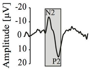
C.R. Jutzeler, L.D. Linde, J. Rosner et al. NeuroImage 225 (2021) 117473

Fig. 1. Representative traces of conventional averaging and single trial averaging analysis methods. A) Individual contact heat evoked potential (CHEP) waveforms,
filtered and re-referenced. B) Conventional averaging of individual CHEP waveforms, from which N2 and P2 outcome are derived. C) Individual CHEP waveforms
following single trial analysis, via wavelet filtering and multiple linear regression with dispersion term (Hu et al., 2011, 2010). D) Averaged CHEP outcomes
determined from single trial analysis.

to enhance the signal to noise ratio and facilitate the estimation of la- up via linear mixed models for each analysis method, to determine re-
tency and amplitude of single trial evoked potential peaks (Hu et al., spective differences in CHEP outcomes and the relationship to subject
2011, 2010; Kramer et al., 2013). To perform an unbiased single trial characteristics between conventional and single-trial averaging. Based
analysis, an automated approach using multiple linear regression with on preliminary linear mixed model analysis, multiple linear regressions
a dispersion term (MLRd) is then implemented (Hu et al., 2011). The between age, rating of perceived intensity, and CHEP outcomes were
dispersion term enhances the ability of multiple linear regressions to further explored for both analysis methods. Bonferroni correction was
detect changes in waveform morphology and provides a more accurate applied to adjust for multiple comparisons. An alpha level of 0.05 was
measure of latency and amplitude of single trial evoked potential peaks used for all statistical tests. R Statistical Software (version 3.5.3, MacOS
(Hu et al., 2011). Waveform peaks were automatically detected within 10.14.6 Mojave) was used for all statistical analyses and producing all
a 100-millisecond window of the wavelet-filtered average N2 and P2. plots (R Core Team, 2019; Wickham, 2016).
Individual trial N2 and P2 waveform parameters were then averaged
together, to provide the single trial analysis outcomes for each partici- 2.7. Simulated data analysis
pant (Fig. 1).
 To further explore the effects of conventional averaging and single
2.6. Statistical analysis trial analysis on CHEPs outcomes, we performed a small simulation
 experiment. The goal here was to systematically manipulate the
 CHEP outcomes from conventional averaging and single-trial aver- amplitudes and latencies of individual trial waveforms to determine
aging were examined using linear mixed effects models. For each CHEP the comparative effects on conventional averaging and single trial
outcome (N2P2 amplitude, N2 latency, P2 latency), fixed effects of der- analysis, respectively. A single waveform was artificially increased
matome (C4, C6, C8), and analysis method (conventional vs single trial in amplitude by 40%, and the latency was shifted 20 ms to the left.
averaging) were assessed in a linear mixed model, with random effects Thus, two waveforms (small and large) were used for our simulation.
of participants. Subject characteristics (age, sex, height, and rating of First, the small waveform was replicated, such that a 15-trial dataset
perceived intensity) were included in these linear mixed models, ex- contained only the small waveform. Then, a single large waveform
amining the overall effect of analysis method and dermatome on CHEP trial was added, with a small waveform removed. This was repeated
outcomes. Subsequent models with an interaction term between each until only large waveforms remained. For each dataset, we performed
subject characteristic and analysis method were included to explore of conventional (across-trial) averaging and single trial analysis, and
influence of analysis method on the relationships between CHEP out- N2P2 outcomes were compared for each dataset. Findings from the
comes and subject characteristics. Significant interactions were followed simulation and example traces of waveforms can be found in Fig. 7.
C.R. Jutzeler, L.D. Linde, J. Rosner et al. NeuroImage 225 (2021) 117473

 Fig. 2. Contact heat evoked potentials (CHEP) N2P2 am-
 plitudes (A), N2 latencies (B), and P2 latencies (C) from
 cervical spine dermatomes (C4, C6, C8). Conventional av-
 eraging (CV) and single trial averaging (STA) analysis
 methods compared within each dermatome / CHEP out-
 comes. Age groups separated into young (18–40 yrs), mid-
 dle (41–60 yrs), and elderly (61–80 yrs). Letters denote
 significant differences between dermatomes for both stim-
 ulation protocols, such that different letters correspond to
 significant differences between dermatomes. For panel B
 specifically, different letters denote significant differences
 between dermatomes and between analysis methods (i.e.
 a is significantly different from b, c, cd, and c; d is signifi-
 cantly different from c, but neither are significantly differ-
 ent from cd). Linear mixed models were adjusted for age,
 sex, and height, with a significance level of alpha
C.R. Jutzeler, L.D. Linde, J. Rosner et al. NeuroImage 225 (2021) 117473

 Table 2
 Contact heat evoked potential (CHEP) summary outcomes (mean ± standard deviation).

 Dermatome
 CHEP Outcome Analysis Method
 C4 C6 C8

 N2P2 Amplitude (uV) CA 27.0 ± 11.7 23.8 ± 7.4 23.2 ± 8.5
 STA 28.2 ± 12.2 25.2 ± 9.8 24.1 ± 11.4
 N2 Latency (ms) CA 361.4 ± 31.3 383.6 ± 31.1 402.3 ± 31.6
 STA 380.8 ± 37.9 414.6 ± 41.2 420.3 ± 53.8
 P2 Latency (ms) CA 504.4 ± 44.2 527.5 ± 61.2 530.0 ± 46.5
 STA 513.9 ± 43.8 536.8 ± 53.6 541.5 ± 62.4

 CA - Conventional averaging.
 STA – Single-trial averaging.

 Fig. 3. Linear regressions between aging and N2P2 amplitude for
 each dermatome (C4, C6, C8) and analysis method (conventional
 averaging and single trial averaging). Strength of linear regression
 (R2 ) is given for each dermatome, across analysis methods.

3.2. Main effects of analysis method p
C.R. Jutzeler, L.D. Linde, J. Rosner et al. NeuroImage 225 (2021) 117473

 Fig. 4. Linear regressions between aging and N2 latency for each
 dermatome (C4, C6, C8) and analysis method (conventional aver-
 aging and single trial averaging). Strength of linear regression (R2 )
 is given for each dermatome, across analysis methods.

latencies (F(1,85.6) = 10.32, p
C.R. Jutzeler, L.D. Linde, J. Rosner et al. NeuroImage 225 (2021) 117473

 Fig. 5. Linear regressions between aging and P2 latency for each
 dermatome (C4, C6, C8) and analysis method (conventional aver-
 aging and single trial averaging). Strength of linear regression (R2 )
 is given for each dermatome, across analysis methods.

between analysis methods revealed significantly longer N2 latencies large amplitude waveforms (Fig. 7B). N2P2 amplitude were most consis-
for single trial averaging compared to conventional averaging for C4 tent between analysis methods with similar number of small and large
(t = −8.4, p
C.R. Jutzeler, L.D. Linde, J. Rosner et al. NeuroImage 225 (2021) 117473

 Fig. 6. Linear regressions between pain rating and N2P2 ampli-
 tude for each dermatome (C4, C6, C8) and analysis method (con-
 ventional averaging and single trial averaging). Strength of linear
 regression (R2 ) is given for each dermatome, across analysis meth-
 ods.

vidual evoked potentials (Mouraux and Plaghki, 2004) and incorporated ences between C6 and C8, as reported previously (Haefeli et al., 2013b;
automated peak detection by multiple linear regression (Mayhew et al., Jutzeler et al., 2016), are an artefact of conventional averaging. Specif-
2006), before ultimately arriving at the method employed in the cur- ically, stimulation of the C8 dermatome at the base of the 5th finger
rent study (Hu et al., 2011, 2010). While demonstrating that single trial may yield more latency jitter due to a less evenly distributed activa-
analysis is clearly possible, decidedly missing, to this point, has been tion of cutaneous thermo-nociceptors, owing to the anatomical structure
evidence that single trial analysis improves the detection of biologically of the skin area and size of the heat stimulator. These challenges may
relevant aspects of nociception. The lack of this knowledge has likely, in also result in unintentional stimulation of glabrous skin, known to result
part, contributed to limited uptake among researchers, for whom single in longer CHEPs latencies (Hüllemann et al., 2019). The development
trial analysis comes at the cost of increased analysis time and greater of smaller, more effective contact heat stimulation devices (De Keyser
complexity compared to conventional averaging. et al., 2018) may improve the assessment of the C8 dermatome.
 Towards shifting the discussion from theory to practice, our results Second, single trial analysis improved the relationship between
demonstrate the inherent value of single trial analysis for nociceptive CHEPs, specifically N2P2 amplitude and P2 latency, and age. Numer-
evoked potentials. This was evidenced by three key observations. The ous studies have highlighted this relationship previously, generally
first is that single trial analysis nullified N2 latency differences be- confirming that nociceptive evoked potentials are smaller and longer
tween C6 and C8 stimulation sites, readily apparent with conventional with advanced age (Creac’H et al., 2015; Di Stefano et al., 2017;
averaging. We have reported significant differences between C6 and Granovsky et al., 2016; Lagerburg et al., 2015; Rosner et al., 2018;
C8 stimulation sites previously based on an analysis of the same data Truini et al., 2005). This is thought to primarily reflect a progressive
(Jutzeler et al., 2016) and also observed trends towards similar differ- loss of nociceptors in the periphery (Ceballos et al., 1999; Ochoa and
ences in other, independent datasets that incorporating conventional Mair, 1969; O’Sullivan and Swallow, 1968; Yezierski, 2012) and a re-
averaging (Haefeli et al., 2013b). Based on matching peripheral con- duction in conduction velocity of the spinothalamic tract (Kakigi and
duction distances and marginal differences centrally, C6 (i.e., base of Shibasaki, 1991), which are both paralleled by changes observed
the thumb) and C8 (i.e., base of the 4th finger) stimulation should, for other measures of pain (e.g., thresholds) (Chakour et al., 1996;
from a neurophysiological perspective, yield similar latencies. The res- Gagliese, 2009; Gibson and Farrell, 2004; Gibson and Helme, 2001).
olution of this discrepancy by single trial analysis suggests that differ- Across studies, however, the details of the relationship between age and
C.R. Jutzeler, L.D. Linde, J. Rosner et al. NeuroImage 225 (2021) 117473

Fig. 7. Simulation study findings: a small waveform and large waveform (40% larger amplitude, 20 ms earlier latency) were used to create 16 datasets. The ratio
of large to small trials is given in the x-axis of panels A, B, and C (e.g. 5:10 equate to 5 large waveforms and 10 small waveforms). Conventional averaging (CA)
and single trial analysis (STA) N2P2 outcomes from each dataset were compared for each dataset. A) N2 latencies across simulated datasets. B) P2 latencies across
simulated datasets. C) N2P2 amplitudes across simulated datasets. D) Individual trials from 5:10 simulated dataset, bold line is conventional (across-trial) average.
E) Individual trials from 10:5 simulated dataset, bold line is conventional (across-trial) average. F) Across-trial averages from all simulation datasets, bold lines are
the all small waveform and all large waveform datasets.

nociceptive evoked potentials are less consistent. For example, stud- by conventional averaging (Mayhew et al., 2006; Warbrick et al., 2009).
ies have reported an association for amplitude only (Frasson et al., More specifically, large amplitude, individual waveforms (i.e., outliers),
2020; Truini et al., 2005), while others observed location dependence which tend also to be shorter (Hu et al., 2011; Iannetti et al., 2005),
(Creac’H et al., 2015; Granovsky et al., 2016; Rosner et al., 2018). Our “pull” the grand average left, biasing interpretation of conventionally
observations suggest the optimal approach to detect genuine age related averaged latency. We demonstrated this phenomenon with simulated
changes in nociceptive evoked potentials, unrelated to slight afferent data. When a low number of large amplitude trials, with earlier latency,
desynchronization and increased latency jitter, is with single trial anal- were included in a dataset with predominately small trials, conventional
ysis. (across-trial) averaged N2 latencies progressively shifted left to a greater
 Finally, ratings were more strongly correlated with CHEPs from extent than single trial averaged latencies (Fig. 7A). This difference in
single trial analysis compared to conventional averaging. As a gen- N2 latency can be explained by the distortion or waveform morphology
eral rule of thumb, N2P2 amplitudes are larger and latencies shorter in conventional averaging, which puts greater weight on larger ampli-
when stimulations are, on average, more intense (Jutzeler et al., 2016; tude waveforms. In contrast, single trial averaging accounts for these
Kramer et al., 2013, 2012a; Linde et al., 2020), albeit within-subjects differences in waveform morphology in the determination individual
(Hu and Iannetti, 2019). While exceptions are commonplace and have trial N2 latencies, which are subsequently averaged. We also provided
a neuroanatomical basis (Kramer et al., 2016), the relationship between an example of this difference between analysis methods as individual
CHEPs and ratings reflects a number of important processes, including trials are added stepwise (Fig. 8), again demonstrated the effect of large
attention, arousal, saliency, and stimulus novelty (Iannetti et al., 2008; amplitude trials influencing across trial averaging. In addition, the non-
Le Pera et al., 2002; Madsen et al., 2014; Ronga et al., 2013). Such linear increase in N2P2 with conventional averaging provided further
endogenous contributions of evoked potentials are thought to be more evidence of distortions of waveform morphology, which have previously
susceptible to latency jitter (Kutas et al., 1977; Legrain et al., 2002; reported (Mayhew et al., 2006; Warbrick et al., 2009).
Siedenberg and Treede, 1996). To this end, methods that better cap- C6 CHEPs appear to have been more affected by single trial averag-
ture a relationship with pain, as was the case for single trial analy- ing compared to C8 (i.e., larger increase in C6 N2 latency), which may
sis, are highly desirable. The inherent value of single trial analysis is relate to physical differences in testing sites. On the dorsum of the hand,
the more accurate portrayal of nociceptive evoked potentials. Similar where stimulation yields more robust CHEPs compared to stimulation of
to previous studies, we observed significantly larger N2P2 amplitudes the palmer surface (Haefeli et al., 2013b), the C8 site is smaller than C6.
(Hu et al., 2011) and longer N2 latencies (Warbrick et al., 2009) follow- The size of the stimulation site is a major issue for contact heat because
ing single trial analysis compared to conventional averaging. Increased the thermode is comparatively large and needs to be subtly shifted after
amplitudes are attributable to phase cancelation resulting from trial to each stimulation whilst remaining in the target dermatome. This is im-
trial waveform variability (i.e., latency jitter), which leads to “flatten- portant for clinical applications, which aim to assess segmental patholo-
ing” of the grand average waveform that serves the basis to interpret gies (Haefeli et al., 2013a; Kramer et al., 2012a). In C6, shifting the ther-
conventionally averaged CHEPs (Hu et al., 2011). Increased N2 laten- mode is more likely to activate novel receptors, in turn producing more
cies are attributed to distortions in waveform morphology introduced “large” amplitude responses in C6 (i.e., outliers), which ultimately leads
C.R. Jutzeler, L.D. Linde, J. Rosner et al. NeuroImage 225 (2021) 117473

Fig. 8. Conventional (arithmetic) average and single trial averaged N2 latencies compared during stepwise addition of individual trials. N2 latency denoted in red.
It can be observed that with subsequent trials, conventional averaging is pulled further left, due to differences in trial amplitude.

to a larger discrepancy between single trial and conventionally averaged ally averaged CHEPs were not wavelet filtered. As such, our findings
CHEPs. are limited to a comparison of two overall methods of data analysis. An-
 other important point is that our findings are limited to a comparison of
 two separate methods of CHEPs analysis, with no objective ‘gold stan-
4.1. Limitations
 dard’. While we provide evidence and support for the use of single trial
 analysis, there remains no true ‘gold standard’ approach for CHEPs anal-
 A major strength of our analysis is our sample size, which is large in
 ysis. Our findings are also limited to healthy subjects. Further research
comparison to previous studies applying single trial analysis (Hu et al.,
 is needed to determine if single trial analysis improves understanding
2010; Kramer et al., 2016, 2013; Mayhew et al., 2006; Warbrick et al.,
 of pathology in patient populations.
2009). Moreover, both men and women of varying ages were included.
Nevertheless, there are limitations. Single trial analysis involves a series
of signal processing steps, which ultimately improve signal clarity of the 5. Conclusion
N2P2 waveform before extracting key amplitude and latency outcomes.
Given there are a series of steps involved, these may, in part, also en- CHEPs provide a method to reliably and safely assess small diam-
hance the signal clarity of N2P2 waveforms. For example, convention- eter nociceptive afferents of the spinothalamic pathway that are typ-
C.R. Jutzeler, L.D. Linde, J. Rosner et al. NeuroImage 225 (2021) 117473

ically involved in peripheral sensitization and chronic pain develop- De Keyser, R., van den Broeke, E.N., Courtin, A., Dufour, A., Mouraux, A., 2018. Event-
ment. When optimal stimulation and data processing parameters are related brain potentials elicited by high-speed cooling of the skin: a robust and non-
 painful method to assess the spinothalamic system in humans. Clin. Neurophysiol. Off.
employed, CHEP outcomes demonstrate clear, robust age- and location- J. Int. Fed. Clin. Neurophysiol. 129, 1011–1019. doi:10.1016/j.clinph.2018.02.123.
dependent changes. Our reported improved associations to aging and de Tommaso, M., Ricci, K., Montemurno, A., Vecchio, E., 2017. Age-related changes in
rating of perceived intensity when using single trial averaging suggest laser-evoked potentials following trigeminal and hand stimulation in healthy subjects.
 Eur J Pain 21, 1087–1097. doi:10.1002/ejp.1010.
a better representation of the underlying physiology of the nociceptive Di Stefano, G., La Cesa, S., Leone, C., Pepe, A., Galosi, E., Fiorelli, M., Valeriani, M.,
system compared to traditional across-trial averaging. While conven- Lacerenza, M., Pergolini, M., Biasiotta, A., Cruccu, G., Truini, A., 2017. Diagnostic
tional (across-trial) averaging offers convenience and ease of use, it may accuracy of laser-evoked potentials in diabetic neuropathy. Pain 158, 1100–1107.
 doi:10.1097/j.pain.0000000000000889.
be more susceptible to waveform distortion and latency shifts when am-
 Frasson, E., Tozzi, M.C., Bordignon, M., Motti, L., Ferrari, F., Torre, G., Graziottin, A.,
plitude variation is present among individual trials are included in anal- Monaco, S., Bertolasi, L., 2020. Laser-evoked potentials to pudendal stimulation
ysis. We recommend the use of single-trial averaging, with freely avail- in healthy subjects: a pilot study. J. Clin. Neurophysiol. Off. Publ. Am. Electroen-
 cephalogr. Soc doi:10.1097/WNP.0000000000000694.
able software (Hu et al., 2011, 2010), to assess the nociceptive system
 Gagliese, L., 2009. Pain and aging: the emergence of a new subfield of pain research. J.
using CHEPs in both clinical and research settings. Pain Off. J. Am. Pain Soc. 10, 343–353. doi:10.1016/j.jpain.2008.10.013.
 Gibson, S.J., Farrell, M., 2004. A review of age differences in the neurophysiology
 of nociception and the perceptual experience of pain. Clin. J. Pain 20, 227–239.
CRediT authorship contribution statement
 doi:10.1097/00002508-200407000-00004.
 Gibson, S.J., Helme, R.D., 2001. Age-related differences in pain perception and report.
 Catherine R. Jutzeler: Conceptualization, Methodology, Investiga- Clin. Geriatr. Med. 17, 433–456 v–vi.
tion, Data curation, Writing - review & editing, Funding acquisition, Granovsky, Y., Anand, P., Nakae, A., Nascimento, O., Smith, B., Sprecher, E., Valls-Solé, J.,
 2016. Normative data for A contact heat evoked potentials in adult population: a
Validation, Visualization. Lukas D. Linde: Conceptualization, Method- multicenter study. Pain 157. doi:10.1097/j.pain.0000000000000495.
ology, Formal analysis, Writing - original draft, Writing - review & edit- Granovsky, Y., Matre, D., Sokolik, A., Lorenz, J., Casey, K.L., 2005. Thermoreceptive in-
ing, Software, Visualization. Jan Rosner: Investigation, Writing - re- nervation of human glabrous and hairy skin: a contact heat evoked potential analysis.
 Pain 115, 238–247. doi:10.1016/j.pain.2005.02.017.
view & editing. Michèle Hubli: Investigation, Writing - review & edit- Haefeli, J., Kramer, J.L.K., Blum, J., Curt, A., 2013a. Assessment of spinothalamic tract
ing. Armin Curt: Supervision, Writing - review & editing. John L.K. function beyond pinprick in spinal cord lesions: a contact heat evoked potential study.
Kramer: Conceptualization, Writing - review & editing, Supervision, Re- Neurorehabil. Neural Repair 28, 494–503. doi:10.1177/1545968313517755.
 Haefeli, J.S., Blum, J., Steeves, J.D., Kramer, J.L.K., Curt, A.E.P., 2013b. Differences in
sources, Project administration. spinothalamic function of cervical and thoracic dermatomes: insights using contact
 heat evoked potentials. J. Clin. Neurophysiol. Off. Publ. Am. Electroencephalogr. Soc.
Acknowledgements 30, 291–298. doi:10.1097/WNP.0b013e31827ed9ee.
 Hatem, S.M., Hu, L., Ragé, M., Gierasimowicz, A., Plaghki, L., Bouhassira, D., Attal, N.,
 Iannetti, G.D., Mouraux, A., 2012. Automated single-trial assessment of laser-evoked
 The authors would like to thank Janosh Rinert for the support in data potentials as an objective functional diagnostic tool for the nociceptive system. Clin.
collection. CRJ is supported by the Swiss National Science Foundation Neurophysiol. 123, 2437–2445. doi:10.1016/j.clinph.2012.05.007.
 Hu, L., Iannetti, G.D., 2019. Neural indicators of perceptual variability of pain across
(Ambizione Grant, PZ00P3_18610). This work was further supported by species. Proc. Natl. Acad. Sci. 116, 1782–1791. doi:10.1073/pnas.1812499116.
the Swiss Spinal Cord Injury Cohort Study Nested Project Grant (J.R. and Hu, L., Liang, M., Mouraux, A., Wise, R.G., Hu, Y., Iannetti, G.D., 2011. Taking into ac-
C.R.J., 2016-N-005). JR is supported by the Clinical Research Priority count latency, amplitude, and morphology: improved estimation of single-trial ERPs
 by wavelet filtering and multiple linear regression. J. Neurophysiol. 106, 3216–3229.
Program of the University of Zurich (CRPP Pain) and through funding
 doi:10.1152/jn.00220.2011.
from the Hartmann Mueller Foundation (grant number 1997). The fun- Hu, L., Mouraux, A., Hu, Y., Iannetti, G.D., 2010. A novel approach for enhancing the
ders had no role in study design, data collection and analysis, decision signal-to-noise ratio and detecting automatically event-related potentials (ERPs) in
 single trials. Neuroimage 50, 99–111. doi:10.1016/j.neuroimage.2009.12.010.
to publish, or preparation of the manuscript.
 Huang, G., Xiao, P., Hung, Y.S., Iannetti, G.D., Zhang, Z.G., Hu, L., 2013. A novel ap-
 proach to predict subjective pain perception from single-trial laser-evoked potentials.
Data and code availability statement Neuroimage 81, 283–293. doi:10.1016/j.neuroimage.2013.05.017.
 Hüllemann, P., Nerdal, A., Sendel, M., Dodurgali, D., Forstenpointner, J., Binder, A.,
 Baron, R., 2019. Cold-evoked potentials versus contact heat-evoked poten-
 Fully anonymized data will be shared at the request from any quali- tials—Methodological considerations and clinical application. Eur. J. Pain 23, 1209–
fied investigator (please contact the Corresponding Author). The code to 1220. doi:10.1002/ejp.1389.
run the analysis as well as create the figures can be found on our Github Iannetti, G.D., Hughes, N.P., Lee, M.C., Mouraux, A., 2008. Determinants of laser-evoked
 EEG responses: pain perception or stimulus saliency? J. Neurophysiol. 100, 815–828.
repository (https://github.com/jutzca/CHEPs_STA). doi:10.1152/jn.00097.2008.
 Iannetti, G.D., Zambreanu, L., Cruccu, G., Tracey, I., 2005. Operculoinsular cortex
References encodes pain intensity at the earliest stages of cortical processing as indicated
 by amplitude of laser-evoked potentials in humans. Neuroscience 131, 199–208.
Atherton, D.D., Facer, P., Roberts, K.M., Misra, V.P., Chizh, B.A., Bountra, C., Anand, P., doi:10.1016/j.neuroscience.2004.10.035.
 2007. Use of the novel Contact Heat Evoked Potential Stimulator (CHEPS) for the Iannetti, G.D., Zambreanu, L., Tracey, I., 2006. Similar nociceptive afferents mediate psy-
 assessment of small fibre neuropathy: correlations with skin flare responses and intra- chophysical and electrophysiological responses to heat stimulation of glabrous and
 epidermal nerve fibre counts. BMC Neurol. 7, 21. doi:10.1186/1471-2377-7-21. hairy skin in humans. J. Physiol. 577, 235–248. doi:10.1113/jphysiol.2006.115675.
Bufacchi, R.J., Iannetti, G.D., 2018. An action field theory of peripersonal space. Trends Jacobs, J.M., Love, S., 1985. Qualitative and quantitative morphology of human sural
 Cogn. Sci. 22, 1076–1090. doi:10.1016/j.tics.2018.09.004. nerve at different ages. Brain 108, 897–924. doi:10.1093/brain/108.4.897.
Bufacchi, R.J., Liang, M., Griffin, L.D., Iannetti, G.D., 2016. A geometric model of defensive Jutzeler, C.R., Curt, A., Kramer, J.L.K., 2015. Effectiveness of high-frequency
 peripersonal space. J Neurophysiol 115, 218–225. doi:10.1152/jn.00691.2015. electrical stimulation following sensitization with capsaicin. J. Pain 16.
Ceballos, D., Cuadras, J., Verdú, E., Navarro, X., 1999. Morphometric and ultrastruc- doi:10.1016/j.jpain.2015.03.005.
 tural changes with ageing in mouse peripheral nerve. J. Anat. 195 (Pt 4), 563–576. Jutzeler, C.R., Rosner, J., Rinert, J., Kramer, J.L.K., Curt, A., 2016. Normative data for the
 doi:10.1046/j.1469-7580.1999.19540563.x. segmental acquisition of contact heat evoked potentials in cervical dermatomes. Sci.
Chakour, M.C., Gibson, S.J., Bradbeer, M., Helme, R.D., 1996. The effect Rep. 6, 34660. doi:10.1038/srep34660.
 of age on A - and C-fibre thermal pain perception. Pain 64, 143–152. Kakigi, R., Shibasaki, H., 1991. Estimation of conduction velocity of the spino-
 doi:10.1016/0304-3959(95)00102-6. thalamic tract in man. Electroencephalogr. Clin. Neurophysiol. 80, 39–45.
Chao, C.C., Hsieh, S.T., Chiu, M.J., Tseng, M.T., Chang, Y.C., 2007. Effects of aging on doi:10.1016/0168-5597(91)90041-u.
 contact heat-evoked potentials: the physiological assessment of thermal perception. Kramer, J.L.K., Curt, A., Steeves, J.D., 2012a. Increased baseline temperature improves
 Muscle Nerve 36, 30–38. doi:10.1002/mus.20815. the acquisition of contact heat evoked potentials after spinal cord injury. Clin. Neu-
Chen, A.C.N., Niddam, D.M., Arendt-Nielsen, L., 2001. Contact heat evoked potentials as rophysiol. 123, 582–589. doi:10.1016/j.clinph.2011.08.013.
 a valid means to study nociceptive pathways in human subjects. Neurosci. Lett. 316, Kramer, J.L.K., Haefeli, J., Jutzeler, C.R., Steeves, J.D., Curt, A., 2013. Improving the
 79–82. doi:10.1016/S0304-3940(01)02374-6. acquisition of nociceptive evoked potentials without causing more pain. Pain 154.
Chen, I.-.A., Hung, S.W., Chen, Y.-.H., Lim, S.-.N., Tsai, Y.-.T., Hsiao, C.-.L., Hsieh, H.-.Y., doi:10.1016/j.pain.2012.10.027.
 Wu, T., 2006. Contact heat evoked potentials in normal subjects. Acta Neurol. Tai- Kramer, J.L.K., Jutzeler, C.R., Haefeli, J., Curt, A., Freund, P., 2016. Discrep-
 wanica 15, 184–191. ancy between perceived pain and cortical processing: a voxel-based morphome-
Creac’H, C., Bertholon, A., Convers, P., Garcia-Larrea, L., Peyron, R., 2015. Effects of aging try and contact heat evoked potential study. Clin. Neurophysiol. 127, 762–768.
 on laser evoked potentials. Muscle Nerve 51, 736–742. doi:10.1002/mus.24458. doi:10.1016/j.clinph.2015.02.054.
C.R. Jutzeler, L.D. Linde, J. Rosner et al. NeuroImage 225 (2021) 117473

Kramer, J.L.K., Taylor, P., Haefeli, J., Blum, J., Zariffa, J., Curt, A., Steeves, J., 2012b. Ochoa, J., Mair, W.G., 1969. The normal sural nerve in man. II. Changes in the ax-
 Test–retest reliability of contact heat-evoked potentials from cervical dermatomes. J. ons and Schwann cells due to ageing. Acta Neuropathol. (Berl.) 13, 217–239.
 Clin. Neurophysiol. 29, 70–75. doi:10.1097/WNP.0b013e318246ada2. doi:10.1007/BF00690643.
Kramer, J.L.K., Taylor, P., Steeves, J., Curt, A., 2009. Assessment of spinothalamic function Perretti, A., Nolano, M., De Joanna, G., Tugnoli, V., Iannetti, G., Provitera, V.,
 in SCI: reliability of contact heat-evoked potentials of cervical dermatomes. J. Spinal Cruccu, G., Santoro, L., 2003. Is Ross syndrome a dysautonomic disorder
 Cord Med. 32 (4), 469. only? An electrophysiologic and histologic study. Clin. Neurophysiol 114, 7–16.
Kutas, M., McCarthy, G., Donchin, E., 1977. Augmenting mental chronometry: the P300 doi:10.1016/S1388-2457(02)00323-1.
 as a measure of stimulus evaluation time. Science 197, 792–795. doi:10.1126/sci- Purves, A.M., Boyd, S.G., 1993. Time-shifted averaging for laser evoked
 ence.887923. potentials. Electroencephalogr. Clin. Neurophysiol. 88, 118–122.
Lagerburg, V., Bakkers, M., Bouwhuis, A., Hoeijmakers, J.G.J., Smit, A.M., Van Den doi:10.1016/0168-5597(93)90062-t.
 Berg, S.J.M., Hordijk-De Boer, I., Brouwer-Van der Lee, M.D.G., Kranendonk, D., R Core Team, 2019. R: A Language and Environment for Statistical Computing. R Foun-
 Reulen, J.P.H., Faber, C.G., Merkies, I.S.J., 2015. Contact heat evoked potentials: dation for Statistical Computing, Vienna, Austria.
 normal values and use in small-fiber neuropathy. Muscle Nerve 51, 743–749. Ragé, M., Van Acker, N., Knaapen, M.W.M., Timmers, M., Streffer, J., Hermans, M.P.,
 doi:10.1002/mus.24465. Sindic, C., Meert, T., Plaghki, L., 2011. Asymptomatic small fiber neuropathy in
Lauria, G., Holland, N., Hauer, P., Cornblath, D.R., Griffin, J.W., McArthur, J.C., 1999. diabetes mellitus: investigations with intraepidermal nerve fiber density, quan-
 Epidermal innervation: changes with aging, topographic location, and in sensory neu- titative sensory testing and laser-evoked potentials. J. Neurol. 258, 1852–1864.
 ropathy. J. Neurol. Sci. 164, 172–178. doi:10.1016/S0022-510X(99)00063-5. doi:10.1007/s00415-011-6031-z.
Le Pera, D., Valeriani, M., Niddam, D., Chen, A.C.N., Arendt-Nielsen, L., 2002. Contact Ronga, I., Valentini, E., Mouraux, A., Iannetti, G.D., 2013. Novelty is not enough: laser-
 heat evoked potentials to painful and non-painful stimuli: effect of attention towards evoked potentials are determined by stimulus saliency, not absolute novelty. J. Neu-
 stimulus properties. Brain Topogr 15, 115–123. doi:10.1023/a:1021472524739. rophysiol. 109, 692–701. doi:10.1152/jn.00464.2012.
Legrain, V., Guérit, J.-.M., Bruyer, R., Plaghki, L., 2002. Attentional modulation of the Rosner, J., Hubli, M., Hostettler, P., Scheuren, P.S., Rinert, J., Kramer, J.L.K.,
 nociceptive processing into the human brain: selective spatial attention, probability Hupp, M., Curt, A., Jutzeler, C.R., 2018. Contact heat evoked potentials: re-
 of stimulus occurrence, and target detection effects on laser evoked potentials. Pain liable acquisition from lower extremities. Clin Neurophysiol 129, 584–591.
 99, 21–39. doi:10.1016/S0304-3959(02)00051-9. doi:10.1016/j.clinph.2017.12.034.
Linde, L.D., Haefeli, J., Jutzeler, C.R., Rosner, J., McDougall, J., Curt, A., Kramer, J.L.K., Sambo, C.F., Forster, B., Williams, S.C., Iannetti, G.D., 2012. To blink or not to blink: fine
 2020. Contact heat evoked potentials are responsive to peripheral sensitization: cognitive tuning of the defensive peripersonal space. J. Neurosci. 32, 12921–12927.
 requisite stimulation parameters. Front. Hum. Neurosci. 13, 459. doi:10.3389/fn- doi:10.1523/JNEUROSCI.0607-12.2012.
 hum.2019.00459. Sambo, C.F., Iannetti, G.D., 2013. Better safe than sorry? The safety margin surrounding
Madsen, C.S., Finnerup, N.B., Baumgartner, U., 2014. Assessment of small fibers using the body is increased by anxiety. J. Neurosci 33, 14225–14230. doi:10.1523/JNEU-
 evoked potentials. Scand J Pain 5, 111–118. doi:10.1016/j.sjpain.2013.11.007. ROSCI.0706-13.2013.
Magerl, W., Treede, R.D., 1996. Heat-evoked vasodilatation in human hairy skin: axon Siedenberg, R., Treede, R.-D., 1996. Laser-evoked potentials: exogenous and endogenous
 reflexes due to low-level activity of nociceptive afferents. J. Physiol 497, 837–848 components. Electroencephalogr. Clin. Neurophysiol. Potentials Sect. 100, 240–249.
 Pt 3. doi:10.1016/0168-5597(95)00255-3.
Mayhew, S.D., Iannetti, G.D., Woolrich, M.W., Wise, R.G., 2006. Automated Staikou, C., Kokotis, P., Kyrozis, A., Rallis, D., Makrydakis, G., Manoli, D., Karandreas, N.,
 single-trial measurement of amplitude and latency of laser-evoked potentials Stamboulis, E., Moschovos, C., Fassoulaki, A., 2016. Differences in pain perception
 (LEPs) using multiple linear regression. Clin. Neurophysiol. 117, 1331–1344. between men and women of reproductive age: a laser-evoked potentials study. Pain
 doi:10.1016/j.clinph.2006.02.017. Med. pnw 167. doi:10.1093/pm/pnw167.
Mouraux, A., Iannetti, G.D., 2008. Across-trial averaging of event-related EEG responses Truini, A., Galeotti, F., Romaniello, A., Virtuoso, M., Iannetti, G.D., Cruccu, G.,
 and beyond. Magn. Reson. Imaging 26, 1041–1054. doi:10.1016/j.mri.2008.01.011. 2005. Laser-evoked potentials: normative values. Clin. Neurophysiol. 116, 821–826.
Mouraux, A., Iannetti, G.D., 2009. Nociceptive laser-evoked brain potentials do doi:10.1016/j.clinph.2004.10.004.
 not reflect nociceptive-specific neural activity. J. Neurophysiol 101, 3258–3269. Warbrick, T., Derbyshire, S.W.G., Bagshaw, A.P., 2009. Optimizing the measure-
 doi:10.1152/jn.91181.2008. ment of contact heat evoked potentials. J. Clin. Neurophysiol. 26, 117–122.
Mouraux, A., Iannetti, G.D., 2018. The search for pain biomarkers in the human brain. doi:10.1097/WNP.0b013e31819d8016.
 Brain J. Neurol. 141, 3290–3307. doi:10.1093/brain/awy281. Wickham, H., 2016. ggplot2: Elegant Graphics For Data Analysis. Springer-Verlag, New
Mouraux, A., Plaghki, L., 2004. Single-trial detection of human brain responses evoked York.
 by laser activation of Adelta-nociceptors using the wavelet transform of EEG epochs. Yezierski, R.P., 2012. The effects of age on pain sensitivity: preclinical studies. Pain Med.
 Neurosci. Lett. 361, 241–244. doi:10.1016/j.neulet.2003.12.110. 13, S27–S36. doi:10.1111/j.1526-4637.2011.01311.x.
O’Sullivan, D.J., Swallow, M., 1968. The fibre size and content of the radial and sural
 nerves. J. Neurol. Neurosurg. Psychiatry 31, 464–470. doi:10.1136/jnnp.31.5.464.
You can also read