Comparative Analysis of Kidney Stone Composition in Patients from Ghana and South Africa: Case Study of Kidney Stones from Accra and Cape Town
←
→
Page content transcription
If your browser does not render page correctly, please read the page content below
Open Journal of Urology, 2021, 11, 53-72
                                                                                             https://www.scirp.org/journal/oju
                                                                                                        ISSN Online: 2160-5629
                                                                                                          ISSN Print: 2160-5440

Comparative Analysis of Kidney Stone
Composition in Patients from Ghana and
South Africa: Case Study of Kidney Stones
from Accra and Cape Town

Evans A. Akpakli1, Lisa Kaestner2, John Lazarus2, Kwame S. Norvixoxo3
1
  Department of Surgery, Korle-Bu Teaching Hospital, Accra, Ghana
2
  School of Medicine, University of Cape Town, Cape Town, South Africa
3
  Faculty of Global Challenges, African Leadership University, Kigali, Rwanda

How to cite this paper: Akpakli, E.A.,        Abstract
Kaestner, L., Lazarus, J. and Norvixoxo,
K.S. (2021) Comparative Analysis of Kid-      Background: Kidney stone disease, also termed nephrolithiasis is associated
ney Stone Composition in Patients from        with significant morbidities such as severe colicky flank pain, haematuria,
Ghana and South Africa: Case Study of
                                              urinary tract infection and kidney failure. Kidney stone disease was perceived
Kidney Stones from Accra and Cape Town.
Open Journal of Urology, 11, 53-72.           as uncommon in developing countries; however, the global prevalence has
https://doi.org/10.4236/oju.2021.113007       been rising over the past two decades due to lifestyle changes. There is very
                                              limited literature on kidney stone composition in Africa, including Ghana
Received: January 20, 2021
                                              and South Africa. It was based on this evidence that this study was underta-
Accepted: March 9, 2021
Published: March 12, 2021                     ken. Aim: The primary aim of this study was to describe and compare the
                                              composition of kidney stone in patients receiving treatment at the Korle-Bu
Copyright © 2021 by author(s) and             Teaching Hospital (KBTH), Accra (Ghana) and Groote Schuur Hospital
Scientific Research Publishing Inc.
This work is licensed under the Creative
                                              (GSH), Cape Town (South Africa). Methods: The study was a retrospective
Commons Attribution International             folder review of patients treated for nephrolithiasis at the Korle-Bu Teaching
License (CC BY 4.0).                          Hospital in Accra (Ghana) and Groote Schuur Hospital in Cape Town (South
http://creativecommons.org/licenses/by/4.0/
                                              Africa). Patients who were treated for kidney stone disease between 1st June
               Open Access
                                              2016 and 31st May 2018 were recruited, and their folder numbers were re-
                                              trieved from theatre logbooks. A total of hundred and sixty-three (n = 163)
                                              folders (n = 30 KBTH; n = 133 GSH) were subsequently retrieved from the
                                              two facilities’ records department. Demographic data and kidney stone analy-
                                              sis results were analyzed using the R statistical software. Results: The age of
                                              KBTH patients ranged from 24 to 75 years and age of 45 years, while that of
                                              GSH ranged 19 to 77 years and median age 48 years respectively. Males were
                                              the majority stone formers for both hospitals [56.7% KBTH; 59.4% GSH].
                                              There was no statistical difference in gender (p = 0.9447) and age (p = 0.2612)

DOI: 10.4236/oju.2021.113007        Mar. 12, 2021               53                                     Open Journal of Urology
E. A. Akpakli et al.

                                 between the two groups. Calcium oxalate (86.7%) and uric acid (90.0%) were
                                 the commonest components of the kidney stones analyzed from the KBTH.
                                 Calcium oxalate (66.2%) and carbonate apatite (40.6%) were the most com-
                                 mon components stones from GSH. Brushite (3.0%), cystine (3.8%) and stru-
                                 vite (19.6%) stones were only found in GSH patients. All kidney stones from
                                 the KBTH were mixed stones. Pure kidney stones were only found among the
                                 GSH dataset constituting 48.9%, also female patients from GSH formed more
                                 mixed stones than their male counterparts (M:F = 40.5%:66.67%) and infec-
                                 tion kidney stones were also predominantly found among female patients.
                                 Conclusion: The findings indicate that the two facilities’ participants are not
                                 different in terms of gender and age. However, the composition of stones was
                                 found to be different between participants from both hospitals. This suggests
                                 that kidney stone composition may be influenced by patients’ geographical
                                 location and or cultural background.

                                 Keywords
                                 Kidney Stones, Comparative Analysis, Korle-Bu, Groote Schuur

                               1. Introduction
                               Stone disease of the urinary tract, also termed urolithiasis, is a debilitating,
                               chronic condition, which has affected people (perhaps) since antiquity. It results
                               in great morbidity like severe colicky flank pain; infection and loss of kidney
                               function may occur when the kidney is obstructed by a stone. It may result in
                               loss of working hours and productivity due to repeated patient visits to the
                               emergency unit or urologist, especially during those acute episodes requiring
                               admission and intervention [1].
                                  Kidney stones have been shown to be associated with other chronic diseases
                               like coronary artery disease, hypertension and chronic kidney disease [2]-[7].
                               This had led to other investigators referring to kidney stone disease as a meta-
                               bolic disorder beyond the obstructive symptoms caused by these stones.
                                  Kidney stone analysis is essential in assessing stone patients, not only in de-
                               termining stone composition, but also in providing a guide as to metabolic
                               anomalies that might be involved in stone formation [8] [9]. The knowledge of
                               stone composition is very important in further investigating and treating pa-
                               tients with kidney stone disease due to the high recurrence of this condition
                               [10].
                                  Kidney stone disease was perceived as uncommon in developing countries
                               until recently [11], but they are now a health concern in many developing na-
                               tions as it is in developed countries. Presently, there is insufficient data in the
                               current literature on the incidence and prevalence of kidney stone disease in
                               Africa and other developing countries [12]. The prevalence of kidney stones has
                               been rising globally over the past two decades due to dietary and lifestyle

DOI: 10.4236/oju.2021.113007                        54                                     Open Journal of Urology
E. A. Akpakli et al.
changes [11] [13] [14]. Although studies have reported a low incidence of kidney
stone disease in people of African descent compared with, for example, Cauca-
sians, there is also the paucity of information regarding kidney stones and stone
composition among Africans [15] [16].
Evidently, stone disease incidence varies with race, ethnicity, occupation, geo-
graphic location, climate and diet [1]. These factors and variations in cultural
practices and diet also affect the chemical composition of kidney stones. For
example, people from South-East Asia consume betel leaves, nuts and calcium
hydroxide paste which have been associated with hypercalciuria and hypocitra-
turia. Individuals from Northern India lack intestinal Oxalobacter formigenes;
hence they are unable to metabolize dietary oxalate which causes absorptive
hyperoxaluria [1] [17].
Anecdotal evidence shows that physicians manage numerous cases of upper
urinary tract stones in clinics across Africa. However, at present, no published
study has compared the stone composition between two different African coun-
tries. On premise, this study was conducted to determine the composition of
kidney stones in our sample (of patients from Korle-Bu Teaching Hospital and
Groote Schuur Hospital) and compare the results of these two countries. The
populace in Ghana is mainly of African descent, while South Africa includes
people of African, European, Asian ancestry and Coloured people. This mul-
ti-race scenario in South Africa also presents an opportunity to understand stone
composition beyond just a single race and if possible, identify the significance of
stone composition and its impact on the nature of kidney stone disease in Africa.
2. Methods
This retrospective study involved patients’ folder review from 1st June 2016 to
31st May 2018 at the Korle-Bu Teaching Hospital (Accra-Ghana) and Groote
Schuur Hospital (Cape Town, South Africa). The study is purely a descriptive
study of kidney stone composition of patients who were treated for kidney stone
disease in the two hospitals over the study period. Patients with kidney stone
disease who were treated at the Urology units of Korle-Bu Teaching hospital
(KBTH) and Groote Schuur Hospital (GSH) over the study period and had their
stones analyzed were included in this study. The study recruited consecutive pa-
tients who were treated for stone disease in the two hospitals.
Ethical approval had been sought and approved from the Surgical Divisional
research committee of Groote Schuur and the Human Research Ethics Commit-
tee (HREC) of both institutions. Patients who had radiological images (X-ray
and CT scan) confirmation of kidney stones and were surgically treated, either
by minimally invasive (URS, PCNL, Laparoscopy) or open surgery were used for
this study. Theatre logbooks between 1st June 2016 and 31st May 2018 were re-
viewed for folder numbers of patients who had stone surgeries. These were used
to source names from HREC approved urology database of the two hospitals and
variable data obtained. Stones collected from patients during surgical procedures
DOI: 10.4236/oju.2021.113007 55 Open Journal of UrologyE. A. Akpakli et al.
were labelled and sent to the laboratory for analysis. Two laboratories (i.e. MDS
Lancet and Pathcare laboratories) were involved in the analysis of stones for this
study. MDS lancet was involved in the analysis of stones from KBTH and Path-
care from GSH. Both laboratories used Fourier transform infrared spectrometer
(FT-IR) to analyze the stones. MDS lancet used Thermo Scientific, Nicolet iS10
FT-IR spectrometer and Pathcare used Agilent Technologies, Cary 630 FT-IR
spectrometer for stone analysis.
There was not a sufficient study population from which a statistics formu-
la-based sample size calculation could be drawn. Thus, we did not calculate a
sample size but used the available folder/data of consecutive patients who were
treated for kidney stone disease and had their stones analyzed. A total of thirty
(n = 30) and one hundred and thirty-three (n = 133) patient folders were ob-
tained from KBTH and GSH respectively. Data on date of surgery and stone
analysis, demography (country of origin, age and gender) and kidney stone
composition after analysis were collected using a data sheet. The data was coded
and captured into Excel spread sheets and then exported into the statistical
package R where it was cleaned and analyzed [18].
Descriptive statistics such as mean, standard deviation, frequencies and per-
centages were used to summarize the data (e.g. age, gender and country of ori-
gin). A student t-test was used to compare differences on age variables between
the South African and Ghanaian samples and a p-value of less than 0.05 is con-
sidered statistically significant. In cases where the variable count is more than
5%, Chi-Square goodness-of-fit tests of proportions were used to assess for sig-
nificance (gender variable between KBTH and GSH). The demographic vari-
ables, kidney stone type and kidney stone chemical composition as function of
demographic indicator was analysed and compared between the Korle-Bu
Teaching Hospital and Groote Schuur Hospital.
3. Results
A total of hundred and sixty-three (n = 163) participants (n = 30 KBTH; n = 133
GSH) were recruited for this study. The age of participants at the KBTH ranged
from 24 to 75 years with a median age of 45 years, while the ages of participants
at the GSH ranged between 19 to 77 years with a median age of 48 years. Males
were the majority stone formers for both hospitals [56.7% KBTH; 59.4% GSH]
with ratio of M:F = 1.3:1 (KBTH) and M:F = 1.5:1 (GSH). However, there was no
significant statistical difference in gender (p = 0.9447) and age (p = 0.2612) be-
tween the two groups as shown in Table 1.
Most of the kidney stones from the dataset were composed of multiple
chemical components.
Calcium apatite was only prevalent among stones of KBTH patients but was
not observed in any of GSH patients. Contrarily, Struvite was only recorded
among patients treated in GSH while none of KBTH stones contained struvite.
With regards to chemical composition of stones, most of the stones from
DOI: 10.4236/oju.2021.113007 56 Open Journal of UrologyE. A. Akpakli et al.
Table 1. Demographic characteristics of kidney stone patients treated at KBTH (Accra,
Ghana) and GSH (Cape Town, South Africa).
Age
Gender
Quantiles Std.
Count Min. Max. Mean
Female Male 25% 50% 75% dev.
Korle-Bu
13 17
Counts 30 24.00 34.00 45.00 53.75 75.00 45.13 14.35
Proportion (%) 43.33 56.67
Groote Schuur
54 79
Counts 133 19.00 38.00 48.00 59.00 77.00 48.44 14.54
Proportion (%) 40.60 59.40
Chi-Squared p-value: 0.9447 Student t-test p-value: 0.2612
Korle-Bu patients (90%) contained uric acid, while only a few (about 20%) of the
stones from Groote Schuur contained the chemical compound. Calcium oxalate
(CaOx) formed 86.67% compared with 66.42% from Korle-Bu patients and
Groote Schuur respectively. Carbonate apatite was more common among GSH
kidney stones 40.30%, while only 16.67% of stones from KBTH contained Car-
bonate apatite. There was however, no apparent difference between stones from
both hospitals with respect to the other kidney stone composition category (10%
and 10.45% for KBTH and GSH respectively). Kidney stones such as Brushite
(3.0%), cystine (3.8%) and struvite (19.6%) were only found in the stones of par-
ticipants receiving treatment at the GSH while no patient from KBTH formed
such stones as shown in Table 2 and Table 3.
More than 49% of the kidney stones from GSH patients were made up of only
one chemical component (pure stones), none of the stones from Korle-Bu on the
other hand were pure stones. The proportion of patients who formed stones
made up of 3 chemical components are the same for the two hospitals but one
patient from Korle-Bu formed a stone made up of four chemical components
(see Figure 1 and Table 4).
The one component stones from GSH, Calcium oxalate was the most com-
mon forming 28.57% of the analysed stones. The other pure stones from GSH
were uric acid (13.53%), struvite (5.26%) and cystine (1.50%) which was classi-
fied among other categories of stones. The majority of the stones examined from
Korle-Bu patients were Calcium oxalate & Uric acid combination stones
(73.33%), only a small fraction (5.26%) of Groote Schuur patients had this type
of stone. Carbonate apatite & uric acid combinations stones (13.33%) were only
observed among Korle-Bu patients. On the other hand, calcium oxalate & car-
bonate apatite (25.56%) and carbonate apatite & struvite (6.77%) component
stones were only identified among stones from GSH as shown in Table 4.
Kidney stones were also analysed as a function of demographic indicators (i.e.
gender and age). In this regard, the age variable was subclassified into two
groups of patients (a) 45 or younger (≤45) and (b) older than 45 (>45) at the
DOI: 10.4236/oju.2021.113007 57 Open Journal of UrologyE. A. Akpakli et al.
Table 2. Chemical components present in kidney stones (number and proportion) in pa-
tients treated at KBTH and GSH.
Kidney Stone Chemical Component
Sodium urate
monohydrate
Ammonium
Carbonate
Unknown
Uric acid
Brushite
Calcium
Calcium
Struvite
Cystine
Apatite
oxalate
apatite
matrix
urate
Count 1 0 3 26 5 0 1 0 27 1
Korle-Bu Proportion
3.33 0.00 10.00 86.67 16.67 0.00 3.33 0.00 90.00 3.33
(%)
Count 5 4 0 88 54 5 0 26 27 0
Groote
Schuur Proportion
3.76 3.01 0.00 66.17 40.60 3.76 0.00 19.55 20.30 0.00
(%)
Table 3. Compressed kidney stone chemical composition: summary of patients treated at
Korle-Bu and Groote Schuur Hospital.
Kidney Stone Chemical Compound Constituent
Calcium Calcium Carbonate Uric
Struvite Other*
Apatite Oxalate Apatite Acid
Count 3 26 5 0 27 3
Korle-Bu
Proportion (%) 10.00 86.67 16.67 0.00 90.00 10.00
Groote Count 0 88 54 26 27 14
Schuur Proportion (%) 0.00 66.17 40.60 19.55 20.30 10.53
*Other: Ammonium urate, Brushite, Cystine, Sodium urate monohydrate and Unknown matrix. NB: This
table is similar to Table 2, except for the new class named “Other”.
time of examination (Table 5). Regarding demographic characteristics, fe-
males tend to have more multi-component (mixed stones) as evident in the
three-component class among both Korle-Bu and GSH patients. While all the
male stone formers from Korle-Bu had two components stones, more than 23%
of the female stone formers had stones containing at least three chemical com-
ponents and the only four-component stone observed was formed by a female
from KBTH. Also, 60.8% of the male stone formers from Groote Schuur formed
one component (pure) stones. However, more than 66% of the female stone
formers from the same hospital formed stones composed of at least two chemical
components (Figure 1).
All Calcium apatite stones, as well as the Other stone category, among
Korle-Bu patients are from the females. The Other stone category is also evi-
dently common among female patients at Groote Schuur. Carbonate apatite was
absent in all the stones from younger patients of Korle-Bu. However, almost half
48.39% of stones from younger patients at Groote Schuur had kidney stones
containing a carbonate apatite component. The prevalence of carbonate apatite
kidney stones was similar for older patients (>45 years) at both hospitals.
DOI: 10.4236/oju.2021.113007 58 Open Journal of UrologyE. A. Akpakli et al.
Table 4. Classification of kidney stone as observed among patients treated at KBTH and
GSH.
Groote
Korle-Bu
Chemical Combinations Stone Schuur
(n = 30) (n = 133)
Count 0 38
Calcium oxalate
Proportion (%) 0.00 28.57
Count 0 7
Struvite
Proportion (%) 0.00 5.26
Count 0 18
Uric acid
Proportion (%) 0.00 13.53
Count 0 2
Other*
Proportion (%) 0.00 1.50
Count 0 34
Calcium oxalate & Carbonate apatite
Proportion (%) 0.00 25.56
Count 22 7
Calcium oxalate & Uric acid
Proportion (%) 73.33 5.26
Count 1 3
Calcium oxalate & Other*
Proportion (%) 3.33 2.26
Count 0 9
Carbonate apatite & Struvite
Proportion (%) 0.00 6.77
Count 4 0
Carbonate apatite & Uric acid
Proportion (%) 13.33 0.00
Count 0 3
Carbonate apatite & Other*
Proportion (%) 0.00 2.26
Count 0 4
Struvite & Other*
Proportion (%) 0.00 3.01
Calcium apatite & Calcium oxalate & Count 1 0
Uric acid Proportion (%) 3.33 0.00
Count 1 0
Calcium apatite & Calcium oxalate & Other*
Proportion (%) 3.33 0.00
Calcium oxalate & Carbonate apatite & Count 0 4
Struvite Proportion (%) 0.00 3.01
Calcium oxalate & Carbonate apatite & Count 0 2
Uric acid Proportion (%) 0.00 1.50
Count 0 2
Carbonate apatite & Struvite & Other*
Proportion (%) 0.00 1.50
Calcium apatite & Calcium oxalate & Count 1 0
Carbonate apatite & Other* Proportion (%) 3.33 0.00
*Other: Ammonium urate, Brushite, Cystine, Sodium urate monohydrate and Unknown matrix.
DOI: 10.4236/oju.2021.113007 59 Open Journal of UrologyE. A. Akpakli et al.
Table 5. Number of chemical components per kidney stone of patients treated at KBTH
and GSH as a function of age and gender.
Renal Stone Constituent
Chemical Count
1 2 3 4
≤45 Count 0 15 1 0
n = 16 Proportion (%) 0.00 93.75 6.25 0.00
Age (year)
>45 Count 0 12 1 1
n = 14 Proportion (%) 0.00 85.71 7.14 7.14
Korle-Bu
Count 0 10 2 1
Female
Proportion (%) 0.00 76.92 15.38 7.69
Gender
Count 0 17 0 0
Male
Proportion (%) 0.00 100.00 0.00 0.00
≤45 Count 22 36 3 0
n = 61 Proportion (%) 36.07 59.02 4.92 0.00
Age (year)
>45 Count 43 24 5 0
Groote n = 72 Proportion (%) 59.72 33.33 6.94 0.00
Schuur Count 18 29 7 0
Female
Proportion (%) 33.33 53.70 12.96 0.00
Gender
Count 47 31 1 0
Male
Proportion (%) 59.49 39.24 1.27 0.00
Figure 1. Number of chemical components per kidney stone as a function of the geo-
graphic location of patients.
DOI: 10.4236/oju.2021.113007 60 Open Journal of UrologyE. A. Akpakli et al.
The participants were diagnosed with kidney stone with abdominopelvic CT
scan or X-ray. Blood investigations (full blood count, blood urea, electrolytes
and creatinine) and urine studies (urinalysis, culture and sensitivity) were done.
Those with evidence of urinary tract infection (UTI) were treated to render the
urine sterile before stone extraction. The patients were counselled and consented
for surgical stone extraction (retrograde ureterorenoscopy, PCNL, laparoscopy
and open surgery). All the participants received prophylactic intravenous antibi-
otics preoperatively after induction of anaesthesia. Postoperatively the patients
were reviewed at 2 weeks, 6 weeks and 12 weeks post-discharge. At first review,
abdominopelvic CT scan or X-rays were done to assess for stone clearance.
Those found with residual fragments were then scheduled for a second proce-
dure to achieve clearance (stone clearance rate and complications data were not
captured, because that did not fall within the scope of this study). To avoid kid-
ney stone recurrence, the patients were offered diet sheet counselling on low-risk
stone regimen and liberal oral fluid intake.
4. Discussion
Our study demonstrated that the male to female ratio from KBTH was 1.3:1 and
that of GSH 1.5:1. This study shows a higher prevalence of kidney stone disease
in males than females in both hospitals. However, there is no gender diversity
among patients treated for urolithiasis in both hospitals. Various epidemiologi-
cal studies showed male preponderance [14] [19] [20] which is also confirmed
by this study as per the gender characteristics. Some studies have, however, re-
ported a higher M:F ratio: 3.5:1 in Nigeria [11], 2.7:1 in Japan [21] and 3.8:1 in
Kenya [22]. In this study, the low male-female ratio is not surprising: Scales et al.
[23], in the United States, using in-patient discharges for kidney stone disease
from 1997 to 2002 in their study, observed a dramatic rise in female discharges.
This demonstrated an increase in the incidence of kidney stone disease in fe-
males and therefore, a change in the prevalence of kidney stone disease among
men and women from a ratio of 1.7:1 to 1.3:1. This change was attributed to life-
style changes associated with risk factors such as obesity which is increasing
globally due to dietary and behavioural changes [24].
The median age of patients from the Korle-Bu was 45 years (range 24 to 75)
and 48 years (range 19 to 77) for GSH patients respectively. The age distribution
for both hospitals was similar, according to the student t-test (p = 0.2612) (Table
1). Kidney stone disease can affect all age groups from as young as 6 months to
older than 90 years, as Alaya et al. [25] observed in their retrospective study in
Tunisia. Even though kidney stone disease affects patients of all age groups, it is
evident from epidemiological studies that the peak incidence for this condition
occurs between the ages of 20 and 60 years, the most productive age bracket
[20], [26] [27] [28]. In this study, the median age of patients is in the 4th decade,
with mean ages of 45.13 (±14.35 SD) and 48.44 (±14.54 SD) for KBTH and GSH
respectively. Studies of kidney stone disease in other African countries observed
DOI: 10.4236/oju.2021.113007 61 Open Journal of UrologyE. A. Akpakli et al.
mean ages of 45 years in Nigeria [11], and 43.5 years in Kenya [22] suggesting a
similar age distribution of kidney stone patients in other African countries con-
sidered in this study.
Calcium oxalate (CaOx) is the most prevalent component of all kidney stones
from both hospitals. This is not unexpected as calcium oxalate has been found to
be the most predominant component, forming 60% to 80%, of all kidney stones
globally [29] [30] [31]. In an Iceland study conducted by Edvardsson et al. [32],
they examined recent trends in the incidence of kidney stone disease in adults
over 24 years: 81% of stones in the study population were CaOx. Ansari et al.
[33], reported pure calcium oxalate stones in 93% of their cohort, of which 80%
were comprised of Calcium oxalate monohydrate (COM) and 20% Calcium
oxalate dihydrate (COD). The high prevalence of CaOx component of stones in
our study is in keeping with a predominance of CaOx stones globally.
Uric acid (UA) kidney stones account for about 5% to 10% of all urinary tract
stones. However, the incidence varies globally ranging from 5% to 40% depend-
ing on the geographic location [34]. It is startling that uric acid was found in
90% of all kidney stones from Korle-Bu. This result should, however, be inter-
preted with caution due to the small sample size from Korle-Bu. Uric acid stone
formation is attributable to low urinary pH (pH < 5.5), low urine volume and
hyperuricosuria which may be associated with metabolic defect or increased in-
gestion of purine diet. Korle-Bu Hospital (Ghana) is located in the tropics with
high daily temperatures and humidity. These conditions obviously will induce
profuse sweating with ensuing dehydration and low urine volumes in the ab-
sence of adequate fluid intake. These hot and humid weather conditions in trop-
ical countries favour UA stone formation as described by some researchers [27]
[34]. The mechanism by which UA and CaOx mixed stones are formed is not
absolutely clear; however, the effect of UA on CaOx stone formation is due to
UA reducing the solubility of CaOx in urine; a process referred to as salting-out
[34] [35]. A study from Nigeria (a country with similar climatic condition to
Ghana) did not identify UA stone among their sample [11]. This might be be-
cause Ekeke et al used a qualitative chemical analysis approach to determine
stone composition instead of FT-IR spectroscopy. Qualitative chemical analysis
of kidney stone has been shown to be beset with many errors in the determining
kidney stone composition. Although the proportion of UA stones in the GSH
dataset is 20.2%, it evident from (Table 4) that two-thirds of these stones
(13.4%) were of pure uric acid. Pure UA stones are often formed in urine with
persistently low pH (pH < 5.5) which is the significant risk factor for their for-
mation even in the presence of normouricosuria. Pure UA kidney stones are
strongly associated with metabolic conditions such as prediabetes, type 2 di-
abetes and obesity [36] [37]. These individuals have renal tubular insulin resis-
tance and a defect in renal tubular ammonia genesis from glutamine with re-
duced excretion of ammonium to buffer hydrogen ions. Some researchers such
as Torricelli et al. [38], have demonstrated that UA stone formers excrete urine
with higher sodium content, lower calcium and lower oxalate contents than
DOI: 10.4236/oju.2021.113007 62 Open Journal of UrologyE. A. Akpakli et al.
CaOx stone formers. The GSH cohort of patients who formed pure UA stones
might have conditions associated with persistent acidic urine production, or ex-
crete urine with higher sodium, lower calcium and lower oxalate content.
From Table 2, cystine kidney stones constituted about 3.76% of stones from
the GSH sample; of these, 2 patients formed 100% cystine stones. None of the
stones analyzed from Korle-Bu contained cystine. Cystine stones are relatively
rare, constituting about 1% to 2% of all kidney stones [39] [40]. Cystine stones
are associated with cystinuria; they are usually an autosomal recessive hereditary
disorder but sometimes heterozygous gene carriers may be autosomal dominant.
The rarity of this condition and the small study population from Korle-Bu,
might explain the lack of cystine stones in the KBTH cohort. In Nigeria, another
West African country, of the eighty-nine stones analyzed in the study by Ekeke
et al. (2018) none were cystine stones. It is plausible that this type of stone might
not be found among West African patients. In a kidney stone composition study
involving over 1000 stones from northern India, none of the analyzed stones
were cystine [33]. This also confirms the rarity of cystine stones. Patients with
cystinuria are often predisposed to the development of high blood pressure and
chronic kidney disease (CKD); hence these patients should be screened for these
diseases and treated accordingly.
As per Table 2, brushite stones were only found among the GSH dataset and
constituted 3.01% of all analyzed stones, but none of the stones from Korle-Bu
were brushite. Calcium phosphate stones exist in three forms: Hydroxyapatite,
Brushite and Carbonate apatite. Approximately 15% of all kidney stones are cal-
cium phosphate, of which a quarter are brushite stones. Brushite stones are
usually the precursor to the formation of Calcium phosphate stones. In the ab-
sence of the conversion of Brushite to Hydroxyapatite, brushite stones are
formed [41] [42]. The most important risk factors associated with brushite stone
formation are hypercalciuria, elevated urine pH and higher urine supersatura-
tion with calcium phosphate [41] [42]. Brushite stone formation has been
strongly linked to the use of Extracorporeal shock wave lithotripsy (ESWL) to
treat kidney stone disease [41]. ESWL is common in Cape Town and widely used
as a treatment modality for small kidney stones. In Accra, this treatment option
is limited or nonexistent. We are unable to tell whether this might explain the
zero brushite stones among patients from Korle-Bu. Another likely explanation
for the lack of brushite stones is that at the very extremes of urine pH (i.e. pH <
4 or >8) brushite can spontaneously be converted to Hydroxyapatite stone. Since
Hydroxyapatite stones existed among the Korle-Bu dataset, it is possible that
conditions (urine pH) might have favoured the conversion of brushite to Hy-
droxyapatite in the patients from Korle-Bu. Brushite stone formers have a higher
risk of developing CKD than CaOx patients with prolonged follow up [43].
Therefore, brushite stone formers should be monitored closely for CKD to avert
progression to end-stage renal disease (ESRD).
Urate kidney stone (ammonium urate and sodium urate monohydrate) com-
ponents were only found among Korle-Bu patients. These stones are usually en-
DOI: 10.4236/oju.2021.113007 63 Open Journal of UrologyE. A. Akpakli et al.
demic bladder stones. Beukes et al. [44], reported a higher prevalence of urate
kidney stones among patients of African descent in their study (from South
Africa). On the contrary and quite remarkably, our study did not observe any
urate component stones among GSH patients.
Struvite, also known as magnesium ammonium phosphate stones, were only
found among the stones from GSH constituting 19.55% of all analyzed stone
samples. Globally, struvite stones constitute 10% to 15% of all renal stones. The
hallmark of these stones is recurrent urinary tract infections associated with urea
splitting organisms. Patients with conditions which predispose them to urinary
stasis (urinary tract obstruction, calyceal diverticulum and neurogenic bladder),
urinary diversion procedures (continent urinary diversion or ileal conduit),
neurologic disorders (spinal injury or pathology), prolonged indwelling urethral
catheter and foreign bodies, are prone to developing recurrent UTI and there-
fore form struvite stones [45]. Over the past decades, the incidence of struvite
stones has been observed to be on the decline in developed countries [46]. This
had been attributed to the use of effective antibiotics in the treatment of UTIs.
Of the 228 renal stones analyzed from the Iceland study, only one of the stones
was struvite [32]. Durgawale et al. (2010), however, reported a very high inci-
dence of struvite stones in their series, of which 71.2% of the analyzed stones
contained struvite. This is not surprising since they included stones of the lower
urinary tract in their work; stones of the lower urinary tract are often associated
with infection. Astoundingly, none of the renal stones analyzed from Korle-Bu
were struvite, this might be due to the unregulated antibiotic usage in Ghana this
might have kept the urine of stone-predisposed patients from Korle-Bu sterile,
hence preventing recurrent UTIs and struvite stone formation. Stone patients
from Korle-Bu do not have indwelling double J (DJ) stent for prolonged periods
before surgery as in GSH patients. This could also be the reason for the lack of
struvite stones from Korle-Bu. Intriguingly, Ansari et al. (2005) reported 180
(20%) of 1050 observed stones as staghorn calculi in their study of stone compo-
sition in northern India. 90% of these staghorn stones were of oxalate composi-
tion (COM and COD) while only 4% were composed of struvite. This contra-
venes the evidence that majority of staghorn kidney stones are of struvite com-
position.
Carbonate apatite stones constitute about 40.60% of stones from GSH while in
Korle-Bu only 16.67% of the stone were of carbonate apatite. This is not sur-
prising as carbonate apatite stones are formed as an extension of the cascade of
events that lead to struvite stones formation. The majority of carbonate apatite
stones are formed in patients with recurrent UTI and persistently high urine pH.
Phosphate is insoluble at high urine pH, and under such conditions, it easily
precipitates out and combines with calcium and carbonate to form carbonate
apatite stones. By extension, carbonate apatite stones are considered infection
stones and are often associated with struvite stones.
From Figure 1, stones analyzed from Korle-Bu were heterogeneous or mixed
stones made up of at least two or more components. It is therefore not surpris-
DOI: 10.4236/oju.2021.113007 64 Open Journal of UrologyE. A. Akpakli et al.
ing that the only 4 component stone came from Korle-Bu. Of the stones from
GSH, 48.87% of the kidney stones were pure stones, and of the mixed stones, the
majority of them were of two components (45.11%). The presence of only mixed
kidney stones from Korle-Bu may probably be associated with ethnicity, dietary
habits and or climatic conditions prevailing in Ghana. The pure stones in the
GSH suggest a possible associated metabolic abnormality in patients who
formed such stones.
As illustrated in Table 4, none of the kidney stones analyzed from the two
hospitals were of pure carbonate apatite. This is not extraordinary as carbonate
apatite seldomly forms pure stones. A carbonated apatite mixed CaOx stone is
often associated with hypercalciuria and relatively alkaline urine in most cases. It
may, however, also be related to conditions such as medullary sponge kidney or
hyperparathyroidism [9] [47]. Thus, individuals who present with such stones
should be screened to exclude the aforementioned disease entities. Carbonate
apatite stones formed in combination with struvite are often associated with urea
splitting organisms; nonetheless, in the absence of struvite, non-urease produc-
ing organisms such as Escherichia coli are usually the causative organism.
Table 5 shows a noticeable difference between gender and stone composition
(i.e. pure or mixed). Although all males from the Korle-Bu formed mixed stones
made up of two chemical components, females from both hospitals were pre-
disposed to forming mixed stones with complex heterogeneity (two or more
component). Struvite stones were only found among the GSH patients and also
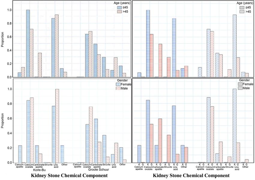
showed a high prevalence among females as illustrated in Figure 2. This is not
unexpected as females are more prone to developing UTI compared with males
and hence are more likely to form infection stones [45] [48] [49]. According to
Jayaraman et al. (2018), globally, the female to male ratio for struvite stone for-
mers is 2:1. Our study contrarily revealed a much higher ratio of 3.3:1 (F:M)
among GSH patients than that observed globally. Notwithstanding this differ-
ence of no struvite stones in Korle-Bu cohort of patients, this study also con-
firmed the predominance of struvite stones in female stone formers. Carbonate
apatite stones from this study were predominantly found in female stone for-
mers in both hospitals. Other authors have also reported a greater prevalence of
carbonate apatite kidney stones in females than men [49] [50].
The category of stones labelled as Other were mostly stones formed by females
from both hospitals. A study from Mayor Clinic Metal Laboratory in the USA
reported that over 43,500 of the kidney stones analyzed showed gender varia-
tions in stone composition. Females were found to form more apatite (calcium
phosphate) and struvite stones while men were prone to forming CaOx and UA
stones [51]. In the same study, Lieske et al. (2014), also observed that females,
particularly younger than 40 years, predominantly formed apatite kidney stones.
Our study also confirmed that apatite stones were largely found among female
stone formers as illustrated in Figure 2. On the contrary, Wathigo et al. (2017)
in Kenya and Ansari et al. (2005) in India did not observe any gender disparity
of stone composition in their studies.
DOI: 10.4236/oju.2021.113007 65 Open Journal of UrologyE. A. Akpakli et al.
Figure 2. Prevalence of chemical component in the kidney stone of patients treated in KBTH(K) and GSH(G) as a function of Age
and Gender.
There is evidence that the incidence of kidney stone disease increases with in-
creasing age and also varies with age stratification as observed in some epidemi-
ological studies [24]. Although, our study stratified age of patients into ≤45 years
and >45 years, we did not study the incidence of stone disease in these age
groups. On the whole, the analysis of kidney stone composition among our co-
hort of patients did not vary between the age groups provided the patients came
from the same geographic location. The observation of no difference in stone
composition regardless of age has also been reported by other researchers in
other countries where kidney stone compositional studies have been conducted
[22] [33] [51].
A study conducted in the late 1980s by Beukes et al. [44], on composition and
racial distribution of kidney stones from Bloemfontein, South Africa, observed
the predominance of CaOx stones among the racial groups studied. This obser-
vation is confirmed by this current study that CaOx stones are the most com-
mon stone type as seen among patients from Ghana (mainly African descent)
and South Africa (mixed race) although race was not directly a subject matter in
our study. Beukes et al. (1987), observed a higher proportion of struvite and
ammonium urate stones (infection stones) among the patients of African des-
DOI: 10.4236/oju.2021.113007 66 Open Journal of UrologyE. A. Akpakli et al.
cent in their patient population at the time. Contrary to their observation none
of the patients from Ghana had struvite stones. We are unable to confirm in this
study whether the infection stones among the South African cohort were more
common in a particular race as race stratification was not considered in the
study.
The limitation of this study was that: the study did not consider the metabolic
abnormalities or the 24-hour urine studies, which are associated with stone for-
mation. If it did, such consideration may have given insight as to why certain
stones where formed. This was not an objective of this study but can however be
the focus of future studies into comparative analysis of stone composition
among different African countries. Furthermore, urinalysis and urine culture
were not included in this study though they also affect stone formation and can
also be used to assess treatment and follow up on patients with regards to urine
pH and specific gravity.
Lastly, the study used results of stone analysis of patients who were treated in
the two hospitals only. Though other institutions also treat patients with stone
disease such patients have not been accounted for in this study. Hence the data
used for the study might not be as representative of the entire population in
Ghana and South Africa. Due to the small data size from Korle-Bu we were una-
ble to stratify age into groups (decades) to determine the incidence of kidney
stone disease among different age groups. Notwithstanding the aforementioned
limitations, this study set the platform for future collaborative studies between
hospitals and laboratories in Africa to determine the incidence of urolithiasis in
Ghana and South Africa (and other countries on the continent) on a much
broader scale.
5. Conclusion
The analyses discussed in this study are based on samples from KBTH and GSH.
Both hospitals are open to the public and offer a variety of services to their pa-
tients. The study shows a similar gender distribution of kidney stone patients
who were treated in Korle-Bu and GSH. There is also no significant difference in
age distribution of patients treated for stone disease in both hospitals. Majority
of the kidney stones from both countries were made up of CaOx stones; how-
ever, the stones from the Ghanaian cohort were mixed stones of which majority
were an admixed calcium oxalate/uric acid composition. Pure kidney stones
such as calcium oxalate, uric acid, struvite and cystine were only present among
the South African patients. Although some patients from Ghana formed carbon-
ate apatite stones, most stones with this component in this study were predomi-
nantly formed by South African patients. Certain kidney stone types, especially
brushite, cystine and struvite were only found among the South African popula-
tion. In contrast, calcium apatite and ammonium urate components stone were
found in kidney stones from Ghanaian patients. Female patients predominantly
formed carbonate apatite and stones with multiple components (mixed stones)
DOI: 10.4236/oju.2021.113007 67 Open Journal of UrologyE. A. Akpakli et al.
from both countries. Notwithstanding the small data especially from Ghana, it is
noteworthy to infer from this study that kidney stone composition in Ghana is
different from South Africa. Therefore, the composition of kidney stone is rec-
ognizably dependent on the demographic region from which the patient origi-
nates.
Authors’ Contributions
EAA and JL conceived and formulated this study. All authors made significant
inputs in drafting the manuscript, in terms of literature search, data analysis and
interpretations, and the paper’s final preparation for submission. All authors
read and approved the final paper.
Availability of Data and Materials
The data supporting the conclusions of this manuscript is available in Figshare
repository
(https://figshare.com/articles/dataset/Data_for_Paper_Comparative_Analysis_of
_kidney_stone_composition_among_patients_from_Ghana_and_South_Africa_
/13519787).
Ethics Approval
Ethical approval was sought and granted by the human research committee of the
faculty of health sciences, UCT (HREC REF:601/2018) and the Korle-Bu Teaching
Hospital institutional review Board, Korle-Bu, Accra (KBTH-IRB/00002/2019).
Conflicts of Interest
All the authors declare no conflict of interest.
References
[1] López, M. and Hoppe, B. (2010) History, Epidemiology and Regional Diversities of
Urolithiasis. Pediatric Nephrology, 25, 49-59.
https://doi.org/10.1007/s00467-008-0960-5
[2] Ferraro, P.M., Taylor, E.N., Eisner, B.H., Gambaro, G., Rimm, E.B., Mukamal, K.J.,
et al. (2013) History of Kidney Stones and the Risk of Coronary Heart Disease.
Journal of the American Medical Association, 310, 408-415.
https://doi.org/10.1001/jama.2013.8780
[3] Rule, A.D., Roger, V.L., Joseph Melton, L., Bergstralh, E.J., Li, X.J., Peyser, P.A., et
al. (2010) Kidney Stones Associate with Increased Risk for Myocardial Infarction.
Journal of the American Society of Nephrology, 21, 1641-1644.
https://doi.org/10.1681/ASN.2010030253
[4] Rule, A.D., Bergstralh, E.J., Melton, L.J., Li, X., Weaver, A.L. and Lieske, J.C. (2009)
Kidney Stones and the Risk for Chronic Kidney Disease. Clinical Journal of the
American Society of Nephrology, 4, 804-811. https://doi.org/10.2215/CJN.05811108
[5] Sigurjonsdottir, V.K., Runolfsdottir, H.L., Indridason, O.S., Palsson, R. and Ed-
vardsson, V.O. (2015) Impact of Nephrolithiasis on Kidney Function. BMC Ne-
phrology, 16, Article No. 149. https://doi.org/10.1186/s12882-015-0126-1
DOI: 10.4236/oju.2021.113007 68 Open Journal of UrologyE. A. Akpakli et al.
[6] Todd Alexande, R., Hemmelgarn, B.R., Wiebe, N., Bello, A., Morgan, C., Samuel, S.,
et al. (2012) Kidney Stones and Kidney Function Loss: A Cohort Study. BMJ, 345,
e5287. https://doi.org/10.1136/bmj.e5287
[7] Worcester, E.M. and Coe, F.L. (2010) Calcium Kidney Stones. New England Journal
of Medicine, 363, 954-963. https://doi.org/10.1056/NEJMcp1001011
[8] Pak, C., Poindexter, J., Adams-Huet, B. and Pearle, M. (2003) Predictive Value of
Kidney Stone Composition in the Detection of Metabolic Abnormalities. American
Journal of Medicine, 115, 26-32. https://doi.org/10.1016/S0002-9343(03)00201-8
[9] Cloutier, J., Villa, L., Traxer, O. and Daudon, M. (2014) Kidney Stone Analysis:
“Give Me Your Stone, I Will Tell You Who You Are!” World Journal of Urology,
33, 157-169. https://doi.org/10.1007/s00345-014-1444-9
[10] Samuell, C.T. and Kasidas, G.P. (1995) Biochemical Investigations in Renal Stone
Formers. Annals of Clinical Biochemistry, 32, 112-122.
https://doi.org/10.1177/000456329503200202
[11] Ekeke, O.N. and Okpani, C.P. (2018) Management of Urinary Stone Disease in a
Resource Limited Tertiary Hospital. IOSR Journal of Dental and Medical Sciences,
17, 38-45.
[12] Raheem, O.A., Khandwala, Y.S., Sur, R.L., Ghani, K.R., Denstedt, J.D. and Catto, J.
(2017) Burden of Urolithiasis: Trends in Prevalence, Treatments, and Costs. Euro-
pean Urology Focus, 3, 18-26. https://doi.org/10.1016/j.euf.2017.04.001
[13] Afshar, K., Jafari, S., Marks, A., Eftekhari, A. and MacNeily, A. (2015) Nonsteroidal
Anti-Inflammatory Drugs (NSAIDs) and Non-Opioids for Acute Renal Colic.
Cochrane Database of Systematic Reviews, No. 6, Article No. CD006027.
https://doi.org/10.1002/14651858.CD006027.pub2
[14] Scales, C.J., Smith, A., Hanley, J. and Saigal, C. (2012) Prevalence of Kidney Stones
in the United States Charles. European Urology, 62, 160-165.
https://doi.org/10.1016/j.eururo.2012.03.052
[15] Lewandowski, S. and Rodgers, A.L. (2004) Renal Response to Lithogenic and
Anti-Lithogenic Supplement Challenges in a Stone-Free Population Group. Journal
of Renal Nutrition, 14, 170-179. https://doi.org/10.1053/j.jrn.2004.04.007
[16] Rodgers, A.L. (2006) The Riddle of Kidney Stone Disease: Lessons from Africa.
Urological Research, 34, 92-95. https://doi.org/10.1007/s00240-005-0017-1
[17] Campschroer, L.M.T., Zhu, Y., Duijvesz, D., Grobbee, D.E. and Lock, M.T. (2014)
Alpha-Blockers as Medical Expulsive Therapy for Ureteral Stones. Cochrane Data-
base of Systematic Reviews, No. 4, Article No. CD008509.
https://doi.org/10.1002/14651858.CD008509.pub2
[18] R Core Team (2017) R: A Language and Environment for Statistical Computing. R
Foundation for Statistical Computing, Vienna.
[19] Adamu, B., Alhassan, S.U. and Effa, E.E. (2015) Adjunctive Medical Expulsive
Therapy for Kidney and Ureteral Stone Fragments Following Shock Wave Lith-
otripsy. Cochrane Database of Systematic Reviews, No. 7, Article No. CD009156.
https://doi.org/10.1002/14651858.CD009156.pub3
[20] Bensalah, K., Pearle, M. and Lotan, Y. (2008) Cost-Effectiveness of Medical Expul-
sive Therapy Using Alpha-Blockers for the Treatment of Distal Ureteral Stones.
European Urology, 53, 411-419. https://doi.org/10.1016/j.eururo.2007.09.012
[21] Hossain, R.Z., Ogawa, Y., Hokama, S., Morozumi, M. and Hatano, T. (2003) Uro-
lithiasis in Okinawa, Japan: A Relatively High Prevalence of Uric Acid Stones. In-
DOI: 10.4236/oju.2021.113007 69 Open Journal of UrologyE. A. Akpakli et al.
ternational Journal of Urology, 10, 411-415.
https://doi.org/10.1046/j.1442-2042.2003.00656.x
[22] Wathigo, F.K., Hayombe, A. and Maina, D. (2017) Urolithiasis Analysis in a Multi-
ethnic Population at a Tertiary Hospital in Nairobi, Kenya. BMC Research Notes,
10, Article No. 158. https://doi.org/10.1186/s13104-017-2474-3
[23] Scales, C.D., Curtis, L.H., Norris, R.D., Patrick Springhart, W., Sur, R.L., Schulman,
K.A., et al. (2007) Changing Gender Prevalence of Stone Disease. Journal of Urol-
ogy, 177, 979-982. https://doi.org/10.1016/j.juro.2006.10.069
[24] Romero, V., Akpinar, H. and Assimos, D.G. (2010) Kidney Stones: A Global Picture
of Prevalence, Incidence, and Associated Risk Factors Kidney Stones: A Global
Perspective. Reviews in Urology, 12, e86-e96.
[25] Alaya, A., Nouri, A., Belgith, M., Saad, H., Jouini, R. and Fadhel Najjar, M. (2012)
Changes in Urinary Stone Composition in the Tunisian Population: A Retrospective
Study of 1,301 Cases. Annals of Laboratory Medicine, 32, 177-183.
https://doi.org/10.3343/alm.2012.32.3.177
[26] Phillips, R., Hanchanale, V.S., Myatt, A., Somani, B. and Nabi, G. (2015) Citrate
Salts for Preventing and Treating Calcium Containing Kidney Stones in Adults.
Cochrane Database of Systematic Reviews, No. 9, Article No. CD010057.
https://doi.org/10.1002/14651858.CD010057.pub2
[27] Sofia, N.H., Walter, T.M. and Sanatorium, T. (2016) Prevalence and Risk Factors of
Kidney Stone. Global Journal for Research Analysis, 5, 183-187.
[28] Worster, A. and Supapol, B.W. (2012) Fluids and Diuretics for Acute Ureteric Colic.
Cochrane Database of Systematic Reviews, No. 2, Article No. CD004926.
https://doi.org/10.1002/14651858.CD004926.pub3
[29] Letavernier, E. and Daudon, M. (2018) Vitamin D, Hypercalciuria and Kidney
Stones. Nutrients, 10, Article No. 366. https://doi.org/10.3390/nu10030366
[30] Sakhaee, K., Capolongo, G., Maalouf, N.M., Pasch, A., Moe, O.W., Poindexter, J., et
al. (2012) Metabolic Syndrome and the Risk of Calcium Stones. Nephrology Dialysis
Transplantation, 27, 3201-3209. https://doi.org/10.1093/ndt/gfr703
[31] Tsujihata, M. (2008) Mechanism of Calcium Oxalate Renal Stone Formation and
Renal Tubular Cell Injury. International Journal of Urology, 15, 115-120.
https://doi.org/10.1111/j.1442-2042.2007.01953.x
[32] Edvardsson, V.O., Indridason, O.S., Haraldsson, G., Kjartansson, O. and Palsson, R.
(2013) Temporal Trends in the Incidence of Kidney Stone Disease. Kidney Interna-
tional, 83, 146-152. https://doi.org/10.1038/ki.2012.320
[33] Ansari, M.S., Gupta, N.P., Hemal, A.K., Dogra, P.N., Seth, A., Aron, M., et al. (2005)
Spectrum of Stone Composition: Structural Analysis of 1050 Upper Urinary Tract
Calculi from Northern India. International Journal of Urology, 12, 12-16.
https://doi.org/10.1111/j.1442-2042.2004.00990.x
[34] Abou-Elela, A. (2017) Epidemiology, Pathophysiology, and Management of Uric
acid Urolithiasis: A Narrative Review. Journal of Advanced Research, 8, 513-527.
https://doi.org/10.1016/j.jare.2017.04.005
[35] Xu, H., Zisman, A.L., Coe, F.L. and Worcester, E.M. (2013) Kidney Stones: An Up-
date on Current Pharmacological Management and Future Directions. Expert
Opinion on Pharmacotherapy, 14, 435-447.
https://doi.org/10.1517/14656566.2013.775250
[36] Chu, F.Y., Chang, C.-C., Huang, P.-H., Lin, Y.-N., Ku, P.-W., Sun, J.-T., et al. (2017)
DOI: 10.4236/oju.2021.113007 70 Open Journal of UrologyE. A. Akpakli et al.
The Association of Uric Acid Calculi with Obesity, Prediabetes, Type 2 Diabetes
Mellitus, and Hypertension. BioMed Research International, 2017, Article ID:
7523960. https://doi.org/10.1155/2017/7523960
[37] Sakhaee, K. (2014) Epidemiology and Clinical Pathophysiology of Uric Acid Kidney
Stones. Journal of Nephrology, 27, 241–245.
https://doi.org/10.1007/s40620-013-0034-z
[38] Torricelli, F.C.M., Gebreselassie, S., Calle, J., Monga, M., De, S. and Liu, X. (2014)
Can 24-Hour Urine Stone Risk Profiles Predict Urinary Stone Composition? Jour-
nal of Endourology, 28, 735-738. https://doi.org/10.1089/end.2013.0769
[39] Chandirika Jayaraman, U. and Gurusamy, A. (2018) Review on Uro-Lithiasis
Pathophysiology and Aesculapian Discussion. IOSR Journal of Pharmacy, 8, 30-42.
[40] Shima, M. and Parks, J.H. (2014) Multimodal Treatments of Cystine Stones: An
Observational, Retrospective Single-Center Analysis of 14 Cases. Korean Journal of
Urology, 55, 515-519. https://doi.org/10.4111/kju.2014.55.8.515
[41] Krambeck, A.E., Handa, S.E., Evan, A.P. and Lingeman, J.E. (2010) Profile of the
Brushite Stone Former. Journal of Urology, 184, 1367-1371.
https://doi.org/10.1016/j.juro.2010.05.094
[42] Moreira, D.M., Friedlander, J.I., Hartman, C., Elsamra, S.E., Smith, A.D. and Okeke,
Z. (2013) Differences in 24-Hour Urine Composition between Apatite and Brushite
Stone Formers. Urology, 82, 768-772. https://doi.org/10.1016/j.urology.2013.04.025
[43] Rivera, M., Jaeger, C., Yelfimov, D. and Krambeck, A.E. (2017) Risk of Chronic
Kidney Disease in Brushite Stone Formers Compared With Idiopathic Calcium Ox-
alate Stone Formers. Urology, 99, 23-26.
https://doi.org/10.1016/j.urology.2016.08.041
[44] Beukes, G.J., De Bruiyn, H. and Vermaak, W.J.H. (1987) Effect of Changes in Epi-
demiological Factors on the Composition and Racial Distribution of Renal Calculi.
British Journal of Urology, 60, 387-392.
https://doi.org/10.1111/j.1464-410X.1987.tb05000.x
[45] Diri, A. and Diri, B. (2018) Management of Staghorn Renal Stones. Renal Failure,
40, 357-362. https://doi.org/10.1080/0886022X.2018.1459306
[46] Daudon, M., Jungers, P., Bazin, D. and Williams, J.C. (2018) Recurrence Rates of
Urinary Calculi According to Stone Composition and Morphology. Urolithiasis, 46,
459-470. https://doi.org/10.1007/s00240-018-1043-0
[47] Daudon, M., Dessombz, A., Frochot, V., Letavernier, E., Haymann, J.-P., Jungers,
P., et al. (2016) Comprehensive Morpho-Constitutional Analysis of Urinary Stones
Improves Etiological Diagnosis and Therapeutic Strategy of Nephrolithiasis. Comptes
Rendus Chimie, 19, 1470-1491. https://doi.org/10.1016/j.crci.2016.05.008
[48] Alelign, T. and Petros, B. (2018) Kidney Stone Disease: An Update on Current
Concepts. Advances in Urology, 2018, Article ID: 3068365.
https://doi.org/10.1155/2018/3068365
[49] Ma, R., Luo, X., Li, Q. and Zhong, H. (2018) Systemic Analysis of Urinary Stones
from the Northern, Eastern, Central, Southern and Southwest China by a
Multi-Center Study. BMC Urology, 18, Article No. 114.
https://doi.org/10.1186/s12894-018-0428-2
[50] Carpentier, X., Daudon, M., Traxer, O., Jungers, P., Mazouyes, A., Matzen, G., et al.
(2009) Relationships between Carbonation Rate of Carbapatite and Morphologic
Characteristics of Calcium Phosphate Stones and Etiology. Urology, 73, 968-975.
https://doi.org/10.1016/j.urology.2008.12.049
DOI: 10.4236/oju.2021.113007 71 Open Journal of UrologyE. A. Akpakli et al.
[51] Lieske, J.C., Rule, A.D., Krambeck, A.E., Williams, J.C., Bergstralh, E.J., Mehta,
R.A., et al. (2014) Stone Composition as a Function of Age and Sex. Clinical Journal
of the American Society of Nephrology, 9, 2141-2146.
https://doi.org/10.2215/CJN.05660614
DOI: 10.4236/oju.2021.113007 72 Open Journal of UrologyYou can also read