Surgical Management of Primary Cutaneous Melanoma: Excision Margins and the Role of Sentinel Lymph Node Examination
←
→
Page content transcription
If your browser does not render page correctly, please read the page content below
Surg Oncol Clin N Am
                                      15 (2006) 301–318

      Surgical Management of Primary
   Cutaneous Melanoma: Excision Margins
      and the Role of Sentinel Lymph
             Node Examination
              John F. Thompson, MDa,b,*,
   Richard A. Scolyer, MBBSa,c, Roger F. Uren, MDa,b
  a
      Sydney Melanoma Unit, Level 3, Gloucester House, Sydney Cancer Centre, Royal Prince
             Alfred Hospital, Missenden Road, Camperdown, NSW 2006, Australia
                b
                 The University of Sydney, Camperdown, NSW 2006, Australia
              c
               Department of Anatomical Pathology, Royal Prince Alfred Hospital,
                     Missenden Road, Camperdown, NSW 2006, Australia

    Thirty years ago, most melanomas were widely excised with 3-cm to 5-cm
margins, and many centers treated regional lymph nodes with routine elective
lymph node dissection (ELND). These management policies were not
evidence-based, they simply represented a continuation of surgical practices
that had been accepted for many decades. These policies were perhaps rein-
forced by the observation that patients whose melanomas had been managed
by excision with narrow rather than wide margins sometimes developed local
recurrence of their disease, whereas others who did not undergo an ELND
sometimes presented later with metastatic disease in regional lymph nodes
that was difficult or impossible to treat surgically. Retrospective studies also
suggested that ELND conferred a survival advantage. In some centers, wide
excision of the primary melanoma and ELND were performed in continuity,
with excision of a broad strip of skin, subcutaneous tissue, and sometimes deep
fascia between the primary melanoma site and the nearest anatomic group of
lymph nodes. This policy was originally based on Hogarth Pringle’s [1] advice
in 1908; extrapolating from his experience with three cases, he stated, ‘‘All that
is removed should be in one continuous strip as far as possible.’’

  * Corresponding author. Syndney Melanoma Unit, Level 3, Gloucester House, Sydney
Cancer Centre, Royal Prince Alfred Hospital, Missenden Road, Camperdown, NSW 2006,
Australia.
  E-mail address: thompson@smu.org.au (J.F. Thompson).

1055-3207/06/$ - see front matter Ó 2006 Elsevier Inc. All rights reserved.
doi:10.1016/j.soc.2005.12.007                                            surgonc.theclinics.com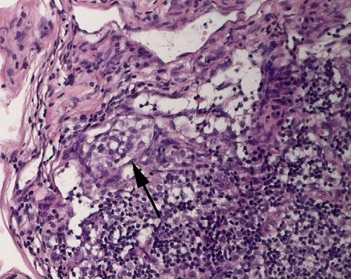
302                               THOMPSON   et al

   Over the past 30 years, however, surgical strategies for the management
of patients who have primary cutaneous melanoma have changed dramati-
cally. These changes occurred because large clinical trials indicated that ex-
cising melanomas with very wide margins conferred little or no benefit and
that routine ELND did not confer an overall survival benefit. The other im-
portant development was the introduction and validation of the sentinel
node (SN) biopsy procedure, with accurate preoperative lymphatic mapping
obtained using lymphoscintigraphy.

Diagnostic biopsy
   The role of surgery in melanoma management begins with excision-
biopsy of the lesion. Even if a confident diagnosis of melanoma is made on
clinical grounds, preliminary excision of the entire tumor with narrow
(2 mm) clearance margins is desirable so that appropriate treatment can
be planned. A decision about the definitive excision margins can then be
made based on information obtained from the pathologist in relation to
the thickness of the primary tumor. In addition to thickness, primary tumor
characteristics, such as ulceration, regression, or a high mitotic rate, will also
indicate whether the likelihood of metastasis to regional lymph nodes is suf-
ficiently high to warrant lymphatic mapping and an SN biopsy for staging
purposes.
   Partial biopsies (eg, incision, punch, shave) of melanocytic lesions can re-
sult in many problems. Nevertheless, when a suspicious pigmented lesion is
very large, an incision biopsy or a punch biopsy may need to be considered,
bearing in mind that these partial biopsies can make it difficult for the pa-
thologist to provide a reliable diagnosis because they represent only a small
part of the entire lesion. Shave biopsies of pigmented lesions may be un-
reliable, because if melanoma is diagnosed, establishing either the Breslow
tumor thickness or the completeness of excision may be difficult or impos-
sible [2]. The decision-making process in relation to clinical management
in such situations can be difficult. Furthermore, surgeons must be aware
that a very high proportion of medical malpractice claims in relation to mel-
anoma involve incomplete biopsy specimens [3].

Definitive managementdexcision margins
   Debate about the excision margins necessary for melanoma dates back to
1885, when Joseph Coats [4] advocated extensive excision of primary mela-
nomas. In 1892, Herbert Snow [5] also recommended wide excision of the
primary tumor (and a ‘‘fairly radical’’ regional lymph node dissection).
However, for a long time William Handley [6] mainly influenced surgical
thinking about excision margins, with his persuasive statements that very
wide clearance of primary tumors was necessary. In the 1960s, the long-held
SURGICAL MANAGEMENT OF PRIMARY CUTANEOUS MELANOMA            303

concept that a melanoma required very wide excision seemed to be rein-
forced by Greta Olsen’s [7] report that atypical melanocytes were often
found within 5 cm of the primary melanoma. She suggested that unless all
of these atypical melanocytes were removed by excision with very wide
clearance margins, local recurrence was likely to occur.
   Although the 5-cm excision margins originally proposed by Handley, Ol-
sen, and others are no longer considered appropriate, debate continues
about the actual clearance margins that should now be used. Experts agree
that complete surgical excision of a primary cutaneous melanoma is re-
quired, and some have even proposed that complete excision of the tumor
is all that is necessary [8]. However, others have suggested that clearance
margins of at least 3 cm are still required, particularly for thick primary
melanomas [9]. Several large retrospective studies reported in the 1980s
indicated that local recurrence was very uncommon in patients whose
melanomas were excised with margins of 2 cm or more. These studies
showed that the risk for local recurrence was principally dependant on the
Breslow thickness of the primary melanoma. One such study, published in
1985, reported the experience of the Sydney Melanoma Unit (SMU) [10]
and determined the recurrence rates in 1839 patients who underwent long-
term follow-up. For thick tumors (defined as R3 mm in this study), the local
recurrence rate was 21% when the excision margin was less than 2 cm and
9% when the excision margin was 2 cm or more. For thin tumors (defined as
0.1–0.7 mm), the local recurrence rates were 2% when excision margins less
than 2 cm were used, and less than 1% when excision margins of 2 cm or
more were used.
   Eventually it became clear that large, prospective, randomized trials were
needed to clarify the excision margins required to minimize the risk for local
recurrence and avoid any adverse effect on survival outcome. The World
Health Organization Melanoma Program undertook such a trial, in which
results were compared for 612 patients who had primary melanomas less
than 2 mm in thickness and who had their tumors excised with margins
of either 1 or 3 cm [11]. Disease-free and overall survival rates did not differ
between the two groups, thus melanomas 1 mm or less in thickness were ad-
equately treated by excision with a 1-cm margin. Another trial was initiated
in the United States, which randomized 486 patients who had intermediate-
thickness melanomas (1–4 mm) to be treated with either 2- or 4-cm excision
margins [12]. Again, the recurrence rates were similar for the two groups,
and no significant difference in overall 5-year survival occurred. However,
as expected, the 4-cm margin group had significantly greater treatment mor-
bidity and length of hospital stay.
   Two subsequent trials in Europe compared the results of treating primary
melanomas with 2- or 5-cm excision margins. The Swedish Study Group un-
dertook one trial involving 989 patients who had melanomas 0.8 to 2 mm in
thickness [13]. The French Group for Research on Malignant Melanoma
undertook the other trial [14] involving 326 patients who had primary
304                             THOMPSON   et al

melanomas 2 mm or more in Breslow thickness. Neither study produced any
evidence that 5-cm margins reduced the local recurrence rate or improved
survival outcome. A large trial undertaken in Britain more recently reex-
amined the question of margins [9]. Excision margins of 1 and 3 cm were
compared in 900 patients who had melanomas 2 mm or more in Breslow
thickness. A 1-cm margin was found to be associated with a slightly in-
creased risk for local recurrence compared with a 3-cm margin, but after
a median follow-up period of 16 months, no difference in survival outcome
occurred between the two groups.
   Although some uncertainties remain, requiring further trial-based evi-
dence for clarification [15], the situation in relation to excision margins
for primary cutaneous melanomas is certainly much clearer than it was 30
years ago. For tumors that are 1 mm or less in Breslow thickness, the gen-
eral consensus is that a 1-cm minimum clearance margin should be adequate
[16]. For tumors between 1 mm and 2 mm in thickness, some evidence sug-
gests that excision margins of more than 1 cm are desirable, but this evi-
dence is not conclusive [17]. For melanomas that exceed 2 mm in Breslow
thickness, available evidence suggests that excision margins of at least 2 cm
are required to minimize the risk for local recurrence, but whether a
2-cm margin is adequate or a margin of 3-cm is required is unclear because
appropriate trials have not been conducted. Most of the available evidence
has been obtained in clinical trials involving patients who had melanomas
located on the trunk or a proximal extremity. Most studies have not in-
cluded patients who had melanomas in the head and neck region or on a dis-
tal extremity, locations where more complex reconstructive techniques may
be required if wider excisions margins are used and cosmetic implications
must also be considered.
   Even if excision margins of more than 2 cm do produce slightly lower
recurrence rates for patients who have intermediate thickness melanomas
and may even have an impact on survival, these benefits are likely to be
small [18] and must be weighed against the increased surgical morbidity
and disfigurement that are likely to be associated with wider margins at
any body site. A persuasive argument has been made that some compro-
mise on excision margins may be reasonable in situations where the risks
for surgical morbidity and cosmetic deformity are high [19]. Some experts
have suggested that a slightly higher risk for local recurrence may be ac-
ceptable to avoid the additional morbidity and cosmetic deformity that
a wider margin will produce. Others have pointed out that if local recur-
rence occurs, it can usually be removed surgically without great difficulty.
Furthermore, although local recurrence is associated with a reduced sur-
vival outcome, the assumption that performing a wider excision to reduce
the risk for local recurrence will necessarily improve survival is scientifi-
cally inappropriate. Careful evaluation of all the available evidence sug-
gests that tumor biology rather than the extent of local treatment
ultimately determines outcome.
SURGICAL MANAGEMENT OF PRIMARY CUTANEOUS MELANOMA           305

Management of regional lymph nodesdlymphatic mapping and sentinel
node biopsy
    In a lecture given in 1892, Herbert Snow [5] established long-lasting sur-
gical thinking on the management of regional lymph nodes by declaring that
operative measures addressed only to the primary lesion were ‘‘utterly fu-
tile,’’ and emphasizing that ‘‘the paramount importance of securing, when-
ever possible, the perfect eradication of those lymph glands which would be
necessarily first infected before enlargement takes place.’’ He suggested that
elective removal of the regional nodes was ‘‘a safe and easier measure.’’
Snow’s recommendation that routine complete lymph node dissection
(CLND) should be performed in patients who have melanoma and who
have no clinical evidence of regional node metastasis was based on the as-
sumption that metastatic melanoma progresses sequentially from primary
melanoma site to regional lymph nodes before passage to more distant sites.
This concept implies that early removal of regional nodes by ELND should
interrupt the metastatic cascade.
    Since Snow’s original proposal, experts have disagreed about the value of
ELND for patients who have melanomas and who present with no clinical
evidence of regional node metastasis. Although retrospective studies seemed
to indicate an overall survival benefit after ELND in those who had inter-
mediate-thickness tumors, randomized trials did not [20–23]. Other consid-
erations were that the long-term morbidity of ELND was significant,
particularly for patients who had primary melanomas of the lower limbs,
and only approximately 20% of patients were found to have metastatic dis-
ease in regional lymph nodes when ELND was performed. Nieweg discusses
ELND in greater detail elsewhere in this issue.
    In 1990, at the height of this controversy, Morton and associates [24]
from the John Wayne Cancer Institute proposed a potential solution at
a meeting of the Society of Surgical Oncology in the United States. They
suggested that it was possible to confidently assess the status of regional
lymph nodes by performing lymphatic mapping to identify an SN in each
patient, and then to remove that node for histologic examination. Technical
details of the procedure were published in 1992 [24], and Morton and col-
leagues proposed that SN biopsy would avoid the need for ELND in
80% of patients. It would, however, identify the other 20% of patients
most likely to benefit from the procedure. This proposal was initially greeted
with great skepticism, but within 3 years, prospective studies undertaken in
the United States [25] and in Australia [26] confirmed its accuracy in iden-
tifying regional node metastasis. Lymphatic mapping and SN biopsy were
performed in each of these studies, followed by immediate completion
CLND so that all remaining nodes in the node field could be assessed by
the pathologist. The results of the two studies were remarkably similar to
those of Morton and colleagues, and established conclusively that the SN
concept was valid. The hypothesis was thus confirmed that if no evidence
306 THOMPSON et al
of micrometastatic disease in SNs was found, metastatic disease was very
rarely present in other nodes in that node field. Other validation studies
with similar results were subsequently reported [27], all indicating that SN
status accurately reflects the status of the entire node field in patients who
have primary cutaneous melanoma.
The SN concept was not new, although its practical importance had not
been fully appreciated until Morton and colleagues’ report. The pathologist
Virchow [28] had clearly described the concept in the mid-19th Century.
However, it seems that the first recorded use of the term sentinel in relation
to lymph nodes was by a British surgeon named Braithwaite, who in 1923
described ‘‘glands sentinel’’ in the upper abdomen, guarding the stomach
and duodenum by trapping bacteria in lymph draining from the terminal
ileum and cecum [29]. Like Morton and his colleagues 67 years later,
Braithwaite used vital dye injections to study lymphatic anatomy in a feline
model and in man. Braithwaite recognized that the descriptions of lym-
phatic drainage pathways that were currently accepted were often at vari-
ance with the lymphatic drainage pathways he observed. In 1960, Gould
and colleagues [30] reported a constant pattern of lymphatic drainage to
a ‘‘sentinel node’’ in the upper neck in patients who had parotid carcinomas;
these authors based their decision of whether to proceed with a radical neck
dissection on the result of frozen section examination of this SN. In 1966,
Sayegh and colleagues [31] described an intra-abdominal ‘‘sentinel node’’
to which lymphatic drainage from the testis occurred. In 1977, Cabanas
[32] reported ‘‘sentinel nodes’’ that received direct lymphatic drainage
from primary tumors of the penis, testis, rectum, anus, breast, and skin.
Nearly 3 decades later, evidence shows that the SN concept holds true in pa-
tients who have a wide range of other malignancies that spread through lym-
phatics [33], including cancer of the breast, thyroid, lung, stomach, colon,
vulva, uterus, prostate, and penis, and non-melanoma skin malignancies
(squamous cell carcinoma and Merkel cell carcinoma) [34–41].
The SN concept is simple. It proposes that lymph draining from a tumor
passes first to an SN before passing to other nodes in the regional node field.
If tumor cells enter lymphatic collectors, they are carried by the lymphatic
flow until they are arrested in a draining lymph node. This first lymph
node encountered is by definition an SN, and tumor cells are thus most
likely to be found in the SN. Although Morton and colleagues [24] originally
defined an SN as ‘‘the lymph node nearest the site of the primary melanoma,
on the direct drainage pathway,’’ more than one lymph node may be present
in a regional node field that receives direct lymphatic drainage from a given
tumor site. SNs may also be present in more than one regional node field
that receives drainage from a tumor site. Therefore, an unambiguous defini-
tion of a SN may be more appropriate, such as: ‘‘A sentinel node is any node
that receives direct lymphatic drainage from a primary tumor site’’ [42]. No-
des that receive lymph that has previously passed through the physiological
filter function of a SN are designated second tier or second echelon nodes.SURGICAL MANAGEMENT OF PRIMARY CUTANEOUS MELANOMA 307 The SN concept is consistent with the well-established fact that the tumor status of the regional lymph nodes is the most powerful predictor of out- come in patients who have a clinically localized primary cutaneous mela- noma [43]. In Morton and colleagues’ early SN studies at the John Wayne Cancer Institute [43], blue dye was injected intradermally at a primary melanoma site, and blue-stained afferent lymphatics were traced to a blue-stained SN in the regional node field. This was a tedious and moderately invasive pro- cess, but subsequently it has shown that preoperative lymphoscintigraphy could not only provide valuable information preoperatively but also facili- tate more rapid SN identification with less extensive dissection by intraoper- ative use of a hand-held gamma probe [44,45]. A report from the SMU showed that residual radioactivity could be used in the SN up to 30 hours after preoperative lymphoscintigraphy for intraoperative SN identification with a gamma probe [46]. This procedure eliminated the need for patients to receive a second dose of radioisotope in the anesthetic bay or operating room. It also offered important logistic advantages by allowing lymphoscin- tigraphy to be performed on the day before surgery. As melanoma treatment centers around the world began acquiring expe- rience with the SN biopsy technique, surgeons quickly recognized that use of the three methods provided the most rapid and reliable identification of SNs. This procedure, including (1) a preoperative lymphoscintigram (LSG), (2) blue-dye injection at the primary melanoma site immediately pre- operatively, and (3) intraoperative use of a gamma probe, is now widely ac- cepted as the standard technique for SN biopsy. A number of studies have shown that results obtained using only blue dye for SN identification are less accurate than those obtained when a hand-held gamma probe is also used intraoperatively [47]. The importance of preoperative lymphoscintigraphy Although simple in concept, the SN biopsy procedure can present many technical challenges. Unless the procedure is undertaken with great care and precision, SN identification may be inaccurate. This misidentification pro- vides misleading information that may not only provide an unrealistic esti- mate of prognosis but also result in inadequate or inappropriate treatment. The key to accurate SN identification is high-quality preoperative lym- phoscintigraphy using a radiolabeled colloid. This procedure, if performed correctly, will not only identify the node field or fields to which lymphatic drainage from a primary melanoma occurs but also determine precisely which nodes are SNs. The location and removal of one SN is insufficient if more than one is present. Micrometastatic melanoma can be found in any true SN, even if it is not the most prominent node on the preoperative lym- phoscintigram or most obviously blue-stained node at surgery (Fig. 1) [26].
308                                   THOMPSON   et al

Fig. 1. Lymphoscintigram showing a prominent lymphatic channel draining from a primary
melanoma site on the thigh to an SN in the groin, but a second, very faint channel draining
to another SN (arrow). The latter node contained micrometastatic melanoma, but the more
prominent node did not.

   The nuclear medicine physician should carefully scrutinize early dynamic
images acquired after tracer injection at the primary melanoma site, which
will usually show with certainty which nodes are receiving direct lymphatic
drainage from that site. Each one of these nodes is by definition an SN. Ex-
perience with more than 4000 patients who have undergone a preoperative
lymphoscintigram for cutaneous melanoma at the SMU has shown clearly
that the lymphatic drainage in an individual patient is completely unpredict-
able [48–50]. This experience has also revealed several previously unrecog-
nized lymphatic drainage patterns. Knowledge of these unusual patterns
and documented ‘‘ectopic’’ SN sites is helpful in minimizing the possibility
that an SN will be missed. The usual and unusual cutaneous lymphatic
drainage patterns from various body sites seen in the SMU series are de-
tailed by Uren and colleagues elsewhere in this issue.

Is sentinel node biopsy of value?
   The SN status of a patient who presents with a primary cutaneous
melanoma has been shown to be an important prognostic factor for long-
term survival [51–58]. Studies have shown a large difference in 5-year sur-
vival probability between patients who are SN-positive and those who are
SN-negative, independent of other prognostic variables. In a study of 991
patients who underwent an SN biopsy procedure at the SMU, the 5-year
survival for patients who were SN-negative was 90%, whereas the 5-year
survival for SN-positive patients was only 56% (Fig. 2) [59].
   Some have suggested that routine performance of an SN biopsy proce-
dure is inappropriate, even improper, because clinical trial evidence has
not shown that SN biopsy, with immediate CLND if a positive SN is found,
results in an improvement in survival outcome [60–62]. However, the
SURGICAL MANAGEMENT OF PRIMARY CUTANEOUS MELANOMA                        309

Fig. 2. (A) Melanoma-specific survival in patients who are sentinel node–positive (n ¼ 139) ver-
sus patients who are sentinel node–negative (n ¼ 836) (P ! .001). (B) Disease-free survival in
patients who are sentinel node–positive (n ¼ 139) versus patients who are sentinel node–nega-
tive (n ¼ 836) (P ! .001). (From Yee V, Thompson JF, McKinnon JG, et al. Outcome in 846
cutaneous melanoma patients from a single center after a negative sentinel node biopsy. Ann
Surg Oncol 2005;12:432; with permission.)

Multicenter Selective Lymphadenectomy Trial (MSLT), a randomized
phase III study involving 2001 patients [47,63], provides strongly suggestive
evidence of a survival benefit in patients who have metastatic disease in their
regional nodes. Survival outcome data from an interim analysis of the
MSLT results were presented recently, but not published when this article
was written [64]. This data showed that the 5-year survival for patients iden-
tified as having metastatic melanoma in regional lymph nodes, either by SN
biopsy or by later presenting with clinical disease in the node field, was sig-
nificantly better if early CNLD was performed when the metastatic disease
was diagnosed through SN biopsy, compared with patients who had a wide
310 THOMPSON et al
excision only as their initial definitive treatment and then underwent a re-
gional node dissection later when disease in the node field became clinically
apparent [64].
SN biopsy as a minimally invasive staging procedure has value even if no
overall survival benefit is shown by the MSLT or other studies [65,66]. Hav-
ing the most reliable estimate of prognosis available is important for most
patients, and stratification into risk groups must be as accurate as possible
for those entering adjuvant therapy trials.
Also of great relevance, although unquantifiable, is the psychologic dev-
astation that can occur when melanoma recurrence occurs in a regional
lymph node field months or years after a patient has undergone initial defin-
itive melanoma treatment and has come to terms with the implications of
a melanoma diagnosis. Lastly, the importance of achieving regional node
field control through an SN biopsy should not be underestimated. If effec-
tive adjuvant therapies are developed, knowledge of SN status will be
needed to identify patients most likely to benefit.
Some believe SN biopsy should not be performed because reports have
shown that in-transit metastasis rates increase [67,68]. However, two large
series showed no increase in in-transit metastasis rates when allowance
was made for primary tumor characteristics [69,70]. Analysis of all available
evidence indicates that regional node surgery does not increase the risk for
in-transit metastasis, but that primary tumor biology alone seems to deter-
mine that risk [71]. This conclusion is supported by the recently reported re-
sults of MSLT-I [64]. After a median follow-up interval of almost 5 years,
the in-transit metastasis rates were virtually identical for patients random-
ized in this trial to undergo treatment with wide excision only or wide exci-
sion plus SN biopsy.
Management of patients who have a positive sentinel node
The SN biopsy procedure was originally introduced so that routine
CLND could be avoided. It identified the small group of patients (approx-
imately 20% in most series) most likely to benefit from CLND, and spared
the remaining patients the inconvenience and morbidity of the procedure.
Ironically, a similar 20% issue now arises for patients found to be SN pos-
itive: do all of them require a CLND, with its attendant morbidity? This
question arises because only about 20% of patients who have a positive
SN will have any evidence of further disease in non-SNs if a CLND is per-
formed. In other words, in approximately 80% of patients who have meta-
static disease in a regional lymph node field, the disease seems to be confined
to the SN or SNs that are removed during the SN biopsy procedure. A fur-
ther large-scale randomized clinical trial is needed to answer this new ques-
tion. Such a trial, MSLT-II, has already commenced. In this new
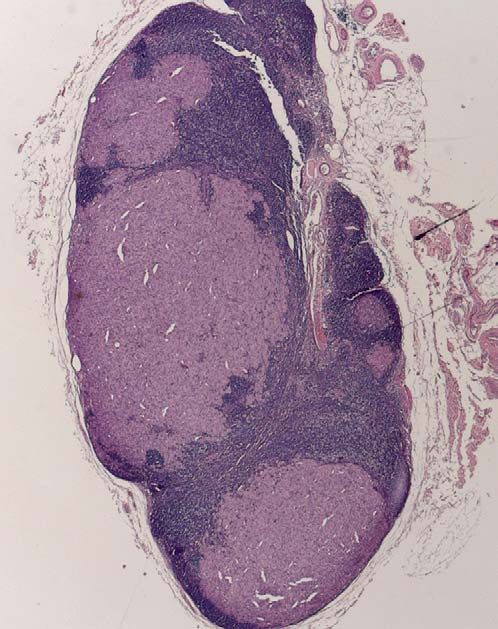
multicenter phase III study, patients who are found to have a positive SNSURGICAL MANAGEMENT OF PRIMARY CUTANEOUS MELANOMA 311 either through conventional histopathology or assessment using reverse transcriptase polymerase chain reaction technology are randomized to re- ceive no further immediate treatment or to have a CLND. Those assigned to the observation-only arm are followed-up with regular high-resolution ultrasound examination of the node field and will undergo CLND later if disease in residual nodes in the node field is identified clinically or through ultrasound. This international study commenced patient accrual in December 2004, with a target of 1925 randomized patients who have a positive SN. Meanwhile, experts are attempting to determine the likelihood that indi- vidual patients will have metastatic disease in non-SNs. Patient characteris- tics (such as age and gender) and features of the primary tumor (such as its thickness, ulcerative state, Clark level of invasion, and histologic subtype) and of the tumor deposits in SNs (such as their number and the distribution and extent of tumor deposits within them) have been assessed in several studies to identify patients with a low probability of having tumor in their non-SNs [72–81]. Consistent with the concept of orderly progression of lymph node metastases, the risk for tumor spread from SNs to non-SNs seems to depend mostly on the extent of SN involvement. Investigations focusing on the nature and extent of involvement of the SN by metastatic disease have already shown that tumor volume and loca- tion in the SN also provide important prognostic information. If just a few metastatic cells are found in the subcapsular sinus of an SN (Fig. 3), the chance of finding additional metastatic disease in a CLND specimen is ex- tremely small and the patient’s prognosis is good. If, however, multiple, large foci of tumor extend deeply into the central part of the SN (Fig. 4), the chance of finding disease in non-SNs in a CLND specimen is high, Fig. 3. Sentinel node containing a small focus of micrometastatic melanoma (arrow) in the sub- capsular sinus region. (From Yee V, Thompson JF, McKinnon JG, et al. Outcome in 846 cu- taneous melanoma patients from a single center after a negative sentinel node biopsy. Ann Surg Oncol 2005;12:42; with permission.)
312                                    THOMPSON   et al

Fig. 4. Sentinel node containing extensive metastatic melanoma, extending into the medulla of
the node.

and the prognosis for the patient is much worse. However, which correlate
of tumor burden or combination of other factors best predicts a low prob-
ability of metastatic tumor appearing in non-SNs, which in turn indicates
patient prognosis, is still unknown. Starz and colleagues [82] measured the
centripetal thickness of the tumor in the positive SN (defined as the maxi-
mum distance of melanoma cells from the inner margin of the SN capsule).
They found non-SN involvement in 9 of 15 patients whose centripetal thick-
ness was more than 1 mm but in only 2 of 25 patients whose centripetal
thickness was less than 1 mm [82]. Furthermore, they suggested that centrip-
etal thickness of melanoma deposits in the SLNs may also provide addi-
tional prognostic information complementing the TNM staging system for
primary cutaneous melanoma.
   Dewar and colleagues [74] assessed whether the location (subcapsular, in-
traparenchymal, multifocal, extensive, or combined) of the tumor deposits
within the SN predicted involvement of non-SNs. They found that metasta-
ses confined to a subcapsular site (35% of cases) were not associated with
further metastases in non-SNs. In a study from the SMU, Scolyer and col-
leagues [80] assessed various pathologic features of positive SNs and found
that the presence of non-SN metastases was best predicted by the presence
of tumor penetrative depth greater than 2 mm (this refers to exactly the
same measurement terms as the centripetal thickness used by Starz and col-
leagues, but is a more accurate description of this feature); tumor deposit
SURGICAL MANAGEMENT OF PRIMARY CUTANEOUS MELANOMA           313

size greater than 10 mm2 as measured microscopically in the histologic tissue
sections; effacement of nodal architecture; and the presence of melanoma
cells in perinodal lymphatic channels. Cochran and colleagues [73] found
that the relative tumor area in the positive SN (as determined by com-
puter-assisted image analysis), Breslow thickness of the primary tumor,
and density of dendritic cells in the nodal paracortex were highly significant
predictors of non-SN positivity. They used these features to develop an al-
gorithm that may help assess the risk for non-SN positivity for individual
patients and could be used to guide management.
   Further studies are needed to define the most accurate and practical
method for predicting which patients have a low probability of having met-
astatic tumor in non-SNs, if this information is to be used to select patients
who can be spared a CLND. In addition, clear and precise definitions of
terms, good interobserver reproducibility, and ease of assessment must be
considered to determine micromorphometric features of tumor within SNs
that can be used to predict prognosis and as possible determinants of patient
management. The MSLT-II clinical trial is measuring tumor burden within
SNs, and the results may determine which patients who have a positive SN
may safely be spared CLND.

The future of sentinel node assessment
   SN biopsy is a minor surgical procedure that causes little discomfort or
inconvenience to patients. However, it is not entirely free of complications,
and 10.1% of patients who underwent SN biopsy in MSLT-I had some form
of morbidity related to SN removal. Most complications were not serious,
and included seroma/hematoma (5.5%), infection (4.6%), and wound sepa-
ration (1.2%) [83]. Clinically detectable lymphedema developed in 0.6% of
patients who underwent SN biopsy, but also occurred in 0.3% of those who
underwent treatment with wide excision only. Nevertheless, minimally inva-
sive and completely noninvasive methods of SN assessment are being sought
to simplify patient management and avoid the risk for any complications.
High-resolution ultrasound examination allows detection of foci as small
as 2 mm in diameter in an SN identified by lymphoscintigraphy [84], but
cannot detect truly micrometastatic disease. However, rapid and accurate
SN assessment can be achieved using magnetic resonance spectroscopy
(MRS) to examine material obtained from fine needle aspiration biopsy
[85]. The spectra obtained when melanoma is present in a fine needle aspi-
rate from an SN show characteristic peaks of choline, taurine, and other me-
tabolites that are not present in nodes free of metastatic melanoma (Fig. 5)
[86]. Completely noninvasive in vivo SN assessment using MRS should also
be possible using appropriately designed surface coils of high magnetic-field
strength [86,87]. Assessment of SN using either material obtained from fine
needle aspiration biopsy or a surface coil will require preliminary SN
314                                    THOMPSON   et al

Fig. 5. Magnetic resonance spectra from (A) normal skin and (B) melanoma, showing promi-
nent choline and taurine peaks. (From Thompson JF, Scolyer RA, Kefford RF. Cutaneous mel-
anoma. Lancet 2005;365:695; with permission.)

localization by lymphoscintigraphy and high-resolution ultrasound. The
practical applicability of these techniques is currently being assessed, and
early results are encouraging.
   For the immediate future, however, SN biopsy using the established and
now well-refined techniques of combined blue-dye and radiocolloid map-
ping, with open surgical removal of identified SNs, will likely continue to
provide important benefits for patients who have primary melanomas.
CLND can safely be avoided in those who are found to be SN-negative,
and when further information becomes available, this procedure may also
become unnecessary in most patients who are SN-positive because they
are unlikely to have metastatic disease in any nodes other than SNs.

Acknowledgments
   The support of the Melanoma Foundation of the University of Sydney is
gratefully acknowledged.

References
 [1] Pringle JH. A method of operation in melanotic tumours of the skin. Edinb Med J 1908;23:
     496–9.
 [2] Scolyer R, Thompson JF, McCarthy SW, et al. Incomplete biopsy of melanocytic lesions can
     impair the accuracy of pathological diagnosis. Australas J Dermatol, in press.
 [3] Troxel DB. Pitfalls in the diagnosis of malignant melanoma: findings of a risk management
     panel study. Am J Surg Pathol 2003;27:1278–83.
SURGICAL MANAGEMENT OF PRIMARY CUTANEOUS MELANOMA 315
[4] Coats J. On a case of multiple melanotic sarcoma with remarks on the mode of growth and
extension of such tumours. Glasgow Med J 1885;24:92–7.
[5] Snow H. Melanotic cancerous disease. Lancet 1892;2:872–4.
[6] Handley WS. The pathology of melanotic growths in relation to their operative treatment.
Lancet 1907;1:927–33, 996–1003.
[7] Olsen G. The malignant melanoma of the skin. New theories based on a study of 500 cases.
Acta Chir Scand Suppl 1966;365:1–222.
[8] Heenan PJ. Local recurrence of melanoma. Pathology 2004;36:491–5.
[9] Thomas JM, Newton-Bishop J, A’Hern R, et al. Excision margins in high-risk malignant
melanoma. N Engl J Med 2004;350:757–66.
[10] Milton GW, Shaw HM, McCarthy WH. Resection margins for melanoma. Aust N Z J Surg
1985;55:225–6.
[11] Veronesi U, Cascinelli N. Narrow excision (1-cm margin). A safe procedure for thin cutane-
ous melanoma. Arch Surg 1991;126:438–41.
[12] Balch CM, Urist MM, Karakousis CP, et al. Efficacy of 2-cm surgical margins for interme-
diate-thickness melanomas (1 to 4 mm). Results of a multi-institutional randomized surgical
trial. Ann Surg 1993;218:262–7.
[13] Cohn-Cedermark G, Rutqvist LE, Andersson R, et al. Long term results of a randomized
study by the Swedish Melanoma Study Group on 2-cm versus 5-cm resection margins for
patients with cutaneous melanoma with a tumor thickness of 0.8–2.0 mm. Cancer 2000;
89:1495–501.
[14] Khayat D, Rixe O, Martin G, et al. Surgical margins in cutaneous melanoma (2 cm versus
5 cm for lesions measuring less than 2.1-mm thick). Cancer 2003;97:1941–6.
[15] Johnson TM, Sondak VK. Melanoma margins: the importance and need for more evidence-
based trials. Arch Dermatol 2004;140:1148–50.
[16] NIH Consensus conference. Diagnosis and treatment of early melanoma. JAMA 1992;268:
1314–9.
[17] Haigh PI, DiFronzo LA, McCready DR. Optimal excision margins for primary cu-
taneous melanoma: a systematic review and meta-analysis. Can J Surg 2003;46:
419–26.
[18] Barzilai DA, Singer ME. The potential impact on melanoma mortality of reducing rates of
suboptimal excision margins. J Invest Dermatol 2003;120:1067–72.
[19] Ross MI, Balch CM. Surgical treatment of primary melanoma. In: Balch CM, Houghton
AN, Sober AJ, et al, editors. Cutaneous melanoma. St Louis: Quality Medical Publishing;
1998. p. 141–53.
[20] Sim FH, Taylor WF, Ivins JC, et al. A prospective randomized study of the efficacy of rou-
tine elective lymphadenectomy in management of malignant melanoma. Preliminary results.
Cancer 1978;41:948–56.
[21] Veronesi U, Adamus J, Bandiera DC, et al. Delayed regional lymph node dissection in stage I
melanoma of the skin of the lower extremities. Cancer 1982;49:2420–30.
[22] Balch CM, Soong SJ, Bartolucci AA, et al. Efficacy of an elective regional lymph node dis-
section of 1 to 4 mm thick melanomas for patients 60 years of age and younger. Ann Surg
1996;224:255–63.
[23] Cascinelli N, Morabito A, Santinami M, et al. Immediate or delayed dissection of regional
nodes in patients with melanoma of the trunk: a randomised trial. WHO Melanoma Pro-
gramme. Lancet 1998;351:793–6.
[24] Morton DL, Wen DR, Wong JH, et al. Technical details of intraoperative lymphatic map-
ping for early stage melanoma. Arch Surg 1992;127:392–9.
[25] Reintgen D, Cruse CW, Wells K, et al. The orderly progression of melanoma nodal metas-
tases. Ann Surg 1994;220:759–67.
[26] Thompson JF, McCarthy WH, Bosch CM, et al. Sentinel lymph node status as an indicator
of the presence of metastatic melanoma in regional lymph nodes. Melanoma Res 1995;5:
255–60.316 THOMPSON et al
[27] Reintgen D, Ross MI, Essner R. Sentinel lymph node biopsy: results to date. In: Thompson
JF, Morton DL, Kroon BB, editors. Textbook of melanoma. London: Martin Dunitz; 2004.
p. 365–72.
[28] Virchow R. Lecture IX: pyaemia and leucocytosis. Chance F, translator. Cellular pathology
as based upon physiology and pathological history. New York: Dover Publications, Inc.;
1971. p. 211–29.
[29] Braithwaite LR. The flow of lymph from the ileocaecal angle, and its possible bearing on the
cause of duodenal and gastric ulcer. Br J Surg 1923;11:7–26.
[30] Gould EA, Winship T, Philbin PH, et al. Observations on a ‘‘sentinel node’’ in cancer of the
parotid. Cancer 1960;13:77–8.
[31] Sayegh E, Brooks T, Sacher E, et al. Lymphangiography of the retroperitoneal lymph nodes
through the inguinal route. J Urol 1966;95:102–7.
[32] Cabanas RM. An approach for the treatment of penile carcinoma. Cancer 1977;39:456–66.
[33] Bilchik AJ, Giuliano A, Essner R, et al. Universal application of intraoperative lymphatic
mapping and sentinel lymphadenectomy in solid neoplasms. Cancer J Sci Am 1998;4:
351–8.
[34] de Hullu JA, Doting E, Piers DA, et al. Sentinel lymph node identification with technetium-
99m-labeled nanocolloid in squamous cell cancer of the vulva. J Nucl Med 1998;39:1381–5.
[35] Kelemen PR, Van Herle AJ, Giuliano AE. Sentinel lymphadenectomy in thyroid malignant
neoplasms. Arch Surg 1998;133:288–92.
[36] Keshtgar MR, Amin A, Taylor I, et al. The sentinel node in anal carcinoma. Eur J Surg On-
col 2001;27:113–4.
[37] Kitagawa Y, Fujii H, Mukai M, et al. The role of the sentinel lymph node in gastrointestinal
cancer. Surg Clin North Am 2000;80:1799–809.
[38] Little AG, DeHoyos A, Kirgan DM, et al. Intraoperative lymphatic mapping for non-small
cell lung cancer: the sentinel node technique. J Thorac Cardiovasc Surg 1999;117:220–4.
[39] Maccauro M, Lucignani G, Aliberti G, et al. Sentinel lymph node detection following the
hysteroscopic peritumoural injection of 99mTc-labelled albumin nanocolloid in endometrial
cancer. Eur J Nucl Med Mol Imaging 2005;32:569–74.
[40] Saha S, Ganatra BK, Gauthier J. Localization of sentinel lymph node in colon cancer. A fea-
sibility study. SSO 50th Annual Cancer Symposium. Chicago, March 20–23, 1997.
[41] Wawroschek F, Vogt H, Weckermann D, et al. The sentinel lymph node concept in prostate
cancer - first results of gamma probe-guided sentinel lymph node identification. Eur Urol
1999;36:595–600.
[42] Uren RF, Howman-Giles R, Thompson JF, et al. Lymphoscintigraphy to identify sentinel
lymph nodes in patients with melanoma. Melanoma Res 1994;4:395–9.
[43] Morton DL, Wanek L, Nizze JA, et al. Improved long-term survival after lymphadenectomy
of melanoma metastatic to regional nodes. Analysis of prognostic factors in 1134 patients
from the John Wayne Cancer Clinic. Ann Surg 1991;214:491–9.
[44] Alex JC, Weaver DL, Fairbank JT, et al. Gamma-probe-guided lymph node localization in
malignant melanoma. Surg Oncol 1993;2:303–8.
[45] Essner R, Foshag LJ, Morton DL. Intraoperative radiolymphoscintigraphy: a useful
adjuvant to intraoperative lymphatic mapping and selective lymphadenectomy in patients
with clinical stage I melanoma. Presented at the Annual Meeting of the Society of Surgical
Oncology. Houston (TX), March 17–20, 1994.
[46] Thompson JF, Niewind P, Uren RF, et al. Single-dose isotope injection for both preopera-
tive lymphoscintigraphy and intraoperative sentinel lymph node identification in melanoma
patients. Melanoma Res 1997;7:500–6.
[47] Morton DL, Thompson JF, Essner R, et al. Validation of the accuracy of intraoperative lym-
phatic mapping and sentinel lymphadenectomy for early-stage melanoma: a multicenter
trial. Ann Surg 1999;230:453–63.
[48] Thompson JF, Uren RF, Shaw HM, et al. Location of sentinel lymph nodes in cutaneous
melanoma: new insights into lymphatic anatomy. J Am Coll Surg 1999;189:195–206.SURGICAL MANAGEMENT OF PRIMARY CUTANEOUS MELANOMA 317
[49] Uren RF, Howman-Giles R, Thompson JF. Patterns of lymphatic drainage from the skin in
patients with melanoma. J Nucl Med 2003;44:570–82.
[50] Uren RF, Howman-Giles R, Thompson JF. Nuclear medicine. In: Hunt KI, Ross MI, edi-
tors. Mastery of lymphatic mapping, in press.
[51] Clary BM, Mann B, Brady MS, et al. Early recurrence after lymphatic mapping and sentinel
node biopsy in patients with primary extremity melanoma: a comparison with elective lymph
node dissection. Ann Surg Oncol 2001;8:328–37.
[52] Doting MHE, Hoekstra HJ, Plukker JT, et al. Is sentinel node biopsy beneficial in melanoma
patients? A report on 200 patients with cutaneous melanoma. Eur J Surg Oncol 2002;28:
673–8.
[53] Gadd MA, Cosimi AB, Yu J, et al. Outcome of patients with melanoma and histologically
negative sentinel lymph nodes. Arch Surg 1999;134:381–7.
[54] Gershenwald JE, Colome MI, Lee JE, et al. Patterns of recurrence following a negative sen-
tinel lymph node biopsy in 243 patients with stage I or II melanoma. J Clin Oncol 1998;16:
2253–60.
[55] Harlow SP, Krag DN, Ashikaga T, et al. Gamma probe guided biopsy of the sentinel node in
malignant melanoma: a multicentre study. Melanoma Res 2001;11:45–55.
[56] Jansen L, Nieweg OE, Peterse JL, et al. Reliability of sentinel lymph node biopsy for staging
melanoma. Br J Surg 2000;87:484–9.
[57] Statius Muller MG, van Leeuwen PA, de Lange-De Klerk ES, et al. The sentinel lymph node
status is an important factor for predicting clinical outcome in patients with Stage I or II cu-
taneous melanoma. Cancer 2001;91:2401–8.
[58] Essner R, Conforti A, Kelley MC, et al. Efficacy of lymphatic mapping, sentinel lymphade-
nectomy, and selective complete lymph node dissection as a therapeutic procedure for early-
stage melanoma. Ann Surg Oncol 1999;6:442–9.
[59] Yee VS, Thompson JF, McKinnon JG, et al. Outcome in 846 cutaneous melanoma patients
from a single center after a negative sentinel node biopsy. Ann Surg Oncol 2005;12:429–39.
[60] Thomas JM, Patocskai EJ. The argument against sentinel node biopsy for malignant mela-
noma. BMJ 2000;321:3–4.
[61] Thomas JM, Clark MA. Sentinel lymph node biopsy: not yet standard of care for melanoma.
BMJ 2004;329:170–1.
[62] Medalie NS, Ackerman AB. Sentinel lymph node biopsy has no benefit for patients with pri-
mary cutaneous melanoma metastatic to a lymph node: an assertion based on comprehen-
sive, critical analysis: part I. Am J Dermatopathol 2003;25:399–417.
[63] Morton DL, Cochran AJ, Thompson JF, et al. Sentinel node biopsy for early-stage mela-
noma: Accuracy and morbidity in MSLT-I, an international multicenter trial. Ann Surg
2005;242:302–13.
[64] Morton DL, Thompson JF, Cochran AJ, et al. Interim results of the Multicenter Selective
Lymphadenectomy Trial (MSLT-I) in clinical stage I melanoma [ASCO abstract 7500].
J Clin Oncol 2005;23(16 Suppl):710.
[65] Morton DL, Cochran AJ. The case for lymphatic mapping and sentinel lymphadenectomy in
the management of primary melanoma. Br J Dermatol 2004;151:308–19.
[66] McMasters KM. Sentinel lymph node biopsy has no benefit for patients with primary cuta-
neous melanoma: an assertion based on comprehensive, critical analysis. Reply. J Am Acad
Dermatol 2004;51:152–4.
[67] Estourgie SH, Nieweg OE, Valdes Olmos RA, et al. Review and evaluation of sentinel node
procedures in 250 melanoma patients with a median follow-up of 6 years. Ann Surg Oncol
2003;10:681–8.
[68] Thomas JM, Clark MA. Selective lymphadenectomy in sentinel node-positive patients may
increase the risk of local/in-transit recurrence in malignant melanoma. Eur J Surg Oncol
2004;30:686–91.
[69] Pawlik TM, Ross MI, Johnson MM, et al. Predictors and natural history of in-transit mela-
noma after sentinel lymphadenectomy. Ann Surg Oncol 2005;12:587–96.318 THOMPSON et al
[70] van Poll D, Thompson JF, Colman MH, et al. A sentinel node biopsy does not increase the
incidence of in-transit metastasis in patients with primary cutaneous melanoma. Ann Surg
Oncol 2005;12:597–608.
[71] Pawlik TM, Ross MI, Thompson JF, et al. The risk of in-transit melanoma metastasis de-
pends on tumor biology and not the surgical approach to regional lymph nodes. J Clin Oncol
2005;23:4588–90.
[72] Carlson GW, Murray DR, Lyles RH, et al. The amount of metastatic melanoma in a sentinel
lymph node: does it have prognostic significance? Ann Surg Oncol 2003;10:575–81.
[73] Cochran AJ, Wen D-R, Huang R-R, et al. Prediction of metastatic melanoma in nonsentinel
nodes and clinical outcome based on the primary melanoma and the sentinel node. Mod
Pathol 2004;17:747–55.
[74] Dewar DJ, Newell B, Green MA, et al. The microanatomic location of metastatic melanoma
in sentinel lymph nodes predicts nonsentinel lymph node involvement. J Clin Oncol 2004;22:
3345–9.
[75] Fink AM, Weihsengruber F, Spangl B, et al. S-classification of sentinel lymph node biopsy
predicts the results of complete regional lymph node dissection. Melanoma Res 2005;15:
267–71.
[76] Lee JH, Essner R, Torisu-Itakura H, et al. Factors predictive of tumor-positive nonsentinel
lymph nodes after tumor-positive sentinel lymph node dissection for melanoma. J Clin Oncol
2004;22:3677–84.
[77] Ranieri JM, Wagner JD, Azuaje R, et al. Prognostic importance of lymph node tumor bur-
den in melanoma patients staged by sentinel node biopsy. Ann Surg Oncol 2002;9:975–81.
[78] Sabel MS, Griffith K, Sondak VK, et al. Predictors of nonsentinel lymph node positivity in
patients with a positive sentinel node for melanoma. J Am Coll Surg 2005;201:37–47.
[79] Salti GI, Das Gupta TK. Predicting residual lymph node basin disease in melanoma patients
with sentinel lymph node metastases. Am J Surg 2003;186:98–101.
[80] Scolyer RA, Li L-XL, McCarthy SW, et al. Micromorphometric features of positive sentinel
lymph nodes predict involvement of nonsentinel nodes in patients with melanoma. Am J Clin
Pathol 2004;122:532–9.
[81] Vuylsteke RJ, Borgstein PJ, van Leeuwen PA, et al. Sentinel lymph node tumor load: an in-
dependent predictor of additional lymph node involvement and survival in melanoma. Ann
Surg Oncol 2005;12:440–8.
[82] Starz H, Balda BR, Kramer KU, et al. A micromorphometry-based concept for routine clas-
sification of sentinel lymph node metastases and its clinical relevance for patients with mel-
anoma. Cancer 2001;91:2110–21.
[83] Morton DL, Cochran AJ, Thompson JF, et al. Sentinel node biopsy for early-stage mela-
noma: Accuracy and morbidity in MSLT-I, an international multicenter trial. Ann Surg
2005;242:302–13.
[84] Starritt EC, Uren RF, Scolyer RA, et al. Ultrasound examination of sentinel nodes in the
initial assessment of patients with primary cutaneous melanoma. Ann Surg Oncol 2005;
12:18–23.
[85] Lean CL, Bourne R, Thompson JF, et al. Rapid detection of metastatic melanoma in lymph
nodes using proton magnetic resonance spectroscopy of fine needle aspiration biopsy spec-
imens. Melanoma Res 2003;13:259–61.
[86] Bourne RM, Stanwell P, Stretch JR, et al. In vivo and ex vivo proton MR spectroscopy of
primary and secondary melanoma. Eur J Radiol 2005;53:506–13.
[87] Stretch JR, Somoraji R, Bourne R, et al. Melanoma metastases in regional lymph nodes
accurately detected by proton magnetic resonance spectroscopy of fine needle aspiration
biopsies. Ann Surg Oncol 2004;11:S60.You can also read