Influence of Environmental Conditions on the Fusion of Cationic Liposomes with Living Mammalian Cells - MDPI
←
→
Page content transcription
If your browser does not render page correctly, please read the page content below
nanomaterials

Article
Influence of Environmental Conditions on the Fusion
of Cationic Liposomes with Living Mammalian Cells
Rejhana Kolašinac 1 , Sebastian Jaksch 2 , Georg Dreissen 1 , Andrea Braeutigam 3 ,
Rudolf Merkel 1 and Agnes Csiszár 1, *
 1   Forschungszentrum Jülich GmbH, Institute of Complex Systems: ICS-7 Biomechanics, 52428 Jülich, Germany
 2   Forschungszentrum Jülich GmbH, Jülich Centre for Neutron Science (JCNS) at Heinz Maier-Leibnitz
     Zentrum (MLZ), 85748 Garching, Germany
 3   Forschungszentrum Jülich GmbH, Institute of Complex Systems: ICS-2 Theoretical Soft Matter and
     Biophysics, 52428 Jülich, Germany
 *   Correspondence: a.csiszar@fz-juelich.de

 Received: 6 June 2019; Accepted: 12 July 2019; Published: 17 July 2019

 Abstract: Lipid-based nanoparticles, also called vesicles or liposomes, can be used as carriers for
 drugs or many types of biological macromolecules, including DNA and proteins. Efficiency and
 speed of cargo delivery are especially high for carrier vesicles that fuse with the cellular plasma
 membrane. This occurs for lipid mixture containing equal amounts of the cationic lipid DOTAP and
 a neutral lipid with an additional few percents of an aromatic substance. The fusion ability of such
 particles depends on lipid composition with phosphoethanolamine (PE) lipids favoring fusion and
 phosphatidyl-choline (PC) lipids endocytosis. Here, we examined the effects of temperature, ionic
 strength, osmolality, and pH on fusion efficiency of cationic liposomes with Chinese hamster ovary
 (CHO) cells. The phase state of liposomes was analyzed by small angle neutron scattering (SANS).
 Our results showed that PC containing lipid membranes were organized in the lamellar phase. Here,
 fusion efficiency depended on buffer conditions and remained vanishingly small at physiological
 conditions. In contrast, SANS indicated the coexistence of very small (~50 nm) objects with larger,
 most likely lamellar structures for PE containing lipid particles. The fusion of such particles to cell
 membranes occurred with very high efficiency at all buffer conditions. We hypothesize that the
 altered phase state resulted in a highly reduced energetic barrier against fusion.

 Keywords: cationic liposomes; membrane fusion; lipid phases; fusion conditions

1. Introduction
     Artificial vesicles or liposomes are frequently investigated due to many applications in
pharmacology and medicine. They have been used as carrier particles for various compounds, e.g.,
DNA, RNA, proteins, and anti-cancer therapeutics [1,2]. Cargo delivery can be substantially improved
using vesicle carriers that are able to fuse with the first cellular barrier, the plasma membrane. To this
end, viral membranes, which naturally contain fusogenic, membrane-associated peptides [1,3,4] and
peptide-free cationic liposomes have been used with similar high-fusion efficiency [2]. In viral carriers,
proteins embedded into the lipid bilayer or associated with it are directly involved in fusion induction,
as well as in processes that precede or follow membrane coalescence [5–8]. Synthetic liposomes can
also benefit from fusion peptides and proteins [9,10], but their presence is not mandatory for efficient
membrane fusion induction. Cholesterol [11], as well as DNA in lipoplexes [12], have also been reported
as membrane fusion initiators. Moreover, pure lipid bilayers assembled from phosphoethanolamines,
for example, or its derivatives, are also known in the literature as fusogenic [13,14]. Such phospholipids,
in general, have a small head group and long unsaturated chains with an inverse conical molecular

Nanomaterials 2019, 9, 1025; doi:10.3390/nano9071025                      www.mdpi.com/journal/nanomaterials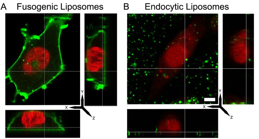
Nanomaterials 2019, 9, 1025                                                                             2 of 16

shape, preferably forming three-dimensional phases like inverse hexagonal (HII ) or cubic instead of
a lamellar phase. During the phase transition from one into the other phase, a fusion-intermediate
structure can form [14–19]. In some cases, two-dimensional lipid mixing entropy is the driving force
regulating the spontaneous formation of multicomponent membranes [20].
      Although such liposomes prevailed as fusogenic, they reach significant fusion efficiencies with
living cells only in combination with a cationic lipid, e.g., DOTAP, and an aromatic molecule, such as
fluorescence dyes [21]. Such so-called fusogenic liposomes (FLs) are able to fuse with mammalian cells
with high efficiency of about 80–100% within 1–5 min without any toxic effect. As shown previously,
the cargo molecules, e.g., proteins [22], nucleic acids [23], or different kind of lipids [24], can efficiently
be delivered to many different cell types due to successful membrane mixing of the liposomal and the
plasma membranes and subsequent content release. Here, membrane fusion success correlated with
the delivery efficiency of the cargo. Therefore, we systematically varied the composition of empty FLs
and identified the components essential for fusion with biological membranes [25]. Thereby, we found
that a cationic lipid with an inverted conical shape and an aromatic molecule are mandatory for fusion
induction. Although the addition of neutral lipids, e.g., DPPC or DOPE, is not essential, it can be
used to tune fusion efficiency. The simple exchange of DOPE with DOPC can turn the fusion ability
of liposomes from 90% into 10% without nominative changes either in particle size or zeta potential
characteristic for the surface charge of the liposomes.
      The central event during membrane fusion is the merging of two membranes, which is defined by
the lipid matrix itself. Beside embedded proteins and lipid composition, environmental conditions
determine fusion ability. For example, Zimmerberg and coworkers examined membrane fusion in
a living cell system and found that it is driven by an osmotic gradient [26], while Akimov et al. showed
for a protein-free model system that changes of the environmental pH in the physiologically relevant
range between 4.0 and 7.5, notably affected the membrane fusion rate [27].
      Because membrane fusion, in general, is in many instances dependent on environmental conditions
we systematically studied the role of temperature, osmolality, pH and ionic concentration of the buffer
on the fusion efficiency of cationic liposomes with living cells in vitro. Chinese hamster ovary
(CHO) cells were used as mammalian cells, and fusion efficiency of cationic liposomes containing
phosphoethanolamine (PE) or phosphatidyl-choline (PC) as neutral compounds and an aromatic
molecule were analyzed. Additionally, structural investigation of liposomes was carried out using
small angle neutron scattering (SANS) [28] to study the influence of thermotropic lipid phases for
efficient membrane fusion.

2. Materials and Methods

2.1. Chemicals
      We used the cationic lipid 1,2-dioleoyl-3-trimethylammonium-propane (chloride salt)
(DOTAP), and the neutral lipids 1,2-dipalmitoyl-sn-glycero-3-phosphoethanolamine (C16(0)PE),
1,2-dipalmitoleoyl-sn-glycero-3-phosphoethanolamine (C16(1)PE), 1,2-dipalmitoyl-sn-glycero-3-
phosphocholine (C16(0)PC), 1,2-dipalmitoleoyl-sn-glycero-3-phosphocholine (C16(1)PC), 1,2-distearoyl-
sn-glycero-3-phosphoethanolamine (C18(0)PE), 1,2-dioleoyl-sn-glycero-3-phosphoethanolamine
(C18(1)PE), 1,2-distearoyl-sn-glycero-3-phosphocholine (C18(0)PC), and 1,2-dioleoyl-sn-glycero-3-
phosphocholine (C18(1)PC). As fluorescently labeled lipids 1,2-dioleoyl-sn-glycero-3-
phosphoethanolamine-N-(dipyrrometheneborondifluoride)butanoyl (TFPE-head) and 1-palmitoyl-2-
(dipyrrometheneboron difluoride)-undecanoyl-sn-glycero-3-phosphoethanolamine (TFPE-chain) were
applied. All mentioned lipids were purchased from Avanti Polar Lipids, Inc. (Alabaster, AL, USA)
and used without further purification. The fluorescently labelled lipid N-(4,4-difluoro-5,7-dimethyl-
4-bora-3a,4a-diaza-s-indacene-3-propionyl)-1,2-dihexadecanoyl-sn-glycero-3-phosphoethanolamine
(triethylammonium salt) (BODIPY FL-DHPE) and the lipid analogue 1,10 -dioctadecyl-3,3,30 ,30 -
Nanomaterials 2019, 9, 1025                                                                       3 of 16

tetramethylindotricarbocyanine iodide also called DiIC18 (7) (DiR) was ordered from Thermo Fisher
Scientific (Eugene, OR, USA).

2.2. Preparation of Liposomes

2.2.1. Liposomes for Treatment of CHO Cells and Microscopy
      Liposomes were prepared according to the method described by Kolasinac et al. with few
modifications [25]. In brief, lipid components, like neutral and cationic lipids, and the fluorescent
compound were mixed in chloroform (EMSURE grade, VWR, Darmstadt, Germany) at a ratio of
1/1/0.1 mol/mol. Chloroform was evaporated under vacuum for 0.5 h. Afterward, lipids were dispersed
in 20 mM N-2-hydroxyethylpiperazine-N-2 ethane sulfonic acid (HEPES) buffer (VWR, Darmstadt,
Germany) at a total lipid concentration of 2 mg/mL and pH 7.4 (osmolality 30 mOsm/kg) or in distilled
and deionized water at the same concentration. Buffer osmolality was determined using a freezing
point osmometer (Osmomat 030 from Gonotec, Berlin, Germany). The solution was vortexed for
1–2 min to produce multilamellar liposomes. After homogenization in an ultrasonic bath (Sonocool,
Bandelin electronic GmbH, Berlin, Germany) for 20 min at 5 ◦ C, mainly unilamellar vesicles were
formed. Before usage, liposomes were kept at 4 ◦ C for no longer than two days.

2.2.2. Liposomes for Small Angle Neutron Scattering (SANS)
     For SANS, preparation of liposomes was slightly altered. For these experiments, BODIPY-FL-DHPE
was used as a dye. The total lipid concentration was set to 10 mg/mL. After evaporation of chloroform,
the lipid film was resuspended in 20 mM HEPES dissolved in D2 O (99 atom % D, Sigma-Aldrich,
Taufkirchen, Germany) and vortexed vigorously without additional sonication. Samples were stored
at −20 ◦ C until usage. One hour before measurements, samples were thawed and vortexed vigorously
before being transferred into quartz cuvettes (110-QS, quartz glass, Suprasil, 1 mm path length, Hellma,
Müllheim, Germany) for SANS measurements.

2.3. Cell Culture
      Experiments were performed on Chinese Hamster Ovary K1 cells (CHOs) purchased from
American Type Culture Collection (ATTC, Manassas, VA, USA). They were maintained in DMEM-F12
(Sigma-Aldrich, Taufkirchen, Germany) supplemented with 10% fetal bovine serum (FBS) (Thermo
Fisher Scientific, Waltham, MA, USA), 10,000 units penicillin and 10 mg/mL streptomycin (both
Sigma-Aldrich). During culture, cells were kept at 37 ◦ C and 5% CO2 in a saturated humid atmosphere.
Cell density never exceeded 80% confluence. Before microscopy, glass surfaces (∅ = 3.5 cm Petri dish)
were coated with human fibronectin (10 µg/mL, BD Biosciences, San Jose, CA, USA) for 30 min at 37 ◦ C
and 50,000 cells were seeded on them and cultivated for 24 h. Cell nuclei were stained for 15 min at
37 ◦ C with DRAQ5 (red fluorescence, Thermo Fischer Scientific, Waltham, MA, USA) or Hoechst 33342
(blue fluorescence, Thermo Fischer Scientific, Waltham, MA, USA) according to the manufacturer’s
protocols. Furthermore, 10 µL of the liposome stock solutions were diluted 1/50 with phosphate buffer
saline (PBS, 290 mOsm/kg, 137 mM NaCl; 2.7 mM KCl; 1.47 mM KH2 PO4 ; 8.1 mM Na2 HPO4 ) at
pH 5–9 (adjusted by addition of 1 M NaOH or HCl), phosphate buffer (PB, 30 mOsm/kg, 2.7 mM
KCl; 1.47 mM KH2 PO4 ; 8.1 mM Na2 HPO4 ), or glucose solution of defined osmolality (30 mOsm/kg
and 290 mOsm/kg) and cells on glass substrates (~100,000 of cells) were incubated in these solutions
for 5 min in the incubator (37 ◦ C and 5% CO2 , saturated humid atmosphere). After the treatment,
the suspension of liposomes was exchanged with fresh, pre-warmed cell culture medium. In some
control experiments, cells were first washed three times using a sodium heparin solution at 2 mg/mL
concentration in PBS buffer before medium exchange to eliminate remaining liposomes from the cell
surfaces. For temperature-dependent experiments, 10 µL of the liposomal stock solutions, stored
at 4 ◦ C no longer than 24 h, were diluted 1/50 with phosphate buffer saline (PBS, 290 mOsm/kg) at
room temperature. Before, liposomes and cells were left for 5 min at room temperature. Immediately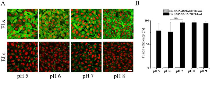
Nanomaterials 2019, 9, 1025                                                                       4 of 16

afterward, the cells were incubated with liposomal solutions at 4 ◦ C, 20 ◦ C, 30 ◦ C or 37 ◦ C for
5 min. Thereafter, they were washed by fresh, pre-warmed cell culture medium, and samples were
immediately analyzed by laser scanning microscopy.

2.4. Characterization of Size and Zeta Potential Distribution of Liposomes
     Both particle size and ζ-potential distributions were measured using a zetasizer (Nano ZS from
Malvern Instruments, Malvern, UK) equipped with a HeNe laser (633 nm). Scattered laser light was
collected at a constant angle of 173◦ . Prior to measurements liposome stock solutions were diluted
using the appropriate buffer. The temperature was set using the instrument thermostat. Data were
collected from three independently prepared samples and analyzed using the instrument software
(DTS from Malvern Instruments). Reported data are mean peak position and its standard deviation
(mean (s.d.))

2.5. Microscopy
     Samples were imaged using a confocal laser scanning microscope (LSM 710 from Carl Zeiss
MicroImaging GmbH, Jena, Germany) equipped with a near UV laser (405 nm), an argon ion laser
(488 nm), and a helium-neon laser (633 nm). Both TopFluor lipid derivatives were excited at 488 nm,
and their fluorescence emissions were detected using a bandpass filter BP 495–550 nm (green channel).
The lipid analog DiR and the nuclear stain DRAQ5 were excited using the 633 nm laser line, and the
emitted signal was collected through the long pass filter LP 650 nm. Hoechst 33342 was excited by the
405 nm laser line, and its emission was detected using a BP 505/90. For imaging, a Plan-Apochromat
40×/1.40 Ph3 (Carl Zeiss, Oberkochen, Germany) objective was used. To maintain appropriate culture
conditions, the confocal microscope was equipped with an incubator (Incubator XL 2, Carl Zeiss,
Oberkochen, Germany). Temperature and CO2 were kept constant at 37 ◦ C and 5%, respectively. The
experiments were repeated at least three times. For each experiment minimum five images were taken,
with at least one hundred cells per image. Overall, fusion efficiency was determined from no less than
1500 cells at each condition.

2.6. Analysis of Images
     An algorithm was developed to quantify fusion efficiency from fluorescence micrographs. It is
based on staining intensity. The code was implemented in Matlab (R2017, Mathworks, Natick, MA,
USA). In the first step, individual cells were segmented. To this end, the nuclei channel was used. This
image was first smoothed (Gaussian filter, standard deviation 3 pixels, pixel size 173 nm throughout)
and morphological opening (disk-shaped structuring element of radius 9 pixels) was performed.
The bright nuclei were segmented using the mean grey value of the image as an intensity threshold.
Subsequently, morphological opening and closing (both with a disk of radius 5 pixels) were performed
on the mask. Next, the watershed transformation was used to separate overlapping nuclei and to
segment cells. Therefore, the distance transform of the negative mask was calculated and multiplied
by −1. Then, local minima (depth less than 2) were eliminated from this resulting image, and finally,
the watershed transform was applied to that image. Next, to the separated nuclei labels, the dividing
lines between the “watershed” areas of neighboring nuclei, computed by the watershed transform,
were used as the shape of the corresponding cell.
     Afterward, the fluorescent lipid signal was analyzed. Since the many small, bright spots in the
images indicated lumps, endocytic, or non-fused liposomes, they were removed. Therefore, the image
was first smoothed (Gaussian filter, the standard deviation of 3 pixels). Then, local bright spots were
detected using the algorithm described in the work of Hersch and colleagues [29]. In brief, the local
z-score of each pixel was calculated within a 91 × 91 environment and segmented using a threshold of
2 for the z-score. Only regions with the area below 100,000 pixels × pixels were accepted. Each such
region was then enlarged by morphological dilation (the disk of radius 3 pixels). On the smoothed
fusion signal image, all spots identified by the z-score segmentation were replaced by pixel values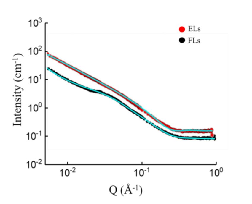
Nanomaterials 2019, 9, 1025                                                                            5 of 16

calculated by inward interpolation from the grey scale values at the rim of the spot (MatLab function
region fill). Spot detection and interpolation of grey values were performed twice. Using the processed
intensity image from the second part of the program and the cell label image from the first part, the
average grey value intensity for each cell was calculated individually. Using the same manually chosen
threshold for all images, all cells were separated into fused (all cells with an average grey value above
the threshold) and non-fused (all cells below the threshold) cells.

2.6.1. Small Angle Neutron Scattering (SANS)
     SANS experiments were carried out at small angle scattering set-up KWS-2, operated by JCNS at
Forschungsneutronenquelle Heinz Maier-Leibnitz, FRM II, in Garching (Germany) [30]. A wavelength
of λ = 7 Å (∆ λ/λ = 10%) and sample-detector distances (SDD) of 1.58, 7.58, and 19.48 m were used to
cover a q-range of 0.002–0.221 Å−1 . The detector was a 3 He detector with a resolution of 8 mm. Exposure
times were 5 min at SDD = 1.58 m, 10 min at SDD = 7.58, and 20 min at SDD = 19.48 m. Samples were
placed in an aluminum holder with plastic cover, and the temperature was controlled by a Peltier
element combined with a counter cooling by a water thermostat. The cuvettes were Hellma quartz
glass cuvettes with a 1 mm sample thickness. Experiments were performed at 37 ◦ C. The scattering
of D2 O and the empty cell were subtracted from the sample scattering taking the transmissions into
account. The resulting intensities were azimuthally averaged. Good agreement was found wherever
curves at different SDDs overlapped. All data corrections were performed with the software QtiKWS
provided by JCNS. Fitting was done by SasView software version 4.2.0 (http://sasview.org).

2.6.2. Model Functions Used for the Data Fitting
       All fit functions contained a scale factor I0 and background Ib , i.e., they were of the form

                                             I (q) = I0 f (q) + Ib                                        (1)

where q denotes the scattering vector. The scale factor contains the scattering volume and the scattering
length density difference between solvent and structure.
     The lamellar model provides the scattering intensity, I(q), for a lyotropic lamellar phase. A uniform
scattering length density and random distribution in solution are assumed, which results in

                                                       1 − cos(qδ)
                                             f (q) ∝                                                      (2)
                                                            δq4

where δ denotes bilayer thickness [31].
     The ellipsoid model is calculated from the form factor for randomly oriented ellipsoids of
revolution with uniform scattering length density. This results in

                                                  [sin(qr) − qr cos(qr)]2
                                        f (q) =                                                           (3)
                                                          9(qr)6

with                                       q
                                        r = Rb 2 sin2 α + Ra 2 cos2 α                                     (4)

where α denotes the angle between the rotational axis of the ellipsoid and the q-vector, Ra is its radius
along this axis, and Rb the radius perpendicular to it. The orientation of the ellipsoid is numerically
averaged over a sphere to give the final fit model [28,32].
    As the final model, we used a general power law

                                                  f (q) = kqk−m                                           (5)
Nanomaterials 2019, 9, 1025 6 of 16
Nanomaterials 2019, 9, x FOR PEER REVIEW 6 of 16
2.7.Statistical
2.7. StatisticalAnalysis
Analysis
Statisticalanalyses
Statistical analysesof
ofdata
datawere
wereperformed
performedbybyone-way
one-wayANOVA
ANOVAusingusingOrigin
Origin9.0
9.0(OriginLab
(OriginLab
Co.,Northampton,
Co., Northampton,MA, MA,USA).
USA).ppNanomaterials 2019, 9, 1025 7 of 16
Nanomaterials 2019, 9, x FOR PEER REVIEW 7 of 16
polydispersity
3.1. index (PDI) in Table 1) with a hydrodynamic diameter of about 115 nm and zeta potential
Influence of Temperature
of + 50 mV without significant changes in the temperature range from 4 °C to 37 °C (see Table 1). Within
Liposomes
statistical with the
significance, composition
endocytic described
liposomes (ELs) above were characterized
gave similar fromTable
results as FLs (see the physicochemical
1).
point of view depending on the temperature. Here, FLs showed a homogeneous population (see
polydispersity index (PDI) in
Table 1. Temperature Table 1) with
dependence a hydrodynamic
of the hydrodynamic diameter
diameter, of about 115 nm
polydispersity and (PDI),
index zeta potential
and
of + 50 mV without significant changes in the temperature range from 4 ◦ C to 37 ◦ C (see Table 1).
zeta potential of fusogenic (DOPE/DOTAP/TFPE-head 1/1/0.1 mol/mol) (FLs) and endocytic
Within(DOPC/DOTAP/TFPE-head
statistical significance, endocytic liposomes
1/1/0.1 mol/mol) (ELs)
(ELs) gave similar
liposomes. resultsare
Experiments asperformed
FLs (see Table 1).
in a PBS
buffer at physiological pH 7.4. Given are averages over three independent measurements and, in
Table 1. Temperature
parentheses, dependence
their standard of the hydrodynamic diameter, polydispersity index (PDI),
deviations.
and zeta potential of fusogenic (DOPE/DOTAP/TFPE-head 1/1/0.1 mol/mol) (FLs) and endocytic
Hydrodynamic Diameter (nm) (s.d.)
(DOPC/DOTAP/TFPE-head
Liposomal Type 1/1/0.1 mol/mol) (ELs) liposomes. Experiments are performed in a PBS
4 °C 20 °C 30 °C 37 °C
buffer at physiological pH 7.4. Given are averages over three independent measurements and, in
(FLs) 110 (13) 116 (13) 118 (16) 117 (13)
parentheses, their standard deviations.
(ELs) 142 (42) 154 (68) 158 (71) 155 (72)
PDI (s.d.)
(FLs)Liposomal Type 0.26 (0.06) Hydrodynamic
0.23 (0.02)Diameter (nm)0.23 (s.d.)
(0.02) 0.23 (0.02)
(ELs) ◦
0.34 (0.12)
4 C 0.32
20 C◦ (0.11) ◦
30 C 0.35 (0.11) 37 ◦ C 0.33 (0.11)
Zeta Potential (mV) (s.d.)
(FLs)
(FLs) 110 (13)
59 (4)
11648(13)
(8)
118 (16) 51 (3)117 (13) 43 (13)
(ELs) (ELs) 142 (42)
68 (13) 15464(68)
(10) 158 (71) 56 (8)155 (72) 58 (8)
PDI (s.d.)
(FLs) 0.26 (0.06) 0.23 (0.02) 0.23 (0.02) 0.23 (0.02)
Upon variation of(ELs)
temperature,0.34
two(0.12)
main trends were 0.35
0.32 (0.11) observed.
(0.11) Liposomes
0.33 (0.11) containing DOPE
as neutral lipid homogenously stained the plasma membranes of CHO
Zeta Potential (mV) (s.d.) cells in the whole temperature
range from 4 °C to 37 °C (see Figure 2A). Here, fusion efficiencies of above(13)
(FLs) 59 (4) 48 (8) 51 (3) 43 80% were determined
(ELs) (Figure 68
from fluorescent micrographs (13)
2B). 64 (10)
In contrast, liposomes56 (8)
containing58 a
(8)phosphocholine, here
DOPC, as neutral lipid stuck to the cell surface resulting in an inhomogeneous dotted fluorescence
pattern,
Uponand afterward
variation were taken up
of temperature, twobymain
endocytosis.
trends wereInternalization by endocytosis
observed. Liposomes was detected
containing DOPE asin
the whole
neutral lipidtemperature
homogenously range from 4the
stained °C plasma
to 37 °Cmembranes
(Figure 2B).ofDespite the temperature
CHO cells in the wholeshock inherent
temperature
in the ◦
procedure, no ◦
indications for cell stress were observed.
range from 4 C to 37 C (see Figure 2A). Here, fusion efficiencies of above 80% were determined
In all cases,micrographs
from fluorescent the headgroup of the2B).
(Figure neutral lipid, PE
In contrast, or PC, respectively,
liposomes containing controlled fusion ability,
a phosphocholine, here
DOPC, as neutral lipid stuck to the cell surface resulting in an inhomogeneous dotted fluorescenceof
while chain length or saturation had much lower effects (see Table 2). Moreover, the replacement
the head
pattern, labeled
and lipid (TFPE-head)
afterward were taken up asbytheendocytosis.
aromatic component with a by
Internalization chain labeled lipid
endocytosis was(TFPE-chain)
detected in
or whole
the a fluorescent lipid analog
temperature range from ◦ C to
(DiR)4 did not ◦ C (Figure 2B).
37significantly influence
Despitefusion efficiency of
the temperature liposomes
shock in the
inherent in
analyzed temperature range (see Table S1).
the procedure, no indications for cell stress were observed.
Figure 2. (A) Fluorescence micrographs of CHO cells treated with fusogenic liposomes
Figure
(FLs) 2. (A) Fluorescence micrographs
DOPE/DOTAP/TFPE-head of CHO upper
(1/1/0.1 mol/mol), cells treated
row, andwithendocytic
fusogenicliposomes
liposomes(ELs)
(FLs)
DOPE/DOTAP/TFPE-head (1/1/0.1 mol/mol), upper row, and endocytic
DOPC/DOTAP/TFPE-head (1/1/0.1 mol/mol), lower row. Green: TFPE-head signal, red: Nuclei liposomes (ELs)
DOPC/DOTAP/TFPE-head
staining with DRAQ5. Scale bar, (1/1/0.1
20 µm,mol/mol),
applies to lower row. Green:
all. Experiments wereTFPE-head
done in PBSsignal,
buffer. red: Nuclei
(B) Fusion
staining with DRAQ5. Scale bar, 20 µm, applies to all. Experiments were done in PBS buffer.
efficiencies. Whiskers indicate standard deviations of at least three independent experiments. In total, (B)
Fusion efficiencies. Whiskers indicate standard
more than 1500 cells were analyzed at each condition. deviations of at least three independent experiments.
In total, more than 1500 cells were analyzed at each condition.Nanomaterials 2019, 9, 1025 8 of 16
In all cases, the headgroup of the neutral lipid, PE or PC, respectively, controlled fusion ability,
while chain length or saturation had much lower effects (see Table 2). Moreover, the replacement of the
head labeled lipid (TFPE-head) as the aromatic component with a chain labeled lipid (TFPE-chain) or
a fluorescent lipid analog (DiR) did not significantly influence fusion efficiency of liposomes in the
analyzed temperature range (see Table S1).
Table 2. Temperature dependence of fusion efficiencies of liposomes containing the cationic lipid
DOTAP, different neutral lipids, and TFPE-head as an aromatic molecule (1/1/0.1 mol/mol). Experiments
are performed in a PBS buffer at physiological pH 7.4 and osmolality 280 mOsm/kg. Given are averages
over at least three independent measurements and, in parentheses, their standard deviations.
Fusion Efficiency % (s.d.)
Liposomal Composition
4 ◦C 20 ◦ C 30 ◦ C 37 ◦ C
C16(0)PE/DOTAP/TFPE-head 97 (1) 98 (2) 98 (1) 99 (1)
C16(0)PC/DOTAP/TFPE-head 0 (0) 0 (0) 0 (0) 0 (0)
C16(1)PE/DOTAP/TFPE-head 88 (11) 93 (6) 96 (3) 98 (2)
C16(1)PC/DOTAP/TFPE-head 13 (3) 16 (7) 14 (3) 17 (1)
C18(0)PE/DOTAP/TFPE-head 92 (6) 96 (1) 89 (9) 95 (5)
C18(0)PC/DOTAP/TFPE-head 37 (8) 38 (11) 47 (6) 62 (5)
C18(1)PE/DOTAP/TFPE-head 83 (9) 90 (3) 87 (4) 92 (6)
C18(1)PC/DOTAP/TFPE-head 3 (2) 5 (4) 2 (1) 4 (2)
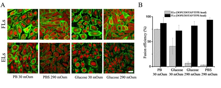
3.2. Phase States of Endocytic and Fusogenic Liposomes
To find the reasons underlying the very different fusion behaviors of PC and PE containing
cationic liposomes, SANS analyses were carried out at the physiologically most relevant temperature of
37 ◦ C. For these measurements, liposomes were formed in HEPES buffered heavy water (cf. Materials
and methods for details). With this method, multi-dispersed, multilayered vesicles were formed.
Nanomaterials 2019, 9, x FOR PEER REVIEW 9 of 16
Experimental results and fits are shown in Figure 3.
Figure 3. Scattering curves of DOPE/DOTAP/BODIPY-FL-DHPE (1/1/0.1 mol/mol) (black circles)
Figure 3. Scattering curves of DOPE/DOTAP/BODIPY-FL-DHPE (1/1/0.1 mol/mol) (black circles)
liposomes and DOPC/DOTAP/BODIPY-FL-DHPE (1/1/0.1 mol/mol) (red circles) liposomes measured
liposomes and DOPC/DOTAP/BODIPY-FL-DHPE (1/1/0.1 mol/mol) (red circles) liposomes measured
at 37 ◦ C. Cyan lines indicate corresponding fits of a single measurement.
at 37 °C. Cyan lines indicate corresponding fits of a single measurement.
The scattering profile of DOPC containing liposomes was adequately modeled by a lamellar lipid
3.3. Influence of Ionic Concentration
phase, with a bilayer thickness of 42.7 Å. DOPE containing liposomes, however, displayed a different
The pattern
scattering influence of the
with surroundingshoulder
a characteristic at Q = 0.015 was
ionic concentration Å−1 .examined
The latterfor variousthe
indicated ionic strengths
presence of
of thescale
small medium. Forand
features the could
purpose
notofbethese experiments,
described the lipid
by a lamellar filmalone.
phase was hydrated
The bestinfitultrapure water
was achieved
instead
by of a bufferoftothe
a superposition avoid the presence
scattering functionofofions in theparticles
ellipsoid liposomalandstock solution.
a power law. TheSubsequently,
curve of
liposomes
DOPE were liposomes
containing diluted incould
phosphate bufferwith
not be fitted (PB)a cylinder
at low total
model.ionThis
concentration
argues against(30the
mM) or in
familiar
phosphate buffer containing
hexagonal/inverted additional
hexagonal phase salineAlso,
of DOPE. (PBS)theatcombination
high total ion concentration
of the cylinder model(280combined
mM). The
presence of ions drastically increased the hydrodynamic size of both types of liposomes (compare
Tables 1 and 3) and reduced the liposomal homogeneity, as shown in Table 3. No significant
differences were detected between FLs and ELs (Table 1). The analysis of liposomal zeta potential
showed a significant reduction in liposomal charges of both liposomes in the presence of PBS and a
moderate decrease in PB buffer compared to glucose solutions.Nanomaterials 2019, 9, 1025 9 of 16
with ellipsoid failed to fit the curve. Additionally, combinations of models (lamellar with ellipsoid and
lamellar with power law) did not fit perfectly the specific region at Q = 0.015 Å−1 . Apart from that part
of the scattering curve, the lamellar model fitted nicely, which indicates the presence of bilayers. The
observed power law contribution is a common occurrence if the probed length scale is smaller than the
scattering object; it reflects local structures of the object [33]. Therefore, the agreeable fit of this model
combining a power law with an ellipsoid and separate fitting of most of the pattern by the lamellar
model leads to the following hypothesis on the structure: We propose small micelle-like structures
are embedded into the lipid bilayers. The best fitting power-law exponent was 2.99, which can be
attributed to large solid vesicles with a rough surface. The best fitting ellipsoidal particles had a polar
radius of 24.8 Å and an equatorial radius of 88.5 Å. The fitting functions are described in the section
Materials and Methods.
3.3. Influence of Ionic Concentration
The influence of the surrounding ionic concentration was examined for various ionic strengths of
the medium. For the purpose of these experiments, the lipid film was hydrated in ultrapure water
instead of a buffer to avoid the presence of ions in the liposomal stock solution. Subsequently, liposomes
were diluted in phosphate buffer (PB) at low total ion concentration (30 mM) or in phosphate buffer
containing additional saline (PBS) at high total ion concentration (280 mM). The presence of ions
drastically increased the hydrodynamic size of both types of liposomes (compare Tables 1 and 3) and
reduced the liposomal homogeneity, as shown in Table 3. No significant differences were detected
between FLs and ELs (Table 1). The analysis of liposomal zeta potential showed a significant reduction
in liposomal charges of both liposomes in the presence of PBS and a moderate decrease in PB buffer
compared to glucose solutions.
Table 3. Ionic concentration dependence of the hydrodynamic diameter, polydispersity index (PDI)
and zeta potential of fusogenic (DOPE/DOTAP/TFPE-head 1/1/0.1 mol/mol) (FLs) and endocytic
(DOPC/DOTAP/TFPE-head 1/1/0.1 mol/mol) (ELs) liposomes. Experiments are performed in a PBS
buffer at physiological pH 7.4. Given are averages over three independent measurements and,
in parentheses, their standard deviations.
Hydrodynamic Diameter (nm) (s.d.)
Liposomal Type
PB (30 mOsm/kg) PBS (290 mOsm/kg) Glucose (30 mOsm/kg) Glucose (290 mOsm/kg)
(FLs) 568 (145) 567 (102) 537 (71) 493 (157)
(ELs) 460 (277) 551 (357) 524 (296) 515 (357)
PDI (s.d.)
(FLs) 0.25 (0.05) 0.36 (0.13) 0.41 (0.09) 0.32 (0.01)
(ELs) 0.34 (0.07) 0.32 (0.11) 0.35 (0.11) 0.33 (0.11)
Zeta Potential (mV) (s.d.)
(FLs) 36 (6) 28 (4) 72 (3) 69 (11)
(ELs) 62 (1) 36 (1) 78 (3) 73 (7)
Fusion efficiency of CHO cells with the same liposomes was also determined. The fluorescence
signal of liposomes after internalization was monitored by confocal microscopy. Liposomes containing
DOPE as neutral lipid diluted in PB buffer showed homogeneous membrane staining with high
fusion efficiencies of approximately 90%. When the same liposomes were diluted in PBS buffer, they
remained fusogenic with similar or slightly higher efficiencies (Figure 4). However, when DOPE was
replaced with DOPC as a neutral component, liposomes displayed different behaviors depending on
ionic strength. In PB, they fused with CHO cells with high efficiencies, while no significant fusion
was detected in PBS (Figure 4). During the treatment with low ionic strength, cells reacted on these
hypo-osmotic conditions by membrane blebbing. Nevertheless, cells recovered immediately after the
treatment without any signs of damage. The same trends were observed in the case of all investigated
PE or PC containing liposomes irrespective of chain length or unsaturation of the neutral component
(see Table 4) or exchange of the aromatic component (see Table S2).C16(0)PC/DOTAP/TFPE-head 93 (6) 3 (3) 66 (2) 5 (4)
C16(1)PE/DOTAP/TFPE-head 86 (4) 99 (1) 98 (1) 99 (1)
C16(1)PC/DOTAP/TFPE-head 33 (3) 0 (0) 27 (7) 10 (2)
C18(0)PE/DOTAP/TFPE-head 76 (9) 89 (2) 82 (12) 98 (2)
C18(0)PC/DOTAP/TFPE-head 89 (3) 51 (1) 64 (8) 65 (8)
C18(1)PE/DOTAP/TFPE-head
Nanomaterials 2019, 9, 1025 87 (12) 94 (5) 72 (11) 82 (8) 10 of 16
C18(1)PC/DOTAP/TFPE-head 75 (1) 3 (1) 41 (16) 7 (3)
Figure 4. (A) Fluorescence micrographs of CHO cells after treatment with fusogenic (DOPE/DOTAP/
TFPE-head
Figure 4. 1/1/0.1
(A) mol/mol) uppermicrographs
Fluorescence row, and endocytic
of CHO liposomes,
cells (DOPC/DOTAP/TFPE-head 1/1/0.1
after treatment with fusogenic
mol/mol) lower row. Green: 1/1/0.1
(DOPE/DOTAP/TFPE-head TFPE-head signal, red:
mol/mol) upperNucleic
row,staining
and with DRAQ5. liposomes,
endocytic Scale bar,
20 µm, applies to all. (B) Fusion efficiencies. Whiskers indicate standard deviations
(DOPC/DOTAP/TFPE-head 1/1/0.1 mol/mol) lower row. Green: TFPE-head signal, red: Nucleic of at least three
independent experiments.
staining with DRAQ5. Scale bar, 20 µm, applies to all. (B) Fusion efficiencies. Whiskers indicate
standard
Table deviations
4. Fusion of at least
efficiencies three independent
of liposomes experiments.
containing the cationic lipid DOTAP, different helper lipids,
and TFPE-head as a dye molecule (1/1/0.1 mol/mol) depending on the osmolality of the solution at 37 ◦ C.
3.4. Influence of Osmolality
Average values of at least three independent experiments and their standard deviations are given.
To test if the remarkably different behavior ofFusion DOPC containing
Efficiency % (s.d.) liposomes in PB and PBS
Liposomal Composition
originated from electrostatic PBor
(30osmotic
mOsm/kg)effects, wemOsm/kg)
PBS (290 varied osmolality
Glucose (30by an uncharged
mOsm/kg) Glucose solute. To this
(290 mOsm/kg)
end, liposomes were
C16(0)PE/DOTAP/TFPE-head prepared as
98 (1) previously described
85 (5) and diluted
95 (4) subsequently in
79 (2)a low (30
C16(0)PC/DOTAP/TFPE-head 93 (6) 3 (3) 66 (2)
mOsm/kg) or a high (290 mOsm/kg) osmolality glucose solution without any addition of salts. 5 (4)
C16(1)PE/DOTAP/TFPE-head 86 (4) 99 (1) 98 (1) 99 (1)
Liposomes containing DOPE as
C16(1)PC/DOTAP/TFPE-head neutral lipid diluted
33 (3) 0 (0) in 30 mOsm/kg 27 (7) or 290 mOsm/kg 10 (2) glucose
C18(0)PE/DOTAP/TFPE-head
solutions 76 (9)
fused with the cell membrane of CHO 89 (2) with similar high
cells 82 (12) efficiencies of approximately
98 (2)
C18(0)PC/DOTAP/TFPE-head 89 (3) 51 (1) 64 (8) 65 (8)
80%. In contrast, liposomes containing
C18(1)PE/DOTAP/TFPE-head 87 (12) DOPC as 94 neutral
(5) component72again (11) showed fusion efficiencies
82 (8)
depended on osmolality. 75
C18(1)PC/DOTAP/TFPE-head
that If (1)such liposomes 3 (1)were diluted in 41 (16)
30 mOsm/kg glucose 7 (3) solution,
significant fusion (approx. 50% efficiency) was detected, while almost no fusion events were observed
3.4. Influence
in 290 mOsmofglucose
Osmolality
solution (see Figure 4 and Table 2). During the treatment with low osmolality
buffer, cells reacted on hypo-osmotic
To test if the remarkably differentconditions
behavior of byDOPC
membrane blebbing.
containing Nevertheless,
liposomes in PB and afterPBS
the
treatment, cells recovered immediately without any obvious damage. Upon
originated from electrostatic or osmotic effects, we varied osmolality by an uncharged solute. To this exchange of neutral
lipids
end, with various
liposomes chain lengths
were prepared and saturation,
as previously describedour andresults
dilutedindicated
subsequentlya universal
in a lowtrend valid for
(30 mOsm/kg)
liposomes containing PE or PC neutral lipids, as shown in Table 2. PE-containing
or a high (290 mOsm/kg) osmolality glucose solution without any addition of salts. Liposomes liposomes fused
containing DOPE as neutral lipid diluted in 30 mOsm/kg or 290 mOsm/kg glucose solutions fused
with the cell membrane of CHO cells with similar high efficiencies of approximately 80%. In contrast,
liposomes containing DOPC as neutral component again showed fusion efficiencies that depended on
osmolality. If such liposomes were diluted in 30 mOsm/kg glucose solution, significant fusion (approx.
50% efficiency) was detected, while almost no fusion events were observed in 290 mOsm glucose
solution (see Figure 4 and Table 2). During the treatment with low osmolality buffer, cells reacted on
hypo-osmotic conditions by membrane blebbing. Nevertheless, after the treatment, cells recovered
immediately without any obvious damage. Upon exchange of neutral lipids with various chain lengths
and saturation, our results indicated a universal trend valid for liposomes containing PE or PC neutral
lipids, as shown in Table 2. PE-containing liposomes fused very efficiently with the plasma membrane
of CHO cells independent of chain length, saturation, or the aromatic component (see also Table S2),
while the fusion efficiency of PC containing liposomes strongly depended on buffer osmolality.
3.5. Influence of pH
Both types of liposomes, fusogenic and endocytic, were characterized in the pH range of buffer
between 5 and 9. Even though, the PBS buffer capacity was not ideal in the whole range, allvery efficiently with the plasma membrane of CHO cells independent of chain length, saturation, or
the aromatic component (see also Table S2), while the fusion efficiency of PC containing liposomes
strongly depended on buffer osmolality.
Nanomaterials 2019, 9, 1025 11 of 16
3.5. Influence of pH
Both types of liposomes, fusogenic and endocytic, were characterized in the pH range of buffer
experiments were carried out in a PBS buffer, where the pH was adjusted to the appropriate value. FLs
between 5 and 9. Even though, the PBS buffer capacity was not ideal in the whole range, all experiments
(DOPE/DOTAP/TFPE-head 1/1/0.1 mol/mol), as well as ELs (DOPC/DOTAP/TFPE-head 1/1/0.1 mol/mol),
were carried out in a PBS buffer, where the pH was adjusted to the appropriate value. FLs
became less homogeneous with larger particles formed at higher pH values. Zeta potential of FLs was
(DOPE/DOTAP/TFPE-head 1/1/0.1 mol/mol), as well as ELs (DOPC/DOTAP/TFPE-head 1/1/0.1 mol/mol),
reduced when pH was increased, while ELs did not show any pH-dependent changes (see Table 5).
became less homogeneous with larger particles formed at higher pH values. Zeta potential of FLs was
reduced when
Table 5. pH was increased,
pH dependence while ELs did
of the hydrodynamic not show
diameter, any pH-dependent
polydispersity index (PDI)changes
and zeta(see Tableof5).
potential
fusogenic (DOPE/DOTAP/TFPE-head 1/1/0.1 mol/mol) (FLs) and endocytic (DOPC/DOTAP/TFPE-head
Tablemol/mol)
1/1/0.1 5. pH dependence of the hydrodynamic
(ELs) liposomes. Experimentsdiameter, polydispersity
were performed in a index (PDI) and
PBS buffer zeta potential
titrated to the
of fusogenic (DOPE/DOTAP/TFPE-head 1/1/0.1 mol/mol) (FLs) and endocytic (DOPC/DOTAP/TFPE-
indicated pH. Given are averages over three independent measurements and, in parentheses, their
head 1/1/0.1
standard mol/mol) (ELs) liposomes. Experiments were performed in a PBS buffer titrated to the
deviations.
indicated pH. Given are averages over three independent measurements and, in parentheses, their
standard deviations. Hydrodynamic Dyameter (nm) (s.d.)
Liposomal Type
pH 5 pH 6
Hydrodynamic pH 7
Dyameter (nm)pH 8
(s.d.) pH 9
Liposomal Type
(FLs) pH 5 221 (193)pH 6 179 (124) pH 7177 (36) pH 8(52)
276 pH 9
333 (117)
(FLs) (ELs) 221 (193) 118 (24)
179 (124)117 (19) 177 (36)108 (23) 276
186(52)
(22) 333 (117)
197 (16)
(ELs) 118 (24) 117 (19) 108 (23) 186 (22)
PDI (s.d.) 197 (16)
(FLs) 0.30 (0.10) 0.30 (0.11) 0.35 (0.11) PDI (s.d.)
0.31 (0.21) 0.35 (0.21)
(FLs) (ELs) 0.30 (0.10)
0.21 (0.03) 0.22 (0.02) 0.19 (0.02) 0.31
0.30 (0.11) 0.35 (0.11) 0.23(0.21)
(0.09) 0.35 (0.21)
0.32 (0.18)
(ELs) 0.21 (0.03) 0.22 (0.02) 0.19 Zeta
(0.02)Potential0.23
(mV)(0.09)
(s.d.) 0.32 (0.18)
(FLs) 42 (7) 39 (2) Zeta Potential (mV)
36 (4) 26 (4)(s.d.) 15 (5)
(FLs) (ELs) 42 (7) 35 (4) 39 (2) 36 (3) 36 (4) 38 (3) 2635(4)(3) 38 (5)15 (5)
(ELs) 35 (4) 36 (3) 38 (3) 35 (3) 38 (5)
We Wealso analyzed
also analyzed thethe
pH pHdependence
dependence ofof
membrane
membrane fusion
fusionofof
cationic
cationicliposomes
liposomes with CHO
with CHO cells
cells
atat ◦ C. Liposomes containing PE, here DOPE, as neutral lipid homogenously stained the cellular
3737 °C. Liposomes containing PE, here DOPE, as neutral lipid homogenously stained the cellular
plasma
plasma membrane
membrane of CHO
of CHO cellscells
at allatpH
allvalues in theinrange
pH values from 5from
the range to 9. Throughout the whole
5 to 9. Throughout therange,
whole
tested
range,fusion efficiency
tested fusion exceeded
efficiency75% (Figure 5).
exceeded In contrast,
75% (Figure liposomes containing
5). In contrast, phosphocholine,
liposomes containing
here DOPC, as neutral
phosphocholine, lipid
here adhered
DOPC, to the cell
as neutral surface,
lipid whichtoresulted
adhered the cellinsurface,
an inhomogeneous speckled
which resulted in an
fluorescence
inhomogeneouspattern. In thefluorescence
speckled whole pH range from
pattern. In 5the
to whole
9, internalization
pH range fromby endocytosis was rarelyby
5 to 9, internalization
detected with awas
endocytosis fusion efficiency
rarely detectedabove
with 1% (Figure
a fusion 5).
efficiency above 1% (Figure 5).
Figure 5. (A) Fluorescence micrographs of CHO cells after treatment with fusogenic (DOPE/DOTAP/
Figure 5. 1/1/0.1
TFPE-head, (A) Fluorescence
mol/mol), upper micrographs of CHO liposomes
row, and endocytic cells after(DOPC/DOTAP/TFPE-head,
treatment with fusogenic
(DOPE/DOTAP/TFPE-head,
1/1/0.1 1/1/0.1 pHmol/mol),
mol/mol), lower row, at different upper
values. Green: row, signal,
TFPE-head and red:
endocytic liposomes
Nucleic staining
(DOPC/DOTAP/TFPE-head,
with DRAQ5. Scale bar, 20 µm, 1/1/0.1 mol/mol),
applies lower
to all. (B) row,efficiencies.
Fusion at different Whiskers
pH values.indicate
Green: standard
TFPE-head
signal, red:
deviations Nucleic
of at staining
least three with DRAQ5.
independent Scale bar, 20 µm, applies to all. (B) Fusion efficiencies.
experiments.
Whiskers indicate standard deviations of at least three independent experiments.
4. Discussion
In the absence of fusogenic peptides or proteins, membrane fusion occurs in the following
sequence [4,13,18,34,35]. First, the two membranes must be brought into close contact. This step
requires the removal of tightly bound hydration water. Second, the locally disrupted outer membraneNanomaterials 2019, 9, 1025 12 of 16
leaflets melt together, forming hemifusion intermediate structures called membrane stalks. Third,
the inner lipid monolayers reorganize, which results in pore opening, as well as full membrane and
content mixing. Each of the intermediate states is characterized by a well-defined free enthalpy that
together defines the activation energy barrier for fusion. Some chemical compounds, such as ions,
drugs, or distinct lipids, as well as conditions like temperature, pH, or osmolality of the buffer, can
alter this barrier [18,35]. Because we recently analyzed the influence of the chemical composition of
cationic liposomes on their fusion efficiency with mammalian cell membranes [25], we tackled here the
effect of environmental conditions on fusion efficiency.
Changes of the environmental conditions such as ionic concentration, pH, or buffer osmolality
mainly influence the first step in the fusion process. For overcoming the first barrier and bringing the
two membranes into close contact, the reduction of the water interface between the fusion partners
is mandatory. For example, ions bound to the membranes (Ca2+ , Na+ , and K+ ) can modify their
surface polarities, which in turn reduces the hydration-dependent intermembrane repulsion [34,36–38].
Nevertheless, our results showed that in the case of FLs, fusion efficiency was not influenced by the
ionic composition of the surrounding buffer, suggesting the presence of another factor that is more
important for the fusion process than the electrostatic interaction. Additionally, even though we
observed no differences between size or zeta potential of fusogenic and endocytic liposomes, both types
of liposomes were taken up by very different cellular pathways. Also, model membrane experiments
by other groups demonstrated that a mixture of giant unilamellar vesicles (GUVs) with opposite
surface charge (e.g., DOTAP containing liposomes and DOPS containing liposomes) aggregate readily
and the lipid mixing efficiency does not change with increasing ionic strength [39,40]. In the case of ELs,
however, the ionic environment played a crucial role in membrane fusion induction. Here, an increased
fusion efficiency was detected only at a low salt concentration (30 mOsm/kg). At physiological salt
concentration (280 mOsm/kg), the fusion efficiency was reduced again.
Additionally, the same trend was observed when buffer osmolality was changed from low to
high (from 30 mOsm/kg to 280 mOsm/kg) by adding sugar instead of salt. The only exception
of PC containing liposomes showing elevated fusion capacity were liposomes with DSPC. This
abnormal fusion behavior can most likely be explained by the particular phase state of DSPC in the
presence of phospholipids with notably different properties. For example, DSPC mixed with DMPC
forms a non-ideal mixture with a broad gel-fluid coexisting region [41,42]. We assume that such
a phase coexistence, also present in a DOTAP/DSPC mixture, is favorable for the formation of fusion
intermediates. Despite their extraordinary high-fusion ability, DSPC containing liposomes still showed
the same trend of higher fusion efficiency at low osmolality and ionic strength buffers compared to
physiological conditions. Therefore, we suggest that the increased membrane fusion was caused by
osmotic destabilization of CHO cells, rather than by the ionic interaction between the liposomal and
the cellular membrane. In this context, the following recent observation is of interest. Middel et al.
showed that repair of membrane lesions in skeletal muscle is accompanied by transiently increased
concentrations of negatively charged phosphatidylserine lipids [43]. A similar mechanism might cause
enhanced electrostatic attraction in osmotically stressed cell membranes.
After a close contact of the two membranes, a transient disturbance of the bilayers structure and
subsequent reorganization is required to overcome the energy barrier of the different steps and form
hemifusion intermediates [44]. It has been proposed that the phase transition between a lamellar (L) and
an inverted hexagonal (HII ) phase is essential for the formation of such intermediate structures [45–47].
Several molecules have been described as initiators for the HII phase, such as drugs, surfactants, and
lipids, e.g., PEs. However, for our fusogenic lipid mixture that contains DOPE, SANS measurements
are not compatible with a HII phase (Figure 2), but rather suggest a mixture of lamellar membranes
with membrane compartments of high curvature (with polar radii of 24.8 Å and an equatorial radius
of 88.5 Å, see Figure 2). Bulavin and Lebovka reported similar fitting models for rough interfaces of
microcapsules carrying five or eight polymer bilayers [33]. We propose here that membrane fusion
is facilitated by the observed micelle-like inclusions in the membranes. As they exhibit curvaturesNanomaterials 2019, 9, 1025 13 of 16
of similar magnitude than lipid HII phases, we expect an effect of comparable size. Because the
fusogenic lipid mixture studied here can fuse with any biological membrane [21] without any influence
of physical conditions such as temperature, osmolality, ionic strength, or pH of the buffer, as shown
above, we hypothesize that the observed structure generally occurs for this lipid mixture and causes
the observed high fusogenicity. Lipid mixtures in the lamellar phase, here the ELs, have been found to
be non-fusogenic.
For a better understanding of the second and the third steps of membrane fusion, we have to
take into consideration the effective shape of the membrane-forming molecules [13]. Lipids with an
effective cylindrical shape (e.g., PC) form bilayers with zero spontaneous curvature. Lipids obtaining
inverted conical shapes lead to positive membrane curvature, while lipids with conical effective
molecular shape (e.g., PE) form membrane structures with negative curvature. Such membranes are
postulated as more fusogenic [48,49]. Our results corroborate this hypothesis, namely, that the lipid
composition has a high impact on liposomal behavior [25]. Here, we found that cationic liposomes
containing PEs with a conical effective molecular shape fuse with the highest efficiency with the
plasma membrane of CHO cells, therefore, they are assigned as fusogenic, while PC containing cationic
liposomes remain rather non-fusogenic. Similar behavior was also found for PE and PC containing
cationic liposomes when they were incubated with red blood cells [50]. The fact that such FLs do
not show any considerable differences in fusion efficiency with cells depending on the temperature,
osmolality, or ionic concentration of the buffer indicates that the presence of a 3D lipid phase formed by
spherical membrane structures with high curvatures lowers the energy barrier for fusion significantly,
and thus, efficiently facilitates membrane fusion. In this context, the additional structures within the
vesicle lamellae can be considered a pre-formation of the intermediate structures that are necessary for
the lamellar fusion. Since they are already present, they do not need to be formed during the fusion
process, and therefore, lower the barrier for the occurrence of fusion.
5. Conclusions
For a lipid formulation that fuses very efficiently with cell membranes, we find a structure
characterized by the simultaneous presence of lipid bilayers and small micelle-like structures with high
surface curvatures. We propose that this peculiar structure is present at a broad range of conditions
and gives rise to efficient fusion. In contrast, for lipids that are mainly organized in a lamellar phase,
like the endocytic liposomes analyzed in our study, buffer conditions strongly influence membrane
fusion. However, under physiological conditions, overall fusion efficiencies remain very low.
Supplementary Materials: The following are available online at http://www.mdpi.com/2079-4991/9/7/1025/s1,
Figure S1: Fluorescence and phase contrast micrographs of CHO cells upon treatment with fusogenic (FLs)
(DOPE/DOTAP/TFPE-head 1/1/0.1 mol/mol) and endocytic (ELs) (DOPC/DOTAP/TFPE-head 1/1/0.1 mol/mol)
liposomes, Table S1: Temperature dependence of the fusion efficiencies of liposomes containing the cationic lipid
DOTAP, different helper lipids, and TFPE-chain or DiR as an aromatic molecule (1/1/0.1 mol/mol), Table S2: Fusion
efficiencies of liposomes containing the cationic lipid DOTAP, different helper lipids, and TFPE-chain or DiR as
dye molecule (1/1/0.1 mol/mol) depending on the osmolarity and ionic strength of the buffer.
Author Contributions: Conceptualization, A.C. and R.M.; measurements: R.K.; data analysis: R.K., G.D., A.B.
and S.J.; data curation: R.K., A.C., R.M. and S.J.; writing—original manuscript preparation: R.K., A.C., and R.M.
Funding: This research received no external funding.
Acknowledgments: The authors gratefully acknowledge the granting of beam time at MLZ. The authors thank
the reviewers for many useful suggestions that improved reported work. This work benefited from the use of the
SasView application, originally developed under NSF award DMR-0520547. SasView contains code developed
with funding from the European Union’s Horizon 2020 research and innovation program under the SINE2020
project, grant agreement No 654000.
Conflicts of Interest: The authors report no conflicts of interest. The funders had no role in the design of the
study; in the collection, analyses, or interpretation of data; or in the writing of the manuscript.Nanomaterials 2019, 9, 1025 14 of 16
References
1. Vaccaro, L.; Cross, K.J.; Kleinjung, J.; Straus, S.K.; Thomas, D.J.; Wharton, S.A.; Skehel, J.J.; Fraternali, F.
Plasticity of influenza haemagglutinin fusion peptides and their interaction with lipid bilayers. Biophys. J.
2005, 88, 25–36. [CrossRef] [PubMed]
2. Faneca, H.; Cardoso, A.; Trabulo, S.; Duarte, S.; Pedroso de Lima, M. Cationic liposome-based systems for
nucleic acid delivery: From the formulation development to therapeutic applications. In Drug Delivery Systems:
Advanced Technologies Potentially Applicable in Personalised Treatment; Springer: Dordrecht, The Netherlands,
2013; Volume 4, pp. 153–184.
3. Donald, J.E.; Zhang, Y.; Fiorin, G.; Carnevale, V.; Slochower, D.R.; Gai, F.; Klein, M.L.; DeGrado, W.F.
Transmembrane orientation and possible role of the fusogenic peptide from parainfluenza virus 5 (PIV5) in
promoting fusion. Proc. Natl. Acad. Sci. USA 2011, 108, 3958–3963. [CrossRef] [PubMed]
4. Shmulevitz, M.; Epand, R.F.; Epand, R.M.; Duncan, R. Structural and functional properties of an unusual
internal fusion peptide in a nonenveloped virus membrane fusion protein. J. Virol. 2004, 78, 2808–2818.
[CrossRef] [PubMed]
5. Boonstra, S.; Blijleven, J.S.; Roos, W.H.; Onck, P.R.; van der Giessen, E.; van Oijen, A.M. Hemagglutinin-
mediated membrane fusion: A biophysical perspective. Annu. Rev. Biophys. 2018, 47, 153–173. [CrossRef]
[PubMed]
6. Sharma, S.; Lindau, M. The fusion pore, 60 years after the first cartoon. FEBS Lett. 2018, 592, 3542–3562.
[CrossRef] [PubMed]
7. Burger, K.N.J. Chapter 11: Morphology of membrane fusion. In Current Topics in Membranes; Epand, R.M.,
Ed.; Academic Press: Cambridge, MA, USA, 1997; Volume 44, pp. 403–445.
8. Chernomordik, L.V.; Kozlov, M.M. Protein-lipid interplay in fusion and fission of biological membranes.
Annu. Rev. Biochem. 2003, 72, 175–207. [CrossRef] [PubMed]
9. Kim, H.; Nobeyama, T.; Honda, S.; Yasuda, K.; Morone, N.; Murakami, T. Membrane fusogenic high-density
lipoprotein nanoparticles. Biochim. Biophys. Acta Biomembr. 2019. [CrossRef]
10. Bartomeu Garcia, C.; Shi, D.; Webster, T.J. Tat-functionalized liposomes for the treatment of meningitis:
An in vitro study. Int. J. Nanomed. 2017, 12, 3009–3021. [CrossRef] [PubMed]
11. Pozzi, D.; Marchini, C.; Cardarelli, F.; Amenitsch, H.; Garulli, C.; Bifone, A.; Caracciolo, G. Transfection
efficiency boost of cholesterol-containing lipoplexes. Biochim. Biophys. Acta Biomembr. 2012, 1818, 2335–2343.
[CrossRef]
12. Caracciolo, G.; Pozzi, D.; Amenitsch, H.; Caminiti, R. Multicomponent cationic lipid−DNA complex
formation: Role of lipid mixing. Langmuir 2005, 21, 11582–11587. [CrossRef] [PubMed]
13. Chernomordik, L.V.; Kozlov, M.M. Mechanics of membrane fusion. Nat. Struct. Mol. Biol. 2008, 15, 675–683.
[CrossRef] [PubMed]
14. Siegel, D.P. The modified stalk mechanism of lamellar/inverted phase transitions and its implications for
membrane fusion. Biophys. J. 1999, 76, 291–313. [CrossRef]
15. Siegel, D. The relationship between bicontinuous inverted cubic phases and membrane fusion. Surfactant Sci.
Ser. 2005, 127, 59–98.
16. Siegel, D.P.; Green, W.J.; Talmon, Y. The mechanism of lamellar-to-inverted hexagonal phase transitions:
A study using temperature-jump cryo-electron microscopy. Biophys. J. 1994, 66, 402–414. [CrossRef]
17. Siegel, D.P. Inverted micellar intermediates and the transitions between lamellar, cubic, and inverted
hexagonal lipid phases. I. Mechanism of the L alpha—-HII phase transitions. Biophys. J. 1986, 49, 1155–1170.
[CrossRef]
18. Siegel, D.P.; Burns, J.L.; Chestnut, M.H.; Talmon, Y. Intermediates in membrane fusion and bilayer/nonbilayer
phase transitions imaged by time-resolved cryo-transmission electron microscopy. Biophys. J. 1989, 56,
161–169. [CrossRef]
19. Siegel, D.P.; Epand, R.M. The mechanism of lamellar-to-inverted hexagonal phase transitions in
phosphatidylethanolamine: Implications for membrane fusion mechanisms. Biophys. J. 1997, 73, 3089–3111.
[CrossRef]
20. Caracciolo, G.; Pozzi, D.; Caminiti, R.; Amenitsch, H. Two-dimensional lipid mixing entropy regulates the
formation of multicomponent lipoplexes. J. Phys. Chem. B 2006, 110, 20829–20835. [CrossRef] [PubMed]You can also read