Human Cancer Antigen Globo H Is a Cell-Surface Ligand for Human Ribonuclease
←
→
Page content transcription
If your browser does not render page correctly, please read the page content below
This is an open access article published under an ACS AuthorChoice License, which permits
copying and redistribution of the article or any adaptations for non-commercial purposes.
Research Article
http://pubs.acs.org/journal/acscii
Human Cancer Antigen Globo H Is a Cell-Surface Ligand for Human
Ribonuclease 1
Chelcie H. Eller,† Tzu-Yuan Chao,† Kiran K. Singarapu,†,‡ Ouathek Ouerfelli,§ Guangbin Yang,§
John L. Markley,†,‡ Samuel J. Danishefsky,∥,⊥ and Ronald T. Raines*,†,#
†
Department of Biochemistry, ‡National Magnetic Resonance Facility at Madison, and #Department of Chemistry, University of
WisconsinMadison, Madison, Wisconsin 53706, United States
§
Organic Synthesis Core Facility and ∥Laboratory for Bioorganic Chemistry, Memorial Sloan Kettering Cancer Center, New York,
New York 10021, United States
⊥
Department of Chemistry, Columbia University, New York, New York 10027, United States
*
S Supporting Information
ABSTRACT: Pancreatic-type ribonucleases are secretory enzymes that
catalyze the cleavage of RNA. Recent efforts have endowed the
homologues from cow (RNase A) and human (RNase 1) with toxicity
for cancer cells, leading to a clinical trial. The basis for the selective
toxicity of ribonuclease variants for cancerous versus noncancerous cells
has, however, been unclear. A screen for RNase A ligands in an array of
mammalian cell-surface glycans revealed strong affinity for a hexasac-
charide, Globo H, that is a tumor-associated antigen and the basis for a
vaccine in clinical trials. The affinity of RNase A and RNase 1 for
immobilized Globo H is in the low micromolar−high nanomolar range.
Moreover, reducing the display of Globo H on the surface of human
breast adenocarcinoma cells with a small-molecule inhibitor of biosynthesis or a monoclonal antibody antagonist decreases the
toxicity of an RNase 1 variant. Finally, heteronuclear single quantum coherence (HSQC) NMR spectroscopy showed that
RNase 1 interacts with Globo H by using residues that are distal from the enzymic active site. The discovery that a systemic
human ribonuclease binds to a moiety displayed on human cancer cells links two clinical paradigms and suggests a mechanism for
innate resistance to cancer.
■ INTRODUCTION
Pancreatic-type ribonucleases (RNases) are small cationic
endocytosis more rapidly than do matched noncancerous
cells.13 These two factors could enhance the cellular uptake of
proteins that are secreted by vertebrate cells.1 RNase A, a RNases.13,14 Indeed, reducing the negative charge on a cell
renowned enzyme from cows, and RNase 1, its most prevalent surface by diminishing the biosynthesis of heparan sulfate and
human homologue, are highly efficient catalysts of RNA chondroitin sulfate decreases net internalization, as does
cleavage.2 Moreover, when engineered to evade the cytosolic decreasing the positive charge of an RNase.15,16 These data
ribonuclease inhibitor protein (RI3), both RNase A and provide some basis for the preferential susceptibility of cancer
RNase 1 are endowed with cytotoxicity.4−8 The putative cells to RNase-mediated cytotoxicity. Still, we suspected that
mechanism for this cytotoxicity involves internalization of an other factors were likely to contribute.
RNase via endosomes, translocation into the cytosol, and Eukaryotic cells are covered by a glycocalyx: an extensive
cleavage of cellular RNA, which leads to apoptosis.9 network of polysaccharides.17 The glycocalyx serves as a rich
Surprisingly, the cytotoxic activity of RI-evasive RNases is source of binding sites for receptors and ligands, as well as
specific for cancer cells, and a variant of RNase 1 is undergoing pathogens and toxins. The mammalian glycome is estimated to
clinical trials as a cancer chemotherapeutic agent.10 The basis consist of a few hundred unique glycan structures on
for the specificity of RI-evasive variants for cancerous versus glycoproteins and glycolipids.18 One such glycan is Globo H.
noncancerous cells has been unclear. Both normal and Globo H is a neutral hexasaccharide glycosphingolipid. As a
cancerous cells contain RI at similar levels.11 Thus, RI evasion component of a glycolipid or glycoprotein, Globo H is located
is unlikely to play a major role in specific toxicity for cancer endogenously on the outer membrane of epithelial cells from
cells. mammary, uterine, pancreas, and kidney tissues.19,20 Impor-
The surface of cancer cells is more anionic than that of tantly, immunohistological analyses have detected high levels of
noncancerous cells due to increases in glycosaminoglycan
profile, phospholipid composition, and glycosphingolipid Received: April 21, 2015
exposure.12 In addition, cancer cells undergo constitutive Published: July 13, 2015
© 2015 American Chemical Society 181 DOI: 10.1021/acscentsci.5b00164
ACS Cent. Sci. 2015, 1, 181−190
ACS Central Science                                                                                                              Research Article

Figure 1. Histogram of the binding of RNase A to a printed array of mammalian cell-surface glycans. The array had 264 synthetic and natural amine-
functionalized glycoconjugates immobilized on N-hydroxysuccinimide-activated glass slides.62 In the synthesis of the glycan array, R = (CH2)5NH2
for Globo H and R = (CH2)2NH2 for SSEA-4. Binding was detected by fluorescence using α-RNase A and a fluorescently labeled secondary
antibody. Glycans are listed in Table S1.

Table 1. Prominent Ligands for RNase A in Mammalian Glycan Array
         glycan type                                  glycan                                              glycan modification
     anionic glycoprotein           1−3              human α1-acid glycoprotein          di-, tri-, tetraantennary sialylated
     anionic glycoprotein           4                ceruloplasmin                       bitriantennary N-glycosidic
     anionic glycoprotein           6                transferrin                         two disialylated biantennary
     unbranched glycan              56               Globo H                             Fucα1−2Galβ1−3GalNAcβ1−3Galα1−4Galβ1−4Glc
     unbranched glycan              90                                                   GalNAcβ1−3Galα1−4Galβ1−4GlcNAc
     unbranched glycan              223              SSEA-4                              Neu5Acα2−3Galβ1−3GalNAcβ1−3Galα1−4Galβ1−4Glc

Globo H on the outer membrane of tumor specimens from                        homologue, RNase 1, for Globo H in vitro using surface-
small-cell lung, breast, prostate, lung, pancreas, gastric, ovarian,         binding assays. Then, using two distinct types of antagonists,
and endometrial tissues.21 Moreover, high levels of this tumor-              we show that breast adenocarcinoma cells that display less
associated antigen correlate to a poor prognosis.22,23 Globo H               Globo H are less vulnerable to cytotoxic RNases. Finally, we
could enable cancer cells to escape from immune surveillance,24              use heteronuclear single quantum coherence (HSQC) NMR
and its intracellular binding to translin-associated factor X                spectroscopy to identify those residues in RNase 1 that interact
(TRAX) promotes angiogenesis,25 which plays a critical role in               with Globo H. Together, these data suggest that the interaction
the growth and spread of cancer. For these reasons and because               of RNase 1 and Globo H could underlie a previously unknown
its endogenous expression resides in tissues that are relatively             endogenous anticancer activity in humans and provide a
inaccessible to the immune system, Globo H has become an                     molecular basis for the efficacy of RNase 1 variants as cancer
attractive vaccine target for epithelial tumors.26 This approach             chemotherapeutic agents.
has been validated by the results of clinical trials in which
treatment of cancer patients with up to 16 mg of a high-affinity,
high-specificity27 monoclonal antibody against Globo H
                                                                             ■    RESULTS
                                                                                Glycan Array Screening. A printed array of 264
(MBr1) resulted in no organ toxicity.28 Accordingly, vaccines                mammalian cell-surface glycans (Table S1) was screened for
based on synthetic Globo H are advancing in clinical trials                  ligands for RNase A. Several glycan ligands were discovered by
worldwide.26,29−33 Despite the current therapeutic interest in               this screen (Figure 1), and those fall into two categories:
Globo H, little is known about its functional role.                          glycoproteins and unbranched glycans (Table 1). The
   Here we screen a printed array of mammalian cell-surface                  glycoproteins bound by RNase A are serum proteins with
glycans and discover that RNase A binds to Globo H. We                       complex glycan modifications. Human α1-acid glycoprotein
measure the affinity of bovine RNase A and its human                           (glycans 1−3) contains various forms of di-, tri-, and
                                                                       182                                                DOI: 10.1021/acscentsci.5b00164
                                                                                                                          ACS Cent. Sci. 2015, 1, 181−190
ACS Central Science                                                                                                        Research Article

Figure 2. Isotherms for the binding of RNase A and RNase 1 to surface-bound Globo H and SSEA-4. Biotinylated glycans were immobilized on a
neutravidin plate and incubated with varying concentrations of RNase−BODIPY conjugates in (A) PBS, pH 7.4, containing Tween X-100 (0.005%
v/v), or (B) 20 mM Tris−HCl buffer, pH 5.0, containing NaCl (130 mM). Fluorescence emission data were fitted by nonlinear regression to eq 2.
Values of Kd are listed in Table 2.

tetraantennary sialylated carbohydrate chains; ceruloplasmin             NAcβ1−3Galα1−4Galβ1−4Glc (glycan 127), but did bind to a
(glycan 4) possesses bi- and triantennary N-glycosidic glycans;          similar structure, GalNAcβ1−3Galα1−4Galβ1−4GlcNAc (gly-
and transferrin (glycan 6) contains two disialylated biantennary         can 90). Together, these results suggest that RNase A
glycans.34−36 Each of these glycoproteins is anionic (pI 2.7−            recognizes the core tetrasaccharide GalNAcβ1−3Galα1−
5.5). Thus, their interaction with RNase A (pI 9.3) could arise          4Galβ1−4Glc that constitutes all globo-series glycosphingoli-
largely through nonspecific Coulombic interactions.                       pids.37,38
   Several tetrasaccharides and hexasaccharides were also                   Binding of RNases to Immobilized Globo H. To
recognized by RNase A. Most prominent in the profile are                  characterize the affinity of RNases for Globo H (glycan 56),
the hexasaccharides Fucα1−2Galβ1−3GalNAcβ1−3Galα1−                       we used an assay that displays the glycan on a surface.
4Galβ1−4Glc (glycan 56) and Neu5Acaα2−3Galβ1−3Gal-                       Specifically, glycan−biotin conjugates were immobilized on
NAcβ1−3Galα1−4Galβ1−4Glc (glycan 223), both of which                     avidin plates and incubated with various concentrations of
belong to the globo series of glycosphingolipids.37,38 Glycan 56         fluorophore-labeled RNases. We found that the RNase 1·Globo
is Globo H. Glycan 223 is the stage-specific embryonic antigen-           H complex has Kd = (0.8 ± 0.2) μM (Figure 2A; Table 2). The
4, SSEA-4, which is expressed briefly during early stages of              affinity of RNase A for Globo H was detectable but significantly
development and in certain teratocarcinoma cells.39,40 Surpris-          weaker, consistent with the lower abundance of this particular
ingly, RNase A appeared to have little affinity for the                    glycan in cows (vide infra). We likewise assessed the affinity of
pentasaccharide precursor to these molecules, Galβ1−3Gal-                RNase A and RNase 1 for immobilized SSEA-4 (glycan 223).
                                                                   183                                              DOI: 10.1021/acscentsci.5b00164
                                                                                                                    ACS Cent. Sci. 2015, 1, 181−190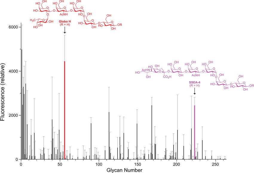
ACS Central Science                                                                                                               Research Article

Table 2. Affinity of RNase A and RNase 1 for Surface-Bound                         tissues. Because RNase A is derived from a cow, we probed cells
Globo H and SSEA-4                                                               from bovine mammary gland epithelial line MAC-T42 for three
                                                                                 globo-series glycans: Globo H, SSEA-4, and SSEA-3. Using
                                                    Kda (μM)
                                                                                 monoclonal antibodies that are specific for these glycans,27 a
        glycan           pH           RNase A                  RNase 1           fluorescently labeled secondary antibody, and confocal
       Globo H           7.4           21   ±   2              0.8 ± 0.2         microscopy, we were unable to detect any of these glycans
       Globo H           5.0           17   ±   2               11 ± 2           on the surface of MAC-T cells (data not shown). Accordingly,
       SSEA-4            7.4           17   ±   1               10 ± 1           we performed subsequent analyses with human cells.
       SSEA-4            5.0           19   ±   1               12 ± 2              Variants of RNase A and RNase 1 that evade cytosolic RI are
a
    Values (±SE) were obtained by fitting the data in Figure 2 to eq 2.           selectively toxic to human cancer cells. For example, variants of
                                                                                 RNase 1 (R39D/N67R/N88R/G89R/R91D, or “DRRRD”)
This glycan shares a pentasaccharide unit with Globo H but                       and RNase A (G88R) demonstrate cytotoxicity against human
appeared to have less affinity for RNase A in the glycan array                     leukemia cells with IC50 values of 10.8 and 6.2 μM,
(Figure 1). We found that RNases did indeed bind more                            respectively.6,15 Hence, we sought to determine the effect of
weakly to SSEA-4 than to Globo H (Figure 2C; Table 2).                           reducing cell-surface Globo H on RNase-mediated cytotoxicity.
   Effect of Globo H on RNase-mediated Cytotoxicity.                                 Small-molecule inhibitors of glycosyltransferases can be used
Globo H has been detected inconsistently in mammals. For                         to modulate the display of glycans on the cell surface.43,44 For
example, this glycan is more abundant in tissues from rat than                   example, 2-fluoro-2-deoxyfucose (2FF) inhibits fucosyltrans-
in those from cats and dogs.41 Analyses of the globo-series                      ferases.43 Of the 20 highest scoring ligands in our screen (Table
glycolipids have not, however, been performed in bovine                          S2), only Globo H has a fucose unit. Intracellular esterases

Figure 3. Effects of Globo H display on RNase-mediated toxicity for human breast adenocarcinoma cells (MCF-7). (A) Confocal microscopy to
visualize the effect of Ac32FF (100 μM in 0.1% v/v DMSO) on Globo H expression. Nucleus: Hoechst 33342 (blue). α-GH: Alexa Fluor-594 (red).
Outer membrane: WGA-488 (green). Scale bar: 5 μm. (B) Flow cytometry to quantify the effect of Ac32FF (100 μM in 0.1% v/v DMSO) on Globo
H expression. (C) Cell viability assay to reveal the effect of diminished Globo H expression due to Ac32FF (100 μM in 0.1% v/v DMSO) on Globo
H expression on the susceptibility of cells to cytotoxic variants of RNase 1 (DRRRD) and RNase A (G88R), n = 3. (D) Cellular DNA synthesis assay
to reveal the effect of blocking Globo H with α-GH (15 ng/mL) on the susceptibility of cells to a cytotoxic variant of RNase 1, n = 3. *, p < 0.01.

                                                                           184                                             DOI: 10.1021/acscentsci.5b00164
                                                                                                                           ACS Cent. Sci. 2015, 1, 181−190
ACS Central Science Research Article
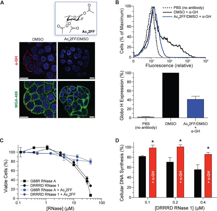
Figure 4. Solution structure of the RNase 1−Globo H interface. (A, B) pH 6.5; (C, D) pH 4.7. A solution of [15N]-RNase 1 was prepared in the
presence and absence of Globo H−ceramide micelles. NMR chemical shift changes (ΔΔδ ppm) were calculated as the average of n = 2 from the
vector change upon addition of Globo H−ceramide and plotted by residue (panels A and C). Changes in chemical shift are displayed with the
program PyMOL (panels B and D). Backbone regions that are colored in red and that are wider indicate greater chemical shift perturbations. Raw
1 15
H, N-HSQC NMR spectra are shown in Figure S3.
convert peracetylated 2-fluoro-2-deoxyfucose (Ac32FF) into and 3B). Using an assay that measures viable cells, we found
2FF.43 We synthesized Ac32FF and used confocal microscopy that cells with lower Globo H levels were less vulnerable to
and flow cytometry to demonstrate that treating MCF-7 cells cytotoxic RNase variants (Figure 3C). Finally, we used a highly
with Ac32FF reduces the surface-display of Globo H (Figure 3A sensitive assay of cellular DNA synthesis to demonstrate that α-
185 DOI: 10.1021/acscentsci.5b00164
ACS Cent. Sci. 2015, 1, 181−190ACS Central Science Research Article
GH antagonizes the cytotoxicity of DRRRD RNase 1 (Figure Specifically, we found that the major secretory ribonuclease
3D). in humans, RNase 1, interacts with a human cell-surface glycan,
Globo H Binding Site on RNase 1. To identify the Globo H, which is a tumor-associated antigen (Figure 1). The
residues in RNase 1 that interact with Globo H, we employed affinity of RNase 1 for this neutral glycan is in the high
1 15
H, N-HSQC NMR spectroscopy. The backbone chemical nanomolar range at physiological pH (Figure 2A; Table 2). As
shifts of RNase 1 exhibited few changes in the presence of both RNase 1 and Globo H are the basis for ongoing clinical
zwitterionic CTAB micelles. In contrast, these shifts were trials,10,33 our discovery links two molecules that are now in the
perturbed markedly by micelles containing a Globo H−lipid clinic. Moreover, our discovery rationalizes the selective
conjugate at 1 equiv relative to RNase 1 (Figures S3A and cytotoxicity of RNase 1 variants for cancerous versus
S3B). These changes in the vector of the chemical shift were noncancerous cells while revealing an endogenous ligand for
calculated with eq 3 and plotted by residue number (Figure 4). Globo H.
At pH 6.5, major shift perturbations were found throughout RNases are known to interact with anionic cell-surface
the amino acid sequence, being greatest at residues Ser23, glycans. Indeed, sialic acid and heparan sulfate play a role in the
Phe46, Val47, Val52, Asn76, Ser77, Tyr92, and Phe120 (Figure cellular uptake of RNases.2,16 These interactions are manifested
4A). These perturbations were mapped onto the structure of primarily through nonspecific Coulombic interactions with
RNase 1 to depict those regions that contribute most to the cationic side chains of RNases. While evidence mounts that
binding of Globo H (Figure 4B). The perturbations at pH 6.5 RNase 1 degrades extracellular RNA and, thus, regulates
were dispersed along the protein, residing mainly in turns and hemostasis and immunity,2,48 no specific cell-surface ligand for
bends (Table 3). RNase 1 is known.
Globo H is a specific ligand for RNase 1. An RNase−Globo
Table 3. Residues of RNase 1 That Interact with Globo H at H interaction was apparent regardless of whether the glycan
pH 6.5 was displayed on a slide (Figure 1), in the well of a plate
(Figure 2), or on a micelle (Figure 4). As these three display
ΔΔδ (ppm) residue structurea modes have only Globo H itself in common, their readout
>0.100 Tyr92 t validates Globo H as a ligand for RNase 1. Moreover, reducing
0.05−0.10 Phe46, Asn76, Phe120 s, b, b the display of Globo H on the surface of breast adenocarcinoma
0.03−0.05 Ser23, Val47, Val52, Ser77 b, s, h, b cells by two distinct methods (i.e., a small-molecule inhibitor of
a
t, turn; b, bend; s, β-strand; h, α-helix. biosynthesis or a monoclonal antibody antagonist) makes cells
less susceptible to a cytotoxic variant of RNase 1 (Figure 3).
RNase 1 undergoes endocytosis, and glycosylaminogylcans We note that cell-surface components change during tumori-
reside on the lumen of endosomes.45 Accordingly, we also used genesis,12 and such changes could amplify selective toxicity that
1 15 is based on the differential display of Globo H.
H, N-HSQC NMR spectroscopy to monitor chemical shift
perturbations at pH 4.7, which is encountered in endo- RNase A and RNase 1 have 82% identity in their amino acid
somes.46,47 Interestingly, we found that the larger overall shift sequence. Correspondingly, the two enzymes appear to interact
perturbations clustered predominantly to a more polar serine- with Globo H in a similar manner, albeit with different affinity
rich loop, Ser18−Thr24, as well as at His80 (Figures 4C and (Table 2). Of the eight residues of RNase 1 that were perturbed
4D; Table 4). As these residues are predominantly polar, the most upon binding to Globo H at neutral pH, six are conserved
in RNase A. Only Val52 and Asn76, which are alanine and
Table 4. Residues of RNase 1 That Interact with Globo H at tyrosine residues in RNase A, differ. Eight of 11 residues of
pH 4.7 RNase 1 that were perturbed most upon binding at low pH are
in RNase A, with Thr24, His80, and Lys102 of RNase 1 being
ΔΔδ (ppm) residue structurea replaced with serine, asparagine, and alanine, respectively, in
>0.100 His80 s RNase A.
0.05−0.10 Ser18, Ser21, Ser22, Ser23 −, −, −, b Like Globo H, glycosaminoglycans reside on the surface of
0.03−0.05 Thr24, Arg39, Val47, Thr82 Asp83, Lys102 b, b, s, s, s, s human cells. Sulfated glycosaminoglycans are known to bind to
a
t, turn; b, bend; s, β-strand, h, α-helix; −, no structure. RNase 1, presumably via favorable Coulombic interactions with
the cationic active site.2,14,16,49 The residues that interact with
Globo H are distal from the enzymic active site (Figures 4B and
interactions are likely due to hydrogen bonding. These data 4D). Accordingly, RNase 1 could employ multivalency in its
suggest that the site on RNase 1 that interacts with Globo H is binding to human cells. This mode of binding is consistent with
smaller at low pH, and that some of the multivalency that an experimental/computational study that concluded that
confers tighter binding is lost. These structural data are specific (though unidentified) cell-surface components and
consistent with the weak affinity of RNase 1 for Globo H at low nonspecific Coulombic interactions both contribute to the
pH (Figure 2B). cellular uptake of RNase 1.14
We also used 1H,15N-HSQC NMR spectroscopy to identify Along with other cell-surface glycans,45 Globo H is likely
the residues in RNase A that interact with Globo H at pH 4.7. displayed on the interior of endosomes following endocytosis.
We found that few residues were altered (Figures S3C and S4), Interestingly, NMR analyses reveal that the binding sites on
consistent with the weak affinity observed with binding assays RNase 1 for Globo H differ with pH (Figure 4). At acidic pH,
(Figure 2B).
■
these sites cluster and become weaker. These data, which are
consistent with observed values of Kd (Figures 2A and 2B;
DISCUSSION Table 2), suggest a mechanism in which RNase 1 is released
We have discovered that a protein and a carbohydrate conspire from lumenal Globo H as endosomes mature, allowing for the
to direct the degradation of RNA within cancer cells. entry of RNase 1 into the cytosol.
186 DOI: 10.1021/acscentsci.5b00164
ACS Cent. Sci. 2015, 1, 181−190ACS Central Science Research Article
Interestingly, other RNase ligands in the glycan array include high vacuum to provide peracetylated 2-fluoro-2-deoxyfucose in
sialic acid glycoproteins (Figure 1 and Table 1). These proteins quantitative yield (17.5 mg; 0.060 mmol). NMR spectra were
include the transporter, α1-glycoprotein, whose role is largely acquired on a Bruker spectrometer operating at 400 (1H) and
unknown aside from being a carrier that can improve the 101 (13C) MHz and are shown in Figures S1 and S2. Chemical
pharmacokinetics of small-molecule drugs.50 Ceruloplasmin is a shift data are reported in units of δ (ppm) relative to residual
copper-binding protein that associates with transferrin, an iron- solvent or TMS.
binding protein. An interaction with these proteins could 1
H NMR (400 MHz, CDCl3, δ): α-anomer, 6.43 (d, J = 3.98
enable an unmodified enzyme to avoid renal filtration in vivo. Hz, 1H, H-1), 5.42 (td, J = 10.64, 3.34 Hz, 1H, H-3), 5.37 (d, J
This mechanism could be more advantageous than PEGylation, = 3.70 Hz, 1H, H-4), 4.88 (ddd, J = 49.38, 10.17, 3.97 Hz, 1H,
which increases the circulation time of RNases in mice but at H-2), 4.25 (q, J = 6.53 Hz 1H, H-5), 2.18 (6H), 2.06 (s, 3H),
the expense of cellular uptake and enzymatic activity.51,52 1.15 (d, J = 6.52 Hz, 3H, H-6); β-anomer, 5.77 (dd, J = 8.05,
For over 50 years, thoughts about pancreatic-type 4.12 Hz, 1H, H-1), 5.31 (s, 1H, H-4), 5.17 (ddd, J = 13.34,
ribonucleases have been dominated by RNase A. This 9.89, 3.47 Hz, 1H, H-3), 4.64 (dt, J = 51.81, 8.90 Hz, 1H, H-2),
renowned enzyme has been the basis for much seminal work 3.99 (q, J = 6.50 Hz, 1H, H-5), 2.23 (s, 3H), 2.18 (s, 3H), 2.06
in biological chemistry, resulting in the first enzymatic reaction (s, 3H), 1.22 (d, J = 6.44 Hz, 3H, H-6). 13C NMR (101 MHz,
mechanism53,54 along with four Nobel prizes.55−57 New work CDCl3, δ): α-anomer (major), δ 170.45, 170.23, 169.22, 89.34
has, however, revealed substantial differences in the biochemical (d, J = 21.62 Hz, C1), 84.36 (d, J = 190.72 Hz, C2), 71.19 (d, J
and biological properties of RNase A and its human = 7.49 Hz, C4), 68.73 (d, J = 19.1 Hz, C3), 67.26 (C5), 21.08,
homologue, RNase 1.2 We have discovered another difference, 20.82, 20.67, 15.88 (C6). HRMS (ESI): calcd for C12H17FO7
and propose that affinity of RNase 1 for Globo H evolved for a [M + NH4]+ 310.1297; found, 310.1299.
specific purpose: to defend the host organism against cancer. Production of RNases. RNase A and RNase 1 were
■ METHODS
Materials. MCF-7 cells were from ATCC (Manassas, VA).
produced by heterologous expression of their cDNA in
Escherchia coli strain BL21(DE3) and purification as described
previously.4 For conjugation of BODIPY, a cysteine residue was
Phosphate-buffered saline (PBS) (Ca2+- and Mg2+-free), installed at residue 19 of the RNases with site-directed
Dulbecco’s modified Eagle’s medium (DMEM), and fetal mutagenesis, and P19C RNase 1 and A19C RNase A were
bovine serum (FBS) were from Life Technologies (Grand produced in a manner similar to that of the wild-type enzymes.
Island, NY). After purification, the nascent thiol was protected as a mixed
A rabbit polyclonal IgG antibody to RNase A (α-RNase A) disulfide by reaction with DTNB. Prior to conjugation, the
was from Biodesign International (Kennebunk, ME); a nascent thiol in 10 mg of protein was deprotected by the
secondary goat α-rabbit IgG−Alexa Fluor 594 conjugate was addition of DTT (4 equiv). The RNase was separated from
from Invitrogen (Carlsbad, CA). A murine monoclonal IgM excess DTT by passage through a column of PD-10 desalting
antibody to Globo H (MBr1 or α-GH) was from Enzo resin from GE Healthcare (Pittsburgh, PA). The deprotected
(Farmingdale, NY); secondary goat α-mouse IgG−Alexa Fluor RNase was reacted with 10 equiv of BODIPY-Fl from Life
594 and IgG−Alexa Fluor 647 conjugates were from Life Technologies dissolved in aqueous DMSO (10% v/v). The
Technologies. BODIPY-Fl solution was added dropwise with stirring, and the
[15N]-NH4Cl was from Cambridge Isotope Laboratories reaction was allowed to proceed at room temperature for 2 h,
(Andover, MA). 2-Fluoro-2-deoxyfucose was from CarboSynth then at 4 °C for 4 h. After overnight dialysis into 50 mM
(San Diego, CA). Dithiothreitol (DTT) was from Goldbio (St. AcOH, pH 5.0, purification by chromatography on a cation-
Louis, MO). Cetyltrimethylammonium bromide, 5,5′-dithiobis- exchange resin (GE Healthcare) yielded RNase−BODIPY
(2-nitrobenzoic acid) (DTNB), isopropyl β-D-1-thiogalactopyr- conjugates. The identity of these conjugates was confirmed
anoside (IPTG), 2-(N-morpholino)ethanesulfonic acid (MES), with matrix-assisted laser desorption/ionization (MALDI) mass
tris(hydroxymethyl)aminomethane (Tris), Tween X-100, spectroscopy at the University of Wisconsin Biotechnology
bovine serum albumin (BSA), and other reagents, solvents, Center and SDS−PAGE imaged by scanning for fluorescence
and buffers were from Sigma-Aldrich (St. Louis, MO) unless and staining with Coomassie.
noted otherwise. [15N]-RNase A and [15N]-RNase 1 were produced in E. coli
Glycan Synthesis. Globo H−biotin and Globo H− as described previously,4 except using a double-growth
ceramide conjugates were synthesized from Globo H as procedure in minimal medium containing [15N]-NH4Cl after
described previously.24,27,58 SSEA-4−biotin conjugate (Com- induction with IPTG.59 Growth conditions yielded an average
pound No. B295, Lot S284-1) was obtained from the of 15 mg of RNases from 1 L of medium. Protein purification
Consortium for Functional Glycomics. was monitored with SDS−PAGE. The purified proteins were
Peracetylated 2-Fluoro-2-deoxyfucose (Ac32FF). Per- analyzed with MALDI mass spectroscopy. The observed masses
acetylated 2-fluoro-2-deoxyfucose was prepared from 2-fluoro- of 14790.1 and 13809.2 Da indicated that isotope incorporation
2-deoxyfucose by a procedure similar to that described had been (13809−13681)/172 = 74% and (14790−14,604)/
previously.43 Briefly, 2-fluoro-2-deoxyfucose (10.0 mg, 0.060 192 = 97% for RNase A and RNase 1, respectively.
mmol) was dissolved in pyridine (0.35 mL), and the resulting Ribonucleolytic Activity Assays. RNases were assayed for
solution was cooled to 0 °C. Acetic anhydride (0.020 mL, 0.22 catalytic activity by monitoring cleavage of a fluorogenic RNA
mmol) was added, and the reaction vial was covered in foil and substrate, 6-FAM−dArUdGdA−6-TAMRA from IDT (Coral-
allowed to warm to room temperature overnight. The reaction ville, IA).60 Assays were performed in 0.10 M MES−NaOH
mixture was then diluted with dichloromethane (5 mL) and [oligo(vinylsulfonic acid)-free61] buffer, pH 6.0, containing
washed with 1 M HCl (3 × 2 mL), saturated aqueous NaHCO3 NaCl (0.10 M). The addition of RNases yielded a linear
(3 mL), and brine (3 mL). The organics were dried over increase of fluorescence that can be converted into activity with
Na2SO4(s), concentrated under reduced pressure, and dried by the equation
187 DOI: 10.1021/acscentsci.5b00164
ACS Cent. Sci. 2015, 1, 181−190ACS Central Science Research Article
ΔI /Δt TE2000-U laser-scanning confocal microscope from Nikon
kcat /KM =
(Imax − Io)[RNase] (1) equipped with an Axio Camdigital camera from Carl Zeiss.
Flow Cytometry. Globo H on live cells was quantified with
where ΔI/Δt is the initial reaction velocity, Imax is the maximum flow cytometry. MCF-7 cells (1 × 10⁵/well) in medium were
detected fluorescence after saturating substrate with excess grown for 3 days in the presence of Ac32FF (100 μM in 0.1%
RNase A, and I0 is the initial background fluorescence after v/v DMSO)43,44 or DMSO (0.1% v/v) alone. The cells were
incubation of substrate. The values of kcat/KM calculated with incubated with α-GH and a fluorescently labeled secondary
eq 1 were (31 ± 6) μM−1 s−1 and (2.1 ± 1.2) μM−1 s−1 for antibody. Fluorescence was measured with a FACSCalibur flow
RNase A and RNase 1, respectively, and neither fluorescence cytometer from BD Bioscience (San Jose, CA). Data were
labeling nor isotopic incorporation had a significant effect on analyzed with FlowJo software from Tree Star (Ashland, OR).
these values. Cell Viability Assay. The effect of Globo H on live cells
Glycan Array Screening. A printed array of mammalian was measured with an assay for cell viability. MCF-7 cells
glycans was screened for RNase A ligands by the standard (5000/well) in medium were added to the wells of a 96-well
procedure of Core H of the Consortium for Functional plate from Corning (Corning, NY) and grown for 3 days in the
Glycomics (CFG).62 The array was version 2.0 and comprised presence of Ac32FF (100 μM in 0.1% v/v DMSO),43,44 DMSO
264 synthetic and natural glycans that are found on the surface (0.1% v/v) alone, or H2O2 (1 mM). The medium was replaced
of mammalian cells. The gycans were functionalized with an with serum-free medium, and RNases in PBS were added at
amino group and immobilized to N-hydroxysuccinimide- various concentrations. Cells were then incubated for 44 h. The
activated glass slides.62 medium was removed, and the cells were incubated in CellTiter
Briefly, RNase A was diluted to a concentration of 200 μg/ 96 MTS reagent from Promega (Madison, WI) for 2 h. The
mL in 20 mM Tris−HCl buffer, pH 7.4, containing NaCl (150 absorbance was then measured at 490 nm and normalized to
mM), CaCl2 (2 mM), MgCl2 (2 mM), Tween 20 (0.05% v/v), that from cells treated with 0.1% v/v DMSO alone (100%) and
and BSA (1% w/v). The binding of RNase A was detected by 1 mM H2O2 (0%).
using α-RNase A (1 μg/mL) and a fluorescently labeled Cellular DNA Synthesis Assay. The effect of Globo H on
secondary antibody. The antibodies alone were also screened live cells was also measured with a sensitive assay for cellular
against the array, and the resulting fluorescence values were DNA synthesis. MCF-7 cells (5000/well) in medium were
subtracted from values in the presence of RNase A. added to the wells of a 96-well plate and grown overnight in
Solid-Phase Glycan Binding Assay. Quantification of medium. The medium was replaced with serum-free medium,
binding between RNases and immobilized glycans was and α-GH (final concentration: 15 ng/mL) was added to the
monitored using a fluorescence surface-binding assay. Briefly, wells. Then, DRRRD RNase 1 in PBS was added to the wells at
a 96-well plate coated in NeutrAvidin from Pierce (Rockford, three concentrations, and the cells were incubated for 44 h. Cell
IL) was washed with 3× PBS and then treated with 10 equiv of proliferation was assessed by monitoring the incorporation of
Globo H−biotin or SSEA-4−biotin. To reduce nonspecific [methyl- 3 H]thymidine into cellular DNA, as described
interactions, wells were incubated with aqueous milk (5% v/v) previously.9
and washed with PBS (3×). RNase−BODIPY conjugates were Preparation of Samples for 1H,15N-HSQC NMR Spec-
incubated to equilibrium in PBS, pH 7.4, containing Tween X- troscopy. Samples were prepared in 600 μL with 100 mM
100 (0.005% v/v) or 20 mM Tris−HCl buffer, pH 5.0, KH2PO4 buffer, pH 6.5 or pH 4.7, containing [15N]-RNase
containing NaCl (130 mM). After 3 washes, the fluorescence of (250 μM), cetyltrimethylammonium bromide (25 mM), and
an RNase−BODIPY conjugate was detected by emission at 530 D2O (10% v/v). Globo H−ceramide conjugate was resus-
nm after excitation at 490 nm. The fluorescence was corrected pended in CHCl3/MeOH/H2O 65:35:5. An aliquot containing
for that from a well treated with unconjugated biotin, and data 0.15 μmol of conjugate (1 equiv compared to the [15N]-RNase)
were analyzed by nonlinear regression to the equation was dried under N2(g) and then under high vacuum for 2 h.
The conjugate was resuspended in the RNase-containing
Bmax [RNase]h solution, which was then placed in an 8 in. glass tube from
B= Wilmad-LabGlass (Vineland, NJ).
Kd h + [RNase]h (2) NMR data were recorded at 25 °C with a 600 MHz Varian
NMR spectrometer. 1H,15N-HSQC NMR spectra were
where B is the normalized relative fluorescence (RFU), Bmax is
measured and peak assignments were made with the program
the maximum percent fluorescence, and h is a Hill coefficient.
Sparky 3 (T. D. Goddard and D. G. Kneller, University of
Cell Culture. Cells from the MCF-7 human breast
California, San Francisco) using the assignments determined
adenocarcinoma line were grown in DMEM (high glucose)
from the solution structures of RNase 163 and RNase A.59 The
containing FBS (10% v/v) and pen/strep from Invitrogen.
vector change of chemical shift (ΔΔδ) upon addition of the
Cells were maintained at 37 °C in 5% CO2.
Globo H−ceramide conjugate (1 equiv) was determined with
Confocal Microscopy. Globo H on live cells was visualized
the equation
with confocal microscopy. MCF-7 cells (1 × 10⁵/well) in
medium were grown for 3 days in the presence of Ac32FF (100
μM in 0.1% v/v DMSO)43,44 or DMSO (0.1% v/v) alone. The ⎛ 1 15 ⎞2
ΔΔδ = (Δδ1H)2 + ⎜ Δδ N⎟
medium was replaced with serum-free medium, and cells were ⎝5 ⎠ (3)
plated in an 8-well microscopy slide from Ibidi (Verona, WI).
The cell surface was stained with α-GH and a fluorescently where 1H and 15N chemical shifts (Δδ) were determined by
labeled secondary antibody, or with WGA-488 from Life subtracting the peak chemical shifts of RNase in the absence of
Technologies. Nuclei were stained with Hoechst 33342 from the Globo H−ceramide conjugate. To depict the chemical shift
Life Technologies. Images were captured with an Eclipse perturbations, images of PDB entry 2k1163 were created with
188 DOI: 10.1021/acscentsci.5b00164
ACS Cent. Sci. 2015, 1, 181−190ACS Central Science Research Article
the program PyMOL from Schrödinger (New York, NY) in human ribonuclease inhibitor protein. J. Mol. Biol. 2007, 367, 434−
which values of ΔΔδ were inserted as the β-factor. 449.
■
(8) Rutkoski, T. J.; Raines, R. T. Evasion of ribonuclease inhibitor as
ASSOCIATED CONTENT a determinant of ribonuclease cytotoxicity. Curr. Pharm. Biotechnol.
2008, 9, 185−199.
* Supporting Information
S (9) Lomax, J. E.; Eller, C. H.; Raines, R. T. Rational design and
The following file is available free of charge on the ACS evaluation of mammalian ribonuclease cytotoxins. Methods Enzymol.
Publications website at DOI: 10.1021/acscentsci.5b00164. 2012, 502, 273−290.
(10) ClinicalTrials.gov Identifier: NCT00818831.
Identity of the 264 glycans in the glycan array, spectral (11) Haigis, M. C.; Kurten, E. L.; Raines, R. T. Ribonuclease inhibitor
characterization of peracetylated 2-fluoro-2-deoxyfucose, as an intracellular sentry. Nucleic Acids Res. 2003, 31, 1024−1032.
raw 1H,15N-HSQC NMR spectra, and chemical shift (12) Dube, D. H.; Bertozzi, C. R. Glycans in cancer and
perturbations of RNase A in the presence of Globo H− inflammationpotential for therapeutics and diagnostics. Nat. Rev.
ceramide micelles at pH 4.7 (PDF) Drug Discovery 2005, 4, 477−488.
■
(13) Levine, M. N.; Hoang, T. T.; Raines, R. T. Fluorogenic probe
for constitutive cellular endocytosis. Chem. Biol. 2013, 20, 614−618.
AUTHOR INFORMATION (14) Sundlass, N. K.; Eller, C. H.; Cui, Q.; Raines, R. T. Contribution
Corresponding Author of electrostatics to the binding of pancreatic-type ribonucleases to
*E-mail: rtraines@wisc.edu. membranes. Biochemistry 2013, 52, 6304−6312.
(15) Johnson, R. J.; Chao, T.-Y.; Lavis, L. D.; Raines, R. T. Cytotoxic
Notes
ribonucleases: The dichotomy of Coulombic forces. Biochemistry 2007,
The authors declare the following competing financial 46, 10308−10316.
interest(s): R.T.R. is a founder of Quintessence Biosciences, (16) Chao, T.-Y.; Lavis, L. D.; Raines, R. T. Cellular uptake of
Inc. (Madison, WI), which is developing cancer chemo- ribonuclease A relies on anionic glycans. Biochemistry 2010, 49,
therapeutic agents based on ribonucleases. 10666−10673.
■ ACKNOWLEDGMENTS
We are grateful to Dr. M. R. Levengood (Seattle Genetics) for
(17) Varki, A. P.; Baum, L. G.; Bellis, S. L.; Cummings, R. D.; Esko, J.
D.; Hart, G. W.; Linhardt, R. J.; Lowe, J. B.; McEver, R. P.; Srivastava,
A.; Sarkar, R. Working group report: The roles of glycans in
hemostasis, inflammation and vascular biology. Glycobiology 2008, 18,
suggesting the use of peracetylated 2-fluoro-2-deoxyfucose and 747−749.
to M. R. Aronoff (University of Wisconsin Madison) for its (18) Drickamer, K.; Taylor, M. E. Glycan arrays for functional
synthesis, and to Drs. W. M. Westler and M. Tonelli glycomics. Genome Biol. 2002, 3, 1034.
(University of Wisconsin Madison) for advice with HSQC (19) Mariani-Costantini, R.; Barbanti, P.; Colnaghi, M. I.; Menard, S.;
NMR spectroscopy. We thank Prof. C. J. Czuprynski Clemente, C.; Rilke, F. Reactivity of a monoclonal antibody with
(University of Wisconsin Madison) for providing transformed tissues and tumors from the human breast. Immunohistochemical
(MAC-T) bovine mammary gland epithelial cells. This work localization of a new antigen and clinicopathologic correlations. Am. J.
was supported by Grant R01 CA073808 (NIH). The glycan Pathol. 1984, 115, 47−56.
array screen and SSEA-4 biotin were provided by the (20) Mariani-Costantini, R.; Colnaghi, M. I.; Leoni, F.; Menard, S.;
Consortium for Functional Glycomics, which was supported Cerasoli, S.; Rilke, F. Immunohistochemical reactivity of a monoclonal
by Grant U54 GM062116 (NIH). This study made use of the antibody prepared against human breast carcinoma. Virchows Archiv.
Organic Synthesis Core Facility at the Memorial Sloan A: Pathol. Anat. Histopathol. 1984, 402, 389−404.
(21) Zhang, S.; Cordon-Cardo, C.; Zhang, H. S.; Reuter, V. E.;
Kettering Cancer Center, which was supported by Grant P30
Adluri, S.; Hamilton, W. B.; Lloyd, K. O.; Livingston, P. O. Selection
CA008748 (NIH), and the National Magnetic Resonance of tumor antigens as targets for immune attack using immunohis-
Facility at Madison, which was supported by Grant P41 tochemistry: I. Focus on gangliosides. Int. J. Cancer 1997, 73, 42−49.
GM103399 (NIH). T.-Y.C. was supported by the Dr. James (22) Martignone, S.; Menard, S.; Bedini, A.; Paccagnella, A.; Fasolato,
Chieh-Hsia Mao Wisconsin Distinguished Graduate Fellow- S.; Veggian, R.; Colnaghi, M. I. Study of the expression and function of
ship. the tumour-associated antigen CaMBr1 in small cell lung carcinomas.
■ REFERENCES
(1) Raines, R. T. Ribonuclease A. Chem. Rev. 1998, 98, 1045−1066.
Eur. J. Cancer 1993, 29A, 2020−2025.
(23) Perrone, F.; Menard, S.; Canevari, S.; Calabrese, M.; Boracchi,
P.; Bufalino, R.; Testori, S.; Baldini, M.; Colnaghi, M. I. Prognostic
significance of the CaMBr1 antigen on breast carcinoma: Relevance of
(2) Eller, C. H.; Lomax, J. E.; Raines, R. T. Bovine brain ribonuclease
is the functional homolog of human ribonuclease 1. J. Biol. Chem. the type of recognised glycoconjugate. Eur. J. Cancer 1993, 29A,
2014, 289, 25996−26006. 2113−2117.
(3) Dickson, K. A.; Haigis, M. C.; Raines, R. T. Ribonuclease (24) Tsai, Y.-C.; Huang, J.-R.; Cheng, J.-Y.; Lin, J.-J.; Hung, J.-T.; Wu,
inhibitor: Structure and function. Prog. Nucleic Acid Res. Mol. Biol. Y.-Y.; Yeh, K.-T.; Yu, A. L. A prevalent cancer associated glycan, Globo
2005, 80, 349−374. H ceramide, induces immunosuppression by reducing Notch1
(4) Leland, P. A.; Schultz, L. W.; Kim, B.-M.; Raines, R. T. signaling. J. Cancer Sci. Ther. 2013, 5, 264−270.
Ribonuclease A variants with potent cytotoxic activity. Proc. Natl. Acad. (25) Cheng, J.-Y.; Wang, S.-H.; Lin, J.; Tsai, Y.-C.; Yu, J.; Wu, J.-C.;
Sci. U.S.A. 1998, 95, 10407−10412. Hung, J.-T.; Lin, J.-J.; Wu, Y.-Y.; Yeh, K.-T.; Yu, A. L. Globo-H
(5) Leland, P. A.; Staniszewski, K. E.; Kim, B.-M.; Raines, R. T. ceramide shed from cancer cells triggers translin-associated factor X−
Endowing human pancreatic ribonuclease with toxicity for cancer cells. dependent angiogenesis. Cancer Res. 2014, 74, 6856−6866.
J. Biol. Chem. 2001, 276, 43095−43102. (26) Danishefsky, S. J.; Shue, Y.-K.; Chang, M. N.; Wong, C.-H.
(6) Rutkoski, T. J.; Kurten, E. L.; Mitchell, J. C.; Raines, R. T. Development of Globo-H cancer vaccine. Acc. Chem. Res. 2015, 48,
Disruption of shape-complementarity markers to create cytotoxic 643−652.
variants of ribonuclease A. J. Mol. Biol. 2005, 354, 41−54. (27) Eller, C. H.; Yang, G.; Ouerfelli, O.; Raines, R. T. Affinity of
(7) Johnson, R. J.; McCoy, J. G.; Bingman, C. A.; Phillips, G. N., Jr.; monoclonal antibodies to globo-series glycans. Carbohydr. Res. 2014,
Raines, R. T. Inhibition of human pancreatic ribonuclease by the 397, 1−6.
189 DOI: 10.1021/acscentsci.5b00164
ACS Cent. Sci. 2015, 1, 181−190ACS Central Science Research Article
(28) Cascinelli, N.; Doci, R.; Belli, F.; Nava, M.; Marolda, R.; Costa, (46) Tycko, B.; Maxfield, F. R. Rapid acidification of endocytic
A.; Menard, S.; Terno, G. Evaluation of toxic effects following vesicles containing α2-macroglobulin. Cell 1982, 28, 643−651.
administration of monoclonal antibody MBr1 in patients with breast (47) Yamashiro, D. J.; Tycko, B.; Fluss, S. R.; Maxfield, F. R.
cancer. Tumori 1986, 72, 267−271. Segregation of transferrin to a mildly acidic (pH 6.5) para-Golgi
(29) Kim, I. J.; Park, T. K.; Hu, S. H.; Abrampah, K.; Zhang, S. L.; compartment in the recycling pathway. Cell 1984, 37, 789−800.
Livingston, P. O.; Danishefsky, S. J. Defining the molecular recognition (48) Fischer, S.; Nishio, M.; Dadkhahi, S.; Gansler, J.; Saffarzadeh,
of Globo-H (human breast-cancer) antigen through probe structures M.; Shibamiyama, A.; Kral, N.; Baal, N.; Koyama, T.; Deindl, E.;
prepared by total synthesis. J. Org. Chem. 1995, 60, 7716−7717. Preissner, K. T. Expression and localisation of vascular ribonucleases in
(30) Bilodeau, M. T.; Park, T. K.; Hu, S.; Randolph, J. T.; endothelial cells. Thromb. Haemostasis 2011, 105, 345−355.
Danishefsky, S. J.; Livingston, P. O.; Zhang, S. Total synthesis of a (49) Chao, T.-Y.; Raines, R. T. Mechanism of ribonuclease A
human breast tumor associated antigen. J. Am. Chem. Soc. 1995, 117, endocytosis: Analogies to cell-penetrating peptides. Biochemistry 2011,
7840−7841. 50, 8374−8382.
(31) Ouerfelli, O.; Warren, J. D.; Wilson, R. M.; Danishefsky, S. J. (50) Toyama, Y.; Ueyama, J.; Nomura, H.; Tsukiyama, I.; Saito, H.;
Synthetic carbohydrate-based antitumor vaccines: Challenges and Hisada, T.; Matsuura, K.; Hasegawa, T. Contribution of plasma
opportunities. Expert Rev. Vaccines 2005, 4, 677−685. proteins, albumin and α1-acid glycoprotein, to pharmacokinetics of a
(32) Zhu, J.; Wan, Q.; Lee, D.; Yang, G.; Spassova, M. K.; Ouerfelli, multi-targeted receptor tyrosine kinase inhibitor, sunitinib, in
O.; Ragupathi, G.; Damani, P.; Livingston, P. O.; Danishefsky, S. J. analbuminemic rats. Anticancer Res. 2014, 34, 2283−2289.
From synthesis to biologics: Preclinical data on a chemistry derived (51) Rutkoski, T. J.; Kink, J. A.; Strong, L. E.; Raines, R. T. Site-
anticancer vaccine. J. Am. Chem. Soc. 2009, 131, 9298−9303. specific PEGylation endows a mammalian ribonuclease with antitumor
(33) ClinicalTrials.gov Identifiers: NCT01248273 and activity. Cancer Biol. Ther. 2011, 12, 208−214.
(52) Rutkoski, T. J.; Kink, J. A.; Strong, L. E.; Raines, R. T. Human
NCT01349647.
ribonuclease with a pendant poly(ethylene glycol) inhibits tumor
(34) Endo, M.; Suzuki, K.; Schmid, K.; Fournet, B.; Karamanos, Y.;
growth in mice. Transl. Oncol. 2013, 6, 392−397.
Montreuil, J.; Dorland, L.; van Halbeek, H.; Vliegenthart, J. F. The
(53) Findlay, D.; Herries, D. G.; Mathias, A. P.; Rabin, B. R.; Ross, C.
structures and microheterogeneity of the carbohydrate chains of
A. The active site and mechanism of action of bovine pancreatic
human plasma ceruloplasmin. A study employing 500-MHz 1H-NMR ribonuclease. Nature 1961, 190, 781−784.
spectroscopy. J. Biol. Chem. 1982, 257, 8755−8760. (54) Cuchillo, C. M.; Nogués, M. V.; Raines, R. T. Bovine pancreatic
(35) Charlwood, J.; Birrell, H.; Tolson, D.; Camilleri, P. Two- ribonuclease: Fifty years of the first enzymatic reaction mechanism.
dimensional chromatography in the analysis of complex glycans from Biochemistry 2011, 50, 7835−7841.
transferrin. Anal. Chem. 1998, 70, 2530−2535. (55) Anfinsen, C. B. Principles that govern the folding of protein
(36) Nakano, M.; Kakehi, K.; Tsai, M. H.; Lee, Y. C. Detailed chains. Science 1973, 181, 223−230.
structural features of glycan chains derived from α1-acid glycoproteins (56) Moore, S.; Stein, W. H. Chemical structures of pancreatic
of several different animals: The presence of hypersialylated, O- ribonuclease and deoxyribonuclease. Science 1973, 180, 458−464.
acetylated sialic acids but not disialyl residues. Glycobiology 2004, 14, (57) Merrifield, R. B. Solid phase synthesis. Science 1984, 232, 341−
431−441. 347.
(37) Chester, M. A. Nomenclature of glycolipids. Pure Appl. Chem. (58) Jeon, I.; Iyer, K.; Danishefsky, S. J. A practical total synthesis of
1997, 69, 2475−2487. Globo-H for use in anticancer vaccines. J. Org. Chem. 2009, 74, 8452−
(38) D’Angelo, G.; Capasso, S.; Sticco, L.; Russo, D. Glycosphingo- 8455.
lipids: Synthesis and functions. FEBS J. 2013, 280, 6338−6353. (59) Tonelli, M.; Eller, C. H.; Singarapu, K. K.; Lee, W.; Bahrami, A.;
(39) Kannagi, R.; Cochran, N. A.; Ishigami, F.; Hakomori, S.; Westler, W. M.; Raines, R. T.; Markley, J. L. Assignments of RNase A
Andrews, P. W.; Knowles, B. B.; Solter, D. Stage-specific embryonic by ADAPT-NMR and enhancer. Biomol. NMR Assignments 2015, 9,
antigens (SSEA-3 and −4) are epitopes of a unique globo-series 81−88.
ganglioside isolated from human teratocarcinoma cells. EMBO J. 1983, (60) Kelemen, B. R.; Klink, T. A.; Behlke, M. A.; Eubanks, S. R.;
2, 2355−2361. Leland, P. A.; Raines, R. T. Hypersensitive substrate for ribonucleases.
(40) Venable, A.; Mitalipova, M.; Lyons, I.; Jones, K.; Shin, S.; Pierce, Nucleic Acids Res. 1999, 27, 3696−3701.
M.; Stice, S. Lectin binding profiles of SSEA-4 enriched, pluripotent (61) Smith, B. D.; Soellner, M. B.; Raines, R. T. Potent inhibition of
human embryonic stem cell surfaces. BMC Dev. Biol. 2005, 5, 15. ribonuclease A by oligo(vinylsulfonic acid). J. Biol. Chem. 2003, 278,
(41) Adobati, E.; Zacchetti, A.; Perico, M. E.; Cremonesi, F.; Rasi, G.; 20934−20938.
Vallebona, P. S.; Hagenaars, M.; Kuppen, P. J.; Pastan, I.; Panza, L.; (62) Blixt, O.; Head, S.; Mondala, T.; Scanlan, C.; Huflejt, M. E.;
Russo, G.; Colnaghi, M. I.; Canevari, S. Expression profile of Alvarez, R.; Bryan, M. C.; Fazio, F.; Calarese, D.; Stevens, J.; Razi, N.;
saccharide epitope CaMBr1 in normal and neoplastic tissue from Stevens, D. J.; Skehel, J. J.; van Die, I.; Burton, D. R.; Wilson, I. A.;
dogs, cats, and rats: Implication for the development of human-derived Cummings, R.; Bovin, N.; Wong, C.-H.; Paulson, J. C. Printed covalent
cancer vaccines. Histochem. J. 1999, 31, 729−737. glycan array for ligand profiling of diverse glycan binding proteins.
(42) Huynh, H. T.; Robitaille, G.; Turner, J. D. Establishment of Proc. Natl. Acad. Sci. U.S.A. 2004, 101, 17033−17038.
bovine mammary epithelial cells (MAC-T): An in vitro model for (63) Köver, K. E.; Bruix, M.; Santoro, J.; Batta, G.; Laurents, D. V.;
bovine lactation. Exp. Cell Res. 1991, 197, 191−199. Rico, M. The solution structure and dynamics of human pancreatic
(43) Rillahan, C. D.; Antonopoulos, A.; Lefort, C. T.; Sonon, R.; ribonuclease determined by NMR spectroscopy provide insight into its
Azadi, P.; Ley, K.; Dell, A.; Haslam, S. M.; Paulson, J. C. Global remarkable biological activities and inhibition. J. Mol. Biol. 2008, 379,
metabolic inhibitors of sialyl- and fucosyltransferases remodel the 953−965.
glycome. Nat. Chem. Biol. 2012, 8, 661−668.
(44) Okeley, N. M.; Alley, S. C.; Anderson, M. E.; Boursalian, T. E.;
Burke, P. J.; Emmerton, K. M.; Jeffrey, S. C.; Klussman, K.; Law, C. L.;
Sussman, D.; Toki, B. E.; Westendorf, L.; Zeng, W. P.; Zhang, X. Q.;
Benjamin, D. R.; Senter, P. D. Development of orally active inhibitors
of protein and cellular fucosylation. Proc. Natl. Acad. Sci. U.S.A. 2013,
110, 5404−5409.
(45) van Meer, G.; Voelker, D. R.; Feigenson, G. W. Membrane
lipids: Where they are and how they behave. Nat. Rev. Mol. Cell Biol.
2008, 9, 112−124.
190 DOI: 10.1021/acscentsci.5b00164
ACS Cent. Sci. 2015, 1, 181−190You can also read