The role of Fc gamma receptors in the activity of immunomodulatory antibodies for cancer
←
→
Page content transcription
If your browser does not render page correctly, please read the page content below
Stewart et al. Journal for ImmunoTherapy of Cancer 2014, 2:29
http://www.immunotherapyofcancer.org/content/2/1/29

 REVIEW                                                                                                                                              Open Access

The role of Fc gamma receptors in the activity of
immunomodulatory antibodies for cancer
Ross Stewart1*, Scott A Hammond2, Michael Oberst2 and Robert W Wilkinson1

  Abstract
  Antibodies targeting T-cell inhibitory pathways, such as CTLA-4 and PD-1/PD-L1, are emerging as an important class of
  cancer therapeutics, and a next generation of immunomodulatory therapies targeting alternative inhibitory (e.g. TIM-3,
  LAG-3, B7-H4, B7-H3, VISTA, A2aR), as well as co-stimulatory (e.g. CD27, OX40, GITR, CD137), pathways are poised to join
  them. Most of these immunomodulatory antibodies are of IgG isotypes that have low, or no, binding to the Fc gamma
  receptors (FcγRs) that trigger cell-mediated cytotoxic effector functions such as antibody dependent cellular cytotoxicity
  (ADCC) and phagocytosis (ADCP). These isotypes were selected to minimise the risk of depleting the T cells upon which
  such antibodies depend for their mechanism of action. However, recent preclinical data highlight a potential role for
  FcγR engagement in the activity of such antibodies. Here we review the biology of the FcγRs and IgG isotypes in both
  humans and mice, detail the potential roles that FcγR interactions can play in the activity of monoclonal antibodies in
  general, and of immunomodulatory antibodies in particular, and discuss how preclinical studies on these interactions
  might be best interpreted and translated to a human setting.
  Keywords: Fc gamma receptors, Anti-CTLA-4, Anti-GITR, Immunomodulatory antibodies

Introduction                                                                             TIM-3, LAG-3, B7-H4, B7-H3, VISTA, A2aR), as well as
Antibodies of the IgG sub-class are bi-functional molecules,                             co-stimulatory (e.g. CD27, OX40, GITR, CD137), pathways
possessing a F(ab) domain, variable in sequence and respon-                              are poised to join them. In contrast to agents such as rituxi-
sible for the binding of antigen, and an Fc domain, constant                             mab, most of these immunomodulatory antibodies are of
in sequence and responsible for mediating a range of anti-                               IgG isotypes that have low, or no, binding to the Fc gamma
body effector functions [1]. These functions are primarily                               receptors (FcγRs), and effects such as ADCC have not been
triggered through interaction with the complement compo-                                 considered to be critical to their activity. Recent studies in
nent C1q or with a family of FcγRs expressed, primarily, on                              mice have, however, highlighted a potential role for FcγR
the surface of leukocytes. Each IgG isotype has a different                              binding, and FcγR mediated effector functions in the activity
binding profile to the various FcγRs, and each FcγR has a                                of antibodies targeting a number of these pathways.
different cellular expression pattern. These differences en-                               Below we review the findings of these recent studies and
able the breadth, flexibility and control of function required                           discuss how they may change our understanding of the role
to mount an effective and regulated humoral response.                                    FcγR interactions play in the activity of immunomodulatory
   Effector functions mediated by the FcγRs, such as ADCC                                antibodies in mice. We begin, however, by reviewing the
and ADCP, are believed to play an important role in the ac-                              biology of the FcγRs and IgG isotypes in mice and humans,
tivity of several therapeutic antibodies; including a number                             highlighting the key similarities and differences, which need
used in Oncology, for example rituximab and trastuzumab.                                 to be appreciated when thinking about how to translate
Antibodies targeting T-cell inhibitory pathways, such as                                 these findings to a human setting, in order to select the op-
CTLA-4 and PD-1/PD-L1, are emerging as an important                                      timal isotype for any potential therapeutic.
class of cancer therapeutics, and a next generation of immu-
nomodulatory therapies targeting alternative inhibitory (e.g.
                                                                                         Review
                                                                                         The Fc gamma receptor family
* Correspondence: stewartr@medimmune.com
1
 MedImmune, AKB Building, Granta Park, CB21 6GH England, UK                              Humans and mice possess two classes of FcγRs, the acti-
Full list of author information is available at the end of the article                   vating and inhibitory receptors. In both species FcγRI is
                                           © 2014 Stewart et al.; licensee BioMed Central Ltd. This is an Open Access article distributed under the terms of the Creative
                                           Commons Attribution License (http://creativecommons.org/licenses/by/4.0), which permits unrestricted use, distribution, and
                                           reproduction in any medium, provided the original work is properly credited. The Creative Commons Public Domain
                                           Dedication waiver (http://creativecommons.org/publicdomain/zero/1.0/) applies to the data made available in this article,
                                           unless otherwise stated.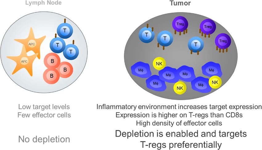
Stewart et al. Journal for ImmunoTherapy of Cancer 2014, 2:29                                                                                      Page 2 of 10
http://www.immunotherapyofcancer.org/content/2/1/29

an activating receptor with high affinity for IgG, and is                          immunogenicity risk respectively) and will not be con-
expressed on monocytic DCs and on monocytes/macro-                                 sidered further in this review. Of the remaining isotypes
phages broadly in humans but in select locations in                                hIgG1 demonstrates the highest affinity for FcγRs, with a
mouse (Table 1) [2,3]. FcγRI is the only FcγR with a                               nM or low μM binding affinity to all receptors (estimated
functionally meaningful binding affinity for monomeric                             by surface plasmon resonance [SPR]), and is considered a
IgG, the remaining FcγRs all exhibit such low affinity for                         potent inducer of all cell mediated effector functions, such
the Fc region of IgG that functional engagement is only                            as ADCC and ADCP. hIgG4 demonstrates a nM affinity
expected to occur upon binding of antibody complexed                               for FcγRI but μM affinities for all other receptors and is
through antigen engagement (Table 2). Both species pos-                            considered a poor inducer of such effector functions.
sess a single inhibitory FcγR, FcγRIIb, which is expressed                         hIgG2 has no measurable affinity for FcγRI, a high nM
on B cells, DCs and basophils in both species, and found                           affinity for the H131 form FcγRIIa and μM affinities for
additionally on monocytes, macrophages and all granulo-                            all other FcγRs (Table 2).
cyte populations in mice (Table 1). The remaining FcγRs                              These absolute affinities are a useful guide as to the
are all activating, but more divergent between species.                            binding of the hIgG isotypes to hFcγRs relative to each
In mice there are only 2 additional activating receptors                           other. However, as noted above, the functional inter-
FcγRIII, which is expressed broadly on NK cells, mono-                             action between IgG and most FcγRs is not monomeric
cytes, macrophages, granulocytes and DCs, and FcγRIV,                              in nature, as is modelled by SPR. In a study [5] examin-
which is found only on monocytes, macrophages and neu-                             ing the more functionally relevant binding of complexed
trophils. In humans there are four additional activating                           IgG to FcγRs on cells, the relative ranking of the isotypes
FcγRs, the best characterised being FcγRIIa and FcγRIIIa.                          with respect to FcγR binding was retained, however no
FcγRIIa is expressed on granulocytes, monocytes and                                binding of IgG2 to cells expressing the inhibitory FcγRIIb
monocyte-derived cells such as macrophages and DCs.                                or the F158 form of the activating FcγRIIIa could be de-
FcγRIIIa is expressed primarily on NK cells, but is also                           tected using this methodology, despite the interaction be-
found on monocytes and macrophages under some cir-                                 tween IgG2 and these proteins demonstrated by SPR.
cumstances (Table 1). Each of these receptors exists as                              Mice also have four IgG isotypes but they are not
two allotypic variants with differing binding affinity for                         equivalent to those of human in either nomenclature or
IgG (Table 2). For FcγRIIIa the more common F158 allo-                             properties. Mouse (m)IgG1, in contrast to hIgG1, is con-
type has a lower affinity than the V158 allotype. For                              sidered a low effector function isotype, since while it
FcγRIIa the more common H131 allotype has a higher                                 binds inhibitory FcγRIIb and activating FcγRIII with nM
affinity than the R131 allotype [4]. The other two acti-                           affinity, it has no measurable binding to activating FcγRIV.
vating receptors are far less well characterised. FcγRIIc                          In mice it is the two IgG2 isotypes, mIgG2a and mIgG2b,
is expressed in only 20% of people and is closely related to,                      that have the highest binding affinities to FcγRs, particu-
but expressed more restrictedly than, FcγRIIa. FcγRIIIb is                         larly to FcγRIV (Table 2). In mice there is no IgG4 isotype,
a GPI linked receptor expressed mainly on the sur-                                 and mIgG3 has no measurable FcγR interactions [6].
face of neutrophils.                                                                 These differences in FcγR and antibody isotype inter-
   Mice and humans also have a divergent family of IgG                             actions and expression, summarised in Tables 1 and 2,
isotypes. In humans there are 4 isotypes, IgG1-4, each of                          mean that there are no direct homologs between human
which has differing binding affinities to the various                              and mouse with respect to FcγR and antibody isotype
FcγRs. Human (h)IgG3 demonstrates relatively high af-                              biology. However, it is possible to draw some parallels
finity binding to most FcγRs, but is not used routinely                            between the species, for example mIgG2a and hIgG1 are
as a therapeutic format (due to its long hinge region                              often considered equivalent functionally due to their
and polymorphic nature which represent a stability and                             broad and high FcγR binding, while the generally lower

Table 1 Expression pattern of the major FcγRs in human and mouse
Cell type                                  FcγRI                    FcγRIIb               hFcγRIIa             mFcγRIII            hFcγRIIIa            mFcγRIV
                                      h            m            h             m
B cells                                                         +             +
DCs                                   +            +            +             +               +                    +
NK cells                                                                                                           +                   +
Monocytes/Macrophages                 +            ~            ~             +               +                    +                   +                    +
Neutrophils                          (+)                        ~             +               +                    +                                        +
Italics indicates an inhibitory receptor. Normal text indicates an activating receptor. Human receptors are denoted by h and mouse receptors by m. + indicates
expression, (+) indicates inducible expression and ~ indicates expression on only a sub-set or on cells at specific physiological locations [2,3].
Stewart et al. Journal for ImmunoTherapy of Cancer 2014, 2:29 Page 3 of 10
http://www.immunotherapyofcancer.org/content/2/1/29
Table 2 Relative affinity of binding between the major Nevertheless a significant effort has been made to engineer
FcγRs and IgG Isotypes in human and mouse antibodies with improved affinity for the activating FcγRs
Human Mouse [10-12], with the aim of enhancing these cytotoxic func-
FcγR IgG1 IgG2 IgG4 FcγR IgG1 IgG2a IgG2b tions and improving therapeutic efficacy, and the outcome
I ++++ - ++++ I - ++++ ++++ of clinical trials with such agents may provide further clar-
IIa H131 +++ ++ ++ III ++ ++ ++
ity as to the true contribution of FcγR mediated cytotox-
icity in activity.
R131 +++ + ++
An alternative role for FcγRs is in the activity of anti-
IIb ++ - ++ IIB +++ ++ +++ bodies that function by agonising cell surface receptors,
IIIa V158 +++ + ++ IV - +++ ++ for example those of the tumor necrosis factor receptor
F158 ++ - ++ (TNFR) super family. In this context binding of IgG Fc
Italics indicates an inhibitory receptor. Normal text indicates an activating to FcγRs results in higher order clustering of antibody.
receptor. Rankings are based on KA x105M−1 for each interaction reported in This in turn results in higher order clustering of the target
published studies [1,5,6]. ++++ indicates a KA greater than 100 x 105 M−1, +++
indicates a KA between 20 and 99 x 105 M−1, ++ indicates a KA between 1 and receptor when bound by the F(ab) domain, which triggers
19 × 105 M−1, + indicates a KA between 0.5 and 0.9 × 10⁵ M−1, − indicates a KA downstream signalling (Figure 1, right). This clustering
that was undetectable or less than 0.5 × 10⁵ M−1.
effect has been shown to be critical to the activity of
antibodies specific for the TNFR family members CD40
and more select binding profiles of mIgG1 and hIgG4 [13,14] death receptor 5 (DR5) [15,16] and CD95 (Fas) [17]
make them close functional analogs. Additionally the ac- in preclinical mouse models, and has been suggested to
tivating mFcγRIII and hFcγRIIa are often considered play a similar role in the activity of other TNFR agonists
analogous based on affinities and expression pattern, as such as anti-OX40 and anti-CD27 [18].
are activating mFcγRIV and hFcγRIIIa. These parallels
are frequently imperfect though, for example: hFcγRIIIa The canonical role of FcγRs in immunomodulatory
is expressed at high levels on NK cells, while mFcγRIV antibodies
is not; hFcγRIIa is not expressed on NK cells while Immunomodulatory antibodies mediate their beneficial
mFcγRIII is; mIgG1 is a more functionally restricted iso- effects by promoting anti-tumor immunity. There are a
type than hIgG4, based on their relative affinities for ac- number of potential ways to achieve this, but one area
tivating FcγRI, mFcγRIV and hFcγRIIa. These differences showing particular promise is the use of antibodies that
need to be kept in mind, and pose challenges, when try- antagonise inhibitory receptors on the surface of T cells.
ing to translate findings in mouse preclinical models to One such antibody, the anti-CTLA-4 ipilimumab, was
humans. approved based on its ability to improve survival in
metastatic melanoma patients [19], while a number of
The potential roles of FcγRs in the activity of therapeutic antibodies targeting PD-1 are showing highly encour-
antibodies aging results in phase 1 clinical trials [20,21]. An alterna-
The FcγR mediated functions most commonly associated tive approach, which is earlier in clinical development
with therapeutic antibodies are those that mediate target but also showing promise, is the use of antibodies that
cell elimination; ADCC, or the related ADCP. These agonise co-stimulatory receptors. The agonistic anti-CD40
functions are triggered when antibody binding to antigen antibody, CP870,893, has demonstrated early signs of activ-
on the surface of a target cell generates sufficient avidity ity in pancreatic cancer [22], and tumor shrinkage was also
to trigger signalling through FcγRs on effector cells such observed in some patients treated with agonistic antibodies
as NK cells and macrophages, which then eliminate tar- directed against OX40 [23] and CD137 [24].
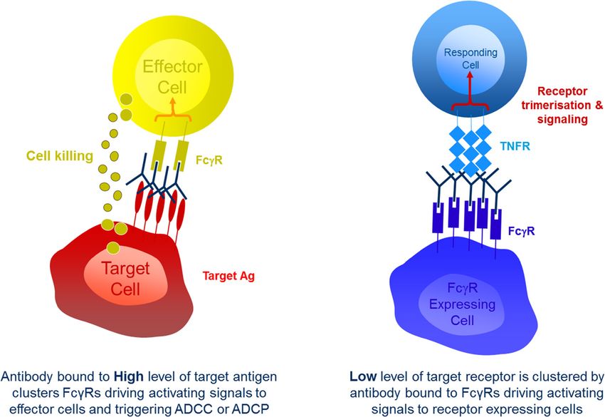
get cells through direct killing or phagocytosis (Figure 1, As mentioned above FcγR engagement has been
left). Preclinical models show that these forms of FcγR- shown to be critical for the activity of antibodies that ag-
mediated cytotoxicity are a significant component of the onise the TNFR CD40 in mice. The anti-tumor activity of
mechanism of action for tumor targeted antibodies such anti-mouse CD40 in these studies is lost upon introduc-
as the anti-CD20 antibody rituximab and the anti-HER2 tion of the D265A mutation into its Fc domain [13],
antibody trastuzumab [7]. The clinical evidence for the which results in an antibody with no measurable FcγR
role of such interactions, however, is more mixed. A binding [6]. It has also been shown that the in vitro activ-
number of relatively small studies have shown improved ity of antibodies directed against other TNFRs in human
outcomes in patients that are homozygous for the higher cells, such as OX40, GITR, CD137 and CD27, requires,
affinity allotypic forms of human FcγRIIIa and FcγRIIa or is greatly enhanced by FcγR engagement or immobil-
[8,9], supporting a role for FcγR-mediated cytotoxicity isation [25,26]. While the evidence suggests that a re-
in this setting, but several larger, randomized studies quirement for cross linking is a common property of
have generally failed to show the same associations [4]. antibodies targeting TNFRs receptors, there are someStewart et al. Journal for ImmunoTherapy of Cancer 2014, 2:29 Page 4 of 10 http://www.immunotherapyofcancer.org/content/2/1/29 Figure 1 The two primary forms of interaction between therapeutic IgGs and FcγRs. On the left: when IgG is bound to high levels of antigen on target cells, such as tumor cells, the resulting clustered Fc domains engage with FcγRs on immune effector cells such as NK cells and macrophages. This drives signals through those FcγRs triggering the initiation of cell-mediated effector functions such as ADCC and ADCP. On the right: when IgG is bound by its Fc domain to FcγRs it can act to cluster low levels of target receptors, such as members of the TNFR family. This clustering acts to trigger signals though the TNFR into target cells, such as T cells. exceptions. For example the hCD27.15 anti-human CD27 cells (APCs) critical to mounting an adaptive immune mAb has demonstrable agonistic properties in the soluble response [13]. In the case of the second hypothesis, the phase [27], the anti-CD40 antibody CP870,893 has been balance of FcγR engagement would become critical to shown to function in vitro in solution and as a F(ab)’2 the activity of anti-CD40. Too little engagement, as fragment that lacks any Fc domain [28] and the anti- seen with alglycosyl antibodies, and no agonistic signal- OX40 antibody 9B12, which is a mouse IgG1 with very ling is triggered so no anti-tumor activity is observed. low affinity interactions with FcγRs, has shown early Too much FcγR engagement on effector cells and deple- signs of clinical activity and demonstrated the ability to tion of critical immune cells begins to occur and anti- activate T cells in patients [23]. tumor activity is once again lost. Such a trade off in FcγR Two studies that further explored the role of FcγR en- affinity could also apply to other immunomodulatory anti- gagement for the activity of anti-mouse CD40 demon- bodies that target TNFRs (CD27, OX40, GITR, CD137), strated that it is the inhibitory mouse FcγRIIb that plays which are also expressed on critical immune cells. In an essential role [13,14]. One of these studies [14] went addition it has been shown that for CD137 antibody medi- on to demonstrate that other mouse FcγRs are capable ated internalisation of receptor can play a role in driving of cross linking anti-CD40 to drive activity in vitro, and signalling [29], and it is unclear how changes in Fc isotype proposed that the dominant role of FcγRIIb in vivo was might affect this property. due primarily to its bioavailability at the site of action For those antibodies that target inhibitory receptors for anti-CD40, namely the secondary lymphoid organs. like PD-1 and CTLA-4 there has been little reason to Both studies also went on to show that the activity of believe, based on their antagonistic mechanism of action, anti-mouse CD40 was lost upon class switching from a that FcγR engagement plays an important role in their mouse (m)IgG1 to a mIgG2a. Two alternative, and non- activity, and no significant evidence to contradict this mutually exclusive, hypotheses exist to explain this obser- belief exists in the clinical setting. Since all of the anti- vation. The first is that the reduced affinity of mIgG2a for gens targeted by these antibodies are expressed either on FcγRIIb results in reduced cross linking at the relevant site T cells or on APCs, the preference has been to select iso- of action and so limits activity [14]. The second is that the types that will avoid the risk of depleting these cells loss of activity occurs due to increased depletion of CD40 through Fc mediated mechanisms; an outcome that would positive cells, which are predominantly antigen presenting likely negate any beneficial immune activation. As a result,
Stewart et al. Journal for ImmunoTherapy of Cancer 2014, 2:29                                                                                 Page 5 of 10
http://www.immunotherapyofcancer.org/content/2/1/29

a common feature of the immunomodulatory antibodies                               engagement of the activating FcγRs in mouse tumor
currently in clinical development is that they are low or                         models. Further examination of the mechanism for this
null effector function isotypes (Table 3). Main exceptions                        dependence revealed that in the lymph nodes anti-mouse
include ipilimumab (anti-CTLA-4), MSB0010718C (anti-                              CTLA-4 acted to increase the number of CD4 T cells, in-
PD-L1) and antibodies like MGA271 (anti-B7-H3), CDX-                              cluding regulatory T cells (T-regs), but that within the
1127 (anti-CD27) and alemtuzumab (anti-CD52) which                                tumor it acted to selectively deplete only T-regs [33]. In
employ ADCC of tumor cells as a part of their mechanism                           one of the three studies, a comparison of several different
of action.                                                                        anti-mouse CTLA-4 antibodies showed that the extent of
  However, several recent preclinical studies are calling                         T-reg depletion observed correlated with activity [33].
into question the view that all TNFR agonist antibodies                           The depletion of T-regs was mediated predominantly
will behave as anti-CD40 does, or that all antibodies tar-                        by mouse FcγRIV, but was independent of NK cells, and
geting inhibitory receptors will be maximally effective as                        considered likely to rely on the activity of intratumoral
a low or null effector function isotype.                                          CD11b + macrophages [33]. In all three studies the level of
                                                                                  CTLA-4 expression was shown to be increased on intratu-
The evolving role of FcγRs in immunomodulatory                                    moral T cells relative to those in lymph nodes, and was
antibodies                                                                        higher on T-regs than on other T cell sub-sets.
Three recently published studies [31-33] have examined                              In their study Bulliard et al. [31] also examined the
the role of mouse FcγRs in the activity of anti-mouse                             role of mouse FcγRs in the activity of the anti-mouse
CTLA-4 antibodies. In these studies anti-mouse CTLA-4                             GITR antibody DTA-1. In contrast to the observations
antibodies were studied in a range of syngeneic tumor                             made for anti-mouse CD40, DTA-1 activity was minim-
models where they have previously shown activity, includ-                         ally affected by the absence of inhibitory FcγRIIb, but
ing MC38 (colon), CT26 (colon) and B16 (melanoma). The                            was dependent on the presence of the activating mouse
approach taken to examine the role of FcγRs varied be-                            FcγRs [31]. In further contrast to anti-CD40, a mouse
tween studies and either involved the use of FcγR knock                           (m)IgG2a version of DTA-1 was equal in activity to the
out mice [31,33] or reformatting of the anti-mouse CTLA-                          parental rat IgG2b version; although no mIgG1 was
4 to a variety of different antibody isotypes [32]. Irrelevant                    tested. Analogous to the mechanism observed for anti-
of the approach taken the result was the same; the activity                       mouse CTLA-4, it was shown that while mIgG2a or rat
of anti-mouse CTLA-4 was shown to be dependent upon                               IgG2b forms of DTA-1 treatment resulted in expansion of

Table 3 List of immunomodulatory antibodies currently in clinical development and their respective isotypes
Antibody                        Target               Company                                          Latest active clinical phase           Isotype
Ipilimumab                      CTLA-4               BMS                                              Approved                               IgG1
Tremelimumab                    CTLA-4               MedImmune/AstraZeneca                            II                                     IgG2
Nivolumab                       PD-1                 BMS                                              Approved                               IgG4
MK-3475                         PD-1                 Merck                                            III                                    IgG4
CT-011                          PD-1                 CureTech                                         II                                     IgG1
MPDL3280A                       PD-L1                Genentech/Roche                                  III                                    Aglycosyl IgG1
MEDI4736                        PD-L1                MedImmune/AstraZeneca                            I                                      IgG1 TM
MDX-1105                        PD-L1                BMS                                              I                                      IgG4
MSB0010718C                     PD-L1                Merck KGaA                                       I                                      IgG1
BMS-663513                      CD137                BMS                                              I                                      IgG4
PF-05082566                     CD137                Pfizer                                           I                                      IgG2
TRX518                          GITR                 GITR Inc                                         I                                      Aglycosyl IgG1
MEDI6469                        OX40                 MedImmune/AstraZeneca/AgonOX                     I                                      Mouse IgG1
CP-870,893                      CD40                 Roche                                            I                                      IgG2
CDX-1127                        CD27                 CellDex                                          I                                      IgG1
Lirilumab; IPH2102              KIR                  BMS/Innate Pharma                                II                                     IgG4
MGA271                          B7-H3                Macrogenics/Servier                              I                                      IgG1
Alemtuzumab                     CD52                 Genzyme/Bayer                                    Approved                               IgG1
IgG1 TM = IgG1 triple mutant, which contains 3 point mutations in the Fc domain that reduce the binding affinity of the mAb to FcγRs [30].
Stewart et al. Journal for ImmunoTherapy of Cancer 2014, 2:29 Page 6 of 10 http://www.immunotherapyofcancer.org/content/2/1/29 all T cell populations in the lymph node, they acted to de- effector function is entirely dependent upon the pres- plete T cells within the tumor. Also similar to anti-mouse ence of FcγR-bearing effector cells at an appropriate CTLA-4, this depletion preferentially occurred in the T-reg density. The studies by both Simpson et al. and Bulliard compartment and, although some reduction in both con- et al. highlight what many of us will have observed in ventional CD4+ and CD8+ T cells was also observed, re- our own work, that there are very few effector cells, such sulted in an increase in the CD8:T-reg ratio within the as NK cells, neutrophils and macrophages, present in tumor. In a more recent study published earlier this year lymph nodes, which are dominated by T, B and dendritic Builliard et al. [34] demonstrated a comparable mechan- cells. What both studies additionally show is that mouse ism, and dependence on activating mouse FcγRs for an tumors are extremely rich in FcγR-expressing effector anti-mouse OX40 antibody. It is however worth noting cells. Simpson et al. evidence greater than 20% NK cells that alternative studies have proposed, and evidenced, and greater than 40% CD11b + cells, which are predom- a mechanism of regulatory T cell re-programming for inantly macrophages, in B16 tumors and Bulliard et al. DTA-1 [35] and for anti-human OX40 mAbs [36], sug- highlight very similar levels of the same cells in CT26 gesting that a loss of FOXP3 expressing T cells following tumors. The coincident presence of high levels of target treatment with agents targeting either pathway may not antigen and a high density of effector cells creates a per- necessarily equate to depletion. fect storm for Fc mediated effector function within the These preclinical studies have brought into question tumor, while the lower target expression and relative two generally held beliefs regarding the role of FcγRs in paucity of effector cells in the periphery makes the same the activity of immunomodulatory antibodies. The first effector function harder to achieve in the lymph node. It is the idea that inhibitory FcγRIIb is the critical receptor is however worth noting that peripheral depletion of im- for the activity of all TNFR agonist antibodies. The data mune cells is possible, as demonstrated by the data on generated for the anti-mouse GITR antibody DTA-1 anti-CD40 [13] and the well documented effects of anti- clearly demonstrate FcγRIIb independent activity, and CD20 [39], suggesting that the nature or expression level our own unpublished data demonstrates similar inde- of the target are likely critical. pendence for other TNFR targeting antibodies. These data suggest, as previously proposed by White et al., that Translating preclinical data to man the specific mouse FcγRs involved in cross linking of As outlined at the start of this review, the significant dif- TNFR agonists may vary depending on the pathway be- ferences between human and mouse FcγR and IgG iso- ing targeted and on the bioavailability of each of the type biology make translation of findings between species FcγRs at the primary site of action. The second is the extremely challenging, and several factors need to be con- idea that effector function enabled antibodies represent sidered and better studied in the human setting before a risk to activity due to depletion of key immune cell these findings can be confidently applied to the activity of sub-sets. Importantly the data highlight the fact that per- therapeutic antibodies in man. ipheral and intratumoral antibody mediated effector Taking anti-CTLA-4 as an example, the first signifi- function are far from equivalent. This difference appears cant translational challenge is the key role for mouse to hinge on two critical differences; the level of target FcγRIV in the T-reg depleting activity demonstrated for antigen expression and the presence of appropriate ef- anti-mouse CTLA-4. FcγRIV does not exist in humans, fector cells (Figure 2). and its closest analog, human (h)FcγRIIIa, is expressed The extent of effector function triggered by antibody predominantly on NK cells, which played no role in the binding has been shown in a number of studies to be mainly macrophage mediated T-reg depletion observed strongly influenced by the level of target antigen expres- with anti-mouse CTLA-4 [33]. In humans macrophages sion [37,38], with antibodies directed against both hu- can express hFcγRIIIa following certain stimuli, such as man CCR4 and CD20 failing to elicit in vitro ADCC IL-10, but primarily express hFcγRIIa/b and hFcγRI, and against human tumor cell lines expressing anything less it is feasible that in human tumors these receptors would than 104 antigen molecules per cell and reaching optimal play the same role as mouse FcγRIV does in mouse tu- activity only in the presence of 105-106 antigen mole- mors. Further study of the intratumoral effector cell and cules [37]. Many of the targets for immunomodulatory FcγR constituent within human tumors would be ex- antibodies are expressed at low to undetectable levels on tremely helpful in understanding which human FcγR resting, peripheral immune cells, but are up-regulated might play the role of mouse FcγRIV in humans. significantly in the presence of pro-inflammatory stimuli, The second challenge for translation lies in the differ- such as those present in the tumor microenvironment. ences between IgG isotypes across species. In mice only As such, and in the light of recent studies, it is clear that mouse (m)IgG2a and mIgG2b demonstrate any measur- in mouse models it is significantly easier to elicit effector able binding to mouse FcγRIV; mIgG1 can therefore function against intratumoral immune cells. Additionally show none of the T-reg depleting activity observed for
Stewart et al. Journal for ImmunoTherapy of Cancer 2014, 2:29                                                                          Page 7 of 10
http://www.immunotherapyofcancer.org/content/2/1/29

 Figure 2 Critical differences between effector function in the periphery relative to the tumor. On the left: in lymph nodes, relatively low
 levels of cell activation and pro-inflammatory cytokines result in low levels of expression for most targets of immunomodulatory antibodies, which
 are generally low on resting immune cells. This low level of target expression, together with a paucity of effector cells in lymph nodes means
 depletion of cells through cell mediated effector functions do not readily occur. On the right: high levels of pro-inflammatory cytokines within
 the tumor microenvironment trigger up-regulation of many targets. This increased target expression together with a high density of effector cells,
 such as macrophages, enables depletion of cells through cell-mediated effector functions. The level of target expression, and the sensitivity to
 depletion varies across cell types and favours depletion of T-regs for targets such as CTLA-4 and GITR.

anti-mouse CTLA-4 in mice. In contrast, while human(h)                      3 year survival rate seen in melanoma patients treated
IgG1 has a significantly higher binding affinity to all human               with the two agents.
FcγRs, hIgG2 and hIgG4 are both capable of interact-                          The third translational challenge relates to the key
ing with human FcγRIIa and IIIa, and while human                            driver for differential depletion of T-regs relative to
FcγRI cannot bind hIgG2, its high affinity for monomeric                    other cell types in all three of the published studies, that
IgG suggests that it would be heavily occupied by serum                     is target expression level. It is clear from the published
IgG and less available to partake in effector function [40].                studies that the levels of GITR and CTLA-4 on mouse
Critically, while hIgG2 and hIgG4 are both very poor                        T-regs is higher than that observed on mouse effector
mediators of NK cell driven ADCC, they are both cap-                        T cells, in particular CD8s. However there are no studies
able of mediating ADCC via PBMCs, monocytes and                             available comparing how the level of target expression
neutrophils [41,42]. It is especially conceivable that in a                 varies between mice and humans, few that compare the
tumor microenvironment, with its increased target anti-                     differential expression of target on human T-regs relative
gen levels and high density of effector cells, that a hIgG2                 to effectors, and none that examine how this expression
or hIgG4 could begin to mediate some degree of ADCC                         changes quantitatively in the tumor versus the periphery.
or ADCP. If one were to assume that a mouse IgG2a                           These are all significant variables that must be studied in
were equivalent to human IgG1 and mouse IgG1 equiva-                        humans before a direct translational conclusion can be
lent to human IgG2 then the data of Selby et al. would                      drawn.
predict significant differences in the two anti-CTLA-4
antibodies that have been tested clinically one of which,                   Conclusions
tremelimumb, is a human IgG2 and the other of which,                        Future directions
ipilimumab, is a human IgG1 currently registered for the                    Immunomodulatory antibodies are showing positive re-
treatment of metastatic melanoma. The complex differ-                       sults in early clinical trials in cancer. These antibodies
ences in FcγR and IgG isotype biology that exist between                    generally fall into two classes. Those that antagonise in-
species, outlined above, suggest however that such as-                      hibitory receptors, such as CTLA-4 and PD-1, and those
sumptions about the comparability of mouse and human                        that agonise stimulatory receptors, such as CD27, OX40,
IgG isotypes may be too reductionist. In relation to                        GITR and CD137.
this, while no definitive comparison of the two agents                        Many groups, including our own, have been working
has been conducted, the toxicity profiles of ipilimumab                     to study the requirement for FcγRs in the activity of TNFR
and tremelimumab are comparable [19,43] and a recent                        agonist antibodies, but the recent study of Bulliard et al.
commentary in the Journal of Clinical Oncology [44]                         [31] has further highlighted the potential complexity of this
highlighted the very limited differences observed in the                    requirement by providing evidence that inhibitory mouse
Stewart et al. Journal for ImmunoTherapy of Cancer 2014, 2:29                                                                                   Page 8 of 10
http://www.immunotherapyofcancer.org/content/2/1/29

FcγRIIb is not required for the activity of all TNFR agonist                   Competing interests
antibodies in mice, as has been previously suggested. Fur-                     Ross Stewart and Robert Wilkinson are salaried employees of MedImmune Ltd.
                                                                               Scott Hammond and Mike Oberst are salaried employees of MedImmune LLC.
ther work is required to pull apart the target specific nature                 All four authors hold shares in the AstraZeneca group.
of FcγR requirements for mAb activity and the relative
contributions of pathway agonism versus targeted deple-                        Authors’ contributions
tion of cells. In the case of TNFR agonists pulling these                      RS, SH and MO conceived of, planned and drafted the initial content of the
apart is made very difficult by the inability to divorce FcγR                  article. RS provided all figures. RW provided substantive review. All authors
                                                                               read and approved the final manuscript.
cross linking, and therefore agonism, from effector func-
tion for the majority of such antibodies.
                                                                               Authors’ information
   The study by Bulliard et al. [31], together with that of                    RS, SH, MO and RW are part of the Oncology research group within
Selby et al. and Simpson et al. [32,33], also highlighted                      MedImmune where they are involved in the discovery and development
the previously underappreciated difference between FcγR-                       of a range of immunomodulatory antibody approaches.

mediated effector function in the periphery as compared to
the tumor microenvironment in mice and highlighted the                         Acknowledgements
                                                                               The authors thank Norman Greenberg for careful review of the final
unexpected potential role of FcγRs in the activity of immu-                    manuscript.
nomodulatory antibodies that target the inhibitory receptor
CTLA-4 in mice. Based on these studies it will be interest-                    Author details
                                                                               1
                                                                                MedImmune, AKB Building, Granta Park, CB21 6GH England, UK.
ing to see how FcγR interactions may impact antibodies                         2
                                                                                MedImmune, One Medimmune Way, Gaithersburg, MD 20878, USA.
targeting PD-1 or PD-L1, which have very different expres-
sion patterns to CTLA-4, despite their shared inhibitory                       Received: 14 February 2014 Accepted: 16 July 2014

function.
   The most important future studies however will be
                                                                               References
those that address the translatability of these findings in                    1. Nimmerjahn F, Ravetch JV: Fcgamma receptors as regulators of immune
mouse preclinical models to man. While their exact                                 responses. Nat Rev Immunol 2008, 8:34–47.
value remains unconfirmed [4], studies exploring the im-                       2. Bruhns P: Properties of mouse and human IgG receptors and their
                                                                                   contribution to disease models. Blood 2012, 119:5640–5649.
pact of human FcγR polymorphisms on clinical activity                          3. Tamoutounour S, Henri S, Lelouard H, de Bovis B, de Haar C, van der
may shed further light on how critical individual recep-                           Woude CJ, Woltman AM, Reyal Y, Bonnet D, Sichien D, Bain CC, Mowat AM,
tors are to the mechanism of specific immunomodula-                                Reis e Sousa C, Poulin LF, Malissen B, Guilliams M: CD64 distinguishes
                                                                                   macrophages from dendritic cells in the gut and reveals the Th1-inducing
tory antibodies. To this end a study presented at ASCO                             role of mesenteric lymph node macrophages during colitis. Eur J Immunol
in 2013 [45] reported no difference in response to the                             2012, 42:3150–3166.
anti-CTLA-4 ipilimumab in patients with the high ver-                          4. Mellor JD, Brown MP, Irving HR, Zalcberg JR, Dobrovic A: A critical review
                                                                                   of the role of Fc gamma receptor polymorphisms in the response to
sus low affinity allotypic forms of human (h)FcγRIIIa or                           monoclonal antibodies in cancer. J Hematol Oncol 2013, 6:1.
hFcγRIIa. The triggering of effector function is however                       5. Bruhns P, Iannascoli B, England P, Mancardi DA, Fernandez N, Jorieux S,
a complex process; the result of an ill-defined interplay                          Daeron M: Specificity and affinity of human Fcgamma receptors and
                                                                                   their polymorphic variants for human IgG subclasses. Blood 2009,
between target expression, IgG affinity for FcγR, FcγR                             113:3716–3725.
expression patterns on different leukocytes and density                        6. Nimmerjahn F, Bruhns P, Horiuchi K, Ravetch JV: FcgammaRIV: a
of those leukocytes that mediate effector function, such                           novel FcR with distinct IgG subclass specificity. Immunity 2005,
                                                                                   23:41–51.
as NK cells and macrophages. These are all variables                           7. Clynes RA, Towers TL, Presta LG, Ravetch JV: Inhibitory Fc receptors modulate
which could diverge significantly between species. Ultim-                          in vivo cytoxicity against tumor targets. Nat Med 2000, 6:443–446.
ately further studies in human in vitro systems and in pri-                    8. Cartron G, Dacheux L, Salles G, Solal-Celigny P, Bardos P, Colombat P,
                                                                                   Watier H: Therapeutic activity of humanized anti-CD20 monoclonal
mary tissue samples, preferably from patients treated with                         antibody and polymorphism in IgG Fc receptor FcgammaRIIIa gene.
immunotherapy agents, will be the most effective way to                            Blood 2002, 99:754–758.
determine the comparability of these variables between hu-                     9. Weng WK, Levy R: Two immunoglobulin G fragment C receptor
                                                                                   polymorphisms independently predict response to rituximab in
man and mouse. It is these studies that will serve to move                         patients with follicular lymphoma. J Clin Oncol 2003, 21:3940–3947.
the translation of this key aspect of biology forward and                      10. Lazar GA, Dang W, Karki S, Vafa O, Peng JS, Hyun L, Chan C, Chung HS,
aid in the design of more effective therapeutics.                                  Eivazi A, Yoder SC, Vielmetter J, Carmichael DF, Hayes RJ, Dahiyat BI:
                                                                                   Engineered antibody Fc variants with enhanced effector function.
                                                                                   Proc Natl Acad Sci U S A 2006, 103:4005–4010.
Abbreviations                                                                  11. Niwa R, Natsume A, Uehara A, Wakitani M, Iida S, Uchida K, Satoh M,
F(ab): Fragment antigen binding; Fc: Fragment crystallisable; FcγR: Fc gamma       Shitara K: IgG subclass-independent improvement of antibody-
receptor; ADCC: Antibody dependent cellular cytotoxicity; ADCP: Antibody           dependent cellular cytotoxicity by fucose removal from Asn297-
dependent cellular phagocytosis; CTLA-4: Cytotoxic T lymphocyte antigen 4;         linked oligosaccharides. J Immunol Methods 2005, 306:151–160.
PD-1: Programmed death 1; PD-L1: Programmed death ligand 1; TIM-3: T-cell      12. Shields RL, Namenuk AK, Hong K, Meng YG, Rae J, Briggs J, Xie D, Lai J,
immunoglobulin domain and mucin domain 3; TNFR: Tumor necrosis factor              Stadlen A, Li B, Fox JA, Presta LG: High resolution mapping of the binding
receptor; GITR: Glucocorticoid-induced TNFR family related gene; KIR:              site on human IgG1 for Fc gamma RI, Fc gamma RII, Fc gamma RIII, and
Killer-cell immunoglobulin-like receptor; LAG-3: Lymphocyte-activation             FcRn and design of IgG1 variants with improved binding to the Fc
gene 3; VISTA: V-domain Ig suppressor of T cell activation.                        gamma R. J Biol Chem 2001, 276:6591–6604.
Stewart et al. Journal for ImmunoTherapy of Cancer 2014, 2:29 Page 9 of 10
http://www.immunotherapyofcancer.org/content/2/1/29
13. Li F, Ravetch JV: Inhibitory Fcgamma receptor engagement drives potential immunotherapeutic tool, Cancer Res, Volume 8 Supplement. 73rd
adjuvant and anti-tumor activities of agonistic CD40 antibodies. edition. 2013:1246.
Science (New York, NY) 2011, 333:1030–1034. 28. Richman LP, Vonderheide RH: Role of crosslinking for agonistic CD40
14. White AL, Chan HT, Roghanian A, French RR, Mockridge CI, Tutt AL, Dixon monoclonal antibodies as immune therapy of cancer. Cancer Immunol
SV, Ajona D, Verbeek JS, Al-Shamkhani A, Cragg MS, Beers SA, Glennie MJ: Res 2014, 2:19–26.
Interaction with FcgammaRIIB is critical for the agonistic activity of anti- 29. Martinez-Forero I, Azpilikueta A, Bolanos-Mateo E, Nistal-Villan E, Palazon A,
CD40 monoclonal antibody. J Immunol 2011, 187:1754–1763. Teijeira A, Perez-Chacon G, Morales-Kastresana A, Murillo O, Jure-Kunkel M,
15. Li F, Ravetch JV: Apoptotic and antitumor activity of death receptor Zapata JM, Melero I: T cell costimulation with anti-CD137 monoclonal
antibodies require inhibitory Fcgamma receptor engagement. Proc Natl antibodies is mediated by K63-polyubiquitin-dependent signals from
Acad Sci U S A 2012, 109:10966–10971. endosomes. J Immunol 2013, 190:6694–6706.
16. Wilson NS, Yang B, Yang A, Loeser S, Marsters S, Lawrence D, Li Y, Pitti R, 30. Oganesyan V, Gao C, Shirinian L, Wu H, Dall’Acqua WF: Structural
Totpal K, Yee S, Ross S, Vernes JM, Lu Y, Adams C, Offringa R, Kelley B, characterization of a human Fc fragment engineered for lack of
Hymowitz S, Daniel D, Meng G, Ashkenazi A: An Fcgamma receptor- effector functions. Acta Crystallogr D Biol Crystallogr 2008, 64:700–704.
dependent mechanism drives antibody-mediated target-receptor 31. Bulliard Y, Jolicoeur R, Windman M, Rue SM, Ettenberg S, Knee DA, Wilson
signaling in cancer cells. Cancer Cell 2011, 19:101–113. NS, Dranoff G, Brogdon JL: Activating Fc gamma receptors contribute to
17. Xu Y, Szalai AJ, Zhou T, Zinn KR, Chaudhuri TR, Li X, Koopman WJ, the antitumor activities of immunoregulatory receptor-targeting anti-
Kimberly RP: Fc gamma Rs modulate cytotoxicity of anti-Fas anti- bodies. J Exp Med 2013, 210:1685–1693.
bodies: implications for agonistic antibody-based therapeutics. 32. Selby MJ, Engelhardt JJ, Quigley M, Henning KA, Chen T, Srinivasan M,
J Immunol 2003, 171:562–568. Korman AJ: Anti-CTLA-4 antibodies of IgG2a isotype enhance antitumor
18. White AL, Chan HT, French RR, Beers SA, Cragg MS, Johnson PW, Glennie MJ: activity through reduction of intratumoral regulatory T cells. Cancer
FcgammaRIotaIotaB controls the potency of agonistic anti-TNFR mAbs. Immunol Res 2013, 1:10.
Cancer Immunol Immunother 2013, 62:941–948. 33. Simpson TR, Li F, Montalvo-Ortiz W, Sepulveda MA, Bergerhoff K, Arce F,
19. Hodi FS, O’Day SJ, McDermott DF, Weber RW, Sosman JA, Haanen JB, Roddie C, Henry JY, Yagita H, Wolchok JD, Peggs KS, Ravetch JV, Allison JP,
Gonzalez R, Robert C, Schadendorf D, Hassel JC, Akerley W, van den Quezada SA: Fc-dependent depletion of tumor-infiltrating regulatory T
Eertwegh AJ, Lutzky J, Lorigan P, Vaubel JM, Linette GP, Hogg D, cells co-defines the efficacy of anti-CTLA-4 therapy against melanoma.
Ottensmeier CH, Lebbe C, Peschel C, Quirt I, Clark JI, Wolchok JD, J Exp Med 2013, 210:1695–1710.
Weber JS, Tian J, Yellin MJ, Nichol GM, Hoos A, Urba WJ: Improved 34. Bulliard Y, Jolicoeur R, Zhang J, Dranoff G, Wilson NS, Brogdon JL: OX40
survival with ipilimumab in patients with metastatic melanoma. engagement depletes intratumoral Tregs via activating FcgammaRs,
N Engl J Med 2010, 363:711–723. leading to antitumor efficacy. Immunol Cell Biol 2014, 92:475–480.
20. Hamid O, Robert C, Daud A, Hodi FS, Hwu WJ, Kefford R, Wolchok JD, 35. Schaer DA, Budhu S, Liu C, Bryson C, Malandro N, Cohen A, Zhong H,
Hersey P, Joseph RW, Weber JS, Dronca R, Gangadhar TC, Patnaik A, Yang X, Houghton AN, Merghoub T, Wolchok JD: GITR pathway
Zarour H, Joshua AM, Gergich K, Elassaiss-Schaap J, Algazi A, Mateus C, activation abrogates tumor immune suppression through loss of
Boasberg P, Tumeh PC, Chmielowski B, Ebbinghaus SW, Li XN, Kang SP, regulatory T cell lineage stability. Cancer Immunol Res 2013, 1:320–331.
Ribas A: Safety and tumor responses with lambrolizumab (anti-PD-1) in 36. Voo KS, Bover L, Harline ML, Vien LT, Facchinetti V, Arima K, Kwak LW,
melanoma. N Engl J Med 2013, 369:134–144. Liu YJ: Antibodies targeting human OX40 expand effector T cells
21. Topalian SL, Hodi FS, Brahmer JR, Gettinger SN, Smith DC, McDermott DF, and block inducible and natural regulatory T cell function. J Immunol 2013,
Powderly JD, Carvajal RD, Sosman JA, Atkins MB, Leming PD, Spigel DR, 191:3641–3650.
Antonia SJ, Horn L, Drake CG, Pardoll DM, Chen L, Sharfman WH, Anders RA, 37. Niwa R, Sakurada M, Kobayashi Y, Uehara A, Matsushima K, Ueda R,
Taube JM, McMiller TL, Xu H, Korman AJ, Jure-Kunkel M, Agrawal S, Nakamura K, Shitara K: Enhanced natural killer cell binding and activation
McDonald D, Kollia GD, Gupta A, Wigginton JM, Sznol M: Safety, activity, and by low-fucose IgG1 antibody results in potent antibody-dependent
immune correlates of anti-PD-1 antibody in cancer. N Engl J Med 2012, cellular cytotoxicity induction at lower antigen density. Clin Cancer
366:2443–2454. Res 2005, 11:2327–2336.
22. Beatty GL, Chiorean EG, Fishman MP, Saboury B, Teitelbaum UR, Sun W, 38. Velders MP, van Rhijn CM, Oskam E, Fleuren GJ, Warnaar SO, Litvinov SV:
Huhn RD, Song W, Li D, Sharp LL, Torigian DA, O’Dwyer PJ, Vonderheide RH: The impact of antigen density and antibody affinity on antibody-
CD40 agonists alter tumor stroma and show efficacy against dependent cellular cytotoxicity: relevance for immunotherapy of
pancreatic carcinoma in mice and humans. Science (New York, NY) carcinomas. Br J Cancer 1998, 78:478–483.
2011, 331:1612–1616. 39. Hamaguchi Y, Xiu Y, Komura K, Nimmerjahn F, Tedder TF: Antibody
23. Curti BD, Kovacsovics-Bankowski M, Morris N, Walker E, Chisholm L, Floyd K, isotype-specific engagement of Fcgamma receptors regulates B
Walker J, Gonzalez I, Meeuwsen T, Fox BA, Moudgil T, Miller W, Haley D, lymphocyte depletion during CD20 immunotherapy. J Exp Med 2006,
Coffey T, Fisher B, Delanty-Miller L, Rymarchyk N, Kelly T, Crocenzi T, 203:743–753.
Bernstein E, Sanborn R, Urba WJ, Weinberg AD: OX40 is a potent 40. van de Winkel JG, Capel PJ: Human IgG Fc receptor heterogeneity:
immune-stimulating target in late-stage cancer patients. Cancer Res molecular aspects and clinical implications. Immunol Today 1993,
2014, 73:7189–7198. 14:215–221.
24. Sznol M, Hodi FS, Margolin K, McDermott DF, Ernstoff MS, Kirkwood JM, 41. Schneider-Merck T, van Bueren JJ L, Berger S, Rossen K, van Berkel PH,
Wojtaszek C, Feltquate D, Logan T: Phase I study of BMS-663513, a fully Derer S, Beyer T, Lohse S, Bleeker WK, Peipp M, Parren PW, van de
human anti-CD137 agonist monoclonal antibody, in patients (pts) with Winkel JG, Valerius T, Dechant M: Human IgG2 antibodies against
advanced cancer (CA). J Clin Oncol 2008, 26:3007. epidermal growth factor receptor effectively trigger antibody-
25. Morris NP, Peters C, Montler R, Hu HM, Curti BD, Urba WJ, Weinberg AD: dependent cellular cytotoxicity but, in contrast to IgG1, only by
Development and characterization of recombinant human Fc:OX40L cells of myeloid lineage. J Immunol 2010, 184:512–520.
fusion protein linked via a coiled-coil trimerization domain. Mol Immunol 42. Isaacs JD, Wing MG, Greenwood JD, Hazleman BL, Hale G, Waldmann H:
2007, 44:3112–3121. A therapeutic human IgG4 monoclonal antibody that depletes target
26. Vitale LA, He LZ, Thomas LJ, Widger J, Weidlick J, Crocker A, O’Neill T, cells in humans. Clin Exp Immunol 1996, 106:427–433.
Storey J, Glennie MJ, Grote DM, Ansell SM, Marsh H, Keler T: 43. Ribas A, Kefford R, Marshall MA, Punt CJ, Haanen JB, Marmol M, Garbe C,
Development of a human monoclonal antibody for potential Gogas H, Schachter J, Linette G, Lorigan P, Kendra KL, Maio M, Trefzer U,
therapy of CD27-expressing lymphoma and leukemia. Clin Cancer Smylie M, McArthur GA, Dreno B, Nathan PD, Mackiewicz J, Kirkwood JM,
Res 2012, 18:3812–3821. Gomez-Navarro J, Huang B, Pavlov D, Hauschild A: Phase III randomized
27. van Eenennaam H, Veraar E, Mulder W, Bastiaanssen E, Driessen L, Hulsik DL, clinical trial comparing tremelimumab with standard-of-care
Vink P, van der Horst G, Xiao Y, Borst J, van Elsas A: Development of an chemotherapy in patients with advanced melanoma. J Clin Oncol
agonistic antibody against the human T-cell costimulatory receptor 2013, 31:616–622.
CD27 as a potential immunotherapeutic tool. In Book Development of an 44. Wilson KS, Kotb R: Is tremelimumab beneficial in advanced melanoma?
agonistic antibody against the human T-cell costimulatory receptor CD27 as a J Clin Oncol 2013, 31:2835–2836.Stewart et al. Journal for ImmunoTherapy of Cancer 2014, 2:29 Page 10 of 10
http://www.immunotherapyofcancer.org/content/2/1/29
45. Korman AJ, Engelhardt J, Shahabi V, Yordanova R, Henning K, Chen T,
Selby MJ: Role of the immunoglobulin constant region in the
antitumor activity of antibodies to cytotoxic T-lymphocyte antigen-4 (CTLA-4).
In Book Role of the immunoglobulin constant region in the antitumor activity of
antibodies to cytotoxic T-lymphocyte antigen-4 (CTLA-4), Journal of Clinical
Oncology, Volume 15_suppl. 31st edition. 2013:9055.
doi:10.1186/s40425-014-0029-x
Cite this article as: Stewart et al.: The role of Fc gamma receptors in the
activity of immunomodulatory antibodies for cancer. Journal for
ImmunoTherapy of Cancer 2014 2:29.
Submit your next manuscript to BioMed Central
and take full advantage of:
• Convenient online submission
• Thorough peer review
• No space constraints or color figure charges
• Immediate publication on acceptance
• Inclusion in PubMed, CAS, Scopus and Google Scholar
• Research which is freely available for redistribution
Submit your manuscript at
www.biomedcentral.com/submitYou can also read