GLI2 but not GLI1/GLI3 plays a central role in the induction of malignant phenotype of gallbladder cancer
←
→
Page content transcription
If your browser does not render page correctly, please read the page content below
ONCOLOGY REPORTS 45: 997-1010, 2021

               GLI2 but not GLI1/GLI3 plays a central role in the
             induction of malignant phenotype of gallbladder cancer
                    SHU ICHIMIYA1, HIDEYA ONISHI1, SHINJIRO NAGAO1, SATOKO KOGA1,
             KUKIKO SAKIHAMA2, KAZUNORI NAKAYAMA1, AKIKO FUJIMURA3, YASUHIRO OYAMA4,
                      AKIRA IMAIZUMI1, YOSHINAO ODA2 and MASAFUMI NAKAMURA4

              Departments of 1Cancer Therapy and Research, 2Anatomical Pathology, 3Otorhinolaryngology and
         4
          Surgery and Oncology, Graduate School of Medical Sciences, Kyushu University, Fukuoka 812‑8582, Japan

                                     Received August 10, 2020; Accepted December 7, 2020

                                                     DOI: 10.3892/or.2021.7947

Abstract. We previously reported that Hedgehog (Hh) signal           Introduction
was enhanced in gallbladder cancer (GBC) and was involved
in the induction of malignant phenotype of GBC. In recent            Gallbladder cancer (GBC) is the seventh most common gastro-
years, therapeutics that target Hh signaling have focused on         intestinal carcinoma and accounts for 1.2% of all cancer cases
molecules downstream of smoothened (SMO). The three tran-            and 1.7% of all cancer‑related deaths (1). GBC develops from
scription factors in the Hh signal pathway, glioma‑associated        metaplasia to dysplasia to carcinoma in situ and then to invasive
oncogene homolog 1 (GLI1), GLI2, and GLI3, function down-            carcinoma over 5‑15 years (2). During this time, GBC exhibits
stream of SMO, but their biological role in GBC remains              few characteristic symptoms, and numerous cases have already
unclear. In the present study, the biological significance of        developed into locally advanced or metastasized cancer by the
GLI1, GLI2, and GLI3 were analyzed with the aim of devel-            time of diagnosis. Gemcitabine (GEM), cisplatin (CDDP), and
oping novel treatments for GBC. It was revealed that GLI2,           5‑fluorouracil (5‑FU) are used as single agents or in combina-
but not GLI1 or GLI3, was involved in the cell cycle‑mediated        tion as chemotherapy for GBC. However, the 5‑year survival
proliferative capacity in GBC and that GLI2, but not GLI1            rate of patients with GBC remains low (at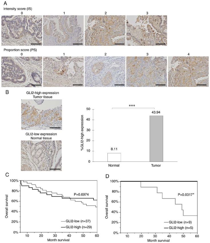
998 ICHIMIYA et al: GLI2 INDUCES THE MALIGNANT PHENOTYPE OF GALLBLADDER CANCER
Materials and methods plates (5.0x10³ cells/well). Cells were then incubated with or
without 1.0 µg/ml recombinant Human Sonic Hedgehog/SHH
Cell lines. The three human GBC cell lines NOZ, (R&D SYSTEMS, Inc.) at 37˚C for 24 and 48 h. In cytotoxic
TGBC2TKB (27), and TYGBK‑1 (28) were used. NOZ and assays, siRNA‑transfected cells were incubated with or without
TYGBK‑1 cells were purchased from the Japanese Collection GEM or CDDP (0‑100 µg/ml) at 37˚C for 48 h. Cell Count
of Research Bioresources (JCRB) bank. The TGBC2TKB cell Reagent SF (Nacalai Tesque, Inc.) at the original concentra-
line was purchased from Riken Cell Bank (Tsukuba, Japan). tion was then added to the cells and incubated at 37˚C for
All cell lines were cultured according to the supplier's specifi- 1 h. Cell proliferation was assessed by absorbance at 492 nm
cations. Absence of mycoplasma contamination in the cell lines using a plate reader (Biotrak visible plate reader; Amersham
was confirmed using a mycoplasma detection kit (Lonza Group, Biosciences; Cytiva) with a reference wavelength 620 nm.
Ltd.). For normoxic conditions, cells were cultured in 5% CO2
and 95% air; for hypoxic conditions, cells were cultured in Cell invasion assay. The invasive capacity of the GBC cell
1% O2, 5% CO2, and 94% N2 in a multi‑gas incubator (Sanyo). lines was assessed by Matrigel invasion assay as previously
described (19). Briefly, siRNA‑transfected cells (2.0x10⁵) were
RNA interference. ON‑TARGETplus™ SMARTpool small inter- placed in the upper chamber of a Transwell chamber with
fering (si)RNA targeting siRNA against GLI1 (cat. no. L‑003896), or without recombinant human SHH and incubated for 18 h.
GLI2 (cat. no. L‑006468), GLI3 (cat. no. L‑011043), and The cells that invaded to the lower side of the filter were fixed
hypoxia‑inducible factor 1α (HIF‑1α) (cat. no. L‑004018) and stained with Diff‑Quik reagent (Sysmex Corporation).
and negative control siRNA (ON‑TARGETplus™ Control Diff‑Quik Fixative, Diff‑Quik Solution I, and then Diff‑Quik
non‑targeting siRNA, cat. no. D‑001810) were purchased from Solution II were used in this order at their original concentra-
Dharmacon; Horizon Discovery Ltd. Cells were seeded in a tions for 10 min at room temperature. The stained cells were
6‑well plate (2.0x10⁵ cells per well) and transfected with 20 µM counted at an x200 magnification under a light microscope
of each construct using Lipofectamine® RNAiMAX (Invitrogen; (Nikon Eclipse TE 300; Nikon Corporation).
Thermo Fisher Scientific, Inc.) at 37˚C for 48 h according to the
manufacturerʼs protocol. The knockdown efficiency of siRNAs Western blot analysis. Western blotting was performed as previ-
for each target gene in each GBC cell line was evaluated by ously described (30). The protein‑transferred membranes were
real time RT‑qPCR analysis (Fig. S1). The time course result of incubated overnight at 4˚C with primary antibodies for GLI1
knockdown efficiency of GLI2 siRNA by real time RT‑qPCR (1:500; cat. no. ab151796; Abcam), GLI2 (1:500; cat. no. ab187386;
analysis is presented in Fig. S2. Abcam), GLI3 (1:200; cat. no. sc‑6154; Santa Cruz Biotechnology,
Inc.), SMO (1:200; cat. no. 20787‑1‑AP; ProteinTech Group, Inc.),
Real time reverse transcription‑quantitative PCR (real time SHH (1:200; cat. no. sc‑365112; Santa Cruz Biotechnology, Inc.),
RT‑qPCR). Total RNA of each GBC cell line was extracted cyclin D1 (1:200; cat. no. sc‑246; Santa Cruz Biotechnology, Inc.),
using a High Pure RNA Isolation kit (Roche Diagnostics) cyclin B1 (1:200; cat. no. sc‑245; Santa Cruz Biotechnology, Inc.),
and quantified by spectrophotometry (NanoDrop 1000; Ki‑67 (1:200; cat. no. sc‑15402; Santa Cruz Biotechnology, Inc.),
Thermo Fisher Scientific, Inc.). RNA (1.0 µg) was reverse E‑cadherin (1:200; cat. no. sc‑7870; Santa Cruz Biotechnology,
transcribed to cDNA with the Verso cDNA Synthesis Kit Inc.), vimentin (1:1,000; cat. no. ab92547; Abcam), SNAI1
(Thermo Fisher Scientific, Inc.) according to the manufac- (1:200, cat. no. sc‑271977; Santa Cruz Biotechnology, Inc.),
turer's protocol. Reactions were run on a 7500 Real‑Time PCR Slug (1:200; cat. no. sc‑15391; Santa Cruz Biotechnology, Inc.),
System™ (Applied Biosystems; Thermo Fisher Scientific, transforming growth factor‑β1 (TGFβ1) (1:200; cat. no. sc‑146;
Inc.) using PowerUp™ SYBR™ Green Master Mix (Applied Santa Cruz Biotechnology, Inc.), programmed cell death
Biosystems; Thermo Fisher Scientific, Inc.). The thermocy- ligand 1 (PD‑L1) (Clone; 29E.22A3) (1:400; cat. no. B7‑H1;
cling parameters were as follows: Initial denaturation at 95˚C BioLegend, Inc.), or HIF‑1α (1:200; cat. no. ab51608; Abcam).
for 2 min, followed by 40 cycles of denaturation at 95˚C for The membranes were then incubated for at least 1 h at room
15 sec and annealing/elongation at 60˚C for 1 min. The primer temperature with horseradish peroxidase‑linked anti‑mouse
sequences used were as follows: Gli1 forward, 5'‑TACATC antibody (1:10,000; cat. no. NA931; Amersham Biosciences;
AACTCCGGCCAATAGG‑3' and reverse, 5'‑CGGCGGCTG Cytiva), horseradish peroxidase‑linked anti‑rabbit antibody
ACAGTATAGGCA‑3'; Gli2 forward, 5'‑CGAGAAACCCTA (1:10,000; cat. no. NA934; Amersham Biosciences; Cytiva), or
CATCTGCAAGA‑3' and reverse, 5'‑GTGGACCGTTTTCAC horseradish peroxidase‑linked anti‑goat antibody (1:10,000;
ATGCTT‑3'; Gli3 forward, 5'‑AAACCCCAATCATGGACT cat. no. sc‑2020; Santa Cruz Biotechnology, Inc.). α‑Tubulin
CAAC‑3' and reverse, 5'‑TACGTGCTCCATCCATTTGGT‑3'; (1:1,000; cat. no. T6199; Sigma‑Aldrich; Merck KGaA) was used
HIF‑1α forward, 5'‑GAAGTGTACCCTAACTAGCCGAGG‑3' as a protein loading control.
and reverse, 5'‑TTTCTTATACCCACACTGAGGT TGG ‑3';
and β ‑actin forward, 5'‑TTGCCGACAG GATGCAGAAGG Cell cycle analysis. Cells (2.0x10⁵ cells/well) were treated for
A‑3' and reverse, 5'‑AGGTGGACAGCGAGGCCAGGAT‑3'. 48 h with GLI2 siRNA. Cells were harvested by trypsinization
The amount of each target gene in a given sample was normal- and fixed in ice‑cold 75% ethanol for at least 1 h. Cell pellets
ized to the level of β‑actin. Relative fold expression of the target were incubated and stained for 30 min at room temperature in
genes was calculated according to the 2‑∆∆Cq method (29). 1 ml PBS containing 50 µg propidium iodide (Sigma‑Aldrich;
Merck KGaA), 0.1% Triton X‑100, 1 mM/l EDTA, and 0.5 mg
Cell proliferation assay. All GBC cell lines were transfected ribonuclease A (Sigma‑Aldrich; Merck KGaA). After staining,
with GLI2 siRNA or control siRNA and seeded onto 96‑well samples were analyzed using FACScan (BD Biosciences)
ONCOLOGY REPORTS 45: 997-1010, 2021                                             999

and BD CellQuest™ Pro software 6.0 (BD Biosciences)                by incubation with Histofine Simple Stain MAX‑PO (M)
at 20,000 events per sample. The percentages of cells in           (4 µg/ml; cat. no. 424131) or Histofine Simple Stain MAX‑PO
G0/G1, S and G2/M phases were calculated for each sample.          (R) (4 µg/ml; cat. no. 424141; both from Nichirei Biosciences,
                                                                   Inc.) for 40 min at room temperature. The labeled antigens were
In vivo xenograft tumor model. Five‑week‑old female athymic        visualized using diaminobenzidine (DAB). Counterstaining
nude mice (BALB/c nu/nu; weight, 16‑18 g; n=8) were purchased      was performed with hematoxylin for 3 min at room tempera-
from Charles River Laboratories Japan and acclimated for           ture. Negative controls were obtained in all cases by omitting
2 weeks. All experimental procedures were approved by              the first antibodies (31). For Masson's trichrome staining, the
the Animal Care and Use Committee of Kyushu University             slides were treated with Bouin's solution (Sigma‑Aldrich;
(permit no. A30‑340‑0). All experiments were performed in          Merck KGaA) for 4 h at room temperature. The samples were
strict accordance with the Guidelines for Proper Conduct of        stained using working Weigert's iron hematoxylin solution
Animal Experiments (Science Council of Japan). Briefly, all        (Muto Pure Chemicals Co., Ltd.) for 10 min at room tempera-
mice were housed and maintained in a specific pathogen‑free        ture and then in 0.9% Biebrich scarlet‑acid fuchsin (Waldeck
animal facility at Kyushu University in housing conditions at a    GmbH & Co. KG), 0.1% acid fuchsin (Sigma‑Aldrich;
temperature of 26‑28˚C, humidity of 40‑70% and lighting time of    Merck KGaA), and 1% acetic acid (Kanto Chemical Co.,
12 h from 8 a.m. to 8 p.m.. Food and water were provided freely.   Inc.) for 20 min at room temperature. Subsequently, the slides
All efforts were made to minimize the number of animals used       were treated with working phosphotungstic/phosphomolybdic
and their suffering. The humane endpoints for euthanasia were      acid solution for 5 min at room temperature and placed in
defined as a tumor diameter exceeding 15 mm or conditions with     Aniline Blue solution (Muto Pure Chemicals Co., Ltd) for
long‑lasting pain. A mouse with a skin ulceration at the tumor     20 min at room temperature. GLI2 expression was evaluated
implantation site was euthanized as an indication for a humane     using the Allred score (32). Total score (TS) ≥3 was used
endpoint. At humane and experimental endpoints, mice were          as a cut‑off value (33,34) and the samples were divided into
euthanized by overdoses of inhaled anesthetics with sevoflurane,   two groups: GLI2‑high expression, with TS ≥3, and GLI2‑low
and the mortality of the mice was verified by cardio‑respiratory   expression, with TS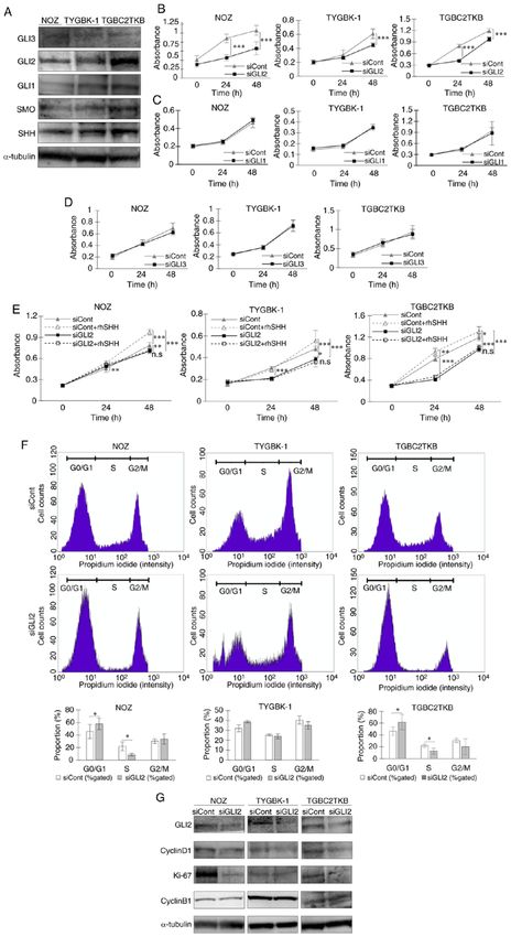
1000                      ICHIMIYA et al: GLI2 INDUCES THE MALIGNANT PHENOTYPE OF GALLBLADDER CANCER

Figure 1. GLI2 is involved in cell proliferation in GBC via regulation of the cell cycle. (A) Expression levels of GLI3, GLI2, GLI1, and Hh signal‑related molecules
(SMO and SHH) were examined in three GBC cell lines by western blot analysis. (B‑D) Proliferation assays in GBC cell lines treated with (B) GLI2 siRNA or
control siRNA, (C) GLI1 siRNA or control siRNA, and (D) GLI3 siRNA or control siRNA for 24 and 48 h. (E) Proliferation assays in GBC cell lines treated with
GLI2 siRNA or control siRNA and then incubated with or without rhSHH for 24 and 48 h. (F) Cell cycle analysis of GBC cell lines treated with GLI2 siRNA or
control siRNA. Percentages of cells in the G0/G1, S, and G2/M phase were calculated. Histograms and the bar graphs are presented. (G) Expression levels of GLI2
and cell cycle‑related molecules (cyclin D1, Ki‑67, and cyclin B1) were examined in GBC cell lines treated with GLI2 siRNA or control siRNA. *P
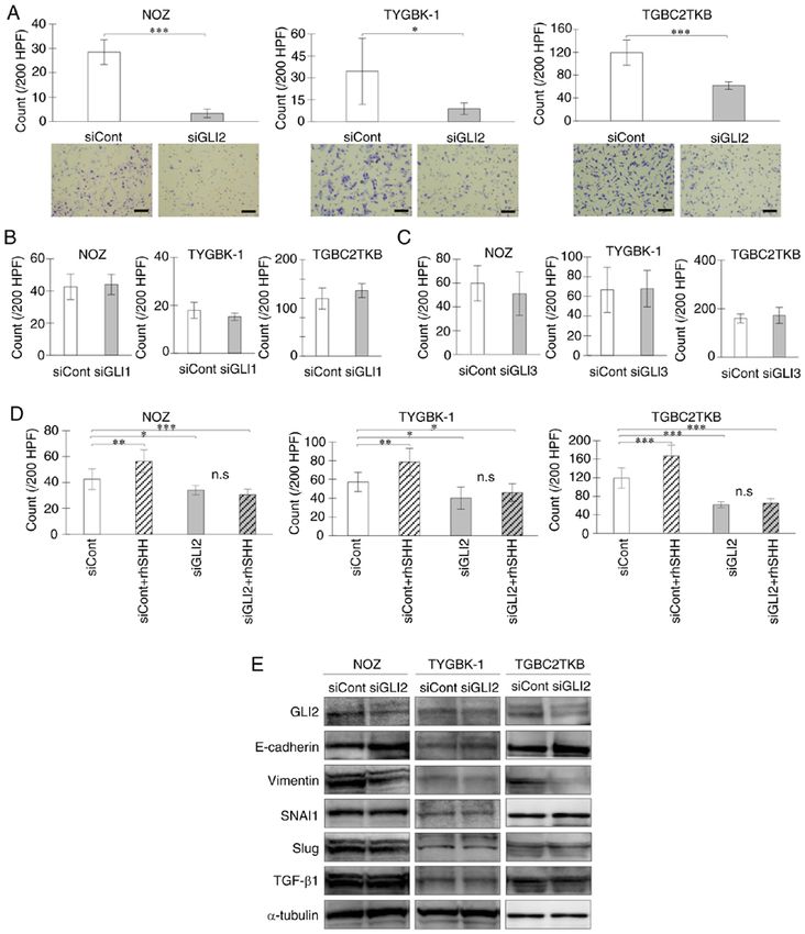
ONCOLOGY REPORTS 45: 997-1010, 2021                                                         1001

Figure 2. GLI2 is required for cell invasion via augmentation of EMT in GBC. Invasion assay of GBC cell lines treated with (A) GLI2 siRNA or control
siRNA, (B) GLI1 siRNA or control siRNA, and (C) GLI3 siRNA or control siRNA for 18 h. (D) Invasion assay of GBC cell lines treated with GLI2 siRNA or
control siRNA and then incubated with or without rhSHH for 18 h. (E) Expression levels of GLI2 and EMT‑related molecules (E‑cadherin, vimentin, SNAI1,
Slug, and TGF‑β1) in GBC cell lines treated with GLI2 siRNA or control siRNA. Scale bar, 100 µm. *P
1002                     ICHIMIYA et al: GLI2 INDUCES THE MALIGNANT PHENOTYPE OF GALLBLADDER CANCER

Figure 3. GLI2 suppression increases the relative survival of GBC cells treated with GEM. Relative absorbance ratio of GBC cell lines transfected with GLI2
siRNA or control siRNA and then treated with (A) GEM and (B) CDDP at 0‑100 µg/ml for 48 h. *P
ONCOLOGY REPORTS 45: 997-1010, 2021                                                        1003

Figure 4. Inhibition of GLI2 suppresses GBC‑cell derived tumor proliferation in vivo. (A) BALB/c nude mice were implanted with control siRNA‑ or GLI2
siRNA‑transfected NOZ cells. (B) GLI2 expression in implanted NOZ cells. (C) Representative images of mice implanted with NOZ cells transfected with
control siRNA (left) or GLI2 siRNA (right), and tumor volumes in the control siRNA group and GLI2 siRNA group. (D) Immunohistochemical staining of
αSMA, MT, Ki‑67, and VEGF in resected xenograft tumors. Original magnification is x200. Positively‑stained areas were quantified using ImageJ. Scale
bar, 100 µm. *P
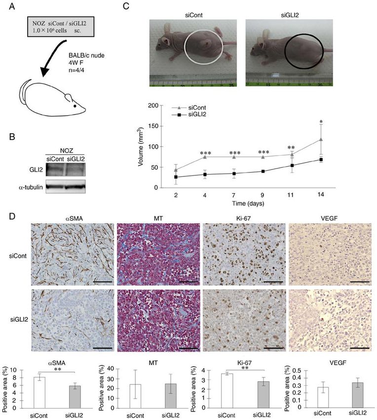
1004                     ICHIMIYA et al: GLI2 INDUCES THE MALIGNANT PHENOTYPE OF GALLBLADDER CANCER

Figure 5. Immunohistochemical analysis of GLI2 in primary GBC specimens. GLI2 expression was evaluated with the Allred Score. (A) Representative
examples of the intensity score (0‑3) and proportion score (0‑4). Original magnification is x200. (B) Representative examples of GLI2‑positive tumor tissue
(upper image) and GLI2‑negative normal tissue (lower image), and GLI2‑positive ratio in normal tissue and tumor tissue. Original magnification is x200.
(C) Kaplan‑Meier survival curves of all GBC patients of all stages according to GLI2 expression. (D) Kaplan‑Meier survival curves of 14 patients who received
adjuvant GEM treatments according to GLI2 expression. Scale bar, 100 µm. *P
ONCOLOGY REPORTS 45: 997-1010, 2021                                          1005

Table I. Associations between clinicopathological characteristics and GLI2 expression.

Variable                            GLI2 low expression (n=37)                  GLI2 high expression (n=29)             P‑value

Age (median=69.5)			                                                                                                    0.4569
1006                  ICHIMIYA et al: GLI2 INDUCES THE MALIGNANT PHENOTYPE OF GALLBLADDER CANCER

Table III. Univariate analysis using 66 GBC patients of overall survival.

Variable                                                    Category (Number of cases)                                        P‑value

GLI2                                      Low expression (37)/High expression (29)                                            0.6974
Age
ONCOLOGY REPORTS 45: 997-1010, 2021                                                          1007

Figure 6. GLI2 expression is inversely correlated to the number of tumor‑infiltrating CD8‑positive T lymphocytes and correlated to PD‑L1 expression.
Comparison of the number of tumor infiltrating (A) CD3‑positive cells and (B) CD8‑positive cells in GLI2‑high and GLI2‑low expression groups. Original
magnification is x400. (C) Comparison of tumor‑infiltrating CD8‑positive/FOXP3‑positive cell ratio in the two groups. (D) Comparison of the number of
tumor‑infiltrating FOXP3‑positive cells. Original magnification is x400. (E) Comparison of the PD‑L1‑positive ratio in the two groups. (F) GLI2 and PD‑L1
expression in GBC cell lines treated with GLI2 siRNA or control siRNA. (G) GLI2 expression of GBC cell lines incubated in normoxic or hypoxic condition
for 48 h. (H) HIF‑1α and GLI2 expression in GBC cell lines treated with HIF‑1α siRNA or control siRNA in hypoxic conditions for 48 h. Scale bar, 50 µm.
*
 P
1008                      ICHIMIYA et al: GLI2 INDUCES THE MALIGNANT PHENOTYPE OF GALLBLADDER CANCER

                                                                                       In view of the in vitro results revealing that GLI2 is
                                                                                  involved in the induction of malignant phenotype in GBC,
                                                                                  we hypothesized that there may be a correlation between
                                                                                  GLI2 expression and GBC patient prognosis. However, no
                                                                                  correlation was revealed between GLI2 expression and overall
                                                                                  survival. The present study included only 66 resectable cases,
                                                                                  and unresectable stage IV cases and elderly patients who
                                                                                  were intolerable to surgery were not included. The 5‑year
                                                                                  survival rate in the patient group was 54.55% (n=36/66),
                                                                                  which is substantially higher than the published 5‑year rate
                                                                                  of
ONCOLOGY REPORTS 45: 997-1010, 2021 1009
treatments for refractory GBC, which has few therapeutic 2. Lazcano‑Ponce EC, Miquel JF, Muñoz N, Herrero R, Ferrecio C,
Wistuba II, Alonso de Ruiz P, Aristi Urista G and Nervi F:
options. Epidemiology and molecular pathology of gallbladder cancer.
CA Cancer J Clin 51: 349‑364, 2001.
Acknowledgements 3. Misra S, Chaturvedi A, Misra NC and Sharma ID: Carcinoma of
the gallbladder. Lancet Oncol 4: 167‑176, 2003.
4. Hebrok M: Hedgehog signaling in pancreas development. Mech
We thank Ms. Emi Onishi for her skillful technical assis- Dev 120: 45‑57, 2003.
tance at Department of Cancer Therapy and Research, 5. Cohen MM Jr: The hedgehog signaling network. Am J Med
Genet A 123A: 5‑28, 2003.
Kyushu University Graduate School of Medical Sciences, 6. Ingham PW and McMahon AP: Hedgehog signaling in animal
Ms. Megumi Kiyota for technical assistance at The Research development: Paradigms and principles. Genes Dev 15:
Support Center, Research Center for Human Disease 3059‑3087, 2001.
7. Kalderon D: Similarities between the hedgehog and Wnt
Modeling, Kyushu University Graduate School of Medical signaling pathways. Trends Cell Biol 12: 523‑531, 2002.
Sciences. 8. Ruel L, Rodriguez R, Gallet A, Lavenant‑Staccini L and
Therond PP: Stability and association of smoothened, costal2
and fused with cubitus interruptus are regulated by hedgehog.
Funding Nat Cell Biol 5: 907‑913, 2003.
9. Onishi H and Katano M: Hedgehog signaling pathway as a
The present study was supported by the Japan Society for the therapeutic target in various types of cancer. Cancer Sci 102:
1756‑1760, 2011.
Promotion of Science KAKENHI grant no. 18K08620. 10. Kinzler KW, Bigner SH, Bigner DD, Trent JM, Law ML,
O'Brien SJ, Wong AJ and Vogelstein B: Identification of an ampli-
Availability of data and materials fied, highly expressed gene in a human glioma. Science 236:
70‑73, 1987.
11. Gailani MR and Bale AE: Developmental genes and cancer:
The datasets used and/or analyzed during the current study are Role of patched in basal cell carcinoma of the skin. J Natl Cancer
available from the corresponding author on reasonable request. Inst 89: 1103‑1109, 1997.
12. Xie J, Murone M, Luoh SM, Ryan A, Gu Q, Zhang C, Bonifas JM,
Lam CW, Hynes M, Goddard A, et al: Activating Smoothened
Authors' contributions mutations in sporadic basal‑cell carcinoma. Nature 391: 90‑92,
1998.
13. Hahn H, Wicking C, Zaphiropoulous PG, Gailani MR, Shanley S,
SI performed the majority of the experiments and wrote the Chidambaram A, Vorechovsky I, Holmberg E, Unden AB,
manuscript. HO, YoO and MN made notable contributions to Gillies S, et al: Mutations of the human homolog of Drosophila
the design, data interpretation and the manuscript revision. patched in the nevoid basal cell carcinoma syndrome. Cell 85:
841‑851, 1996.
SN, SK, KS, KN, AF, YaO and AI performed experiments 14. Johnson RL, Rothman AL, Xie J, Goodrich LV, Bare JW,
and established experimental techniques. KS, YaO, and AI Bonifas JM, Quinn AG, Myers RM, Cox DR, Epstein EH Jr
helped to design and plan the experiments. KS and YaO were and Scott MP: Human homolog of patched, a candidate gene
for the basal cell nevus syndrome. Science 272: 1668‑1671,
involved in the validation of the data. All authors have read 1996.
and approved the manuscript. 15. Kubo M, Nakamura M, Tasaki A, Yamanaka N, Nakashima H,
Nomura M, Kuroki S and Katano M: Hedgehog signaling
pathway is a new therapeutic target for patients with breast
Ethics approval and consent to participate cancer. Cancer Res 64: 6071‑6074, 2004.
16. Thayer SP, di Magliano MP, Heiser PW, Nielsen CM, Roberts DJ,
Protocols involving the use of human tissues were approved Lauwers GY, Qi YP, Gysin S, Fernández‑del Castillo C,
Yajnik V, et al: Hedgehog is an early and late mediator of pancre-
by the Ethical Committees for Clinical Study at Kyushu atic cancer tumorigenesis. Nature 425: 851‑856, 2003.
University (reference no. 30‑230) and conducted in accor- 17. Watkins DN, Berman DM, Burkholder SG, Wang B, Beachy PA
dance with the Ethical Guidelines for Human Genome/Gene and Baylin SB: Hedgehog signalling within airway epithelial
progenitors and in small‑cell lung cancer. Nature 422: 313‑317,
Research enacted by the Japanese Government and the 2003.
Helsinki Declaration. Written informed consent was obtained 18. Berman DM, Karhadkar SS, Maitra A, Montes De Oca R,
from all patients who agreed to the use of their samples in the Gerstenblith MR, Briggs K, Parker AR, Shimada Y, Eshleman JR,
Watkins DN and Beachy PA: Widespread requirement for
present research. Protocols involving the use of animals were Hedgehog ligand stimulation in growth of digestive tract tumours.
approved by the Animal Care and Use Committee of Kyushu Nature 425: 846‑851, 2003.
University (permit no. A30‑340‑0). 19. Matsushita S, Onishi H, Nakano K, Nagamatsu I, Imaizumi A,
Hattori M, Oda Y, Tanaka M and Katano M: Hedgehog signaling
pathway is a potential therapeutic target for gallbladder cancer.
Patient consent for publication Cancer Sci 105: 272‑280, 2014.
20. Dahmane N, Sánchez P, Gitton Y, Palma V, Sun T, Beyna M,
Weiner H and Ruiz I Altaba A: The sonic hedgehog‑gli
Not applicable. pathway regulates dorsal brain growth and tumorigenesis.
Development 128: 5201‑5212, 2001.
Competing interests 21. Berman DM, Karhadkar SS, Hallahan AR, Pritchard JI,
Eberhart CG, Watkins DN, Chen JK, Cooper MK, Taipale J,
Olson JM and Beachy PA: Medulloblastoma growth inhibi-
The authors declare that they have no competing interests. tion by Hedgehog pathway blockade. Science 297: 1559‑1561,
2002.
22. Chen JK, Taipale J, Cooper MK and Beachy PA: Inhibition of
References hedgehog signaling by direct binding of cyclopamine to smooth-
ened. Genes Dev 16: 2743‑2748, 2002.
1. Bray F, Ferlay J, Soerjomataram I, Siegel RL, Torre LA and 23. Rudin CM, Hann CL, Laterra J, Yauch RL, Callahan CA, Fu L,
Jemal A: Global cancer statistics 2018: GLOBOCAN estimates Holcomb T, Stinson J, Gould SE, Coleman B, et al: Treatment of
of incidence and mortality worldwide for 36 cancers in 185 coun- medulloblastoma with hedgehog pathway inhibitor GDC‑0449.
tries. CA Cancer J Clin 68: 394‑424, 2018. N Engl J Med 361: 1173‑1178, 2009.1010 ICHIMIYA et al: GLI2 INDUCES THE MALIGNANT PHENOTYPE OF GALLBLADDER CANCER
24. Yauch RL, Dijkgraaf GJ, Alicke B, Januario T, Ahn CP, 43. Onishi H, Kai M, Odate S, Iwasaki H, Morifuji Y, Ogino T,
Holcomb T, Pujara K, Stinson J, Callahan CA, Tang T, et al: Morisaki T, Nakashima Y and Katano M: Hypoxia activates the
Smoothened mutation confers resistance to a Hedgehog pathway hedgehog signaling pathway in a ligand‑independent manner by
inhibitor in medulloblastoma. Science 326: 572‑574, 2009. upregulation of Smo transcription in pancreatic cancer. Cancer
25. Von Hoff DD, LoRusso PM, Rudin CM, Reddy JC, Yauch RL, Sci 102: 1144‑1150, 2011.
Tibes R, Weiss GJ, Borad MJ, Hann CL, Brahmer JR, et al: 44. Onishi H, Yamasaki A, Kawamoto M, Imaizumi A and
Inhibition of the hedgehog pathway in advanced basal‑cell carci- Katano M: Hypoxia but not normoxia promotes smoothened
noma. N Engl J Med 361: 1164‑1172, 2009. transcription through upregulation of RBPJ and mastermind‑like
26. Sabol M, Trnski D, Musani V, Ozretić P and Levanat S: Role of 3 in pancreatic cancer. Cancer Lett 371: 143‑150, 2016.
GLI transcription factors in pathogenesis and their potential as 45. Duman‑Scheel M, Weng L, Xin S and Du W: Hedgehog regulates
new therapeutic targets. Int J Mol Sci 19: E2562, 2018. cell growth and proliferation by inducing cyclin D and cyclin E.
27. Horiuchi H, Kawamata H, Fujimori T and Kuroda Y: A MEK Nature 417: 299‑304, 2002.
inhibitor (U0126) prolongs survival in nude mice bearing human 46. Day PJ, Cleasby A, Tickle IJ, O'Reilly M, Coyle JE, Holding FP,
gallbladder cancer cells with K‑ras mutation: Analysis in a novel McMenamin RL, Yon J, Chopra R, Lengauer C and Jhoti H:
orthotopic inoculation model. Int J Oncol 23: 957‑963, 2003. Crystal structure of human CDK4 in complex with a D‑type
28. Sekine S, Shimada Y, Nagata T, Moriyama M, Omura T, cyclin. Proc Natl Acad Sci USA 106: 4166‑4170, 2009.
Yoshioka I, Hori R, Matsui K, Sawada S, Okumura T, et al: 47. Rutter M, Wang J, Huang Z, Kuliszewski M and Post M: Gli2
Establishment and characterization of a new human gallbladder influences proliferation in the developing lung through regula-
carcinoma cell line. Anticancer Res 32: 3211‑3218, 2012. tion of cyclin expression. Am J Respir Cell Mol Biol 42: 615‑625,
29. Livak KJ and Schmittgen TD: Analysis of relative gene expres- 2010.
sion data using real‑time quantitative PCR and the 2(‑Delta 48. Zhang D, Liu J, Wang Y, Chen J and Chen T: shRNA‑mediated
Delta) C(T)) method. Methods 25: 402‑408, 2001. silencing of Gli2 gene inhibits proliferation and sensitizes human
30. Suyama K, Onishi H, Imaizumi A, Shinkai K, Umebayashi M, hepatocellular carcinoma cells towards TRAIL‑induced apop-
Kubo M, Mizuuchi Y, Oda Y, Tanaka M, Nakamura M and tosis. J Cell Biochem 112: 3140‑3150, 2011.
Katano M: CD24 suppresses malignant phenotype by downregu- 49. Gerdes J, Lemke H, Baisch H, Wacker HH, Schwab U and
lation of SHH transcription through STAT1 inhibition in breast Stein H: Cell cycle analysis of a cell proliferation‑associated
cancer cells. Cancer Lett 374: 44‑53, 2016. human nuclear antigen defined by the monoclonal antibody
31. Kameda C, Nakamura M, Tanaka H, Yamasaki A, Kubo M, Ki‑67. J Immunol 133: 1710‑1715, 1984.
Tanaka M, Onishi H and Katano M: Oestrogen receptor‑alpha 50. Kawamoto M, Umebayashi M, Tanaka H, Koya N, Nakagawa S,
contributes to the regulation of the hedgehog signalling pathway Kawabe K, Onishi H, Nakamura M and Morisaki T: Combined
in ERalpha‑positive gastric cancer. Br J Cancer 102: 738‑747, gemcitabine and metronidazole is a promising therapeutic
2010. strategy for cancer stem‑like cholangiocarcinoma. Anticancer
32. Allred DC, Harvey JM, Berardo M and Clark GM: Prognostic Res 38: 2739‑2748, 2018.
and predictive factors in breast cancer by immunohistochemical 51. Thompson R and Eastman A: The cancer therapeutic potential of
analysis. Mod Pathol 11: 155‑168, 1998. Chk1 inhibitors: How mechanistic studies impact on clinical trial
33. Harvey JM, Clark GM, Osborne CK and Allred DC: Estrogen design. Br J Clin Pharmacol 76: 358‑369, 2013.
receptor status by immunohistochemistry is superior to the 52. Montano R, Khan N, Hou H, Seigne J, Ernstoff MS, Lewis LD
ligand‑binding assay for predicting response to adjuvant endo- and Eastman A: Cell cycle perturbation induced by gemcitabine
crine therapy in breast cancer. J Clin Oncol 17: 1474‑1481, 1999. in human tumor cells in cell culture, xenografts and bladder
34. Mohsin SK, Weiss H, Havighurst T, Clark GM, Berardo M, cancer patients: Implications for clinical trial designs combining
Roanh le D, To TV, Qian Z, Love RR and Allred DC: gemcitabine with a Chk1 inhibitor. Oncotarget 8: 67754‑67768,
Progesterone receptor by immunohistochemistry and clinical 2017.
outcome in breast cancer: A validation study. Mod Pathol 17: 53. Ou YQ, Zhu Wb, Li Y, Qiu PX, Huang YJ, Xie J, He SM,
1545‑1554, 2004. Zheng XK, Leng TD, Xu D and Yan GM: Aspirin inhibits prolif-
35. Lin J, Long J, Wan X, Chen J, Bai Y, Wang A, Yang X, Wu Y, eration of gemcitabine‑resistant human pancreatic cancer cells
Robson SC, Sang X and Zhao H: Classification of gallbladder and augments gemcitabine‑induced cytotoxicity. Acta Pharmacol
cancer by assessment of CD8+ TIL and PD‑L1 expression. BMC Sin 31: 73‑80, 2010.
Cancer 18: 766, 2018. 54. Bynigeri RR, Jakkampudi A, Jangala R, Subramanyam C,
36. Schindelin J, Arganda‑Carreras I, Frise E, Kaynig V, Longair M, Sasikala M, Rao GV, Reddy DN and Talukdar R: Pancreatic
Pietzsch T, Preibisch S, Rueden C, Saalfeld S, Schmid B, et al: stellate cell: Pandora's box for pancreatic disease biology. World
Fiji: An open‑source platform for biological‑image analysis. Nat J Gastroenterol 23: 382‑405, 2017.
Methods 9: 676‑682, 2012. 55. Oyama Y, Onishi H, Koga S, Murahashi M, Ichimiya S,
37. Cui X, Zhu S, Tao Z, Deng X, Wang Y, Gao Y, Liao Y, Ma W, Nakayama K, Fujimura A, Kawamoto M, Imaizumi A,
Umebayashi M, et al: Patched 1‑interacting peptide represses
Zhang Y and Ma X: Long‑term outcomes and prognostic markers fibrosis in pancreatic cancer to augment the effectiveness of
in gallbladder cancer. Medicine (Baltimore) 97: e11396, 2018. immunotherapy. J Immunother 43: 121‑133, 2020.
38. Valle J, Wasan H, Palmer DH, Cunningham D, Anthoney A, 56. Pardoll DM: The blockade of immune checkpoints in cancer
Ma raveyas A, Madhusuda n S, Iveson T, Hughes S, immunotherapy. Nat Rev Cancer 12: 252‑264, 2012.
Pereira SP, et al: Cisplatin plus gemcitabine versus gemcitabine 57. Sharma P and Allison JP: The future of immune checkpoint
for biliary tract cancer. N Engl J Med 362: 1273‑1281, 2010. therapy. Science 348: 56‑61, 2015.
39. Javle M, Zhao H and Abou‑Alfa GK: Systemic therapy for gall- 58. Wu T and Dai Y: Tumor microenvironment and therapeutic
bladder cancer. Chin Clin Oncol 8: 44, 2019. response. Cancer Lett 387: 61‑68, 2017.
40. Liu CC, Steen CB and Newman AM: Computational approaches 59. Onishi H, Fujimura A, Oyama Y, Yamasaki A, Imaizumi A,
for characterizing the tumor immune microenvironment. Kawamoto M, Katano M, Umebayashi M and Morisaki T:
Immunology 158: 70‑84, 2019. Hedgehog signaling regulates PDL‑1 expression in cancer cells
41. Zhang Y and Zhang Z: The history and advances in cancer to induce anti‑tumor activity by activated lymphocytes. Cell
immunotherapy: Understanding the characteristics of Immunol 310: 199‑204, 2016.
tumor‑infiltrating immune cells and their therapeutic implica-
tions. Cell Mol Immunol 17: 807‑821, 2020.
42. Cho YA, Yoon HJ, Lee JI, Hong SP and Hong SD: Relationship This work is licensed under a Creative Commons
between the expressions of PD‑L1 and tumor‑infiltrating Attribution-NonCommercial-NoDerivatives 4.0
lymphocytes in oral squamous cell carcinoma. Oral Oncol 47: International (CC BY-NC-ND 4.0) License.
1148‑1153, 2011.You can also read