Potential role of mitochondria in gastric cancer detection: Fission and glycolysis (Review)
←
→
Page content transcription
If your browser does not render page correctly, please read the page content below
ONCOLOGY LETTERS 21: 439, 2021

                   Potential role of mitochondria in gastric cancer
                     detection: Fission and glycolysis (Review)
                                             HANG YANG, YAN LI and BING HU

        Department of Gastroenterology, West China Hospital, Sichuan University, Chengdu, Sichuan 610041, P.R. China

                                      Received January 10, 2021; Accepted March 22, 2021

                                                    DOI: 10.3892/ol.2021.12700

Abstract. Gastric cancer (GC) is characterized by high              5. Reprogrammed energy metabolism and GC
morbidity and mortality rates worldwide. Helicobacter pylori        6. Association between mitochondrial dynamics and energy
infection, high salt intake, smoking, alcohol, low fiber intake,       metabolism: Fission and glycolysis
family history of GC, obesity and precancerous lesions,             7. Conclusions
including chronic atrophic gastritis and intestinal metaplasia,
are considered general risk factors for GC. Image enhance‑
ment endoscopy methods, which improve the visualization             1. Introduction
of mucosal structures and vascularity, may be used for the
early diagnosis of GC, such as narrow band imaging, which
can reveal fine details of subtle superficial abnormalities of      Chronic atrophic gastritis (CAG) is considered a common risk
early gastric cancer (EGC). Mitochondria are well‑known for         factor for the development of gastric cancer (GC). Endoscopic
their role in producing ATP via the tricarboxylic acid cycle. In    imaging and biopsy are crucial for early detection and
cancer cells, the energetic metabolism can be reprogrammed          diagnosis of GC (1). Image‑enhanced endoscopy combined
as anaerobic glycolysis for energy production and anabolic          with biopsy, according to the Sydney protocol and regular
growth. In addition to their dominant metabolic functions,          endoscopic surveillance, are recommended for patients with
mitochondria participate in several central signaling pathways,     extensive CAG or intestinal metaplasia (2). A visible lesion
such as the apoptotic pathway and NLRP3 inflammasome                may be treated by endoscopic mucosal resection or endoscopic
activation. Conversely, mitochondrial dynamics, including           submucosal dissection. However, when a lesion is invisible,
fission/fusion and mitophagy, can also contribute to the            regular endoscopic surveillance is required for high‑risk
pathogenesis of cancer. The dysfunction and dysregulation of        patients. The interval between Helicobacter pylori eradica‑
mitochondria have been associated with several ageing and           tion and cancer occurrence may vary from several months to
degenerative diseases, as well as cancer. The present review        >10 years (3). Surveillance endoscopy is one of the methods
focuses on energy metabolism and mitochondrial dynamics,            enabling the early diagnosis of GC (4). Once an existing lesion
and summarizes the changes in gastric carcinogenesis, the           is identified, it can be treated in a timely manner. Interval
diagnosis of EGC and indicates potential targeted treatments.       cancer can occur due to missed lesions or to a newly developed
                                                                    lesion during surveillance (5). Thus, it is essential to identify
                                                                    a molecular biological marker for the detection of invisible
Contents                                                            lesions at the organelle level.
                                                                        Recently, diverse pathophysiological functions of mitochon‑
1.   Introduction                                                   dria have been reported, including mitochondrial dynamics (6),
2.   Mitochondrial dynamics: Fission and fusion                     metabolic reprogramming (7), mitochondria‑released
3.   Fission, mitophagy and GC                                      damage‑associated molecular patterns and NLRP3 inflamma‑
4.   Fusion and GC                                                  some activation (8), mitochondrial DNA (mtDNA), autophagy
                                                                    and mitophagy (9), mitochondrial outer membrane permea‑
                                                                    bilization (10) and mitochondrial aging (11). In addition,
                                                                    mitochondrial (mt)DNA mutations, deletions and impaired
                                                                    DNA replication are the most common causes of mitochon‑
Correspondence     to: Professor Bing Hu, Department of
                                                                    drial dysfunction (12). mtDNA sensing via STING signaling
Gastroenterology, West China Hospital, Sichuan University, 37 Guo
Xue Xiang, Wu Hou, Chengdu, Sichuan 610041, P.R. China              participates in inflammation and cancer (12,13). The effects
E‑mail: hubingnj@163.com                                            of mitochondrial dynamics on carcinogenesis and cancer
                                                                    progression have also been reported, highlighting the poten‑
Key words: diagnosis of early gastric cancer, mitochondrial         tial use of mitochondrial biomarkers in cancer detection and
dynamics, reprogrammed energy metabolism                            prognosis, as well as the potential targeting of mitochondrial
                                                                    dynamics for treating cancer (14). However, there is still a
                                                                    paucity of research associated with GC.
2 YANG et al: FISSION AND GLYCOLYSIS IN GC
The present review summarizes the role of mitochondrial reintegration, thereby facilitating mitophagy, mainly via
dynamics and energy metabolism reprogramming in GC to interactions between Parkin, Bcl‑2/adenovirus E1B 19 kDa
identify potential indicators for biologically complemented protein interacting protein 3 (BNIP3) and Drp1 (24). Increasing
endoscopy and further promote translating discoveries of Drp1 results in excessive mitochondrial fragmentation and
molecular biology. Thus, fission and glycolysis from mitochon‑ deficiencies, decreases mitochondrial motility and shortens
dria may be useful in detecting GC. If an electron microscope mitochondrial length (25), which may be further enhanced in
can be installed on the endoscopy system, the mitochondrial hypoxia (26). Fission can also be triggered by stress stimuli,
dynamics may be observable during the early stages of GC. such as nutrient deprivation, DNA damage, inflammation and
Furthermore, when fission is increased and fusion is decreased, mitochondrial membrane depolarization (27). Given that mito‑
further precision biopsy of the targeted tissue should be chondria‑associated membranes related to the endoplasmic
performed to detect metabolic activity. The combination reticulum at specific regions can facilitate calcium (Ca2+) flux
of both approaches may enable early diagnosis and provide into the mitochondria and further control the homeostasis and
a novel treatment strategy. However, further investigation is metabolism of Ca2+, close coupling of these organelles increases
required. mitochondrial Ca2+ levels, thus initiating apoptosis (28). It has
also been reported that enhanced fission attenuates adherence
2. Mitochondrial dynamics: Fission and fusion to inhibit Ca2+ overload in mitochondria and apoptosis (29).
In terms of mitophagy, this process maintains cellular
Mitochondria are responsible for energy supply and are health by selectively enclosing damaged and depolarized
involved in several biological processes, including cell death mitochondria in autophagic vacuoles for lysosome‑mediated
and proliferation (6). Mitochondria constantly maintain a elimination (30). Mitophagy degrades dysfunctional mito‑
dynamic shape, which may change in response to cellular chondria and further attenuates reactive oxygen species (ROS)
bioenergetic demands, such as nutrient status, which is generation, which in turn promotes cell survival and protects
defined as mitochondrial dynamics (12). The mitochondrial against cell death (31). Increasing evidence suggest that several
morphology is a result of the interplay between rapid fusion modulators of mitophagy are deregulated in human cancer,
and fission events (15). The key components mediating these including Parkinson protein 2 E3 ubiquitin protein ligase,
processes belong to the dynamin family of GTPases that FUN14 domain containing 1, BNIP3 and BNIP3L (32,33).
utilize GTP hydrolysis to drive mechanical work on biological In addition, a study revealed that impaired mitophagy can
membranes (16). Mitofusin proteins, Mfn1 and Mfn2, are enhance the aggressiveness in GC cells under hypoxia by
involved in the fusion of the outer mitochondrial membrane, activating the mtROS/hypoxia‑inducible factor (HIF)‑1α inter‑
while GTPase optic atrophy 1 mediates the fusion of the inner play (34). Mitophagy may also be enhanced by overexpression
mitochondrial membrane (17). Mitochondrial fission is medi‑ of Opa‑interacting protein 5, thus plays an important role in cell
ated by the GTPase dynamin‑related protein 1 (Drp1) following survival and death in docetaxel‑treated GC cells (35). Another
its recruitment by the membrane‑anchored proteins, namely study demonstrated that Drp1 expression is upregulated, and
mitochondrial fission factor and fission protein 1 (Fis1) (18). the expression levels of the mitophagy‑related regulators,
Commonly, the mitochondrial fission/fusion machinery is PTEN‑induced putative kinase 1 and Parkin, are downregu‑
involved in generating new mitochondria, and eliminating old, lated in patients with GC (36). Given that mitophagy can clear
damaged and non‑repairable mitochondria (6). Mitochondrial the damaged part of mitochondria and mtDNA, it protects
fission plays an important role in mitochondrial proliferation, healthy cells from malignant transformation and tumor cells
mitochondrial distribution during cell division and the removal from apoptosis (31). It has been suggested that, in the early
of damaged mitochondria via mitophagy (19). Unopposed stages of GC, mitophagy is associated with tumor suppression,
mitochondrial fission causes mitochondrial fragmentation, whereby it can promote tumor growth at the advanced stages of
which is generally associated with metabolic dysfunction and GC. For example, mitophagy was increased in advanced‑stage
several diseases, such as degenerative diseases and cancer (20). GC to sustain the viability and migration of GC cells (37), since
It has been reported that impaired mitochondrial fission is mitophagy in solids tumor may be activated by two common
associated with mitochondrial elongation (21). In addition, factors, namely hypoxia and low nutrient supply (38) (Fig. 1).
unopposed fusion results in a hyperfused network and serves
to counteract metabolic insults, preserve cellular integrity and 4. Fusion and GC
protect against autophagy (20). It was previously reported that
impaired mitochondrial fusion may promote fission‑induced Mitochondrial fusion results in a more interconnected mito‑
mitochondrial fragmentation (21). Thus, the maintenance of chondrial network and enhances the communication with
mitochondrial fission/fusion balance plays a key role in cell the endoplasmic reticulum (39). Fusion allows the diffusion
cycle progression (6). The dynamics is critical for the effects of matrix content among mitochondria, diluting the accumu‑
of fission/fusion on morphology regulation, content exchange, lated mtDNA mutations and oxidized proteins (40). Fusion is
and the maintenance of mtDNA and mitochondrial oxidative commonly enhanced by starvation by triggering the protein
phosphorylation (OXPHOS) activity (22,23). kinase A‑mediated phosphorylation of Drp1 (at Ser637) to
blunt fission (41). In addition, mitochondrial fusion is required
3. Fission, mitophagy and GC for mtDNA maintenance (22). Thus, impaired mitochondrial
fusion is often accompanied by bioenergetic defects due to
Fission isolates depolarized mitochondria, while it coordinates loss of mtDNA (42). Furthermore, mitochondrial fusion is
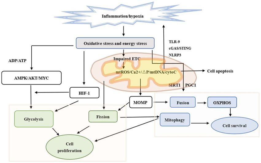
the downregulation of fusion mediators to prevent network also associated with increased OXPHOS and ATP generationONCOLOGY LETTERS 21: 439, 2021 3 Figure 1. Different mitochondrial dynamics and energy metabolism in an epithelial cell subjected to chronic inflammation. Chronic inflammation causes the injury of epithelial cells. Mitochondria is involved in further innate immune responses, including cGAS‑STING signaling, TLR‑9 and NLRP3 inflammasome formation following the release of mtDNA. Mitochondria is also associated with apoptosis. When atrophic epithelial cells preserve their programed cell death ability and sur‑ rounding inflammation is sufficiently severe, cells undergo apoptosis instead of necrosis. Chronic inflammation can also damage mitochondria and lead to changes in mitochondrial metabolism and dynamics via HIF‑1, AMPK and MOMP. Fission and glycolysis promote cell proliferation and invasion. Fusion and OXPHOS are compatible with cell survival. Mitophagy protects both normal and cancer cells by selectively eliminating damaged mitochondria. Green outline represents proliferation, blue outline represents survival and the text without boxes represent apoptosis. cGAS, cyclic GMP–AMP synthase; STING, stimulator of interferon genes; TLR‑9, Toll‑like receptor‑9; NLRP3, NOD‑like receptor family pyrin domain‑containing 3; mtDNA, mitochondrial DNA; HIF‑1, hypoxia‑inducible factor‑1; AMPK, AMP‑activated protein kinase; MOMP, mitochondrial outer membrane permeabilization; OXPHOS, oxidative phosphorylation; ETC, electron transport chain; mtROS, mitochondrial ROS; ΔP, increased potential; cytoC, cytochrome c; PGC1, proliferator‑activated receptor‑γ coactivator. via remodeling of the cristae (43,44), and downregulation which is associated with poor prognosis (50). It has also been of OPA1, which is responsible for fusion, resulted in mito‑ reported that SIRT1 exerts inhibitory effects on chemoresis‑ chondrial dysfunction and mtDNA stress (45). The number tance and cancer stem cell properties via Forkhead box O3 and of mitochondria is regulated by mitochondrial biogenesis to AMP‑activated protein kinase (AMPK) (51). AMPK, another meet the energy demands of the cells and compensate for their key energy metabolic sensor, plays a key role in maintaining damage (46). A study demonstrated that peroxisome prolifer‑ cellular energy homeostasis and is activated upon alterations in ator‑activated receptor gamma coactivator (PGC‑1) and the the cellular AMP/ATP ratio (52). Previous studies have demon‑ protein deacetylase sirtuin 1 (SIRT1) can regulate fusion and strated that, upon energy deficiency, AMPK activation may OXPHOS (14). Thus, activation of PGC‑1α by SIRT1 induces result in increased PGC‑1α expression and phosphorylation to mitochondrial biogenesis and confers metabolic advan‑ modulate the expression of several key players in mitochon‑ tages (14). Another study revealed that PGC‑1β can induce drial biogenesis and OXPHOS of fatty acids (53,54) (Fig. 1). mitochondrial fusion by upregulating Mfn2 expression via estrogen‑related receptor α coactivation (47). Mfn2 expression 5. Reprogrammed energy metabolism and GC is downregulated in GC tissues compared with normal gastric mucosal tissues, and is negatively associated with tumor size, Energy metabolism is essential for maintaining cellular homeo‑ indicating an antitumor role of Mfn2 (48). In vitro experi‑ stasis and biological functions, and includes ATP production ments have demonstrated that overexpression of Mfn2 can in the cytosol (glycolysis) and mitochondria (OXPHOS) (55), suppress gastric cancer cell proliferation and colony forma‑ which can be reprogrammed during carcinogenesis (56). tion (48). SIRT1 is an enzyme that mediates NAD+‑dependent Cancer cells undergo metabolic reprogramming, including deacetylation of target substrates (49). Given that the cellular enhanced glycolysis, mutations in genes encoding tricarbox‑ redox balance of NAD+ and NADH is highly associated with ylic acid (TCA) cycle enzymes, upregulation of de novo lipid catabolic fluxes, SIRT1 can act as a sensor, directly connecting synthesis and glutaminolysis (57). Glycolysis is characterized metabolic perturbations with transcriptional output (49). by an increased rate of glucose uptake and its glycolytic conver‑ SIRT1 expression is significantly downregulated in GC tissues, sion to lactate, even under oxygen‑rich conditions (55). There
4                                            YANG et al: FISSION AND GLYCOLYSIS IN GC

are several pathways and transcriptional regulators involved in      phenotype (12,73,74). Several studies have been performed in
the regulation of metabolic reprogramming, such as PI3K/AKT          different cell types that alter their mitochondrial morphology to
pathway and HIF‑1 (58,59). The PI3K/AKT pathway can regu‑            meet their energy demands, functions and behaviors. Conversely,
late several aspects of this metabolic program (58). A previous      certain cells, such as T cells and stem cells, have higher energy
study demonstrated that AKT activation was sufficient to             demands to perform their metabolic and cell‑specific func‑
induce glycolysis by promoting glucose transporter 1 and phos‑       tions (75,76). When T cells recognize major histocompatibility
phorylating pyruvate dehydrogenase kinase to inhibit pyruvate        complexes presented by antigen‑presenting cells in response
dehydrogenase and favor lactate dehydrogenase (LDH)                  to infection or tumors, they proliferate and differentiate into
activity (60). It has been reported that HIF‑1 is overexpressed      different T‑cell subsets (23). Effector T cells display looser
in human cancers as a result of intratumoral hypoxia, as well        cristae remodeling via fission with reduced electron transport
as genetic alterations, such as gain‑of‑function mutations in        chain (ETC) complexes, thus attenuating ETC efficiency and
oncogenes and loss‑of‑function mutations in tumor suppressor         promoting aerobic glycolysis (23). Conversely, in memory
genes (61). HIF‑1 may also be triggered by the accumulation          T cells, tight cristae remodeling via fusion with enhanced ETC
of TCA substrates (62), while its degradation is regulated by        complex activity is observed, thus enhancing ETC efficiency and
O2‑dependent prolyl hydroxylation (PHs) (61). HIF‑1α main‑           OXPHOS (23). Endothelial progenitor cells (EPCs) accelerate
tains its stability by avoiding the hydroxylation of PHs in cancer   glycolysis to produce lactate during angiogenesis by upregulating
cells, since PHs can be inhibited by the increased levels of         the expression levels of HIF‐1α and vascular endothelial growth
cytosolic pyruvate, lactate, succinate, fumarate and ROS (59).       factor (77). In human EPCs, downregulation of Fis1 expression is
Most genes encoding glycolytic enzymes and transporters are          associated with mitochondrial dysfunction and may contribute to
the targets of HIF‑1α, and its overexpression in cancer cells is     the impaired activity of EPCs during the senescence process (73).
associated with increased levels of glycolytic proteins (63). A      However, upregulation of Fis1 expression in senescent EPCs
study revealed that HIF‑1α levels were high in certain tumors,       restores the younger phenotype (73). Another study investigated
even under oxygen‑rich conditions, indicating that hormones          the function of mitochondrial fission genes in embryonic stem
or growth factors can cause the stabilization of HIF‑1α expres‑      cells (ESCs). Transmission electron microscopy revealed a
sion, which may serve important roles in carcinogenesis (64).        significant increase in the cytoplasm‑to‑nucleus ratio and mito‑
A previous study suggested that HIF‑1α can act as a negative         chondrial elongation in dynamin‑1‑like protein (‑/‑) ESCs caused
regulator of mitochondrial biogenesis and oxidative phosphory‑       by incomplete fission. In addition, increased OXPHOS and intra‑
lation to inhibit the conversion of pyruvate to acetyl‑CoA and       cellular ATP concentration and reduced glycolysis was observed,
mitochondrial respiration and to promote LDH expression (65).        which were associated with mitochondrial elongation (78). The
HIF‑1α activation can also inhibit MYC transcription to further      proliferation and invasion of tumor cells also require faster
downregulate PGC‑1α and PGC‑1β expression, which in turn             and increased energy supply (79). Thus, Drp1 expression is
regulates mitochondrial biogenesis and OXPHOS (54). In               upregulated in several types of cancer cells, including liver (80),
GC, inhibiting HIF‑1α signaling attenuates the migratory and         breast (81) and lung cancers (82), and may be considered as a
invasive abilities of GC cells, and epithelial‑to‑mesenchymal        biomarker for predicting poor survival in patients with these
transition (66), whereas activation of HIF‑1α signaling promotes     types of cancer. A study on ovarian cancer demonstrated
cell metastasis and glucose metabolism (67).                         that glycolysis is promoted by activating PI3K/AKT/HIF‑1α
    The tumor microenvironment favors the growth and expan‑          signaling, while mitochondrial fission is enhanced by phos‑
sion of cancer and inflammatory cells, which in turn directly or     phorylation of Drp1 at Ser616 (83). As a member of the AMPK
indirectly promotes gastric tumorigenesis by secreting soluble       family, salt‑inducible kinase 2 was demonstrated to be involved
factors or modulating immune responses (68). It has been reported    in both pathways (83). In addition, Drp1 expression was signifi‑
that NF‑κB is activated in chronic inflammation, thus promoting      cantly upregulated in pancreatic cancer (PC) cells and tissues via
the further activation of tumor‑promoting genes, such as IL‑6 and    downregulation of microRNA‑29a expression (74). High Drp1
cyclooxygenase (COX)‑2 (69). NF‑κB and HIF‑1 can link inflam‑        expression was associated with poor survival of patients with
matory signaling to hypoxia and coordinate the activation of both    PC, while Drp1 promoted both the proliferation and metastasis
COX‑2 and IL‑6, and the Janus kinase/STAT3 pathway (70). It          of PC cells, mainly through facilitating aerobic glycolysis (74).
has been reported that STAT3 cooperates with NF‑κB and HIF‑1         Another study revealed that Drp1 may promote KRAS‑driven
in the regulation of both genes (71). NF‑κ B can be strongly         tumor growth by supporting both glycolysis and mitochondrial
induced by hypoxia and chronic inflammation, and is involved         function (84). Taken together, these findings suggest a mutual
in the reprogramming of tumor glycolysis by interacting with         association between Drp1 and glycolysis, and the promoting
HIF‑1α (70). Given that inflammation can induce cells lacking        effect of Drp1 and glycolysis on cancer cell proliferation and
oxygen and upregulate HIF‑1α, glycolysis gradually becomes the       invasion.
main energy source instead of OXPHOS (55) (Fig. 1)
                                                                     7. Conclusions
6. Association between mitochondrial dynamics and en-
ergy metabolism: Fission and glycolysis                              GC is the fifth most common type of cancer and the third most
                                                                     common cause of cancer‑associated mortality, with 784,000
Mitochondrial morphological changes are a type of primary            mortalities reported in 2018 worldwide (85). Early detection
signal to shape metabolic reprogramming during cellular quies‑       and treatment can improve the outcome of patients with GC.
cence or activation (14,72). Recent studies have demonstrated        Innovative endoscopic techniques may be more accurate in
that increased mitochondrial fission promotes a pro‑tumorigenic      achieving cytological or even biological diagnosis. Mitochondria
ONCOLOGY LETTERS 21: 439, 2021 5
are strongly associated with carcinogenesis. The present review 2. Banks M, Graham D, Jansen M, Gotoda T, Coda S, di Pietro M,
Uedo N, Bhandari P, Pritchard DM, Kuipers EJ, et al: British
summarized the role of mitochondria dynamics, reprogramming Society of Gastroenterology guidelines on the diagnosis and
of energy metabolism and their changes in GC. Based on current management of patients at risk of gastric adenocarcinoma.
literature, it can be concluded that mitochondria in GC are char‑ Gut 68: 1545‑1575, 2019.
3. Take S, Mizuno M, Ishiki K, Kusumoto C, Imada T, Hamada F,
acterized by fission and enhanced glycolysis to meet the increased Yoshida T, Yokota K, Mitsuhashi T and Okada H: Risk of gastric
energy requirements of cancer cells, and decrease necrosis via cancer in the second decade of follow‑up after Helicobacter
mitophagy. Upregulated expression levels of Drp1 and HIF‑1α are pylori eradication. J Gastroenterol 55: 281‑288, 2020.
4. Shichijo S and Hirata Y: Characteristics and predictors of
associated with fission and glycolysis, respectively. The balance gastric cancer after Helicobacter pylori eradication. World J
of mitochondrial fission and fusion and the ratio of glycolysis to Gastroenterol 24: 2163‑2172, 2018.
OXPHOS are positively associated with different stages of carci‑ 5. Choi SI, Park B, Joo J, Kim YI, Lee JY, Kim CG, Choi IJ,
Kook MC and Cho SJ: Three‑year interval for endoscopic
nogenesis. When increased fission and glycolysis and decreased screening may reduce the mortality in patients with gastric
apoptosis and fusion are detected in high‑risk patients, they may cancer. Surg Endosc 33: 861‑869, 2019.
indicate that cells are in the process of malignant transformation. 6. Horbay R and Bilyy R: Mitochondrial dynamics during cell
cycling. Apoptosis 21: 1327‑1335, 2016.
Thus, treatment is required to inhibit this process, which may be 7. Kim SY: Cancer Energy Metabolism: Shutting Power off Cancer
a promising approach to the detection of early gastric cancer via Factory. Biomol Ther (Seoul) 26: 39‑44, 2018.
organelle‑ and molecular‑level endoscopy in the future. 8. Zhou R, Yazdi AS, Menu P and Tschopp J: A role for mitochondria
in NLRP3 inflammasome activation. Nature 469: 221‑225, 2011.
9. Jung S, Jeong H and Yu SW: Autophagy as a decisive process for
Acknowledgements cell death. Exp Mol Med 52: 921‑930, 2020.
10. Bock FJ and Tait SWG: Mitochondria as multifaceted regulators
of cell death. Nat Rev Mol Cell Biol 21: 85‑100, 2020.
Not applicable. 11. Sun N, Youle RJ and Finkel T: The Mitochondrial Basis of
Aging. Mol Cell 61: 654‑666, 2016.
Funding 12. Srinivasan S, Guha M, Kashina A and Avadhani NG: Mitochondrial
dysfunction and mitochondrial dynamics‑The cancer connection.
Biochim Biophys Acta Bioenerg 1858: 602‑614, 2017.
The present study was supported by the 1·3·5 project for 13. Liu S, Feng M and Guan W: Mitochondrial DNA sensing by
disciplines of excellence Clinical Research Incubation Project, STING signaling participates in inflammation, cancer and
beyond. Int J Cancer 139: 736‑741, 2016.
West China Hospital, Sichuan University, China (grant 14. Maycotte P, Ma r ín‑Her nández A, Goyr i‑Agui r re M,
no. 20HXFH016). Anaya‑Ruiz M, Reyes‑Leyva J and Cortés‑Hernández P:
Mitochondrial dynamics and cancer. Tumour Biol: May 4, 2017
(Epub ahead of print). doi: 10.1177/1010428317698391.
Availability of data and materials 15. Liu X, Weaver D, Shirihai O and Hajnóczky G: Mitochondrial
‘kiss‑and‑run’: Interplay between mitochondrial motility and
Not applicable. fusion‑fission dynamics. EMBO J 28: 3074‑3089, 2009.
16. Chan DC: Mitochondrial dynamics and its involvement in
disease. Annu Rev Pathol 15: 235‑259, 2020.
Authors' contributions 17. Schrepfer E and Scorrano L: Mitofusins, from mitochondria to
metabolism. Mol Cell 61: 683‑694, 2016.
18. Cantó C: Mitochondrial dynamics: Shaping metabolic adap‑
HY designed the present review and drafted the initial manuscript. tation. Int Rev Cell Mol Biol 340: 129‑167, 2018.
BH contributed to designing and reviewing the manuscript. HY, 19. Ni HM, Williams JA and Ding WX: Mitochondrial dynamics
YL and BH contributed to revising the manuscript for important and mitochondrial quality control. Redox Biol 4: 6‑13, 2015.
20. Wai T and Langer T: Mitochondrial dynamics and metabolic
intellectual content. Data authentication is not applicable. All regulation. Trends Endocrinol Metab 27: 105‑117, 2016.
authors have read and approved the final manuscript. 21. Bhatia D, Capili A and Choi ME: Mitochondrial dysfunction in
kidney injury, inflammation, and disease: Potential therapeutic
approaches. Kidney Res Clin Pract 39: 244‑258, 2020.
Ethics approval and consent to participate 22. Yan C, Duanmu X, Zeng L, Liu B and Song Z: Mitochondrial
DNA: Distribution, mutations, and elimination. Cells 8: 379, 2019.
Not applicable. 23. Buck MD, O'Sullivan D, Klein Geltink RI, Curtis JD, Chang CH,
Sanin DE, Qiu J, Kretz O, Braas D, van der Windt GJ, et al:
Mitochondrial dynamics controls T cell fate through metabolic
Patient consent for publication programming. Cell 166: 63‑76, 2016.
24. Friedman JR and Nunnari J: Mitochondrial form and function.
Nature 505: 335‑343, 2014.
Not applicable. 25. Campello S, Lacalle RA, Bettella M, Mañes S, Scorrano L and
Viola A: Orchestration of lymphocyte chemotaxis by mito‑
Competing interests chondrial dynamics. J Exp Med 203: 2879‑2886, 2006.
26. Wu W, Li W, Chen H, Jiang L, Zhu R and Feng D: FUNDC1
is a novel mitochondrial‑associated‑membrane (MAM) protein
The authors declare that they have no competing interests. required for hypoxia‑induced mitochondrial fission and
mitophagy. Autophagy 12: 1675‑1676, 2016.
27. Twig G, Elorza A, Molina AJ, Mohamed H, Wikstrom JD,
References Walzer G, Stiles L, Haigh SE, Katz S, Las G, et al: Fission and
selective fusion govern mitochondrial segregation and elimi‑
1. Pimentel‑Nunes P, Libânio D, Marcos‑Pinto R, Areia M, Leja M, nation by autophagy. EMBO J 27: 433‑446, 2008.
Esposito G, Garrido M, Kikuste I, Megraud F, Matysiak- 28. Kumar V and Maity S: ER Stress‑sensor proteins and
Budnik T, et al: Management of epithelial precancerous conditions ER‑mitochondrial crosstalk‑signaling beyond (ER) stress
and lesions in the stomach (MAPS II): European Society of response. Biomolecules 11: 173, 2021.
Gastrointestinal Endoscopy (ESGE), European Helicobacter and 29. Szabadkai G, Simoni AM, Chami M, Wieckowski MR, Youle RJ
Microbiota Study Group (EHMSG), European Society of Pathology and Rizzuto R: Drp‑1‑dependent division of the mitochondrial
(ESP), and Sociedade Portuguesa de Endoscopia Digestiva (SPED) network blocks intraorganellar Ca2+ waves and protects against
guideline update 2019. Endoscopy 51: 365‑388, 2019. Ca2+‑mediated apoptosis. Mol Cell 16: 59‑68, 2004.6 YANG et al: FISSION AND GLYCOLYSIS IN GC
30. Zhang J and Ney PA: Reticulocyte mitophagy: Monitoring 53. Shi HJ, Xu C, Liu MY, Wang BK, Liu WB, Chen DH, Zhang L,
mitochondrial clearance in a mammalian model. Autophagy 6: Xu CY and Li XF: Resveratrol improves the energy sensing
405‑408, 2010. and glycolipid metabolism of blunt snout bream megalobrama
31. Palikaras K, Lionaki E and Tavernarakis N: Mechanisms of amblycephala fed high‑carbohydrate diets by activating the
mitophagy in cellular homeostasis, physiology and pathology. AMPK‑SIRT1‑PGC‑1α network. Front Physiol 9: 1258, 2018.
Nat Cell Biol 20: 1013‑1022, 2018. 54. Scarpulla RC: Metabolic control of mitochondrial biogenesis
32. Xu Y, Shen J and Ran Z: Emerging views of mitophagy in through the PGC‑1 family regulatory network. Biochim Biophys
immunity and autoimmune diseases. Autophagy 16: 3‑17, 2020. Acta 1813: 1269‑1278, 2011.
33. Xu HM and Hu F: The role of autophagy and mitophagy in 55. Yang H, Du L and Zhang Z: Potential biomarkers in septic shock
cancers. Arch Physiol Biochem: Oct 9, 2019 (Epub ahead of besides lactate. Exp Biol Med (Maywood) 245: 1066‑1072, 2020.
print). doi: 10.1080/13813455.2019.1675714. 56. Bose S and Le A: Glucose metabolism in cancer. Adv Exp Med
34. Shida M, Kitajima Y, Nakamura J, Yanagihara K, Baba K, Biol 1063: 3‑12, 2018.
Wakiyama K and Noshiro H: Impaired mitophagy activates 57. Pavlova NN and Thompson CB: The emerging hallmarks of
mtROS/HIF‑1α interplay and increases cancer aggressiveness in cancer metabolism. Cell Metab 23: 27‑47, 2016.
gastric cancer cells under hypoxia. Int J Oncol 48: 1379‑1390, 58. Alzahrani AS: PI3K/Akt/mTOR inhibitors in cancer: At the
2016. bench and bedside. Semin Cancer Biol 59: 125‑132, 2019.
35. Kim TW, Lee SJ, Park YJ, Park SY, Oh BM, Park YS, Kim BY, 59. Rodríguez‑Enríquez S, Marín‑Hernández Á, Gallardo-
Lee YH, Cho HJ, Yoon SR, et al: Opa‑interacting protein Pérez JC, et al: Transcriptional regulation of energy metabolism
5 modulates docetaxel‑induced cell death via regulation of in cancer cells. Cells 8: 1225, 2019.
mitophagy in gastric cancer. Tumour Biol: Oct 15, 2017 (Epub 60. Gonnella R, Santarelli R, Farina A, Granato M, D'Orazi G,
ahead of print). Faggioni A and Cirone M: Kaposi sarcoma associated
36. Marzetti E, Lorenzi M, Landi F, Picca A, Rosa F, Tanganelli F, herpesvirus (KSHV) induces AKT hyperphosphorylation,
Galli M, Doglietto GB, Pacelli F, Cesari M, et al: Altered bortezomib‑resistance and GLUT‑1 plasma membrane exposure
mitochondrial quality control signaling in muscle of old gastric in THP‑1 monocytic cell line. J Exp Clin Cancer Res 32: 79, 2013.
cancer patients with cachexia. Exp Gerontol 87: 92‑99, 2017. 61. Semenza GL: Targeting HIF‑1 for cancer therapy. Nat Rev
37. Yan H, Qiu C, Sun W, Gu M, Xiao F, Zou J and Zhang L: Yap Cancer 3: 721‑732, 2003.
regulates gastric cancer survival and migration via SIRT1/Mfn2/ 62. Gottlieb E and Tomlinson IP: Mitochondrial tumour suppressors:
mitophagy. Oncol Rep 39: 1671‑1681, 2018. A genetic and biochemical update. Nat Rev Cancer 5: 857‑866,
38. Ferro F, Servais S, Besson P, Roger S, Dumas JF and Brisson L: 2005.
Autophagy and mitophagy in cancer metabolic remodelling. 63. Koch A, Ebert EV, Seitz T, Dietrich P, Berneburg M, Bosserhoff A
Semin Cell Dev Biol 98: 129‑138, 2020. and Hellerbrand C: Characterization of glycolysis‑related gene
39. de Brito OM and Scorrano L: An intimate liaison: Spatial orga‑ expression in malignant melanoma. Pathol Res Pract 216:
nization of the endoplasmic reticulum‑mitochondria relationship. 152752, 2020.
EMBO J 29: 2715‑2723, 2010. 64. Hägg M and Wennström S: Activation of hypoxia‑induced tran‑
40. Santel A, Frank S, Gaume B, Herrler M, Youle RJ and Fuller MT: scription in normoxia. Exp Cell Res 306: 180‑191, 2005.
Mitofusin‑1 protein is a generally expressed mediator of mito‑ 65. Gogvadze V, Orrenius S and Zhivotovsky B: Mitochondria in
chondrial fusion in mammalian cells. J Cell Sci 116: 2763‑2774, cancer cells: What is so special about them? Trends Cell Biol 18:
2003. 165‑173, 2008.
41. Yu R, Liu T, Ning C, Tan F, Jin SB, Lendahl U, Zhao J and Nistér M: 66. Zhou Y, Xu Q, Shang J, Lu L and Chen G: Crocin inhibits the
The phosphorylation status of Ser‑637 in dynamin‑related migration, invasion, and epithelial‑mesenchymal transition of
protein 1 (Drp1) does not determine Drp1 recruitment to mito‑ gastric cancer cells via miR‑320/KLF5/HIF‑1α signaling. J Cell
chondria. J Biol Chem 294: 17262‑17277, 2019. Physiol 234: 17876‑17885, 2019.
42. Amati‑Bonneau P, Valentino ML, Reynier P, Gallardo ME, 67. Gan L, Meng J, Xu M, Liu M, Qi Y, Tan C, Wang Y, Zhang P,
Bornstein B, Boissière A, Campos Y, Rivera H, de la Aleja JG, Weng W, Sheng W, et al: Extracellular matrix protein 1 promotes
Carroccia R, et al: OPA1 mutations induce mitochondrial DNA cell metastasis and glucose metabolism by inducing integrin
instability and optic atrophy ‘plus’ phenotypes. Brain 131: β 4/FAK/SOX2/HIF‑1α signaling pathway in gastric cancer.
338‑351, 2008. Oncogene 37: 744‑755, 2018.
43. Elezaby A, Sverdlov AL, Tu VH, Soni K, Luptak I, Qin F, 68. Oya Y, Hayakawa Y and Koike K: Tumor microenvironment in
Liesa M, Shirihai OS, Rimer J, Schaffer JE, et al: Mitochondrial gastric cancers. Cancer Sci 111: 2696‑2707, 2020.
remodeling in mice with cardiomyocyte‑specific lipid overload. 69. Bruning U, Fitzpatrick SF, Frank T, Birtwistle M, Taylor CT and
J Mol Cell Cardiol 79: 275‑283, 2015. Cheong A: NFκ B and HIF display synergistic behaviour during
44. Yao CH, Wang R, Wang Y, Kung CP, Weber JD and Patti GJ: hypoxic inflammation. Cell Mol Life Sci 69: 1319‑1329, 2012.
Mitochondrial fusion supports increased oxidative phosphory‑ 70. D'Ignazio L, Bandarra D and Rocha S: NF‑κ B and HIF crosstalk
lation during cell proliferation. Elife 8: e41351, 2019. in immune responses. FEBS J 283: 413‑424, 2016.
45. Rodríguez‑Nuevo A, Díaz‑Ramos A, Noguera E, Díaz-Sáez F, 71. Lavecchia A, Di Giovanni C and Cerchia C: Novel inhibitors
Duran X, Muñoz JP, Romero M, Plana N, Sebastián D, of signal transducer and activator of transcription 3 signaling
Tezze C, et al: Mitochondrial DNA and TLR9 drive muscle pathway: An update on the recent patent literature. Expert Opin
inflammation upon Opa1 deficiency. EMBO J 37: e96553, 2018. Ther Pat 24: 383‑400, 2014.
46. Yang H and Zhang Z: Sepsis‑induced myocardial dysfunction: 72. Mishra P and Chan DC: Metabolic regulation of mitochondrial
the role of mitochondrial dysfunction. Inflamm Res: Mar 8, 2021 dynamics. J Cell Biol 212: 379‑387, 2016.
(Epub ahead of print). doi: 10.1007/s00011-021-01447-0. 73. Wang HH, Wu YJ, Tseng YM, Su CH, Hsieh CL and Yeh HI:
47. Liesa M, Borda‑d'Agua B, Medina‑Gómez G, Lelliott CJ, Paz JC, Mitochondrial fission protein 1 up‑regulation ameliorates
Rojo M, Palacín M, Vidal‑Puig A and Zorzano A: Mitochondrial senescence‑related endothelial dysfunction of human endothelial
fusion is increased by the nuclear coactivator PGC‑1beta. PLoS progenitor cells. Angiogenesis 22: 569‑582, 2019.
One 3: e3613, 2008. 74. Liang J, Yang Y, Bai L, Li F and Li E: DRP1 upregulation promotes
48. Zhang GE, Jin HL, Lin XK, Chen C, Liu XS, Zhang Q and pancreatic cancer growth and metastasis through increased aerobic
Yu JR: Anti‑tumor effects of Mfn2 in gastric cancer. Int J Mol glycolysis. J Gastroenterol Hepatol 35: 885‑895, 2020.
Sci 14: 13005‑13021, 2013. 75. Almeida L, Lochner M, Berod L and Sparwasser T: Metabolic
49. Tang BL: Sirt1 and the mitochondria. Mol Cells 39: 87‑95, 2016. pathways in T cell activation and lineage differentiation. Semin
50. Li H, He C, Wang X, Wang H, Nan G and Fang L: MicroRNA‑183 Immunol 28: 514‑524, 2016.
affects the development of gastric cancer by regulating 76. Van Wyngene L, Vandewalle J and Libert C: Reprogramming of
autophagy via MALAT1‑miR‑183‑SIRT1 axis and PI3K/AKT/ basic metabolic pathways in microbial sepsis: therapeutic targets
mTOR signals. Artif Cells Nanomed Biotechnol 47: 3163‑3171, at last? EMBO Mol Med 10: e8712, 2018.
2019. 77. Ren R, Guo J, Shi J, Tian Y, Li M and Kang H: PKM2 regulates
51. An Y, Wang B, Wang X, Dong G, Jia J and Yang Q: SIRT1 angiogenesis of VR‑EPCs through modulating glycolysis, mito‑
inhibits chemoresistance and cancer stemness of gastric cancer chondrial fission, and fusion. J Cell Physiol 235: 6204‑6217, 2020.
by initiating an AMPK/FOXO3 positive feedback loop. Cell 78. Seo BJ, Choi J, La H, Habib O, Choi Y, Hong K and Do JT:
Death Dis 11: 115, 2020. Role of mitochondrial fission‑related genes in mitochondrial
52. Gowans GJ and Hardie DG: AMPK: A cellular energy sensor morphology and energy metabolism in mouse embryonic stem
primarily regulated by AMP. Biochem Soc Trans 42: 71‑75, 2014. cells. Redox Biol 36: 101599, 2020.ONCOLOGY LETTERS 21: 439, 2021 7
79. Hsu PP and Sabatini DM: Cancer cell metabolism: Warburg and 84. Nagdas S, Kashatus JA, Nascimento A, Hussain SS, Trainor RE,
beyond. Cell 134: 703‑707, 2008. Pollock SR, Adair SJ, Michaels AD, Sesaki H, Stelow EB, et al:
80. Lin XH, Qiu BQ, Ma M, Zhang R, Hsu SJ, Liu HH, Chen J, Drp1 promotes KRas‑driven metabolic changes to drive
Gao DM, Cui JF, Ren ZG, et al: Suppressing DRP1‑mediated pancreatic tumor growth. Cell Rep 28: 1845-1859.e5, 2019.
mitochondrial fission and mitophagy increases mitochondrial 85. Bray F, Ferlay J, Soerjomataram I, Siegel RL, Torre LA and
apoptosis of hepatocellular carcinoma cells in the setting of Jemal A: Global cancer statistics 2018: GLOBOCAN estimates
hypoxia. Oncogenesis 9: 67, 2020. of incidence and mortality worldwide for 36 cancers in 185
81. Liu B, Fan Y, Song Z, Han B, Meng Y, Cao P and Tan K: countries. CA Cancer J Clin 68: 394‑424, 2018.
Identification of DRP1 as a prognostic factor correlated with
immune infiltration in breast cancer. Int Immunopharmacol 89: This work is licensed under a Creative Commons
107078, 2020. Attribution-NonCommercial-NoDerivatives 4.0
82. Rehman J, Zhang HJ, Toth PT, Zhang Y, Marsboom G, Hong Z, International (CC BY-NC-ND 4.0) License.
Salgia R, Husain AN, Wietholt C and Archer SL: Inhibition of
mitochondrial fission prevents cell cycle progression in lung
cancer. FASEB J 26: 2175‑2186, 2012.
83. Gao T, Zhang X, Zhao J, Zhou F, Wang Y, Zhao Z, Xing J,
Chen B, Li J and Liu S: SIK2 promotes reprogramming of
glucose metabolism through PI3K/AKT/HIF‑1α pathway and
Drp1‑mediated mitochondrial fission in ovarian cancer. Cancer
Lett 469: 89‑101, 2020.You can also read