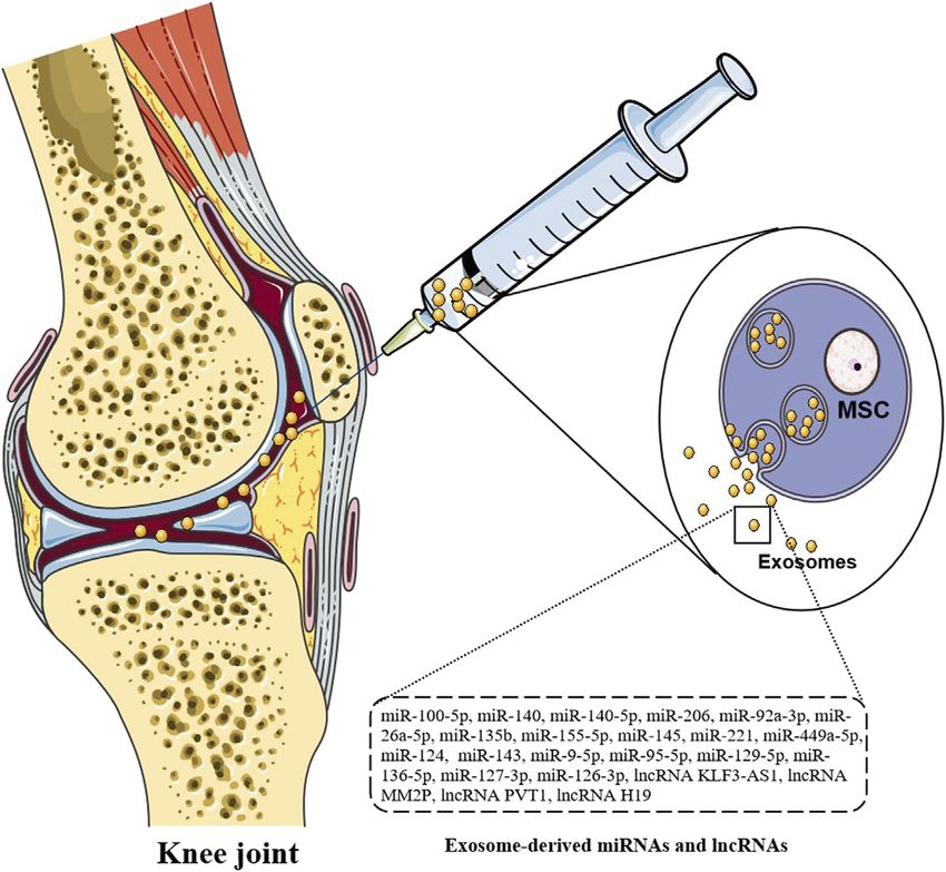
The Research Progress of Exosomes in Osteoarthritis, With Particular Emphasis on the Mediating Roles of miRNAs and lncRNAs - Frontiers
←
→
Page content transcription
If your browser does not render page correctly, please read the page content below
REVIEW
                                                                                                                                                 published: 21 May 2021
                                                                                                                                         doi: 10.3389/fphar.2021.685623

                                           The Research Progress of Exosomes
                                           in Osteoarthritis, With Particular
                                           Emphasis on the Mediating Roles
                                           of miRNAs and lncRNAs
                                           Chenggui Miao 1,2,3, Wanwan Zhou 1, Xiao Wang 4* and Jihong Fang 5,6*
                                           1
                                             Department of Pharmacology, School of Integrated Chinese and Western Medicine, Anhui University of Chinese
                                           Medicine, Hefei, China, 2Department of Pharmacy, School of Life and Health Sciences, Anhui University of Science and
                                           Technology, Fengyang, China, 3 Institute of Prevention and Treatment of Rheumatoid Arthritis of Chinese Medicine, Anhui
                                           University of Chinese Medicine, Hefei, China, 4Department of Clinical Nursing, School of Nursing, Anhui University of
                                           Chinese Medicine, Hefei, China, 5Department of Nursing, Anhui Provincial Children’s Hospital, Affiliated to Anhui Medical
                                           University, Hefei, China, 6 Department of Orthopedics, Anhui Provincial Children’s Hospital, Affiliated to Anhui Medical
                         Edited by:        University, Hefei, China
                             Tao Xu,
      Anhui Medical University, China
                                           Osteoarthritis (OA) is a kind of degenerative disease, which is caused by many factors
                      Reviewed by:
                    Dongdong Wang,
                                           such as aging, obesity, strain, trauma, congenital joint abnormalities, joint deformities.
        McMaster University, Canada        Exosomes are mainly derived from the invagination of intracellular lysosomes, which
                         Guanlan Xu,
                                           are released into the extracellular matrix after fusion of the outer membrane of multi
University of Alabama at Birmingham,
                       United States       vesicles with the cell membrane. Exosomes mediate intercellular communication and
                 *Correspondence:          regulate the biological activity of receptor cells by carrying non-coding RNA, long
                          Xiao Wang        noncoding RNAs (lncRNAs), microRNAs (miRNAs), proteins and lipids. Evidences
            wangxiao@ahtcm.edu.cn
                        Jihong Fang
                                           show that exosomes are involved in the pathogenesis of OA. In view of the
            fangjihong510@126.com          important roles of exosomes in OA, this paper systematically reviewed the roles of
                                           exosomes in the pathogenesis of OA, including the roles of exosomes in OA diagnosis,
                   Specialty section:
                                           the regulatory mechanisms of exosomes in the pathogenesis, and the intervention
        This article was submitted to
        Inflammation Pharmacology,          roles of exosomes in the treatment of OA. Reviewing the roles of exosomes in OA will
              a section of the journal     help to clarify the pathogenesis of OA and explore new diagnostic biomarkers and
           Frontiers in Pharmacology
                                           therapeutic targets.
          Received: 25 March 2021
           Accepted: 10 May 2021           Keywords: exosomes, non-coding RNA, long noncoding RNA, osteoarthritis, microRNA
           Published: 21 May 2021
                            Citation:
Miao C, Zhou W, Wang X and Fang J          Abbreviations: ADAMTS5, disintegrin and metalloproteinase with thrombospondin motifs 5; ADSCs, adipose-derived stem
                                           cells; AFSC, amniotic fluid stem cells; ATF4, transcription factor 4; CAP, chondrocyte-affinity peptide; Col2a1, collagen alpha 1;
    (2021) The Research Progress of
                                           DMM, destabilization of the medial meniscus; E7 exo, E7 peptide; ESC, human embryonic stem cell; HDAC, histone
     Exosomes in Osteoarthritis, With
                                           deacetylase; HDAC3, histone deacetylase 3; IPFP, infrapatellar fat pad; KGN, kartogenin; lncRNAs, long noncoding RNAs;
Particular Emphasis on the Mediating       MSCs, mesenchymal stem cells; OA, osteoarthritis; pBMSCs, polydactyly bone marrow-derived MSCs; PHCs, primary human
      Roles of miRNAs and lncRNAs.         chondrocytes; PRP, platelet-rich plasma; SAGA, Spt-Ada-Gcn5 acetyltransferase; SHED, stem cells from human exfoliated
        Front. Pharmacol. 12:685623.       deciduous teeth; SMMSC-Exos, synovial mesenchymal stem cells; TLR, toll-like receptor; TMJ-OA, temporomandibular joint
     doi: 10.3389/fphar.2021.685623        osteoarthritis; YAP, yes-associated protein.

Frontiers in Pharmacology | www.frontiersin.org                                     1                                             May 2021 | Volume 12 | Article 685623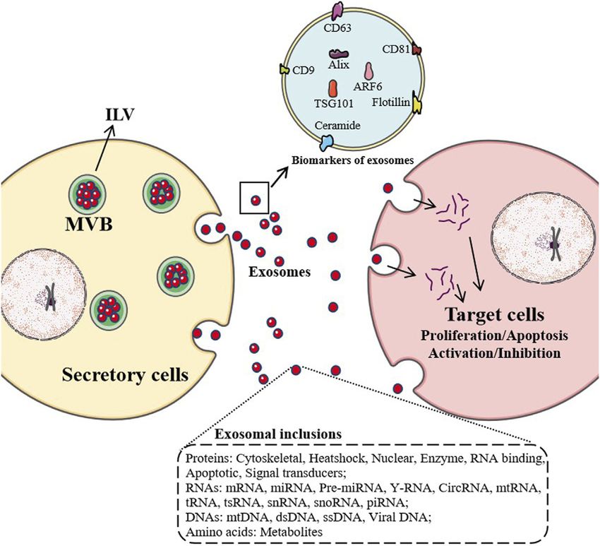
Miao et al.                                                                                                                                       Exosomes in Osteoarthritis

INTRODUCTION                                                                              to target cell membrane proteins and activate signal pathways in
                                                                                          target cells. Second, in the extracellular matrix, exosome
Exosomes are small membrane bubbles (40–150 nm) containing                                membrane proteins can be cleaved by proteases. The cleaved
complex RNAs and proteins. Many cells can secrete exosomes in                             fragments can act as ligands to bind to receptors on the cell
physiological and pathological states. They are mainly derived                            membrane, thus activating intracellular signaling pathways.
from the vesicles formed by the collapse of lysosomal particles,                          Thirdly, the exosome membrane can fuse directly with the
which are released into extracellular matrix after fusion of the                          target cell membrane, and non-selectively release proteins,
outer membrane and cell membrane (Zhang and Yu, 2019;                                     mRNAs, microRNAs (miRNAs) and long noncoding RNAs
Kalluri and LeBleu, 2020).                                                                (lncRNAs) (Familtseva et al., 2019; Jiang et al., 2019).
    Almost all types of cells can secrete exosomes, which naturally                          When exosomes are first discovered, they are considered as a
exist in body fluids, including blood, saliva, urine, cerebrospinal                        way for cells to excrete waste. Nowadays, with a large number of
fluid and milk. The precise molecular mechanisms of their                                  studies on their biological sources, material composition,
secretion    and      uptake,      composition,     “carrier”    and                      transportation, intercellular signal transduction, and
corresponding functions has just begun to be studied                                      distribution in body fluids, exosomes have been found to have
(Meldolesi, 2018). Exosomes are considered as specific vesicles                            a variety of functions (Shan et al., 2019). Exosomes can
and participate in intercellular communication. There is a                                participate in immune response, antigen presentation, cell
growing interest in the study of exosomes, whether to study                               migration, cell differentiation, tumor invasion. Studies have
their functions or to understand how to use them in the                                   shown that exosomes participate in the pathological
development of minimally invasive diagnosis (Mashouri et al.,                             mechanisms of osteoarthritis (OA), and promote the
2019).                                                                                    pathological development of OA (Chen B. Y. et al., 2019;
    When exosomes are secreted from host cells into receptor                              Zhou Q. F. et al., 2020) (Figure 1).
cells, exosomes can regulate the biological activity of receptor cells                       OA is a kind of degenerative disease, which is caused by many
by carrying proteins, nucleic acids and lipids (Gonda et al., 2019).                      factors such as aging, obesity, strain, trauma, congenital joint
Exosome mediated intercellular communication mainly through                               abnormalities, joint deformities (Hunter and Bierma-Zeinstra,
the following ways. First, exosome membrane proteins can bind                             2019; Kan et al., 2019). The disease is more common in the

  FIGURE 1 | Maturation and secretion mechanisms of exosomes. After most endosomes mature to multivesicular bodies (MVB) or late endosomes, their contents,
  RNAs, proteins, lipids are packaged as intraluminal vesicles (ILV) in MVB. With the fusion of MVB and cell membrane, ILV are released as exosomes and enter target cells
  by endocytosis, which affect the physiological and pathological mechanisms of target cells, such as proliferation, apoptosis, activation and inhibition.

Frontiers in Pharmacology | www.frontiersin.org                                       2                                              May 2021 | Volume 12 | Article 685623
Miao et al. Exosomes in Osteoarthritis
middle-aged and elderly people, and occurs in the weight-bearing may be that exosome inclusions promote the chondrocyte
joints and joints with more activity (such as cervical spine, lumbar proliferation and inhibit the chondrocyte apoptosis through
spine, knee joint, hip joint, etc.). Excessive weight bearing or the the Wnt/β-catenin signaling pathway. It also suggests the
use of these joints can promote the occurrence of degenerative feasibility of exosome inclusions as diagnostic markers for OA
changes. The clinical manifestations includes joint swelling, joint (Yuan et al., 2016). In OA chondrocytes treated by IL-1β, platelet-
stiffness, joint movement limitation (Sacitharan, 2019; Abramoff rich plasma (PRP) exosomes can inhibit the release of TNF-α,
and Caldera, 2020). promote the proliferation of OA chondrocytes, and significantly
The main symptom is joint pain, often rest pain, which is reduce the apoptosis rate of OA chondrocytes. PRP exosomes, as
manifested as pain after rest. After a moment of activity, the pain carriers containing PRP derived growth factors, provide a new
is relieved, but after too much activity, the pain is aggravated way for the diagnosis of OA (He et al., 2020).
(Mandl, 2019). Another symptom is joint stiffness, which often The levels of lncRNA PVT1 and high mobility groupprotein
occurs in the morning or after the joint maintains a certain B1 (HMGB1) are up-regulated, while the level of miR-93-5p is
position for a long time in the daytime. Joint swelling can be seen down-regulated in serum and LPS induced C28/I2 cells. PVT1
in the affected joints. There is a sense of friction or “click” sound deletion can reverse the decrease of cell viability, increase of
during activity. Muscle atrophy and joint deformity can be found apoptosis and inflammation induced by LPS in C28/I2 cells.
in severe cases (O’Neill and Felson, 2018; Geyer and Schönfeld, PVT1 regulates the expression of HMGB1 through miR-93-5p.
2018). Inhibition of miR-93-5p can eliminate the apoptosis,
At present, mesenchymal stem cells (MSCs) are considered to inflammatory response and collagen degradation of C28/I2
be related to the development of OA. Some people believe that the cells mediated by PVT1 silencing. The increase of HMGB1
paracrine of nutritional factors, including exosomes mediated reverses the up-regulation of miR-93-5p mediated apoptosis
secretion, plays an important role in MSC based OA treatment and inflammation of C28/I2 cells. In addition, PVT1 regulates
mechanisms (Colombini et al., 2019; Song et al., 2020). There is the TLR4/NF-κB pathway through miR-93-5p/HMGB1 axis.
evidence that the paracrine of exosomes may play important roles Obviously, PVT1 gene knockout by exosomes can inhibit the
in the repair of joint tissue. Exosomes isolated from various stem pathological development of OA through miR-93-5p mediated
cells contribute to tissue regeneration of heart, limbs, skin and HMGB1/TLR4/NF-κB pathway, and these exosomal inclusions
other tissues, and exosomes derived from MSCs may inhibit the may be new diagnostic markers for OA (Xue et al., 2019; Meng
pathological development of OA (Mianehsaz et al., 2019). et al., 2020; Sun et al., 2020).
In this work, we reviewed the role of exosomes in the
pathogenesis of OA, which will help to clarify the Exosomes in Synovial Fluid
pathogenesis of OA and explore new diagnostic biomarkers There are gender differences in exosome proteins in synovial fluid
and therapeutic targets. of patients with OA (Kim et al., 2020). Studies have shown that
exosome-derived miRNAs in synovial fluid of OA patients were
changed, and these changes were gender specific. For example,
CIRCULATING EXOSOMES AS female OA specific miRNAs targeted the estrogen responsive toll-
DIAGNOSTIC BIOMARKERS OF like receptor (TLR) signaling pathway. After OA derived
exosomes treatment, the expression of anabolic genes
OSTEOARTHRITIS decreased, catabolic genes were up-regulated, and the
Exosomes in Peripheral Blood expression of inflammatory genes also increased significantly
Exosomes may play important roles in OA diagnosis, and the (Kolhe et al., 2017).
miRNAs, lncRNAs and other inclusions in exosomes may be new In the synovial fluid exosomes of female OA, the levels of
biomarkers for OA diagnosis (Otahal et al., 2020). In OA, miR- haptoglobin, orosomucoid and ceruloplasmin are up-regulated,
193b-3p regulates the chondrogenesis and chondrocyte while the level of apolipoprotein is down-regulated. In the
metabolism by targeting the histone deacetylase 3 (HDAC3). synovial fluid exosomes of male OA, the levels of β-2-
The expression of miR-193b-3p increases in chondrogenic and glycoprotein and complement component five protein are
hypertrophic human MSCs, but decreases in degenerative significantly up-regulated, while the level of Spt-Ada-Gcn5
cartilage. The level of plasma exosomal miR-193b-3p in OA acetyltransferase (SAGA)-related factor 29 is down-regulated.
patients is significantly lower than that in control group, There are gender differences in synovial fluid exosome protein
suggesting that the exosomal miR-193b-3p may be a new content in OA patients, which indicates that these differentially
diagnostic marker for OA (Meng et al., 2018). expressed proteins may be new diagnostic markers for OA (Kolhe
Plasma exosomes of OA patients can induce OA chondrocytes et al., 2020).
to express cartilage genes and inhibit the release of inflammatory Synovial fluid contains various cytokines, and most of them
cytokines. This highlights the potential of plasma exosome are not only in free form, but also enriched in exosomes (Carlson
inclusions as regulators of extracellular matrix metabolism and et al., 2018). Compared with the cytokine spectrum of synovial
inflammation, and may be candidates for a new approach of cell- fluid, the exosomes of patients with end-stage OA have more
free therapy and diagnosis of OA (Chang et al., 2018). cytokine content, especially chemokines. Synovial fluid derived
Exosomes in plasma of OA patients have been found to have exosomes recruit inflammatory cells, inhibit cartilage
new potential in relieving knee osteoarthritis. The mechanisms proliferation and promote joint degeneration. Synovial fluid
Frontiers in Pharmacology | www.frontiersin.org 3 May 2021 | Volume 12 | Article 685623Miao et al. Exosomes in Osteoarthritis
FIGURE 2 | Injection therapy of exosomes. Direct injection of exosomes carrying miRNAs and lncRNAs can significantly down-regulate arthritis score, inhibit FLS
proliferation and invasion, and reduce inflammatory response and joint damage.
microenvironment and exosomes mediated intercellular cartilage induction, 141 differentially expressed miRNAs are
communication provide a new perspective for OA pathological found, including 35 up-regulated miRNAs, such as miR-92a,
research, and these exosomal cytokines may be new diagnostic miR-193a-5p, miR-320c, miR-1246, miR-1290, 106 down
biomarkers for OA (Gao et al., 2020). -regulated miRNAs, such as miR-377-3p and miR-6891-5p.
Exosomal lncRNAs in synovial fluid are valuable in the MiR-320c in the induced exosomes promotes the proliferation
differential diagnosis of early and late-stage OA. For example, of OA chondrocytes and down-regulates the MMP13 expression
the levels of exosomal lncRNAs in early OA and late-stage OA more than that in control group. These miRNAs can induce
synovial fluid are significantly higher than that in control group. cartilage and may play important roles in cartilage regeneration
The expression of lncRNA PCGEM1 in patients with late-stage and the final treatment of OA (Sun et al., 2019) (Figure 2).
OA is significantly higher than that in patients with early OA, and
the expression of PCGEM1 in early OA is significantly higher
than that in control group. Exosomal PCGEM1 may be a Exosomes Derived From Synovial
powerful indicator to differentiate early OA from late-stage Mesenchymal Stem Cells
OA (Zhao and Xu, 2018). Exosomes derived from OA synovial MSCs with miR-140-5p
overexpression are effective for OA treatment. Wnt5a and wnt5b
in exosomes can activate the Yes-associated protein (YAP)
EXOSOMES IN OSTEOARTHRITIS through the Wnt signaling pathway and promote the
PATHOGENESIS proliferation and migration of chondrocytes. The side effect is
significantly reduction of the ECM secretion, and the high
Exosomes from various tissues play important roles in the expression of miR-140-5p blocks this side effect. In vivo,
pathogenesis of OA. IL-1β induced MSCs exosomes have Exosomes derived from miR-140-5p-overexpressing human
obvious anti-inflammatory activity in OA sw982 cells. The synovial MSCs successfully prevent the OA in model rats (Tao
roles of IL-1β in inducing MSC-derived exosomes is mediated et al., 2017). MiR-129-5p in human synovial MSC exosomes
by miR-147b, leading to the inhibition of NF-κB pathway (Kim attenuates the IL-1β induced OA by targeting the HMGB1. In
et al., 2021). By comparing the expression of miRNAs in the OA patients and IL-1β induced chondrocytes, miR-129-5p
exosomes of human bone marrow MSCs with that without is decreased, while HMGB1 is significantly up-regulated.
Frontiers in Pharmacology | www.frontiersin.org 4 May 2021 | Volume 12 | Article 685623Miao et al. Exosomes in Osteoarthritis
MiR-129-5p targets the 3′UTR of HMGB1 and inhibits the up- improves the cartilage injury induced by IL-1β. KLF3-AS1 may be
regulation of HMGB1. Exosomes rich in miR-129-5p can a potential therapeutic target for OA (Liu et al., 2018b).
significantly reduce the inflammatory response and apoptosis In OA model mice induced by collagenase, bone marrow
of chondrocytes, while exosomes lacking miR-129-5p increases MSC-derived exosomes increase the chondrogenic genes type
the inflammatory response and apoptosis of chondrocytes (Qiu II collagen alpha 1 (Col2a1) and aggrecan, decrease the markers
et al., 2021). of chondrocyte hypertrophy MMP-13 and runt-related
Synovial fluid derived MSCs transplantation is an effective transcription factor 2 (Runx2), and attenuate the IL-1β
method to treat OA cartilage degeneration. kartogenin (KGN) is a induced inhibition of chondrocyte proliferation. Exosomes
small molecule that can induce MSCs to differentiate into derived from KLF3-AS1-overexpressing-MSCs ameliorate the
chondrocytes in vitro and in vivo. It controls the chondrogenic IL-1β induced chondrocyte injury. Interestingly, KLF3-AS1
differentiation of transplanted MSCs. However, the poor water promotes the GIT1 expression by adsorbing the miR-206.
solubility of KGN limits its clinical application. The exosomes Thus bone marrow MSC-derived exosomes promote the
containing KGN can effectively solve this technical problem. osteoarthritis chondrocyte proliferation and inhibit the
MSCs binding peptide E7 is fused with exosome membrane apoptosis through the KLF3-AS1/miR-206/GIT1 axis (Liu
protein Lamp 2b to produce exosomes with surface display E7 et al., 2018a).
peptide (E7 exo). The E7 exo containing KGN could effectively MiR-127-3p is enriched in bone marrow MSC-derived
enter MSCs, and the degree of cartilage differentiation is higher exosomes, and bone marrow MSC-derived exosomes inhibit
than that of KGN alone or without E7 exo (Xu et al., 2021). The the IL-1β induced chondrocyte injury. MiR-127-3p can inhibit
E7 exo containing KGN pretreated bone marrow MSCs have the CDH11 of chondrocytes, thus blocking the activation of Wnt/
higher cartilage repair efficiency, stronger cartilage matrix β-catenin pathway and alleviating the damage of OA
formation and less degradation than those from bone marrow chondrocytes (Dong et al., 2021).
MSCs (Liu et al., 2020). Mao et al. (2018b) investigated the expression of miR-92a-3p
In exosomes derived from synovial fibroblasts, overexpressed in human bone marrow MSC chondrogenesis model and OA
miR-126-3p inhibits the chondrocyte inflammation and cartilage primary human chondrocytes (PHCs) in vitro. Interestingly, the
degradation in OA model rats, which may have certain expression of miR-92a-3p increased in MSC chondrocytes and
therapeutic value for OA patients (Zhou et al., 2021). MiR- decreased in OA chondrocytes. MSC-miR-92a-3p-exos
26a-5p is low expressed in OA patients and synovial promoted the cartilage proliferation and matrix gene
fibroblasts treated with IL-1β, while PTGS2 is high expressed. expression of MSCs and PHCs, while MSC-anti-miR-92a-3p-
PTGS2 is a direct target of miR-26a-5p. Overexpression of miR- exos inhibited the chondrocyte differentiation and cartilage
26a-5p alleviates the injury of synovial fibroblasts by inhibiting matrix synthesis by enhancing the Wnt5a expression. MiR-
the PTGS2. Obviously, overexpression of miR-26a-5p inhibits the 92a-3p regulates the cartilage development and homeostasis by
injury of synovial fibroblasts in OA through the PTGS2, which is directly targeting the Wnt5a, suggesting its important roles in OA
of great significance for the treatment of OA (Jin et al., 2020b). pathology.
Exosomal lncRNA H19 in synovial fibroblasts attenuates the After IL-1β is added to rabbit bone marrow MSCs and
progression of OA through miR-106b-5p/TIMP2 axis. Cartilage chondrocytes cultured in vitro, chondrocyte viability decreases,
repair mediated by exosomes is characterized by increased cell cell apoptosis and mitochondrial membrane potential changes
viability and migration as well as reduced matrix degradation. In significantly. However, these changes disappeared after adding
the process of exosomes mediated cartilage repair, the bone marrow MSC-derived exosomes. Compared with IL-1β
enhancement of cell proliferation and migration is related to group, the exosomes of bone marrow MSCs inhibit the
the regulation of miR-106b-5p/TIMP2 axis. Transfection of phosphorylation of p38 and ERK, and promote the
miR-106-5p mimics in chondrocytes significantly reduces the phosphorylation of Akt. These findings suggest that bone
cell proliferation and migration, promotes the matrix marrow MSC-derived exosomes inhibit the chondrocyte
degradation, increases the expression of MMP13 and apoptosis through p38, ERK and Akt pathways (Qi et al., 2019).
ADAMT5, and decreases the expression of COL2A1 and MSCderived exosomes inhibit the pathogenesis of OA by
ACAN in chondrocytes. TIMP2 is directly regulated by miR- inhibiting syndecan-1. Injection of exosomes containing miR-
106-5p. This suggests that H19 may promotes the chondrocyte 9-5p can alleviate inflammation and OA like injury, down-
proliferation and migration and inhibits the degradation of OA regulate the levels of inflammatory factors, alleviate the
matrix by targeting miR-106b-5p/TIMP2 axis in OA oxidative stress injury, and reduce the levels of OCN, MMP-
pathogenesis (Tan et al., 2020). 13, comp and AKP. Syndecan-1 is the target of miR-9-5p. Up-
regulation of syndecan-1 leads to aggravation of inflammation
and OA like injury. The exosomal miR-9-5p derived from bone
Exosomes Derived From Bone Marrow marrow MSC has anti-inflammatory and cartilage protective
Mesenchymal Stem Cells effects on OA by regulating the syndecan-1 (Jin et al., 2020a).
Exosomal lncRNA KLF3-AS1 from human bone marrow MSCs The expression of ELF3 increases and the expression of miR-
can be used as an effective therapeutic molecule for OA patients. 136-5p decreases in traumatic OA cartilage. MiR-136-5p is
KLF3-AS1 is significantly enriched in the exosomes of MSCs. The confirmed to target the ELF3 and down-regulates its
exosomal KLF3-AS1 inhibits the apoptosis of chondrocytes and expression. After chondrocytes internalizes exosomes, the
Frontiers in Pharmacology | www.frontiersin.org 5 May 2021 | Volume 12 | Article 685623Miao et al. Exosomes in Osteoarthritis
expression of ELF3 decreases. The exosomal miR-136-5p, derived Furthermore, ATF4-OA-Exosomes promote the autophagy and
from bone marrow MSCs, can promote the chondrocyte inhibit the apoptosis in TNF-α or tunicamycin treated
migration in vitro and inhibits the cartilage degeneration in chondrocytes (Wang Y. et al., 2021).
vivo, thereby inhibiting the pathological changes of OA (Chen
et al., 2020).
The stimulating effects of exosomes isolated from osteoblasts Exosomes Derived From Human Embryonic
of coxarthrosis on bone marrow MSCs are mainly manifested in Stem Cells
the catabolism and osteogenic differentiation. Interestingly, this The exosomes derived from human ESC-induced MSCs (ESC-
has nothing to do with donor pathology, reflecting the influence MSCs) are involved in the therapeutic mechanisms of alleviating
of exosomes on tissue microenvironment and cell metabolism in OA. In destabilization of the medial meniscus (DMM) model
coxarthrosis (Niedermair et al., 2020). mice, intra-articular injection of ESC-MSCs reduces the cartilage
destruction and matrix degradation in DMM model, which is
mediated by ESC-MSCs derived exosomes. In the presence of IL-
Exosomes Derived From Primary 1β, these exosomes maintain the chondrocyte phenotype by
Chondrocytes increasing the type II collagen synthesis and decreasing the
The exosomes of primary chondrocytes may play a role in the ADAMTS5 expression. Immunocytochemistry shows that the
treatment of OA. Zheng et al. (2019) isolated exosomes from exosomes are colocalized with type II collagen positive
primary chondrocytes cultured in normal and IL-1β induced chondrocytes. This provides a new target for the development
inflammatory environment, and found that there were more of OA drugs and drug delivery systems (Wang et al., 2017).
mitochondrial proteins in exosomes of chondrocytes in
normal group. Intra-articular injection of exosomes of
chondrocytes in normal group successfully prevented the Exosomes Derived From Chondrogenic
development of OA. Studies have shown that chondrocyte Progenitor Cells
exosomes restored the mitochondrial dysfunction and Chondrogenic progenitor cells have high self-renewal ability and
promoted the macrophages to differentiate into M2 phenotypes. chondrogenic potential. Wang et al. (2020) found that intra-
Drug delivery is the key to the successful clinical application of articular injection of exosomes secreted by chondrogenic
nucleic acid drugs, and exosomal miRNAs in the treatment of OA progenitor cells in MRL/MpJ superhealer mice promoted the
has brought a new perspective to the treatment of OA. MiR-140 is repair of articular cartilage injury in mice. By comparing the
not only significant in promoting the cartilage formation and miRNA expression profiles of control CBA (CBA EVs) and MRL/
inhibiting the degeneration, but also plays an important role in MPJ mouse chondroblasts, the differentially expressed exosomal
cartilage development (Duan et al., 2020). Liang et al. (2020) miRNAs were involved in a variety of biological processes.
fused chondrocyte-affinity peptide (CAP) with lysosome Among them, 80 miRNAs were significantly up-regulated and
associated membrane glycoprotein 2b on the surface of the 100 were down-regulated, and 20 disordered miRNAs linked OA
exosomes to obtain cap exosomes. This exosomes could repair through the AMPK signaling, autophagy regulation and
effectively encapsulate miR-140, specifically enter insulin signaling. The mechanisms of exosomes involved in OA
chondrocytes, and transport miR-140 to chondrocytes. Studies may be more related to miRNAs (Toh et al., 2017).
have shown that cap exosomes could also transfer miR-140 to the
deep cartilage region through the dense mesochondral
membrane, inhibited the cartilage degradation protease, and Exosomes Derived From Vascular
suppress the progress of OA. Endothelial Cells
MiR-95-5p regulates the chondrogenesis and cartilage Exosomes from vascular endothelial cells have been proved to be
degradation through the histone deacetylase (HDAC) 2/8. involved in the pathogenesis of many diseases, and their roles in
MiR-95-5p overexpression of primary chondrocyte derived the OA pathogenesis have also been confirmed. Exosomes
exosomes may be effective in the treatment of OA. HDAC 2/8 derived from vascular endothelial cells promote the
is up-regulated in OA tissues and exosomes secreted by pathological development of OA by inducing the chondrocyte
chondrocytes, and mediates the expression of specific genes in apoptosis. These exosomes can inhibit the autophagy and p21
chondrocytes. MiR-95-5p directly acts on the 3′UTR of HDAC 2/ expression, reduce the ability of chondrocytes to resist the
8 to promote the cartilage formation and prevent OA by directly oxidative stress, increase the content of ROS and induce the
targeting the HDAC 2/8 (Mao et al., 2018a). apoptosis. Exosomes derived from vascular endothelial cells
Transcription factor 4 (ATF4) plays an important role in promote the progress of OA and provide new ideas for the
chondrocyte proliferation and bone formation. It has been diagnosis and treatment of OA (Yang et al., 2021).
proved that the serum derived exosomes of OA mice has
therapeutic effects on OA model mice (Chen D. et al., 2019).
Studies have shown that intra-articular injection of ATF4-OA- Exosomes Derived From Adipose-Derived
Exosomes can reduce the degeneration, damage and Stem Cells
inflammatory reaction of articular cartilage in OA model mice, ADSCs are candidate cells for anti-inflammatory and
and partially restore the autophagy function of knee cartilage. cytoprotective effects on cartilage. Exosomes mediate the
Frontiers in Pharmacology | www.frontiersin.org 6 May 2021 | Volume 12 | Article 685623Miao et al. Exosomes in Osteoarthritis
paracrine effect of ADSCs and down-regulate the aging expression of the MMP1, MMP9, MMP13, ADAMTS5 and
characteristics of OA osteoblasts (Tofiño-Vian et al., 2017). mTOR. On the contrary, miR-100 inhibition up-regulates
ADSCs promote the chondrogenesis and inhibit the these targets. Furthermore, miR-100-5p directly targets the
inflammation. Patients with OA are usually associated with 3′UTR of mTOR, and inhibits the expression of mTOR (Luo
obesity and chronic inflammation. Exosomes isolated from et al., 2019).
ADSCs down-regulate the expression of IL-6, NF-κB and
TNF-α, and up-regulate the expression of IL-10. Exosome
therapy can protect the articular chondrocytes from Exosomes Derived From Monocyte Derived
H2O2 induced apoptosis. In addition, exosome therapy Cells
promotes the chondrogenesis of periosteal cells and LncRNA MM2P and exosomes mediate the Sox9 transfer from
increases the level of chondrogenic markers, including type monocyte derived cells to primary chondrocytes. Treatment of
II collagen and β-catenin. Wnt signaling pathway may be its RAW264.7 mouse macrophages and mouse bone marrow-
downstream signaling pathway. The periosteal cells with derived macrophages with IL-4 or IL-13 up-regulate the
exosomes show high levels of miR-145 and miR-221, and expression of MM2P. MM2P blocks the SHP2 mediated
the miR-145 and miR-221 are related to the enhancement of dephosphorylation of STAT3 at Try705 and interacts with
periosteal cells and chondrogenic potential, respectively RNA binding protein FUS. In turn, p-STAT3 increases the
(Zhao et al., 2020). Sox9 gene expression. These cells release the Sox9 mRNA and
For exosomes derived from MSCs of infrapatellar fat pad exosomes containing proteins. The supernatant of these cells can
(IPFP), miR-100-5p-abundant exosomes protect the articular promote the differentiation of primary chondrocytes, that is, up-
cartilage and improve the gait abnormality by inhibiting regulates the expression of the Col1a2 and Acan genes, and
the mTOR signal of OA. IPFP MSCs can produce a large promotes the secretion of extracellular matrix components.
number of exosomes, which show typical morphological These effects are mediated by Sox9 mRNA and protein
characteristics of exosomes. IPFP MSC-derived exosomes delivered to chondrocytes by exosomes. MM2P and its
can reduce the severity of OA, inhibit the apoptosis, exosomes may be new therapeutic and diagnostic targets for
promote the matrix synthesis and reduce the expression OA (Bai et al., 2020).
of catabolic factors. These exosomes can significantly
enhance the autophagy level of chondrocytes through the
mTOR inhibition. The detection of luciferase reporter gene Exosomes Derived From Amniotic Fluid
shows that mir-100-5p combines with the mTOR’s 3′UTR, Stem Cells
which reverses the mTOR signal pathway. It is important that The exosomes secreted by AFSCs have a certain effect on the
intra-articular injection of antagormir-100-5p significantly treatment of OA. AFSCs can secrete exosomes with growth
reduces the protective effects of MSC-derived exosomes on factors and immune regulatory molecules, which can prevent
articular cartilage. These IPFP MSCs are expected to be a tissue degradation and induce the cartilage repair. Compared
potential therapy for OA (Clockaerts et al., 2010; Wu et al., with the control group, the exosomes treated model animals show
2019). stronger pain tolerance and improved the histological score.
Exosomes containing TGF-β can induce the cartilage recovery,
which has better surface regularity and hyaline cartilage
Exosomes Derived From Human Dental characteristics, and is positively correlated with the content of
Pulp Stem Cells TGF-β. It is easier to detect macrophage markers in exosomes
Exosomes of human DPSCs can inhibit the chondrocyte treated joints, which suggests that AFSC exosomes can regulate
apoptosis in OA model rats. After transfection of DPSCs with the macrophage polarization (Beretti et al., 2018; Zavatti et al.,
miR-140-5p mimics, the exosomal miR-140-5p increased 2020).
significantly. In IL-1β treated human chondrocytes, DPSC-
derived exosomes promote the expression of chondrocyte
related mRNAs, including aggrecan, Col2α1 and Sox9. Exosomes Derived From Polydactyly Bone
Exosomes containing miR-140-5p significantly enhance this Mesenchymal Stem Cells
phenomenon. MiR-140-5p rich exosomes derived from DPSCs Zhou X. et al. (2020) obtained a special kind of MSCs from the
may play an anti-apoptotic role by regulating the expression of bone marrow of patients with polydactyly, and found
apoptosis related proteins. The exosomes of DPSCs may be a that polydactyly bone marrow-derived MSCs (pBMSCs)
potential strategy for the treatment of OA (Lin et al., 2021). played certain roles in the pathological mechanisms of OA.
Exosomes derived from stem cells of human exfoliated It was important that pBMSCs have stronger ability to
deciduous teeth (SHED) have a certain therapeutic effect on differentiate into chondrocytes than BMSCs. Exosomes
temporomandibular arthritis. MiR-100-5p is enriched in these secreted by pBMSCs stimulated the migration and
exsomes. SHED exosomes inhibit the expression of IL-6, IL-8, proliferation of chondrocytes. The expression of BMP4 in
MMP1, MMP3, MMP9, MMP13, disintegrin and pBMSCs was significantly higher than that in BMSCs, and
metalloproteinase with thrombospondin motifs 5 (ADAMTS5). the pBMSCs regulate chondrocyte formation through the
The chondrocytes treated with miR-100 mimics show low BMP4 signal.
Frontiers in Pharmacology | www.frontiersin.org 7 May 2021 | Volume 12 | Article 685623Miao et al. Exosomes in Osteoarthritis
Exosomes Derived From Mesenchymal and mitochondria proliferates, the lysosomes increases and
Stem Cells of Temporomandibular Joint phagocytic function enhances (Meng et al., 2019; Barrett,
2020). Macrophages have a series of continuous functional
Osteoarthritis states. M1 and M2 macrophages are the two extremes of
In the immunocompetent model rats of TMJ-OA, MSC-
this continuous state. M1 macrophages participate in the
derived exosomes play a key role in inflammatory response,
positive immune response and play a role in immune
injury behavior, condylar cartilage and subchondral bone
surveillance by secreting pro-inflammatory cytokines and
healing. Exosomes mediated TMJ repair of OA is
chemokines, and presenting antigens. M2 macrophages
characterized by early inhibition of pain and degeneration,
only have weak antigen presenting ability, and down-
followed by reduced inflammation and sustained proliferation.
regulate the immune response by secreting inhibitory
MSC-derived exosomes gradually improve the matrix
cytokines such as IL-10 or TGF-b, which play an
expression and subchondral bone structure, and promote
important role in immune regulation. Macrophages play
the overall joint repair and regeneration. MSC-derived
important regulatory roles in OA tissue repair, inflammatory
exosomes enhance the synthesis of s-GAG synthesis blocked
response and chondrocyte proliferation (Wang S. et al., 2019;
by IL-1β, and inhibit the production of nitric oxide and
Russell et al., 2019).
MMP13 induced by IL-1β. Interestingly, adenosine receptor
Exosomes of OA chondrocytes promote the production of IL-
activation, Akt, ERK and AMPK phosphorylation inhibitors
1β in macrophages. These exosomes inhibit the LPS induced
can partially eliminate these effects. Obviously, MSC-derived
autophagy by inhibiting the expression of ATG4B through miR-
exosomes promote the repair and regeneration of TMJ in OA
449a-5p. The decrease of autophagy leads to the production of
through a well coordinated mechanism (Cui et al., 2017; Zhang
mitochondria, which further enhances the activation of
et al., 2019).
inflammatory bodies and the subsequent production of IL-1β.
This provides a new perspective for understanding the activation
Exosomes Derived From Mesenchymal of synovial macrophages and OA pathogenesis in patients with
OA (Ni et al., 2019).
Stem Cells of Lumbar Facet Joint MiR-135b is highly expressed in MSC-derived exosomes
Osteoarthritis stimulated by TGF-β1. MiR-135b mimics induce the M2
LFJ OA is one of the common causes of low back pain. In the try polarization of synovial macrophases. The effects of miR-135b
of mouse bone marrow MSC-derived exosome treatment, and TGF-β1-stimulated exosomes on the polarization of M2
exosomes block the abnormal CGRP positive nerves and synovial macrophases will be reversed by the increase of
abnormal H-type angiogenesis in the LFJ subchondral bone, MAPK6. In conclusion, MSC-derived exosomal miR-135b
alleviating the low back pain. Bone marrow MSC-derived promotes the polarization of M2 synovial macrophages by
exosomes reduce the cartilage degeneration, inhibit the targeting the MAPK6, thus alleviating the cartilage damage
expression of tartrate resistant acid phosphatase, reduce the and providing a new target for the treatment of OA (Wang
activation of RANKL-RANK-TRAF6 signal, and promote the and Xu, 2021).
subchondral bone reconstruction. Bone marrow MSC-derived OA is a chronic degenerative disease, which leads to
exosomes have significant protective effects on patients with LFJ- limited activity and even disability. Exosomes derived
OA, which may be a potential choice for the treatment of LFJ-OA from bone marrow MSCs can delay the progression of
(Li et al., 2020). OA (Zhao et al., 2018). Exosomes reduce the cartilage
TGF-β1, transforming growth factor β1, regulates the damage and synovial macrophage infiltration, inhibit the
proliferation of chondrocytes through MSC-derived exosomes. M1 macrophage production and promote the M2
In the OA model, TGF-β1 stimulation enhances the expression of macrophage production. Exosomes reduce the expression
miR-135b in the MSC-derived exosomes and increases the of pro-inflammatory cytokines IL-1β, IL-6 and TNF-α in
survival rate of C5.18 cells. Interestingly, there is a negative synovial fluid, and increase the release of anti-inflammatory
regulatory relationship between miR-135b and Sp1. The IL-10. It is important that macrophages treated by exosomes
combination of TGF-β1 and miR-135b inhibitor lead to the maintain chondrogenic properties of chondrocytes.
decrease of C5.18 cell activity. Obviously, TGF-β1 inhibits the Obviously, bone marrow MSCs-derived exosomes
SP1 through miR-135b derived from MSC-derived exosomes, alleviate the OA by promoting the phenotype
and promotes the chondrocyte proliferation, and then promotes transformation of synovial macrophages from M1 to M2
the cartilage repair (Wang et al., 2018). (Zhang et al., 2020).
The synovial exosomes stimulate the release of many
inflammatory cytokines, chemokines and metalloproteinases by
Effects of Exosomes on Macrophages in macrophages in OA, but do not affect the expression of CD80 and
Osteoarthritis CD86 costimulator molecules. The purified exosomes has
Macrophages are derived from mononuclear cells in the blood obvious functional activity in stimulating the release of pro-
after penetrating blood vessels. After entering connective tissue, inflammatory factors by M1 macrophages (Domenis et al.,
the volume of monocytes increases, the endoplasmic reticulum 2017) (Table 1).
Frontiers in Pharmacology | www.frontiersin.org 8 May 2021 | Volume 12 | Article 685623Miao et al. Exosomes in Osteoarthritis
TABLE 1 | Exosomes reported in the pathogenesis of OA.
Classification of Exosomal Origin of Regulatory roles Targets References
exosomes inclusions exosomes
Monocyte-derived LncRNA MM2P RAW264.7 mouse macrophages Promotes the chondrocyte differentiation Sox9 Bai et al.
exosomes and mouse bone marrow-derived and functions (2020)
macrophages
MSC-derived exosomes MiR-136-5p OA chondrocytes and mouse Inhibits the chondrocyte degeneration in ELF3 Chen et al.
model of post-traumatic OA traumatic osteoarthritis (2020)
MSC-derived exosomes MiR-127-3p Bone marrow MSCs and primary Inhibits the CDH11 in chondrocytes and CDH11-mediated Dong et al.
chondrocytes of model rats relieving the chondrocyte damage in OA. wnt/β-catenin (2021)
pathway
MSC-derived exosomes MiR-9-5p Rat model induced by anterior Has anti-inflammatory and cartilage Syndecan-1 Jin et al.
cruciate ligament/medial collateral protective effects on OA (2020a)
ligament transection
MSC-derived exosomes MiR-26a-5p MSC and synovial fibroblasts of OA Retards the damage of synovial PTGS2 Jin et al.
model rats fibroblasts in vitro and alleviates the OA (2020b)
damage
CAP exosomes MiR-140 Chondrocytes of OA patients Inhibits the cartilage-degrading Cartilage-degrading Liang et al.
proteases, and alleviates the OA proteases (2020)
progression in mode rats
Exosomes derived from mir- MiR-140-5p IL-1β treated human chondrocytes Inhibits the chondrocyte apoptosis and Chondrocyte Lin et al.
140-5p-overexpressing and OA model rats improves the knee joint conditions in rat apoptosis genes (2021)
human DPSCs model
MSC-derived exosomes LncRNA KLF3- IL-1β-induced OA chondrocytes Promotes the proliferation and inhibits KLF3-AS1/miR- Liu et al.
AS1, miR-206 and collagenase-induced mouse apoptosis of chondrocytes 206/GIT1 axis (2018a)
OA model
MSC-derived exosomes LncRNA IL-1β-induced OA chondrocytes Exosomal KLF3-AS1 promotes the — Liu et al.
KLF3-AS1 and collagenase-induced mouse cartilage repair and chondrocyte (2018b)
OA model proliferation
SHED-derived exosomes MiR-100-5p Temporomandibular joint SHED-exosomes suppresses the mTOR Luo et al.
chondrocytes inflammation in chondrocytes (2019)
Primary chondrocyte- MiR-95-5p OA primary chondrocyte Promotes the cartilage formation and HDAC2/8 Mao et al.
derived exosomes prevents the OA (2018a)
MSC-derived exosomes MiR-92a-3p Human MSC and OA primary Enhances the chondrogenesis and WNT5A Mao et al.
human chondrocytes suppresses the cartilage degradation via (2018b)
targeting WNT5A
Exosomes derived from OA LncRNA PVT1 OA patient serum and LPS-induced Alleviates the lipopolysaccharide-induced HMGB1/TLR4/NF- Meng et al.
patient serum and LPS- C28/I2 cells OA progression κB pathway via mir- (2020)
treated C28/I2 cells 93-5p
Exosomes derived from MiR-449a-5p Chondrocytes and macrophages Enhances the mature IL-1β production of ATG4B Ni et al. (2019)
osteoarthritic chondrocyte of OA macrophages and aggravates the
synovitis in OA
Exosomes derived from MiR-124 and OA mouse models Curcumin reinforces the MSC-derived NF-kB and ROCK1/ Qiu et al.
curcumin-treated MSCs miR-143 exosomes in attenuating osteoarthritis TLR9 (2020)
Synovial MSC-derived MiR-129-5p OA patients and IL-1β-induced Relieves the IL-1β induced OA HMGB1 Qiu et al.
exosomes chondrocytes (2021)
Synovial MSC-derived MiR-140-5p Human synovial MSCs Enhances the cartilage tissue RalA Tao et al.
exosomes regeneration and prevents the OA in a rat (2017)
model
Exosomes derived from LncRNA H19 OA chondrocytes Promotes the chondrocyte proliferation MiR-106b-5p/ Tan et al.
synovial fibroblasts and migration and inhibits the degradation TIMP2 (2020)
of OA matrix
MSC-derived exosomes MiR-135b Chondrocyte of OA model rats TGF-β1 promotes the chondrocyte Sp1 Wang et al.
proliferation by regulating the Sp1 through (2018)
MSC-exosomes derived miR-135b
MSC-derived exosomes MiR-135b Cartilage tissues and synovial Attenuates the cartilage injury via MAPK6 Wang and Xu.
macrophages of model rats promoting the M2 synovial macrophage (2021)
polarization
Synovial MSC-derived MiR-155-5p OA chondrocytes and mouse Prevents the OA via enhancing the Runx2 Wang Z. et al.
exosomes model of OA proliferation and migration, attenuating (2021
the apoptosis, and modulating the ECM
secretion
IPFP MSC-derived MiR-100-5p IPFP of OA patients Protects the articular cartilage and 3ʹUTR of mTOR Wu et al.
exosomes ameliorates the gait abnormalities via (2019)
inhibition of mTOR
(Continued on following page)
Frontiers in Pharmacology | www.frontiersin.org 9 May 2021 | Volume 12 | Article 685623Miao et al. Exosomes in Osteoarthritis
TABLE 1 | (Continued) Exosomes reported in the pathogenesis of OA.
Classification of Exosomal Origin of Regulatory roles Targets References
exosomes inclusions exosomes
ADSC-derived exosomes MiR-145, Chondrocytes of OA Promotes the chondrogenesis and Wnt/β-catenin Zhao et al.
miR-221 suppresses inflammation (2020)
Exosomes derived from MiR-126-3p Model rats of OA Suppresses the chondrocyte IL-1β, IL-6, and Zhou et al.
synovial fibroblasts inflammation and cartilage degradation TNF-α (2021)
THE ROLES OF EXOSOMES IN THE regulating the extracellular matrix (ECM) secretion of
TREATMENT OF OSTEOARTHRITIS chondrocytes. Studies have shown that synovial MSC-derived
exosomes promote the proliferation and migration of OA
Curcumin alleviates OA by enhancing MSC-derived exosomes. chondrocytes and inhibit their apoptosis, but has no effect on
Curcumin significantly restores the expression of miR-143 and the secretion of ECM. MiR-155-5p overexpression in exosomes
miR-124, and up-regulate the expression of NF-KB and ROCK1 show common characteristics and further promote the ECM
in OA pathogenesis. The 3′UTR of NF-KB and ROCK1 contains secretion by targeting the Runx2. Exosomal miR-155-5p
the binding sites of miR-143 and miR-124, respectively. effectively prevent the pathological development of OA model
Importantly, curcumin reduces the DNA methylation of miR- mice. Furthermore, overexpression of Runx2 partially reverses
143 and miR-124 promoters, suggesting that curcumin affects the the effect of exosomal miR-155-5p on OA chondrocytes, proving
methylation of these two miRNAs (Qiu et al., 2020). that Runx2 is a direct target of miR-155-5p (Wang Z. et al., 2021).
Injection of bone marrow MSC-derived exosomes can alleviate Zhu et al. (2017) compared the efficacy of MSCs from synovial
the cartilage injury and pain in patients with OA. For example, mesenchymal stem cells (SMMSC-Exos) and induced pluripotent
exosomes treatment significantly attenuates the inhibitory effects stem cells (iMSC-Exos) in the treatment of OA. IMSC-Exos and
of IL-1β on the chondrocyte proliferation and migration. SMMSC-Exos were injected into the joints of OA model mice
Exosomes treatment significantly attenuates the IL-1β-induced induced by collagenase. The difference of therapeutic effects
down-regulation of COL2A1 and ACAN and up-regulation of between them were evaluated by pathology,
MMP13 and ADAMT5. Exosomes treatment significantly immunohistochemistry, cell count and scratch test. Both
reduces the up-regulation of CGRP and iNOS in dorsal root iMSC-Exos and SMMSC-Exos could reduce the symptoms of
ganglion (DRG) of OA model rats. Compared with untreated OA OA model in mice, but the therapeutic effects of iMSC-Exos were
model rats, the paw withdrawal latency (PWL) value of better than that of SMMSC-Exos, and iMSC-Exosomes promoted
exogenous OA model rats was significantly increased (Liu the migration and proliferation of chondrocytes more strongly.
et al., 2019). Since autologous iMSCs are inexhaustible in theory, iMSC-Exos
Cosenza et al. (2017) compared the roles of bone marrow may be a promising new method for the treatment of OA (Qiong
MSC-derived exosomes and microbubbles/microparticles (MPs) et al., 2020).
in OA pathogenesis. In OA chondrocytes, bone marrow MSC-
derived exosomes and MPs both could inhibit the catabolism of
MMP-13, ADAMTS5 and inflammatory marker iNOS, and re- CONCLUSION AND PERSPECTIVES
induce the expression of aggrecan. Both exosomes and MPs could
protect chondrocytes from apoptosis, inhibit the activation of Exosomes are widely distributed in various body fluids, carrying
macrophages, and protect model mice from joint injury. and transmitting important signal molecules, forming a new
Exosomes and MPs replicate the main therapeutic effects of intercellular information transmission system. Exosomes affect
BM MSCs, suggesting that exosomes and MPs may contain the physiological state of cells and are closely related to the
the same substances that mediate intercellular communication. occurrence and process of many diseases (Wang Y. et al.,
In a bone defect treatment experiment, the osteochondral 2019). Almost all types of cells can secrete exosomes, and
defect model was established in the trochlear groove of the exosomes are also widely found in body fluids, including
distal femur of rats. One defect was treated with 100 μg blood, tears, urine, saliva, milk, and ascites. At present, studies
human extracellular exosomes, and the exosomes were injected have found that exosomes contain the nucleic acids (miRNA,
into the joint after operation. Once a week for 12 weeks. lncRNA, circRNA, mRNA, tRNA), protein, cholesterol, etc. The
Compared with control, the appearance of the defect was surface markers of exosomes are CD63, CD81, CD9, TSG101 and
enhanced and the histological score was improved. At HSP70 (Li et al., 2018; Konečná et al., 2019). Therefore, exosomes
12 weeks, the defects treated with exosomes showed complete can be associated with almost any disease and become an
recovery of cartilage and subchondral bone, which confirmed the innovative hot spot in the research of disease biomarkers,
effectiveness of human embryonic MSC-derived exosomes in disease mechanisms and drug development.
cartilage repair (Zhang et al., 2016). Exosomes transport proteins, mRNA, miRNA, lncRNA,
MiR-155-5p overexpression of synovial MSCs in vitro circRNA, and even organelles into receptor cells, and
prevents the OA by reducing the chondrocyte apoptosis and participate in intercellular communication (Zhang et al., 2017).
Frontiers in Pharmacology | www.frontiersin.org 10 May 2021 | Volume 12 | Article 685623Miao et al. Exosomes in Osteoarthritis
Exosomes play key roles in immune response, inflammation, animal models need to be evaluated. Furthermore, we should pay
angiogenesis, apoptosis, coagulation, waste disposal and other attention to the importance of vesicles secreted by cells. Vesicles
physiological processes. Exosomes from different cell sources are not only the stumbling blocks of exosomes research, but also
contain different RNA and protein components, which can be the next research hotspot, because vesicles may play a more
used as early diagnostic biomarkers for a variety of diseases, and special role than exosomes.
can also be used as carrier of drugs for disease treatment (Sung
et al., 2018).
In addition, the characteristics of exosomes indicate its potential AUTHOR CONTRIBUTIONS
value in the treatment of OA. First, exosomes have a relatively long
life span. Exosomes can be isolated from various body fluids and CM was responsible for the design and writing of this review,
stored at −80°C for a long time. Secondly, exosomes carry bioactive including the roles of exosomes in the diagnosis of OA and the
substances, including mRNAs, miRNAs, lncRNAs and proteins, to regulatory roles of exosomes in the pathogenesis of OA. WZ was
protect them from enzymatic degradation, which indicates that responsible for assisting CM in revising grammar. JF was
exosomes have the potential to deliver nucleic acid and protein responsible for assisting CM in writing about the roles of
drugs to target cells. Third, exosomes can be further modified to exosomes in OA diagnosis. XW was responsible for assisting
carry specific drugs to meet the needs of specific treatment in the design and writing of this review, funding the research
regimens. In this review, we provide evidence of exosomes in group to carry out research in this field.
the direct and indirect regulation of OA pathogenesis, with
particular emphasis on the roles of miRNAs and lncRNAs
(Colao et al., 2018; Yamashita et al., 2018). FUNDING
However, the potential feasibility and targets of exosomes as
OA treatment vectors are not fully understood, and the basic and This project was supported by the National Science Foundation of
clinical research still has a long way (Lakshmi et al., 2021; China (No. 81302783), the Anhui Province Key Research and
Stefanius et al., 2021). In the future, the following problems Development Plan (No. 1804a0802218), the Excellent talent
must be solved. First, exosomes will be isolated and purified to project of Anhui Science and Technology University (No.
eliminate the interference of vesicles and other cell metabolites. XJYXRC201801), the special support plan of high-level talent
The targets and mechanisms of exosomes in different tissues need introduction of Anhui University of Chinese Medicine (No.
to be clarified, and the efficacy and safety of exosomes in different 2020rcZD001).
Clockaerts, S., Bastiaansen-Jenniskens, Y. M., Runhaar, J., Van Osch, G. J. V. M.,
REFERENCES Van Offel, J. F., VerhaarOffel., J. A. N., et al. (2010). The Infrapatellar Fat Pad
Should Be Considered as an Active Osteoarthritic Joint Tissue: a Narrative
Abramoff, B., and Caldera, F. E. (2020). Osteoarthritis. Med. Clin. North America Review. Osteoarthritis and Cartilage. 18 (7), 876–882. doi:10.1016/j.joca.2010.
104 (2), 293–311. doi:10.1016/j.mcna.2019.10.007 03.014
Bai, J., Zhang, Y., Zheng, X., Huang, M., Cheng, W., Shan, H., et al. (2020). LncRNA Colao, I. L., Corteling, R., Bracewell, D., and Wall, I. (2018). Manufacturing
MM2P-Induced, Exosome-Mediated Transfer of Sox9 from Monocyte-Derived Exosomes: A Promising Therapeutic Platform. Trends Mol. Med. 24 (3),
Cells Modulates Primary Chondrocytes. Cell. Death Dis. 11 (9), 763. doi:10. 242–256. doi:10.1016/j.molmed.2018.01.006
1038/s41419-020-02945-5 Colombini, A., Perucca Orfei, C., Kouroupis, D., Ragni, E., De Luca, P.,
Barrett, T. J. (2020). Macrophages in Atherosclerosis Regression. Arterioscler ViganÒ, M., et al. (2019). Mesenchymal Stem Cells in the Treatment of
Thromb Vasc. Biol. 40 (1), 20–33. doi:10.1161/ATVBAHA.119.312802 Articular Cartilage Degeneration: New Biological Insights for an Old-
Beretti, F., Zavatti, M., Casciaro, F., Comitini, G., Franchi, F., Barbieri, V., et al. Timer Cell. Cytotherapy. 21 (12), 1179–1197. doi:10.1016/j.jcyt.2019.
(2018). Amniotic Fluid Stem Cell Exosomes: Therapeutic Perspective. 10.004
Biofactors. 44 (2), 158–167. doi:10.1002/biof.1407.Epub2018Jan16 Cosenza, S., Ruiz, M., Toupet, K., Jorgensen, C., and Noël, D. (2017). Mesenchymal
Carlson, A. K., Rawle, R. A., Adams, E., Greenwood, M. C., Bothner, B., and June, Stem Cells Derived Exosomes and Microparticles Protect Cartilage and Bone
R. K. (2018). Application of Global Metabolomic Profiling of Synovial Fluid for from Degradation in Osteoarthritis. Sci. Rep. 7 (1), 16214. doi:10.1038/s41598-
Osteoarthritis Biomarkers. Biochem. Biophys. Res. Commun. 499 (2), 182–188. 017-15376-8
doi:10.1016/j.bbrc.2018.03.117 Cui, D., Li, H., Xu, X., Ye, L., Zhou, X., Zheng, L., et al. (2017). Mesenchymal Stem
Chang, Y.-H., Wu, K.-C., Harn, H.-J., Lin, S.-Z., and Ding, D.-C. (2018). Exosomes Cells for Cartilage Regeneration of TMJ Osteoarthritis. Stem Cell Int. 2017,
and Stem Cells in Degenerative Disease Diagnosis and Therapy. Cell. Transpl. 1–11. doi:10.1155/2017/5979741
27 (3), 349–363. doi:10.1177/0963689717723636 Domenis, R., Zanutel, R., Caponnetto, F., Toffoletto, B., Cifù, A., Pistis, C., et al.
Chen, B.-Y., Sung, C. W.-H., Chen, C., Cheng, C.-M., Lin, D. P.-C., Huang, C.-T., (2017). Characterization of the Proinflammatory Profile of Synovial Fluid-
et al. (2019). Advances in Exosomes Technology. Clinica Chim. Acta. 493, Derived Exosomes of Patients with Osteoarthritis. Mediators Inflamm. 2017,
14–19. doi:10.1016/j.cca.2019.02.021 1–11. doi:10.1155/2017/4814987
Chen, D., Gong, Y., Xu, L., Zhou, M., Li, J., Song, J., et al. (2019). Bidirectional Dong, J., Li, L., Fang, X., and Zang, M. (2021). Exosome-Encapsulated microRNA-
Regulation of Osteogenic Differentiation by the FOXO Subfamily of Forkhead 127-3p Released from Bone Marrow-Derived Mesenchymal Stem Cells
Transcription Factors in Mammalian MSCs. Cell. Prolif. 52 (2), e12540, doi:10. Alleviates Osteoarthritis through Regulating CDH11-Mediated Wnt/
1111/cpr.12540 β-Catenin Pathway. J Pain Res. 14, 297–310. doi:10.2147/JPR.S291472
Chen, X., Shi, Y., Xue, P., Ma, X., Li, J., and Zhang, J. (2020). Mesenchymal Stem Duan, L., Liang, Y., Xu, X., Xiao, Y., and Wang, D. (2020). Recent Progress on the
Cell-Derived Exosomal microRNA-136-5p Inhibits Chondrocyte Degeneration Role of miR-140 in Cartilage Matrix Remodelling and its Implications for
in Traumatic Osteoarthritis by Targeting ELF3. Arthritis Res. Ther. 22 (1), 256. Osteoarthritis Treatment. Arthritis Res. Ther. 22 (1), 194. doi:10.1186/s13075-
doi:10.1186/s13075-020-02325-6 020-02290-0
Frontiers in Pharmacology | www.frontiersin.org 11 May 2021 | Volume 12 | Article 685623You can also read