MicroRNA expression profiling after recurrent febrile seizures in rat and emerging role of miR 148a 3p/SYNJ1 axis - Nature
←
→
Page content transcription
If your browser does not render page correctly, please read the page content below
www.nature.com/scientificreports
OPEN MicroRNA expression profiling
after recurrent febrile seizures
in rat and emerging role
of miR‑148a‑3p/SYNJ1 axis
Jian Xu1,2, Mingqiang Sun2, Xiaodong Li3, Lei Huang4, Zhenzhong Gao3,5*, Jian Gao3,5* &
Anmu Xie1,5*
Febrile seizures (FSs) are common neurological disorders in both infants and children, although
the precise underlying mechanism remains to be explored, especially in the expression pattern and
function of microRNAs (miRNAs). In this report, we aimed to screen new potential miRNAs and
examine the role of miR-148a-3p in hippocampal neurons in FS rats via Synaptojanin-1 (SYNJ1). Thirty
rats were randomly divided into the normal and FS model groups, which were investigated by miRNA
array. This process identified 31 differentially expressed (20 upregulated and 11 downregulated)
miRNAs and potential miRNA target genes. In addition, hippocampal neurons were assigned into five
groups for different transfections. Apoptosis was detected by TUNEL and flow cytometry. SYNJ1 was
identified as a target gene of miR-148-3p. In vitro experiments revealed that inhibition of miR-148a-3p
decreased neuronal cell apoptosis. Moreover, overexpression of miR-148a-3p resulted in activation
of PI3K/Akt signaling pathway and the apoptosis of hippocampal neurons. MiR-148a-3p inhibitor
could reverse the above events. Taken together, our data demonstrated that the hippocampal miRNA
expression profiles of a rat model of FS provide a large database of candidate miRNAs and neuron-
related target genes. Furthermore, miR-148a-3p acted as a apoptosis enhcaner via the activation of
the SYNJ1/PI3K/Akt signaling pathway, highlighting a potential therapeutic target in the treatment of
infants with hyperthermia-induced brain injury.
Febrile seizures (FSs) are common in both infants and children, and they occur in approximately 2% of the child
population and are the most common neurological disease in c hildren1. According to the American Academy
of Pediatrics (AAP), FSs can occur in the absence of metabolic disorders, intracranial infections, or a history of
seizures and can be classified as simple or complex FSs2,3. These types of FSs are associated with temperatures
of 39 °C or higher, although there is a lack of evidence for specific causes, such as acute electrolyte imbalances4.
Developmental delays, viral infections, discharge from the neonatal unit after 28 days, a family history of febrile
spasms, and possible iron and zinc deficiencies have been identified as risk factors for F Ss5. Antipyretic therapy
alone is ineffective; however, this therapy is effective in combination with phenobarbital in preventing the recur-
rence of FSs and is even more effective in combination with phenobarbital and valproic acid in routine antie-
pileptic therapy6. Due to the undesirable side effects of these methods, new biomarkers are urgently needed to
improve FS diagnosis, treatment and prognosis.
In recent years, the in-depth study of noncoding RNAs has prompted the roles of small molecular RNAs in
neurological diseases, and especially the relationships between small molecular RNAs and brain development,
to receive increasing attention7. MicroRNAs (miRNAs) compose a family of noncoding small RNAs and are
essential posttranscriptional regulators that inhibit mRNA translation or directly degrade target m RNA8. They
are essential for the healthy development of neurons and may be involved in many neurological diseases; however,
the mechanisms of such involvement remain to be explored. For example, miRNAs play essential roles in gene
1
Department of Neurology, Maternal and Child Health Hospital of Weifang Medical University, Weifang 261011,
China. 2Department of Clinical Lab, Maternal and Child Health Hospital of Weifang Medical University,
Weifang 261011, China. 3Department of Pediatric, Maternal and Child Health Hospital of Weifang Medical
University, Weifang 261011, China. 4Department of Cancer Blood Disease, Cincinnati Children’s Hospital Medical
Center, Cincinnati, OH 45229, USA. 5These authors contributed equally: Zhenzhong Gao, Jian Gao, and Anmu
Xie. *email: 1395600477@qq.com; gaojian1650@126.com; 763050348@qq.com
Scientific Reports | (2021) 11:1262 | https://doi.org/10.1038/s41598-020-79543-0 1
Vol.:(0123456789)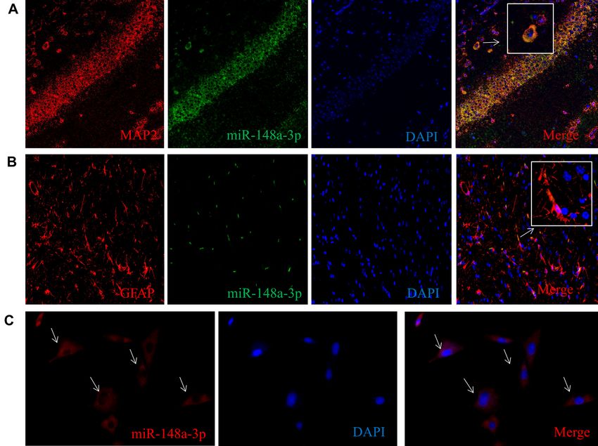
www.nature.com/scientificreports/
regulatory networks involved in brain development and adult neuroplasticity9. MiR-124a and miR-9 regulate
the differentiation of embryonic stem (ES) cells into neurons or glial cells10. while brain-specific miR-9 plays
a crucial role in regulating the cellular behavior of stem-cell-derived neural precursor cells (NPCs)11. Studies
have shown that miRNAs, through epigenetic regulation, are vital participants in nervous system diseases such
as convulsions and epilepsy.
In previous studies, miR-148a-3p has been found to be closely associated with non-small-cell lung cancer, oral
cancer, and other cancers. For example, in non-small-cell lung cancer, miR-148a-3p forms a regulatory pathway
with DNA methyltransferase 1 (DNMT1) to increase tumor p roliferation12. In acute pancreatitis, miR-148a-3p
13
can inhibit cell necrosis by targeting PTEN . In addition, miR-148a-3p can affect apoptosis through targeted
regulation of K LF614,15. Furthermore, studies have shown that miR-148a-3p is processed from the precursor of
miR-148a, and therefore has activity similar to that of miR-148a16. MiR-148a reduces liver damage in mice by
regulating calmodulin-dependent protein kinase II17. New evidence has shown that miR-148-a may regulate
the MAPK pathway during oxygen and glucose deprivation-induced microglial inflammation, which is of vital
importance in ischemic injury induced by hypoxia. Recent studies have suggested that miR-148a-3p may play an
important role in nervous and immune systems18–21. Compared with the diverse mechanisms for miR-148a-3p
functions in cancer or other diseases, how miR-148a-3p regulates specific neuroimmune diseases is poorly under-
stood. Previously, the apoptosis of hippocampal neurons was affected in brain injury by regulating the PI3K/Akt
signaling pathway22. However, the exact role of miR-148a-3p and its underlying mechanism with the neuronal
apoptosis and the effect of the PI3K/Akt signaling pathway in a recurrent FS model has not been clearly explored.
In this report, we investigated the pathogenic roles of miR-148a-3p in developmental convulsive brain injury.
We used miRNA microarray to detect differences in miRNA expression in the hippocampal tissue between FS
model and normal rats. We used a series of methods for data mining, information screening and analysis of
miRNAs associated with the nervous system, and we discuss the roles of miR-148a-3p and its target gene Synap-
tojanin-1 (SYNJ1) in seizure-related brain damage during development. Our findings will help to elucidate the
pathogenic factors of FSs and the subsequent development of epilepsy and provide new insights for the clinical
diagnosis and treatment of seizure diseases.
Results
Differentially expressed miRNA in a rat model of recurrent FS. To investigate how FSs affect
miRNA expression profiles in the developing brain, we placed rats at postnatal day 14 (P14 rats) under hyper-
thermic conditions at 39.5–42.5 ℃ (HT group) (Fig. 1A). In addition, the normothermic group (NT) was not
exposed to hyperthermic conditions.
Hierarchical clustering was performed based mainly on the similarity of the gene expression profile data;
Pearson correlation coefficients were used as the measures of similarity. Finally, the individual samples were
analyzed by cluster analysis. After hierarchical cluster analysis, all the experimental samples were divided into
two main clusters that were consistent with our expected results based on the experimental grouping. Based on
a P-value of < 0.05 and a fold change (FC > 1.5), 31 differentially expressed miRNAs were identified, including
21 upregulated miRNAs and 10 downregulated miRNAs (Fig. 1B,C). All the information is shown in Table S1.
To further validate the altered expression of miRNAs as detected by miRNA microarray, we selected nine
differentially expressed miRNAs and verified them by qRT-PCR. The results of quantitative PCR were consistent
with those of the microarray, confirming the value of the microarray as a fast, high-throughput, and large-scale
gene expression research technique (Fig. 1D). The RT-PCR primer sequences are shown in Table S2.
GO and pathway analysis of the miR‑148a‑3p target genes. Since miR-148a-3p has vital roles in
the regulation of both the nervous and immune systems, we predicted the target genes of the miRNAs using the
TargetScan and miRDB databases. A Venn diagram showed that 186 genes were found to be targeted by miR-
148a-3p (Fig. 2A). The miR-148a-3p target genes are listed in Table S3. In addition, we selected candidate target
genes related to nerve, immune, and epigenetic functions, which may act as key players in FS (Fig. 2B).
The GO functions of the genes were classified according to the NCBI and AmiGO databases. The significant
GO functional classifications obtained mainly included cellular response to nutrient levels, cellular response
to starvation, perinuclear region of cytoplasm, covalent chromatin modification, axon, neuron recognition,
and synaptic vesicle cycle (Fig. 3A). Furthermore, the significant pathways of the target genes were analyzed
according to the screening criterion of P < 0.05. Through the KEGG, BioCarta, Reactome and other databases,
the top 10 signaling pathways with significant differences were obtained by Fisher’s exact test of hypergeometric
distributions. Significant pathways mainly included the FoxO signaling pathway, Calnexin/calreticulin cycle,
TGF-β signaling pathway, PI3K-AKT signaling pathway, downstream TCR signaling, and the hippocampal
signaling pathway (Fig. 3B).
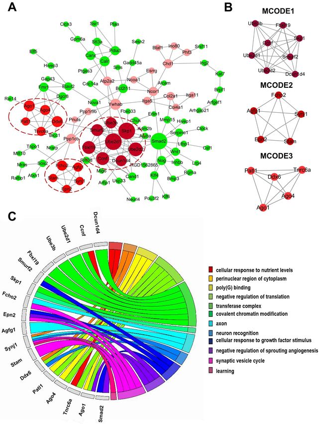
PPI network construction and module and hub gene identification. Protein interaction networks
have proven to be powerful tools for predicting and mining new essential genes in specific GO and KEGG path-
ways. Using the STRING online database and Cytoscape software, a total of 186 miR-148a-3p target genes were
filtered into the PPI network complex (Fig. 4A).
Subsequently, the PPI network of miR-148a-3p target genes was constructed with the most important module
obtained based on the MCODE analysis in Cytoscape (Fig. 4B). When “Node Score ≥ 0.2” was defined as the
cut-off criterion in MCODE, three clusters were identified from the PPI network, and the most significant clus-
ter consisted of 8 nodes and 26 edges. The 18 node degree genes were DCUN1D4, CCNF, UBE2D2, UBE2D1,
UBE3B, FBXL19, SMURF2, SKP1, FCHO2, EPN2, AGFG1, SYNJ1, STAM, DDX6, PATL1, AGO4, TNRC6A,
and AGO1. Furthermore, we analyzed the functions of the 18 genes, 6 of which (FCHO2, SYNJ, AGFG1, DDX6,
Scientific Reports | (2021) 11:1262 | https://doi.org/10.1038/s41598-020-79543-0 2
Vol:.(1234567890)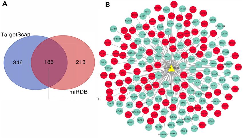
www.nature.com/scientificreports/

                                  Figure 1.  miRNA expression profile in a rat model of recurrent FS. (A) Experimental paradigms. Rats
                                  were treated with a hot-water bath at P14 for 7 consecutive days in the Hyperthermia (HT) group (bottom).
                                  (NT) group (upper) and (HT) group rats were killed at P21, and hippocampal tissues were taken for miRNA
                                  microarray. (B) Hierarchical clustering analysis. Heat map of the differentially expressed miRNAs between
                                  the HT and NT groups. A total of 31 miRNAs (P < 0.05, FC > 1.5) were identified from the hierarchical cluster
                                  analysis, including 21 upregulated miRNAs and 10 downregulated miRNAs. miRNA expression profiles are
                                  shown in the heatmap as upregulated (red color), downregulated (green color) and no change (black color). (C)
                                  Volcano plot analysis. (D) QRT-PCR verification. Nine differentially expressed miRNAs were verified in the HT
                                  group and the NT group.

Scientific Reports |   (2021) 11:1262 |               https://doi.org/10.1038/s41598-020-79543-0                                              3

                                                                                                                                         Vol.:(0123456789)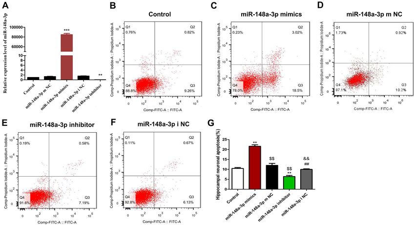
www.nature.com/scientificreports/
Figure 2. MiR-148a-3p target gene analysis. (A) Venn diagram of target genes for candidate miRNAs. (B)
miR-148a-3p target gene network. Each node represents a target gene. Among them, red nodes represent
genes related to nerve, immune and epigenetic pathways. Green nodes represent genes with other functions
(development, gene transcription, etc).
STAM, and EPN2) are mainly involved in axon, neuron recognition, and synaptic vesicle cycle (Fig. 4C). These
findings may play a particularly important role in FS.
Effects of different concentrations of KA on the apoptosis rate of hippocampal neuronal cells
in vitro. Many researches have shown that kainic acid (KA) was uesed to induce hippocampal neuronal
apoptosis, and it has been widely used as an experimental drug for seizures models since then23. Specifically, KA
has been shown to activate glutamate receptors, affect mitochondria function, and cause cell death in neuronal
cells24.
Rat hippocampal neurons in culture were treated with 50, 100, 150 and 200 μM KA for 24 h. The cells were
collected, and the KA-induced apoptosis of the hippocampal neurons was observed by TUNEL staining. The
results showed that the number of apoptotic hippocampal neurons and the apoptosis rate of hippocampal neu-
rons in the 50 μM KA group were significantly higher than those in the control group; the numbers of apoptotic
cells and the apoptosis rates were further increased in the 100, 150 and 200 μM groups, and the apoptosis rates
changed in a dose-dependent manner (Fig. S1). However, higher doses of KA induce greater neurotoxicity, which
can lead to the death of large numbers of nerve cells. Therefore, in the follow-up experiment, we selected 100 μM
KA for treatment of neuronal cells to establish an in vitro seizure model.
Sub‑cellular location of miR‑148a‑3p in vivo and vitro. To further study the function of miR-148a-3p,
we measured the cellular localization of miR-148a-3p in vivo and vitro and found that miR-148a-3p was colocal-
ized with MAP2 (neuronal maker) and GFAP (astrocytes maker) (Fig. 5).
SYNJ1 is a target gene of miR‑148a‑3p. To screen the target genes of miR-148a-3p, which is closely
related to the neuroimmune system, we detected the predicted target genes by qRT-PCR. We found that the FS
model group significantly increased the expression of miR-148a-3p and significantly decreased the mRNA expres-
sion of miR-148a-3p target genes, mainly including SYNJ1, FMR1, JPH3, ARFGEF1, ATP7A, NCOA1(Fig. 6A).
These genes are involved in the nervous and immune systems. Meanwhile, significantly decreased Expression of
SYNJ1 was detected in the hippocampal neurons after KA-induced. In addition, the SYNJ1 gene plays a vital role
in the function and pathways of the nervous and immune systems25, including Parkinson’s26, seizures27, epilepsy.
Based on the reasons above, SYNJ1 was selected as a candidate target gene for verification.
The target relationship between miR-148a-3p and SYNJ1 is displayed in Fig. 6B, which shows that miR-
148a-3p was able to target SYNJ1. To confirm that SYNJ1 was a direct target gene of miR-148a-3p, we performed a
double fluorescein enzyme reporter experiment. Recombinant plasmids of wild-type (WT)-miR-148a-3p/SYNJ1
and mutated (Mut)-miR-miR-148a-3p/SYNJ1 were constructed by inserting the luciferase reporter vector and the
Scientific Reports | (2021) 11:1262 | https://doi.org/10.1038/s41598-020-79543-0 4
Vol:.(1234567890)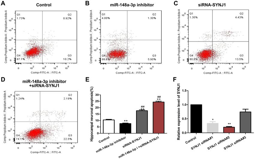
www.nature.com/scientificreports/

                                  Figure 3.  Gene ontology (GO) and pathway analysis for miR-148a-3p target genes. (A) Indicate biological
                                  process (BP), cellular component (CC) and molecular function (MF) predicted miR-148a-3p target genes. The
                                  vertical axis shows the GO terms, and the horizontal axis shows the − log10(P). (B) Significant pathways for
                                  predicted miR-148a-3p target genes. The vertical axis shows the pathway categories, and the horizontal axis
                                  shows the enrichment scores of pathways. P < 0.05 was used as a threshold to select significant GO categories
                                  and KEGG pathways. − Log 10p was the base 10 logarithm of the P-value.

                                  wild-type or mutant 3′ UTR sequence of SYNJ1. WT-miR-148a-3p/SYNJ1 and Mut-miR-148a-3p/SYNJ1 vectors
                                  were co-transfected with miRNA-148a-3p mimic or control plasmids into hippocampal neurons. The results
                                  showed that compared with that of the control plasmid group, the luciferase activities of the miR-148a-3p mimic
                                  and WT-pGL3-SYNJ13′-UTR cotransfection group were inhibited, while the luciferase activities of the miR-
                                  148a-3p mimic and MUT-pGL3-SYNJ13′-UTR co-transfection group were not significantly different (Fig. 6C).
                                  In addition, as we expected, the SYNJ1 mRNA level was changed in response to treatment with miR-148a-3p
                                  mimics or inhibitors. MiR-148a-3p mimic treatment reduced the mRNA level of SYNJ1. Conversely, miR-148a-3p
                                  inhibitors caused accumulation of SYNJ1 (Fig. 6D). Taken together, these results show that SYNJ1 is a novel bona
                                  fide target of miR-148a-3p in rat hippocampus neuronal cells.

                                  MiR‑148a‑3p promotes cell apoptosis of hippocampal neurons after KA‑induction. To further
                                  confirm the correlation between miR-148a-3p level and KA-induced neuron cell apoptosis, we manipulated
                                  miR-148a-3p levels by treating H19-7 rat hippocampal neuronal cells with either miR-148a-3p mimics or miR-
                                  148a-3p inhibitors, followed by 100 μM KA treatment in vitro.
                                      As shown in Fig. 7A, we first determine the effectiveness of miR-148a-3p mimics or inhibitor for the expres-
                                  sion of miR-148a-3p. The results showed that the miR-148a-3p mimics efficiently increased miR-148a-3p levels
                                  (P < 0.001) and the miR-148a-3p inhibitor exhibited the lowest miR-148a-3p expression (P < 0.01).

Scientific Reports |   (2021) 11:1262 |               https://doi.org/10.1038/s41598-020-79543-0                                                  5

                                                                                                                                           Vol.:(0123456789)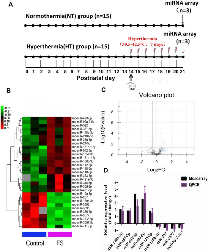
www.nature.com/scientificreports/

                                            Figure 4.  PPI network analysis and functional enrichment analysis of hub genes. (A) PPI network analysis.
                                            Circular nodes represent the miR-148a-3p target genes. Circular nodes in red represent the hub genes. A
                                            larger node size corresponds to more genes that are co-expressed with the gene. Darker colors indicate greater
                                            significance. (B) Three significant modules selected from the PPI network. Each node represents a protein. (C)
                                            GO enrichment analysis of 18 hub genes. Different color bands represent different GO terms.

                                              As shown in Fig. 7B–G, we found that the apoptosis rate of hippocampal neuronal cells transfected with the
                                            miR-148a-3p mimic was significantly higher than that of hippocampal neurons transfected with the control

          Scientific Reports |   (2021) 11:1262 |               https://doi.org/10.1038/s41598-020-79543-0                                                   6

Vol:.(1234567890)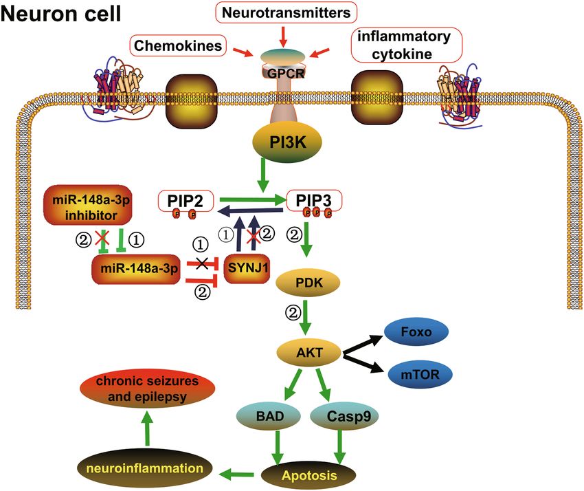
www.nature.com/scientificreports/

                                  Figure 5.  Sub-cellular location of miR-148a-3p in vivo and vitro. (A–C) Cellular location of miR-148a-3p in
                                  neuronal cells in normal rat hippocampus tissue. (D–F) Cellular location of miR-148a-3p in astrocyte cells
                                  in normal rat hippocampus tissue. (G–I) MiR-148a-3p was localized in the cytoplasm of H19-7 hippocampal
                                  neuronal cells.

                                  construct (P < 0.01), while that of hippocampal neurons transfected with the miR-148a-3p inhibitor was sig-
                                  nificantly lower (P < 0.01). Compared with that of the miR-148a-3p mimic group, the apoptosis rates of the
                                  miR-148a-3p mimic NC group and the miR-148a-3p inhibitor group were significantly decreased (P < 0.01). In
                                  addition, compared with that of the miR-148a-3p inhibitor NC group, the apoptosis rate of the miR-148a-3p
                                  inhibitor group was significantly decreased (P < 0.01). All findings strongly support our hypothesis that miR-
                                  148a-3p mediates neuronal cell apoptosis in response to KA treatment in vitro.

                                  MiR‑148a‑3p mediates hippocampal neuronal apoptosis by targeting SYNJ1 after KA‑induc‑
                                  tion. To further confirm whether miR-148a-3p depends on the SYNJ1 to regulate the apoptosis of hippocam-
                                  pal neurons, we studied the effect of SYNJ1 siRNA on the miR-148a-3p-mediated apoptosis of hippocampal
                                  neurons treated with KA. We first detected the silencing efficacy of SYNJ1-siRNA. As shown in the RT-qPCR
                                  results in Fig. 8F, compared with the control, the siRNA-1, siRNA-2, and siRNA-3 groups had decreased mRNA
                                  expression levels of SYNJ1 while the siRNA-2 group exhibited the lowest SYNJ1 expression (P < 0.01). These
                                  findings demonstrated that the silencing efficacy was highest in the siRNA-2 group; thus, sequences in the
                                  siRNA-2 group were selected for cell transfection.
                                      After successful transfection of rat hippocampal neurons with the miR-148a-3p inhibitor and miR-148a-3p
                                  inhibitor + SYNJ1 siRNA for 48 h, the cells were treated with 100 μM KA and cultured for 24 h. The cells were
                                  collected, and hippocampal neuronal apoptosis was detected by flow cytometry. The results showed that the
                                  apoptosis rate of hippocampal neurons after miR-148a-3p inhibitor transfection was significantly decreased
                                  compared to that of the control (P < 0.01). Compared with that of the miR-148a-3p inhibitor group, the apop-
                                  tosis rate of the miR-148a-3p inhibitor + SYNJ1 siRNA group was significantly increased (P < 0.01) (Fig. 8A–E).
                                  Taken together, our results indicate that SYNJ1 acts as a downstream target to mediate miR-148a-3p regulation
                                  of apoptosis in rat hippocampus neuronal cells.

Scientific Reports |   (2021) 11:1262 |               https://doi.org/10.1038/s41598-020-79543-0                                                 7

                                                                                                                                           Vol.:(0123456789)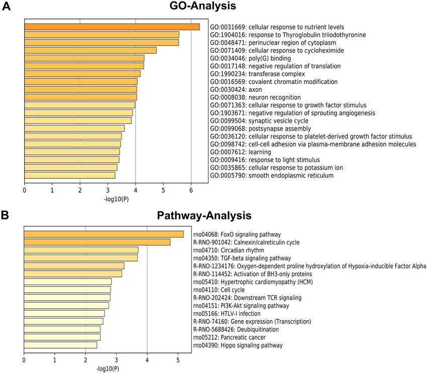
www.nature.com/scientificreports/
Figure 6. SYNJ1 is a bona fide target of miR-148a-3p in rat hippocampal neuronal cells. (A) Detection of
predicted miR-148a-3p target genes between the normal and FS model groups. Relative mRNA expression of
the miR-148a-3p target genes. *P < 0.05 compared with the normal group. (B) Predication that SYNJ1 is a target
gene of miR-148a-3p by TargetScan. (C) Dual-luciferase experiment to verify the targeting relationship between
miR-148a-3p and SYNJ1; **P < 0.01 vs. NC group vs. miR-148a mimics-3p + WT-SYNJ1 group; ##P < 0.01 vs,
miR-148a-3p mimics + WT-SYNJ1 group vs. miR-148a-3p mimics + MUT-SYNJ1 group. The data were analyzed
by one-way ANOVA; the pairwise comparison after ANOVA was analyzed by LSD-t test. (D) Relative SYNJ1
levels after different treatments. **P < 0.01, ***P < 0.001 vs. control group.
MiR‑148a‑3p mediates hippocampal neuronal apoptosis via activation of PI3K‑AKT signaling
pathway. To further study whether miR-148a-3p can activate the PI3K/AKT pathway to regulate the apop-
tosis of hippocampal neurons, western blotting was performed to determine protein expression level of related
proteins of the PI3K/Akt signaling pathway (PI3K, p-PI3K, Akt, and p-Akt) (Fig. 9). and the results indicated
that miR-148a-3p could promote the activation of the PI3K/Akt signaling pathway in hippocampal neurons.
The protein expression of PI3K and Akt was not significantly different in hippocampal neurons of all the
groups (all P > 0.05). In comparison to the control group, the protein expression of p-PI3K and p-Akt were
significantly elevated in miR-148a-3p mimics group (P < 0.01), while that in miR-148a-3p inhibitor group was
significantly decreased (P < 0.01). The protein expression of p-PI3K and p-Akt was not significantly different in
hippocampal neurons of mimics NC and inhibitor NC groups (all P > 0.05).
Compared with the miR-148a-3p mimics group , the protein expression of p-PI3K and p-Akt were signifi-
cantly decreased in the miR-148a-3p inhibitor group (P < 0.01), which indicated that miR-148a-3p expression
promoted the activation of the PI3K/Akt signaling pathway in hippocampal neurons after KA-induced.
Discussion
FSs are most common in children at the age of 5, and a variety of factors may increase the risk of F Ss28. MiRNAs
may play an important role in neural circuitry by functioning in various brain regions and may also play key roles
in neurological diseases, such as seizures and epilepsy29–31. Among these miRNAs, miR-148a-3p is involved in
the development and dynamic balance of the neuron-immune system and in various types of cancers, autoim-
mune diseases, inflammatory diseases, and other pathological p rocesses32,33. Therefore, the primary purpose of
this study was to explore the potential mechanism by which miR-148a-3p contributes to FSs to better elucidate
the pathological mechanism of FSs. This study provided evidence that downregulation of miR-148a-3p inhibited
apoptosis of hippocampal neurons after KA induced through the PI3K/Akt signaling pathway.
In this report, we first established a rat model of recurrent FSs and used miRNA expression profile chip tech-
nology to analyze the hippocampi of rats in the recurrent convulsion group and the normal group. According to
the results of hierarchical cluster analysis, the six groups of experimental samples were divided into two different
clusters, from which it was inferred that the experimental sample classification was reliable and consistent with
the expected results. Through information analysis, 31 differentially expressed miRNAs and related functional
Scientific Reports | (2021) 11:1262 | https://doi.org/10.1038/s41598-020-79543-0 8
Vol:.(1234567890)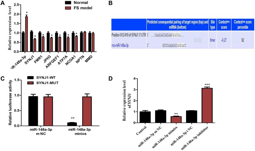
www.nature.com/scientificreports/
Figure 7. Inhibition of miR-148a-3p reduces hippocampal neuronal apoptosis by kainic acid. (A) Relative miR-
148a-3p levels after different treatments. (B–F) Cell apoptosis detection by flow cytometry. (G) Apoptosis rates
of hippocampal neurons. Data are shown as the Mean ± S.E.M. and represent three independent experiments.
**P < 0.01 vs. control group; $$,##P < 0.01 vs. miR-148a-3p mimics group; &&P < 0.01 vs. miR-148a-3p inhibitor
group.
information were obtained. Twenty-one miRNAs were significantly upregulated, including miR-542-5p, miR-
499-5p, miR-148a-3p, and miR-451-5p. Eleven miRNA genes were significantly downregulated, including miR-
207 and miR-130b-3p. Whether these differentially expressed miRNAs participate in the development of FSs
needs further study. In addition, to verify the reliability of the gene chip data, the expression profiles of 9 dif-
ferentially expressed miRNAs were verified by qRT-PCR, and the results were consistent with the chip results,
providing a solid foundation for the subsequent expression profile analysis.
Information mining and comprehensive analysis revealed that the expression level of miR-148a-3p was sig-
nificantly increased in the hippocampus in the FS models, consistent with the results of qRT-PCR. Therefore, we
selected miR-148a-3p for in-depth research and analysis. Studies have shown that the function of differentially
expressed miR-148a-3p is closely related to neuroimmunity and apoptosis. Recent studies have shown that
in ischemic stroke-related diseases, miR-148a-3p can interact with lncRNA-h19 and ROCK2 genes to form a
pathway that regulates the oxidative stress caused by ischemic stroke18. Additionally, miR-148a-3p can target
the KLF6 gene to regulate the proliferation and apoptosis of skeletal muscle cells14,15. As the precursor of miR-
148a-3p, miR-148a has the same a ctivity34. Clinical studies have shown that there are significant abnormalities
in the expression of miR-148a in patients with neurological diseases such as Parkinson’s disease and Alzheimer’s
disease. Other differentially expressed miRNAs screened by bioinformatics analysis are also worth studying.
Previous studies have revealed that the functions of these miRNAs are closely related to neuroimmune and
other factors. For example, studies have shown that miR-499-5p can play a neuroprotective role by regulating the
expression of C-reactive protein in hypoxic-ischemic e ncephalopathy35. MiR-207 may be involved in cognitive
impairment induced by obstructive sleep apnea (OSA)36. Furthermore, miR-130b-3p can inhibit the inflamma-
tory response induced by eCIRP37.
We predicted the target genes of miR148a-3p with TargetScan and miRDB, and a total of 186 overlapping
target genes were obtained. A target gene relationship map was constructed using the relevant database informa-
tion. The diagram confirmed that the DNMT1 gene is a target of miR148a-3p and that there is a close relationship
between DNMT1 and the occurrence and development of e pilepsy38. In addition, epigenetic regulation plays
vital roles in nervous system d iseases39. Among the predicted target genes were many genes strictly related to
neuroimmune function, such as SYNJ1, Noggin (NOG), Neuronplastin (NPTN), Nuclear receptor coactivator
1 (NCOA1), and SOS Ras/Rho guanine nucleotide exchange factor 2 (SOS2). Previous studies have shown that
the NOG gene can restore stem cell populations and neurogenesis in the h ippocampus40. NOG gene can also
41
promote the repair of myelin s heaths . NPTN, a multifunctional neuronal adhesion molecule, is closely related
to long-term synaptic p lasticity42.
Subsequently, we selected the potential target genes of the significantly upregulated miR-148a-3p and analyzed
their GO functions and KEGG signaling pathways. The different GO term and KEGG pathway information
suggests that there is a close relationship between FSs and neuroimmunity, which provides an essential clue for
further study of the role of immune inflammation in FS. The results of GO analysis showed that the functions
Scientific Reports | (2021) 11:1262 | https://doi.org/10.1038/s41598-020-79543-0 9
Vol.:(0123456789)www.nature.com/scientificreports/
Figure 8. miR-148a-3p regulation of hippocampal neuron apoptosis through targeting SYNJ1. (A–D)
Hippocampal neuron apoptosis using flow cytometry. H19-7 hippocampal neuronal cells were incubated
with 100 µM KA for 48 h. miR-148a-3p inhibitor and siRNA-SYNJ1 were added to the culture 4 h prior to KA
treatment. Cells were collected and stained with Annexin V and propidium iodide (PI) and then examined by
flow cytometry. (E) Hippocampal neuron apoptosis rate. Results are shown as the mean ± S.E.M. and represent
three independent experiments. **P < 0.01 vs. control group; ##P < 0.01, vs. miR-148a-3p inhibitors group.
(F) Comparison of the effects of three different SYNJ1 siRNAs. *P < 0.05, SYNJ1 siRNA#1 vs. control group;
**P < 0.01, SYNJ1 siRNA#2 vs. control group.
Figure 9. Protein expression level of PI3K/Akt signaling pathway-related proteins in hippocampal neurons
of rats. (A) protein bands of PI3K/Akt signaling pathway-related proteins detected by western blotting (B)
Comparison on expression of PI3K/Akt signaling pathway-related proteins. **P < 0.01 compared with the
control group; ##P < 0.01 compared with the miR-148a-3p mimics group. PI3K hosphatidylinositol 3-kinase,
p-PI3K phosphorylated PI3K, p-Akt phosphorylated Akt.
Scientific Reports | (2021) 11:1262 | https://doi.org/10.1038/s41598-020-79543-0 10
Vol:.(1234567890)www.nature.com/scientificreports/
of the potential target genes were closely related to nervous system processes, such as neuroprotection, neuron
migration, and hippocampal signaling pathways. Recent studies have shown that immune and inflammatory
processes play essential roles in FSs, especially innate immune system processes and subsequent inflammatory
responses caused by infection, nerve trauma, and other factors related to epilepsy. Consistent with these roles,
the insulin signaling pathway was significantly enriched. Studies have shown that the insulin signaling pathway
is closely related to epilepsy. For example, administration of low-dose intranasal insulin can reduce the frequency
of spontaneous convulsions and epileptic discharges and increase the cognitive abilities of epileptic mice43. Addi-
tionally, under epileptic conditions, neuronal death induced by convulsions is affected by activation of the FoxO
signaling pathway44. Furthermore, the FoxO signaling pathway family can participate in cell death in a variety
of pathological c onditions45. FoxO1 and FoxO2 play essential roles in the development and differentiation of
immune cells. FoxO1 and FoxO3 can promote the formation of regulatory T cells and inhibit the formation of
T-helper 1 (Th1) and T-helper 17 (Th17) c ells46.
The PPI network displayed functional connections of miR-148a-3p target genes, and certain hub genes were
selected based on the MCODE analysis. Most of them were enriched in neuron and synaptic function. Neurons
and synaptogenesis factors have always played a vital role in FS. Based on the module analysis of the PPI network,
6 neural-associated genes were selected: SYNJ1, FCHO2, AGFG1, DDX6, STAM, and EPN2. Thus, our findings
indicate that these screened key genes may be the driving forces underlying the occurrence of FS.
SYNJ1, a polyphosphate inositol phosphatase, exists in presynaptic nerve endings and protein complexes
involved in cellular endocytosis and plays a vital role in the phosphorylation of synaptic vesicles. In addition, the
SYNJ1 gene plays essential roles in the nervous and immune systems. SYNJ1 gene mutations are associated with
two rare nervous system diseases: early-onset Parkinson’s disease and severe neurodegeneration with refractory
seizures and recurrent seizures47. Deletion of the SYNJ1 gene can lead to seizures25. SYNJ1 has been reported
as a potential regulator of allogeneic T cell r esponses48. The level of SYNJ1 mRNA was reduced after allogeneic
stimulation of naive T cells. Knockdown of SYNJ1 in allogeneically stimulated T cells confirmed its role in T cell
proliferation and cytokine responses48. Through comprehensive analysis, we found that miR-148a-3p may affect
the occurrence and development of convulsions through its potential target gene, SYNJ1. In a double luciferase
experiment, it was shown that SYNJ1 can indeed be directly regulated by miR-148a-3p.
Previous studies have revealed that miRNAs are closely related to the apoptosis of hippocampal neurons.
Upregulation of miR-223 expression can inhibit hippocampal neuronal apoptosis and brain damage in rats with
convulsions49. MiR-421 can inhibit the apoptosis of hippocampal neurons in epileptic rats through the TLR
signaling pathway50. Silencing miR-134 can reduce the damage to hippocampal neurons and the frequency of
spontaneous convulsions in epileptic r ats51. Other studies have shown that there is a close relationship between
miR-148a-3p and apoptosis. MiR-148a-3p and DNMT1 form a regulatory pathway that increases tumor pro-
liferation. Other studies have shown that miR-148a-3p can inhibit cell necrosis by bypassing PTEN targeting
in acute p ancreatitis16. In a previous study, we used miRNA arrays and qRT-PCR to confirm that hippocampal
miR-148a-3p was significantly upregulated after recurrent convulsions during development, suggesting that
miR-148a-3p may be involved in the occurrence and development of convulsions. To confirm that miR-148a-3p
is involved in the apoptosis related to convulsion injury, we used rat hippocampal neurons (H19-7 cells) as the
research objects in this study. We transfected hippocampal neurons with a miR-148a-3p mimic and a miR-
148a-3p inhibitor and treated them with KA to establish a neurotoxicity model that approximated convulsions.
We thus observed the effects of overexpression or low expression of miR-148a-3p on the apoptosis of hip-
pocampal neurons treated with KA and investigated the related mechanisms. The results showed that compared
with those in the non-transfected group, the miR-148a-3p expression levels in hippocampal neurons in the
miR-148a-3p mimic group were significantly upregulated while those in the miR-148a-3p inhibitor group were
significantly downregulated. Cell models of miR-148a-3p overexpression and low expression were thus success-
fully constructed and can be used for follow-up studies.
This report showed that the apoptosis rate of hippocampal neurons was significantly higher in the miR-
148a-3p mimic group than in the control group while the apoptosis rate of hippocampal neurons was significantly
lower in the miR-148a-3p inhibitor group than in the miR-148a-3p inhibitor group. It is suggested that over-
expression of miR-148a-3p can promote KA-induced apoptosis of hippocampal neurons, while low expression
of miR-148a-3p can inhibit apoptosis. To further confirm that miR-148a-3p regulates hippocampal neuronal
apoptosis in a SYNJ1-dependent manner, we studied the effects of SYNJ1 siRNA on miR-148a-3p-mediated
regulation of hippocampal neuronal apoptosis under KA treatment. Apoptosis was detected by flow cytometry.
The apoptosis rate of hippocampal neurons in the SYNJ1 siRNA + miR-148a-3p inhibitor group was significantly
higher than that in the SYNJ1 siRNA + miR-148a-3p mimic group, indicating that SYNJ1 siRNA transfection
could block the decrease in hippocampal neuronal apoptosis induced by the miR-148a-3p inhibitor. These
findings further confirmed that the effect of miR-148a-3p on hippocampal neuronal apoptosis depends on the
existence of SYNJ1, which enables the regulation of hippocampal neuronal apoptosis after seizures.
Our study detected the apoptosis rate of hippocampal neurons by Flow cytometry, which suggested that
miR-148a-3p promoted the apoptosis of neurons. Previously, the PI3K/Akt signaling pathway was reported to
modulate the survival response to neuronal apoptosis caused by oxidative stress following epileptic s eizures52.
Additionally, the neuronal inflammatory response induced by interleukin-1β stimulates the activation of the
PI3K/Akt signaling pathway, which is involved in Pathophysiology of S eizures53. In order to further verify
whether miR-148a-3p affects the PI3K/AKT signaling pathway in hippocampal neurons, western blot analysis
demonstrated that levels of phosphorylation of PI3K and AKT are activated when transfected with the miR-
148a-3p. Based on our findings, we propose a novel model wherein miR-148a-3p promote neuronal apoptosis
via activation of PI3K-AKT signaling pathway by targeting SYNJ1 (Fig. 10).
In summary, through microarray analysis of miRNA expression profiles in the hippocampus of rats with
recurrent FSs, a series of differentially expressed miRNAs and candidate target genes along with GO functional
Scientific Reports | (2021) 11:1262 | https://doi.org/10.1038/s41598-020-79543-0 11
Vol.:(0123456789)www.nature.com/scientificreports/
Figure 10. Schematic of the proposed hypothesis model showing that miR-148a-3p positively regulates
neuronal apoptosis via activating the PI3K-AKT signaling pathway by targeting SYNJ1. In chronic seizure or
epilepsy tissue, neurons and astrocytes and even microglia may be affected by neurotransmitters, chemokines
and inflammatory cytokines. Furthermore, activation of the PI3K-AKT signaling pathway play a central role
in the process of the apoptotic death of neural cells and causes neuroinflammation, which eventually leads to
seizures or epilepsy. Mir-148a-3p inhibitor can inhibit the expression of mir-148a-3p, thereby increasing the
expression of the SYNJ1 gene, making dephosphorylating PIP3 into PIP2, inhibiting PI3K-AKT signaling
pathway, and reducing cell apoptosis. Conversely, upregulation of miR-148a-3p could enhance the PI3K-AKT
mediated apoptosis pathway and neuroinflammation and then cause neuroinflammation, leading to seizures
and epilepsy.
and KEGG pathway classifications were obtained. Our findings also demonstrate for the first time that the
miR-148a-3p effectively promoted neuronal cell apoptosis via the activation of PI3K/AKT signaling pathway by
controlling the expression of SYNJ1 in the neuronal cell, which provides new insights into a potential therapeutic
target of miR-148a-3p in the modulation of neuron-mediated apoptosis and may be beneficial for the treatment
of infants with FS brain injury. However, future research should study the effects of miR-148a-3p on specific
epileptic phenotypes, such as abnormal neuronal circuits, spatial learning disruptions, and memory impairment.
Additionally, considering the limiting factors, such as the blood–brain barrier, it is imperative to determine the
clinical feasibility of targeting miR-148a-3p in the treatment of seizures or epilepsy.
Materials and methods
Ethics statement. All protocols complied with the recommendations in the Guide for the Care and Use
of Animals for an Animal Biosafety Level 3 (ABSL-3) Laboratory and were approved by the Animal Ethics
Committee of Wuhan University (Permit Number: SCXK 2016-0004). All surgery was performed under chloral
hydrate anesthesia, and all efforts were made to minimize suffering.
Establishment of the rat models of FS. Sprague–Dawley rats (aged 14 days) were obtained from an
ABSL-3 laboratory. The rats were housed individually under a 12-h light–dark cycle with ad libitum access
to food and water. Hyperthermia-induced seizures were produced using a hot-water bath54. The animals were
Scientific Reports | (2021) 11:1262 | https://doi.org/10.1038/s41598-020-79543-0 12
Vol:.(1234567890)www.nature.com/scientificreports/
placed in the temperature-controlled (39.5–42.5 °C) water bath and removed from the water immediately once
seizures were induced. Seizures were induced once every day for 7 days.
After completion of the whole seizure induction process, the rats were anesthetized with 10% chloral hydrate
(3 ml/kg). The brains were quickly removed and placed on ice, and the hippocampi were dissected, placed into
liquid nitrogen and transferred into a − 80 °C low-temperature freezer for storage and later use.
MiRNA array analysis. RNA was isolated by the TRIzol method (Invitrogen, Thermo Fisher, USA), and
miRNA profiling was performed by Kang Chen Biosciences (Shanghai, China). Briefly, purified RNA was labeled
using a miRCURY Hy3/Hy5 Power labeling kit and hybridized to a miRCURY LNA miRNA array (v.11.0)
(Exiqon, Vedaek, Denmark). Scanning was performed with an Axon GenePix 4000B microarray scanner. Gene-
Pix Pro V6.0 was used to read the raw intensities of the images55. The results were subjected to unsupervised
hierarchical clustering and tree analysis.
MiRNA target prediction. All significant differentially expressed miRNAs were analyzed by bioinformat-
ics algorithms. The potential target genes of these miRNAs were predicted using miRNA target prediction data-
bases, including TargetScan (http://www.targetscan.org) and miRDB (http://www.mirdb.org).
Gene Ontology and pathway enrichment analysis. Functional classification of all differentially
expressed miRNA target genes was performed by DAVID v6.8 Database (https://david.ncifcrf.gov/) to deter-
mine the biological significance of the targets, and the accompanying p-values calculated by Fisher’s exact test
indicated the functions that were overrepresented among the targets. The P-values produced by topGO denoted
the significance of the enrichment of GO terms for the differentially expressed genes. A lower P-value indicated
a more significant GO term (a P-value threshold of ≤ 0.05 is recommended). Moreover, a pathway analysis was
performed using the Kyoto Encyclopedia of Genes and Genomes (KEGG) database to identify the enriched
pathways of the targets56–58, and the P-value (significance) was calculated for each pathway using a hypergeomet-
ric distribution59. Furthermore, to perform the GO and pathway enrichment analyses of the miR-148a-3p target
genes, we used the Metascape Database (http://metascape.org) with the custom analysis60.
Integration of PPI network complex identification. We developed a miR-148a-3p target gene-
encoded protein and PPI network using STRING (http://string-db.org)61. The PPI network was constructed
using Cytoscape software (version 3.6.1) to analyze the interactions between predicted target gene-encoded pro-
teins in FS62. The Node Analyzer was calculated using the Network Analyzer plug-in, which reveals the number
of connections used to filter the PPI hub genes. The corresponding protein identified at the central node may be
a core protein and key candidate gene with important physiological regulatory functions.
Real‑time quantitative PCR (qRT‑PCR) analysis. Briefly, after hippocampal isolation and RNA extrac-
tion, cDNA was prepared using a RevertAid First Strand cDNA Synthesis Kit (Thermo Fisher, USA). qRT-PCR
was performed using TaqMan MicroRNA Assays (Life Technologies, Carlsbad, CA, USA). All real-time reac-
tions were performed in triplicate on an ABI PRISM 7000 sequence detection system (Ambion-Applied Biosys-
tems, Foster City, CA, USA). The relative expression was calculated using the comparative cycle threshold (ct)
method, and the relative expression ratio was determined by the formula 2 −△△ct63.
Terminal deoxynucleotidyl transferase‑mediated dUTP nick‑end labeling (TUNEL)
assay. Apoptosis was determined using a TUNEL assay. The apoptotic index was calculated as the percent-
age of TUNEL-positive cells divided by the total number of cells. TUNEL in situ cell death detection kits were
purchased from Roche64.
Flow cytometry analysis. An annexin V-fluorescein isothiocyanate (FITC)/propidium iodide (PI) apop-
tosis detection kit (Cat No. 70-AP101-100; MultiSciences, Hangzhou, China) was used to evaluate the apoptotic
rates of neuronal cells. After 48 h of transfection, neurons were treated with kainic acid (KA; 100 μM) for 24 h,
and then the cells were collected, washed with PBS, and suspended with 5 µl of Annexin V-FITC and 5 µl of
PI. The cells were then incubated for 30 min in the dark at room temperature. We used a flow cytometer (BD
Biosciences) to analyze apoptosis and evaluate the apoptotic rate65.
Dual‑luciferase reporter assay. The gene SYNJ1 was predicted to be a target of miR-148a-3p by Tar-
getScan (http://www.targetscan.org). To verify that SYNJ1 is a target gene of miR-148a-3p, the 3′UTR region of
SYNJ1 was amplified, the PCR product was cloned into the downstream multiple cloning site of the PGL3 lucif-
erase reporter vector (Promega Corporation, Madison, WI), and then site-directed mutagenesis was performed
on the binding site between miR-148a-3p and the target gene, which was predicted by TargetScan. The Renilla
luciferase reporter vector pRL-TK (TaKaRa Biotechnology Co. Ltd., Dalian, China) was chosen as the internal
reference to adjust for differences in cell number and transfection efficiency. H19-7 hippocampal neuronal cells
were separately cotransfected with a miR-148a-3p mimic or a mimic negative control (NC) and a luciferase
reporter vector. Then, the luciferase activity was examined by the method provided by P romega66.
Immunofluorescence staining and FISH. The procedure was performed as described in a previous
s tudy67. Briefly, segments of hippocampus were rapidly dissected from SD rat brains perfused with 4% para-
formaldehyde, fixed with 4% PFA, and then cryoprotected in 30% sucrose. miR-148a-3p probes (5′-CAAAGTT
Scientific Reports | (2021) 11:1262 | https://doi.org/10.1038/s41598-020-79543-0 13
Vol.:(0123456789)www.nature.com/scientificreports/
CTGTAGTGCACTGA-3′) were synthesized (F03101, Gene Pharma). FISH was performed using the FISH kit
(Guangzhou, Ribobio), and then FISH sections were incubated with MAP2 antibody (1:200, ab5392; Abcam),
and GFAP antibody (1:100, #3670, Cell Signaling Technology), then with fluorescent-conjugated secondary
antibody (1:200, 111-165-003, 115-165-003, Jackson) at 37 °C for 1 h. After the sections were rinsed in 0.01 M
PBS, the slides were mounted on a mounting medium with DAPI and scanned via Ortho-Fluorescent Micros-
copy (Nikon).
Western Blotting. Protein samples from the H19-7 hippocampus neurons were loaded to a 12% SDS-
PAGE. PVDF membranes were used for the SDS-PAGE. The membrane was blotted for 2 h using 5% non-fat
milk in the TBST solution.
Primary antibodies against PI3K, phosphorylate-PI3K (p-PI3K), Akt and phosphorylate Akt (p-Akt), and
β-actin (1: 3000, Abcam, Cambridge, MA) were added for incubation at 4 °C overnight. Then, the membranes
were incubated with a species-matched HRP-conjugated secondary antibody (Abcam) for 1 h at room tem-
perature. Quantitative data of western blots were analysed using Gel-Pro3.0 software. The intensity of the tested
protein bands was normalised to the internal reference.
Statistical analysis. All data were analyzed using SPSS 21.0 integrated software (SPSS Inc. IBM, Chicago,
IL). Measurement data are shown as the Mean ± standard deviation (SD). Comparisons between two groups and
among multiple groups were performed via t test and one-way analysis of variance (ANOVA), respectively. A
value of P < 0.05 was considered statistically significant.
Data availability
All datasets generated during the study are available on request from the corresponding author.
Received: 7 April 2020; Accepted: 4 December 2020
References
1. Pavlidou, E., Hagel, C. & Panteliadis, C. Febrile seizures: Recent developments and unanswered questions. Childs Nerv. Syst. 29,
2011–2017 (2013).
2. Smith, D. K., Sadler, K. P. & Benedum, M. Febrile seizures: Risks, evaluation, and prognosis. Am. Fam. Physician 99, 445–450
(2019).
3. Steering Committee on Quality, I. & Management, S. o. F. S. A. A. o. P. Febrile seizures: Clinical practice guideline for the long-term
management of the child with simple febrile seizures. Pediatrics 121, 1281–1286 (2008).
4. Ozen, F. et al. The prevalence of Familial Mediterranean Fever common gene mutations in patients with simple febrile seizures.
Eur. Rev. Med. Pharmacol. Sci. 18, 657–660 (2014).
5. Ghasemi, F., Valizadeh, F. & Taee, N. Iron-deficiency anemia in children with febrile seizure: A case-control study. Iran J. Child
Neurol. 8, 38–44 (2014).
6. Renda, R., Yuksel, D. & Gurer, Y. K. Y. Evaluation of patients with febrile seizure: Risk factors, reccurence, treatment and prognosis.
Pediatr. Emerg. Care 36, 173–177 (2017).
7. Ferrante, M. & Conti, G. O. Environment and neurodegenerative diseases: An update on miRNA Role. Microrna 6, 157–165 (2017).
8. Bartel, D. P. MicroRNAs: Target recognition and regulatory functions. Cell 136, 215–233 (2009).
9. Esteves, M., Serra-Almeida, C., Saraiva, C. & Bernardino, L. New insights into the regulatory roles of microRNAs in adult neuro-
genesis. Curr. Opin. Pharmacol. 50, 38–45 (2019).
10. Krichevsky, A. M., Sonntag, K. C., Isacson, O. & Kosik, K. S. Specific microRNAs modulate embryonic stem cell-derived neuro-
genesis. Stem Cells 24, 857–864 (2006).
11. Delaloy, C. et al. MicroRNA-9 coordinates proliferation and migration of human embryonic stem cell-derived neural progenitors.
Cell Stem Cell 6, 323–335 (2010).
12. Bai, Y., Lang, L., Zhao, W. & Niu, R. Long non-coding RNA HOXA11-AS promotes non-small cell lung cancer tumorigenesis
through microRNA-148a-3p/DNMT1 regulatory axis. Onco Targets Ther. 12, 11195–11206 (2019).
13. Cai, S. W., Han, Y. & Wang, G. P. miR-148a-3p exhaustion inhibits necrosis by regulating PTEN in acute pancreatitis. Int. J. Clin.
Exp. Pathol. 11, 5647–5657 (2018).
14. Jerez, S. et al. Extracellular vesicles from osteosarcoma cell lines contain miRNAs associated with cell adhesion and apoptosis.
Gene 710, 246–257 (2019).
15. Song, C. et al. miR-148a-3p regulates proliferation and apoptosis of bovine muscle cells by targeting KLF6. J. Cell Physiol. 234,
15742–15750 (2019).
16. He, H. et al. miR-148a-3p promotes rabbit preadipocyte differentiation by targeting PTEN. Vitro Cell Dev. Biol. Anim. 54, 241–249
(2018).
17. Zheng, D. et al. The miR-148a alleviates hepatic ischemia/reperfusion injury in mice via targeting CaMKIIalpha. Xi Bao Yu Fen
Zi Mian Yi Xue Za Zhi 32, 1202–1206 (2016).
18. Zeng, J. et al. Metformin protects against oxidative stress injury induced by ischemia/reperfusion via regulation of the lncRNA-
H19/miR-148a-3p/Rock2 Axis. Oxid. Med. Cell Longev. 2019, 8768327 (2019).
19. Wong, H. A. et al. The cancer genome atlas analysis predicts microRNA for targeting cancer growth and vascularization in glio-
blastoma. Mol. Ther. 23, 1234–1247 (2015).
20. Huang, F. et al. miR-148a-3p mediates notch signaling to promote the differentiation and M1 activation of macrophages. Front.
Immunol. 8, 1327 (2017).
21. Patel, V. et al. The stretch responsive microRNA miR-148a-3p is a novel repressor of IKBKB, NF-kappaB signaling, and inflam-
matory gene expression in human aortic valve cells. FASEB J. 29, 1859–1868 (2015).
22. Shen, M. et al. Dexmedetomidine exerts neuroprotective effect via the activation of the PI3K/Akt/mTOR signaling pathway in rats
with traumatic brain injury. Biomed. Pharmacother. 95, 885–893 (2017).
23. Wang, Q., Yu, S., Simonyi, A., Sun, G. Y. & Sun, A. Y. Kainic acid-mediated excitotoxicity as a model for neurodegeneration. Mol.
Neurobiol. 31, 3–16 (2005).
24. Condorelli, D. F. et al. Connexin-30 mRNA is up-regulated in astrocytes and expressed in apoptotic neuronal cells of rat brain
following kainate-induced seizures. Mol. Cell Neurosci. 21, 94–113 (2002).
Scientific Reports | (2021) 11:1262 | https://doi.org/10.1038/s41598-020-79543-0 14
Vol:.(1234567890)www.nature.com/scientificreports/
25. Hardies, K. et al. Loss of SYNJ1 dual phosphatase activity leads to early onset refractory seizures and progressive neurological
decline. Brain 139, 2420–2430 (2016).
26. Chen, K. H., Wu, R. M., Lin, H. I., Tai, C. H. & Lin, C. H. Mutational analysis of SYNJ1 gene (PARK20) in Parkinson’s disease in
a Taiwanese population. Neurobiol. Aging 36(2905), e2907-2908 (2015).
27. Ben Romdhan, S. et al. A novel SYNJ1 mutation in a Tunisian family with juvenile Parkinson’s disease associated with epilepsy. J.
Mol. Neurosci. 66, 273–278 (2018).
28. Fineberg, S. K., Kosik, K. S. & Davidson, B. L. MicroRNAs potentiate neural development. Neuron 64, 303–309 (2009).
29. Ma, F., Zhang, X. & Yin, K. J. MicroRNAs in central nervous system diseases: A prospective role in regulating blood–brain barrier
integrity. Exp. Neurol. 323, 113094 (2020).
30. Tiwari, D., Peariso, K. & Gross, C. MicroRNA-induced silencing in epilepsy: Opportunities and challenges for clinical application.
Dev. Dyn. 247, 94–110 (2018).
31. Ma, Y. The challenge of microRNA as a biomarker of epilepsy. Curr. Neuropharmacol. 16, 37–42 (2018).
32. Tang, Y., Yang, P., Zhu, Y. & Su, Y. LncRNA TUG1 contributes to ESCC progression via regulating miR-148a-3p/MCL-1/Wnt/
beta-catenin axis in vitro. Thorac. Cancer 11, 82–94 (2020).
33. Ashizawa, M. et al. miRNA-148a-3p regulates immunosuppression in DNA mismatch repair-deficient colorectal cancer by target-
ing PD-L1. Mol. Cancer Res. 17, 1403–1413 (2019).
34. Hu, C. W. et al. Quantitative proteomics reveals diverse roles of miR-148a from gastric cancer progression to neurological develop-
ment. J. Proteome Res. 12, 3993–4004 (2013).
35. Jia, H., Qu, M., Fan, G., Wu, H. & Wang, L. miR-499-5p suppresses C-reactive protein and provides neuroprotection in hypoxic-
ischemic encephalopathy in neonatal rat. Neurosci. Res. 19, S0168–0102 (2019).
36. Gao, H. et al. Intermittent hypoxia caused cognitive dysfunction relate to miRNAs dysregulation in hippocampus. Behav. Brain
Res. 335, 80–87 (2017).
37. Gurien, S. D. et al. Extracellular microRNA 130b–3p inhibits eCIRP-induced inflammation. EMBO Rep. 21, e48075 (2020).
38. de Nijs, L. et al. DNA methyltransferase isoforms expression in the temporal lobe of epilepsy patients with a history of febrile
seizures. Clin. Epigenet. 11, 118 (2019).
39. Dai, Y. J. et al. Prolonged febrile seizures induce inheritable memory deficits in rats through DNA methylation. CNS Neurosci.
Ther. 25, 601–611 (2019).
40. Diaz-Moreno, M. et al. Noggin rescues age-related stem cell loss in the brain of senescent mice with neurodegenerative pathology.
Proc. Natl. Acad. Sci. USA 115, 11625–11630 (2018).
41. Shin, J. A. et al. Iron released from reactive microglia by noggin improves myelin repair in the ischemic brain. Neuropharmacology
133, 202–215 (2018).
42. Beesley, P., Kraus, M. & Parolaro, N. The neuroplastins: Multifunctional neuronal adhesion molecules–involvement in behaviour
and disease. Adv. Neurobiol. 8, 61–89 (2014).
43. Peng, S. et al. Low-dose intranasal insulin improves cognitive function and suppresses the development of epilepsy. Brain Res.
1726, 146474 (2020).
44. Kim, Y. S. et al. Decreased interaction between FoxO3a and Akt correlates with seizure-induced neuronal death. Epilepsy Res. 108,
367–378 (2014).
45. Cokic, B. B., Cokic, V. P., Suresh, S., Wirt, S. & Noguchi, C. T. Nitric oxide and hypoxia stimulate erythropoietin receptor via MAPK
kinase in endothelial cells. Microvasc. Res. 92, 34–40 (2014).
46. Cabrera-Ortega, A. A., Feinberg, D., Liang, Y., Rossa, C. Jr. & Graves, D. T. The role of forkhead box 1 (FOXO1) in the immune
system: Dendritic cells, T cells, B cells, and hematopoietic stem cells. Crit. Rev. Immunol. 37, 1–13 (2017).
47. Al Zaabi, N., Al Menhali, N. & Al-Jasmi, F. SYNJ1 gene associated with neonatal onset of neurodegenerative disorder and intractable
seizure. Mol. Genet. Genomic Med. 6, 109–113 (2018).
48. Sun, Y. et al. Allogeneic T cell responses are regulated by a specific miRNA-mRNA network. J. Clin. Investig. 123, 4739–4754 (2013).
49. Wang, B. et al. Up-regulation of microRNA-223 inhibits brain injury and hippocampal neuron apoptosis of rats after febrile seizure
through the NLRP3-Caspase-1 signaling pathway. Biomed. Pharmacother. 114, 108683 (2019).
50. Sun, Y., Li, Y., Rao, J., Liu, Z. & Chen, Q. Effects of inorganic mercury exposure on histological structure, antioxidant status and
immune response of immune organs in yellow catfish (Pelteobagrus fulvidraco). J. Appl. Toxicol. 38, 843–854 (2018).
51. Gao, X. et al. Silencing microRNA-134 alleviates hippocampal damage and occurrence of spontaneous seizures after intraventricular
kainic acid-induced status epilepticus in rats. Front. Cell Neurosci. 13, 145 (2019).
52. Wu, Q. & Yi, X. Down-regulation of long noncoding RNA MALAT1 protects hippocampal neurons against excessive autophagy
and apoptosis via the PI3K/Akt signaling pathway in rats with epilepsy. J. Mol. Neurosci. 65, 234–245 (2018).
53. Xiao, Z., Peng, J., Yang, L., Kong, H. & Yin, F. Interleukin-1beta plays a role in the pathogenesis of mesial temporal lobe epilepsy
through the PI3K/Akt/mTOR signaling pathway in hippocampal neurons. J. Neuroimmunol. 282, 110–117 (2015).
54. Jiang, W., Duong, T. M. & de Lanerolle, N. C. The neuropathology of hyperthermic seizures in the rat. Epilepsia 40, 5–19 (1999).
55. Liu, Y. et al. Modulation of T cell cytokine production by miR-144* with elevated expression in patients with pulmonary tuber-
culosis. Mol. Immunol. 48, 1084–1090 (2011).
56. Kanehisa, M., Furumichi, M., Sato, Y., Ishiguro-Watanabe, M. & Tanabe, M. KEGG: Integrating viruses and cellular organisms.
Nucleic Acids Res. gkaa970. https://doi.org/10.1093/nar/gkaa970 (2020).
57. Kanehisa, M. Toward understanding the origin and evolution of cellular organisms. Protein Sci. 28, 1947–1951 (2019).
58. Kanehisa, M. & Goto, S. KEGG: Kyoto encyclopedia of genes and genomes. Nucleic Acids Res. 28, 27–30 (2000).
59. Alexa, A., Rahnenfuhrer, J. & Lengauer, T. Improved scoring of functional groups from gene expression data by decorrelating GO
graph structure. Bioinformatics 22, 1600–1607 (2006).
60. Zhou, Y. et al. Metascape provides a biologist-oriented resource for the analysis of systems-level datasets. Nat. Commun. 10, 1523
(2019).
61. Szklarczyk, D. et al. STRING v11: Protein-protein association networks with increased coverage, supporting functional discovery
in genome-wide experimental datasets. Nucleic Acids Res. 47, D607–D613 (2019).
62. Kohl, M., Wiese, S. & Warscheid, B. Cytoscape: Software for visualization and analysis of biological networks. Methods Mol. Biol.
696, 291–303 (2011).
63. An, F. et al. miR-15b and miR-16 regulate TNF mediated hepatocyte apoptosis via BCL2 in acute liver failure. Apoptosis 17, 702–716
(2012).
64. Tu, Y. et al. Overexpression of miRNA-497 inhibits tumor angiogenesis by targeting VEGFR2. Sci. Rep. 5, 13827 (2015).
65. Lu, J., Zhou, N., Yang, P., Deng, L. & Liu, G. MicroRNA-27a-3p downregulation inhibits inflammatory response and hippocampal
neuronal cell apoptosis by upregulating mitogen-activated protein kinase 4 (MAP2K4) expression in epilepsy: In vivo and in vitro
studies. Med. Sci. Monit. 25, 8499–8508 (2019).
66. Nayak, S. et al. Novel internal regulators and candidate miRNAs within miR-379/miR-656 miRNA cluster can alter cellular phe-
notype of human glioblastoma. Sci. Rep. 8, 7673 (2018).
67. Pan, Z. et al. Epigenetic modification of spinal miR-219 expression regulates chronic inflammation pain by targeting CaMKIIg-
amma. J. Neurosci. 34, 9476–9483 (2014).
Scientific Reports | (2021) 11:1262 | https://doi.org/10.1038/s41598-020-79543-0 15
Vol.:(0123456789)You can also read